Embed Size (px)
Citation preview
170
Biochimica et Biophysica Acta, 523 (1978} 1 7 0 - - 1 8 0 © Elsev ie r /Nor th -Hol land Biomedica l Press
BBA 6 8 3 8 2
PURIFICATION AND PROPERTIES OF A R T H R O B A C T E R NEURAMINIDASE
PHILIP WANG, S T U A R T W. T A N E N B A U M and M I C H A E L F L A S H N E R
From the Department o f Chemistry, S U N Y College of Environmental Science and Forestry, Syracuse, N.Y. 13210 (U.S.A.)
(Received A u g u s t 3rd, 1977)
Summary
Neuraminidase (EC 3.2.1.18) from an Arthrobacter species was purified to homogeneity by conventional procedures (yield approx. 1 mg/1) and was judged to be homogeneous by sodium dodecyl sulfate gel electrophoresis. Gel electrofocusing of neuraminidase revealed 1 major band (85--90%), pI 5.35 _+ 0.05, and 6 minor bands, whose pI ranged from 5.25 to 5.70, and each of which had catalytic activity. Arthrobacter neuraminidase is a monomeric glyco- protein of molecular weight 88 000, has an apparent K m of 7.8 • 10 -4 M for N-acetylneuraminlactose, is insensitive to inhibition by N-acetylneuraminic acid, and is about 2% carbohydrate by weight. The amino acid composit ion as well as the galactosamine and glucosamine content was determined. The enzyme can hydrolyze (~, 2--3), (~, 2--6), or (~, 2--8) linkages. The active site of the enzyme appears to be inaccessible, since no inhibition was observed by reagents known to modify sulfhydryl, lysyl, carboxyl, histidinyl, and argininyl residues. In contrast, N-bromosuccinimide at a 60-fold molar ratio to enzyme, gave complete inhibition. These results suggest that a t ryptophan residue is essential for catalysis.
Introduction
Bacterial neuraminidases (EC 3.2.1.18) from various origins have long been utilized by cell biologists and biochemists in order to selectively hydrolyze sialic acid residues from carbohydrates, gangliosides and glycoproteins {reviewed in refs. 1--3). These investigations have taken advantage of the absolute specificity of this enzyme for sialic acids linked via a-ketosidic bonds.
A manuscript describing the morphological, physiological and biochemica l propert ies o f this organism has appeared (Can. J. Microbiol. 23, 1568--1572 (1978)). A subculture of this isolate, designated Arthrobacter sialophilus, has been depos i ted with the American Type Culture Collec- tion (ATCC No . 31253).

171
Such studies optimally require neuraminidase preparations which do not con- tain contaminant toxins, glycohydrolases or proteases. There are, however, numerous reports in the literature which indicate that available preparations contain such unwanted enzymatic activities, even after their purification by affinity chromatography [4--7]. Furthermore, most microorganisms which form neuraminidase are pathogens and in point of fact, produce relatively small quantities of the enzyme.
We have recently reported the isolation of a saprophytic Gram positive organism of the genus Arthrobacter, which can produce a large amount of an inducible, extracellular neuraminidase. General properties of this enzyme as well as optimal conditions for its induction have been delineated [8]. In order to further our studies on structure-function biochemistry of bacterial neuraminidases [ 9,10], and to provide a homogeneous source for application to molecular biological investigations, we wish to report its purification to func- tional homogeneity. A preliminary report of this work has been presented [11].
Materials and Methods
Materials. Reagents used were Bacto-tryptone (Difco Laboratories), yeast extract (Baltimore Biological Laboratories), ammonium sulfate (Schwarz-Mann; enzyme grade), DEAE-cellulose (Bio-Rad, Cellex D), Sephadex G-200 (Pharrnacia Fine Chem.), N-acetylneuraminlactose (Boehringer-Mannheim), electrophoresis reagents (Bio-Rad Laboratories), colominic acid (Calbiochem), and Ampholine (LKB). All other chemicals were of reagent grade. 'Edible bird's nest ' was obtained from a local Chinese grocery store.
Growth o f cells. Cultures of Arthrobacter [8] were grown on 1% (w/v) t ryptone/0.5% (w/v) yeast extract in 20-1 jugs containing 12.5 1 of medium with aeration at 30°C for 24 h. After centrifugation, the cells were washed first with sterile 0.9% NaC1 and then suspended overnight in a minimal salt medium 5 8 g Na2HPO4, 3 0 g KH:PO4, 5 g NaC1, 1 0 g NH4C1, 1 .28g MgSO4 in l l deionized water. This salt t reatment was required to enhance enzyme forma- tion [8]. Induction was carried out for 6 h at 30°C by diluting the salt-shocked cells to a final salt concentrat ion of 2% with acid-treated 'bird's nest ' (final concentrat ion 0.40 mg/ml); this inducer was prepared as described previously [8].
Assay. The standard neuraminidase assay mixture contained 80 gmol of ci trate/phosphate buffer (pH 6.0), 2 .0rag of Collocalia mucoid [12], and enzyme in a final volume of 1.0 ml. Incubations were carried out at 37°C. Aliquots of 0.20 ml were withdrawn at 5 and 10 min, and the N-acetyl- neuraminic acid (AcNeu) was determined essentially as described by Warren [ 13 ]. In all experiments, zero time samples were also run. A unit of enzyme activity is defined as that amount which releases 1 gmol/min under the standard con- ditions. In some experiments, 10 mM sodium acetate buffer (pH 6.0) replaced the ci trate/phosphate buffer. Specific activity is expressed as units/mg protein. Protein was determined by the method of Lowry et al. [14], with crystalline bovine serum albumin as the standard.
Gel electrofocusing. The procedure used was a modification of the method described by Wrigley [15]. Neuraminidase was included directly into the gel.

1 7 2
Focusing of the pH gradient was performed at 4°C at a constant current of 1 mA/gel until a potential of 400 V was reached, which was then maintained for a further 1 h. The gel was fixed in 12.5% trichloroacetic acid, washed with water, stained with Coomassie Blue (0.1% in 45% ethanol and 10% glacial acetic acid), and destained in 7% acetic acid. Enzyme activity and pH measure- ments were determined by slicing a duplicate gel into 1-mm sections using a Bio-Rad slicer, and suspension of each section in 1.0 ml water overnight at 4°C. The pH was determined using a Radiometer pH meter equipped with an expanded scale.
Gel electrophoresis. Analytical polyacrylamide gel electrophoresis was performed at pH 8.5 in 6% gels [16] and stained with Coomassie Blue, as described previously [8]. Gels were stained for neuraminidase activity using methoxyphenol-N-acetyl-a-neuraminate [ 17 ]. SDS polyacrylamide gel electro- phoresis was performed as described by Weber and Osborn [18]. Protein standards were cross-linked marker proteins obtained from Gallard-Schlesinger Chemical Mfg. Corp. The monomer molecular weight was 14 300.
Amino acid analysis. Samples for amino acid analysis were dialyzed extensively against distilled, deionized water, and hydrolyzed in evacuated glass tubes with 6 M HC1 for 22 h at l l 0 °C . Analyses were performed in duplicate on a Technicon NC-2P amino acid analyzer.
Purification procedure for neuraminidase A summary of the steps in purification of neuraminidase is given in Table I. Preparation of induction filtrate (Step I). The induction filtrate from
Arthrobacter cultures was obtained by continuous centrifugation using a Sorvall SS 34 rotor (Step I). All additional procedures were performed at 4°C.
(NH4):S04 fractionation (Step II). To the cell-free induction filtrate, solid (NH4)2SO4 was added slowly to 0.80 saturation (561 g/l). The mixture was stirred for an additional 30 min, and then centrifuged at 10 000 × g for 15 min. The pellet was dissolved in a minimal volume of 0.01 M citrate phosphate buffer (pH 6.0) containing 10-4M phenylmethylsulfonylf luoride (Buffer A). The solution was then dialyzed against a 20-fold excess of Buffer A with several changes. The undissolved material was removed by centrifugation, and the supernatant was decanted and saved (Step II).
TABLE I
PURIFICATION OF ARTHROBACTER NEURAMINIDASE
Step V o l u m e To ta l T o t a l Spec i f ic Yie ld (ml ) ac t iv i ty p ro t e in ac t iv i ty (%)
(uni t s ) (mg) (uni t s / rag)
I. I n d u c t i o n f i l trate 23 270 7 0 3 5 11 0 7 3 0 .626 100 II . ( N H 4 ) 2 S O 4 p rec ip i t a t i on (0 - -80%) * 500 6 8 0 8 1 740 3.91 96 .8 I I I . DEAE-ce l lu lose c h r o m a t o g r a p h y 1 120 6 1 8 4 549 11.27 87.9 IV. Ul t ra - f i l t ra t ion (PM-10) 4 .2 5682 203 27.94 80.8 V. Sephadex G-200 c h r o m a t o g r a p h y 172 3 5 4 7 73.3 48 .4 50.4 VI . 2nd ( N H 4 ) 2 S O 4 prec ip i ta t ion
Precipitate I 5 .05 1485 11.6 110 18.3 Precipitate II 4.1 857 9.5 90 .0 12.2
* Puri f icat ion s teps II-VI were carried o u t at 4°C.

173
DEAE-cellulose chromatography (Step III). The sample from the previous step was passed over a column of DEAE-cellulose (2 × 80 cm) that had been equilibrated with Buffer A. Under these conditions, neuraminidase was not absorbed, whereas 33% of the total protein remained tightly bound.
PM-10 diaflo ultrafiltration (Step IV). The active fractions obtained from the previous step were pooled and concentrated by pressure filtration (nitrogen) using an Amicon ultrafiltration apparatus with a Diaflo PM-10 membrane. This step gave a 2.5 fold purification as a result of the loss of approx. 60% of the Lowry-positive material, which is presumably low molecular weight peptides present in the acid-treated 'bird's nest'.
Sephadex G-200 chromatography (Step V). A column (2.5 × 100 cm) was prepared and washed with Buffer A. The enzyme solution was layered onto the column, and the enzyme was eluted as a sharp activity peak (Fig. 1). The presence of a single symmetrical activity peak is in marked contrast to the multiple activity peaks often observed with other bacterial neuraminidases [10,19--21] . Fractions containing enzyme activity were combined and con- centrated by pressure filtration as described above.
(NH4)~S04 precipitation (Step VI). To remove the remaining impurities, saturated (NH4)2SO4 was added dropwise to Fraction V until the material was slightly cloudy, and the mixture was allowed to stand overnight at 4°C. The precipitate was collected by centrifugation in a Sorvall RC 2B at 27 000 × g for l h. It was dissolved in Buffer A (precipi tateD. Additional saturated (NH4)2SO4 was added to mother liquor as described above, and the precipitate collected by centrifugation (precipitate II). Both preparations were extensively dialyzed against Buffer A and stored at --20°C. Under these conditions, the purified enzyme has remained stable for over a year. The yield of enzyme was
1.6
1.4
1.2
0 1.0
LIJ rO Z 0 .8 ,,~ Q3 n," 0 0 . 6 O3
'=::Ira 0 .4
0
T I I I 5 2 0
28.0
,6.o
N
. . . . 4.~
4 0 - 8 0 - 120 160 2 0 0 2 4 0 ELUTION VOLUME (ML)
Fig. 1. P u r i f i c a t i o n o f n e u r a m i n i d a s e b y gel f i l t r a t i o n on S e p h a d e x G - 2 0 0 . A S e p h a d e x G - 2 0 0 c o l u m n (2 .5 × 1 0 0 c m ) was e q u i l i b r a t e d w i t h B u f f e r A, a n d u s e d a t 4°C. A s o l u t i o n o f t he e n z y m e in B u f f e r A (4 .2 ml ) c o n t a i n i n g 2 0 3 m g of p r o t e i n was a p p l i e d to the c o l u m n . The c o l u m n wa s e l u t e d w i t h B u f f e r A at a f l o w ra te o f 1 2 m l / h , and the fract ion v o l u m e w a s 2 ml . The t r i ang les indicate the ac t iv i ty o f neuramin idase as a f u n c t i o n o f the e lu t ion v o l u m e ; the c ircles represent the prote in as a f u n c t i o n o f e lu- t i o n v o l u m e .

174
approximately 1 mg/l of induction filtrate. The final preparation (purified 174-fold) had a specific activity of 110 units/mg of protein. When the Arthrobacter enzyme (precipitate I) was assayed under conditions of low ionic strength (0.01 M sodium acetate, pH 6.0) it exhibited a specific activity of 225.
Results
Analysis o f purity. Although precipitates I and II obtained by ammonium sulfate crystallization differed slightly in their final specific activities each preparation gave identical analytical gel patterns. The purified protein, after polyacrylamide gel electrophoresis under standard non-denaturating condi- tions displayed one major protein band and a closely affiliated minor band. For localization of enzyme activity, unstained replicate disc gels were cut into 1-mm sections and incubated overnight in Buffer A at 4°C. Neuraminidase was associated with each of the major and minor bands and, in addition, a third trace activity band was also observed. In a separate analysis using the specific neuraminidase activity stain, o-methoxyphenol-N-acetyl-a-neuraminate [ 17 ], the enzyme was also localized as coincident with the major and two minor bands. Densitometer tracings of the stained gel at 550 nm indicated over 95% of the applied protein in association with the major band. An SDS polyacryl- amide gel electrophoresis analysis of the homogeneous enzyme is shown in Fig. 2. In contrast to the results described above, with 50 gg of protein/gel, only a single protein band was observed. Similar results were obtained with 10% gels. These experiments are consistent with the conclusion that the neuraminidase has been purified to greater than 95%.
Gel electrofocusing. The homogeneous enzyme was also analyzed by gel electrofocusing. The results of a pH profile between pH 4 and pH 6 are shown in Fig. 3. Control experiments, using a pH gradient between pH 3 and pH 10, indicated that all of the protein present in the preparation migrated between the pH 4--6 region. As shown in Fig. 3, with a sample size of 10 gg/gel, one major Coomassie Blue band and 6 minor bands were observed. To determine if these protein bands reflected neuraminidase activity, an unstained duplicate gel was sliced into 1 mm sections, suspended in 1.0 water, allowed to stand at 4°C overnight, and was assayed for catalytic activity. Each band manifested neuraminidase activity. Densitometer tracings of the stained gel indicated that the major band contained between 85--90% of the protein applied, with the remaining material distributed among the minor bands. The isoelectric point of the major enzyme form was 5.35-+ 0.05, and the isoelectric points for the minor isofunctional forms ranged from 5.25 to 5.72.
Electrofocusing analyses similar to these were also directly performed on the induction filtrate. Such gel protein-activity patterns were similar to those found for the homogeneous enzyme. This suggests that the presence of multiple activity forms in the homogeneous enzyme is not a result of the purification procedures.
Molecular weight determination and subunit composition. In order to investigate the subunit structure of neuraminidase, the molecular weight of the homogeneous enzyme was determined by SDS polyacrylamide gel electro- phoresis [ 18]. A molecular weight of 89 000 was estimated for the Arthrobacter

175
~! ~ii:~!! ~
6 . 0
5 . 0
"1-
4 . 0
io 20 3-6 4o 5o GEL SLICE
1.6 - - 8 0 I]
I - -
1.2 - 6 0 >,. i t "
LLI r ' r
z o.s ~ - 40 ,~
v 0 03 Z rr, " ~ I-.-
3 . 4 - 2 0 0
13..
0
Fig. 2. Gel e l e c t r o p h o r e s i s o f n e u r a m i n i d a s e in the p r e s e n c e o f 1% S D S / 1 % ~ * m e r c a p t o e t h a n o l . N e u r a m i n i d a s e , p r e c i p i t a t e I, ( 5 0 ~g) w a s app l i ed to a 5% p o l y a c r y l a m i d e gel , s u b j e c t e d to e l e c t r o p h o r e s i s , and s ta ined as d e s c r i b e d u n d e r Materia ls and M e t h o d s .
Fig. 3. Gel e l e c t r o f o c u s i n g o f n e u r a m i n i d a s e . N e u r a m i n i d a s e (prec ip i ta te I, 1 0 pg) w a s s u b j e c t e d to gel e l e c t r o f o c u s i n g at 4°C in 8 .3% gels c o n t a i n i n g 2% a m p h o l i n e ( p H 4---6). The p H and neurarnin idase ac t iv i ty d e t e r m i n e d as d e s c r i b e d u n d e r Materia ls and M e t h o d s . T h e tr iangles ind ica te the act iv i ty o f n e u r a m i n i d a s e as re la ted to the gel s l ice; the c ircles r e p r e s e n t the p H as re la ted to the gel sl ice. T h e p r o t e i n pa t t ern w a s d e t e r m i n e d b y s c a n n i n g a d u p l i c a t e C 0 o m a s s i e - B l u e - s t a i n e d gel w i t h a Gi l ford 2 4 1 0 - S l inear t ransport o n a Gi l ford 2 4 0 s p e c t r o p h o t o m e t e r , and the area u n d e r e a c h p e a k was d e t e r m i n e d w i t h the use o f a p l a n i m e t e r . Inser t - - D u p l i c a t e gel s ta ined w i t h C o o m a s s i e B lue as d e s c r i b e d u n d e r Materials and M e t h o d s .
enzyme. We have previously reported that the molecular weight of this neuraminidase obtained by use of a calibrated Sephadex G-150 column was 87 000 [8]. These results affirm the conclusion [8] that this enzyme is a monomeric protein of molecular weight approx. 88 000.
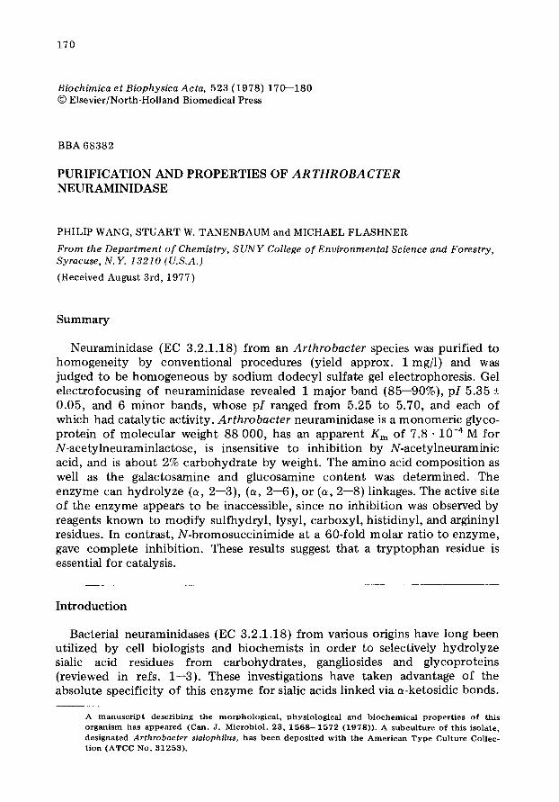
Amino acid analysis. The amino acid composition of the enzyme is shown in Table II. The Arthrobacter enzyme contains significantly more acidic amino acids, than basic ones, which is consistent with its acid isoelectric point deter- mined by gel electrofocusing. It also contains two disulfide bonds and one free cysteinyl residue. The presence of relatively large amounts of proline residues suggests that the Arthrobacter neuraminidase may have little helical structure. The Arthrobacter enzyme also contains a small number of glucosamine and galactosamine residues and is about 2% by weight neutral sugars. Therefore, neuraminidase is a glycoprotein.
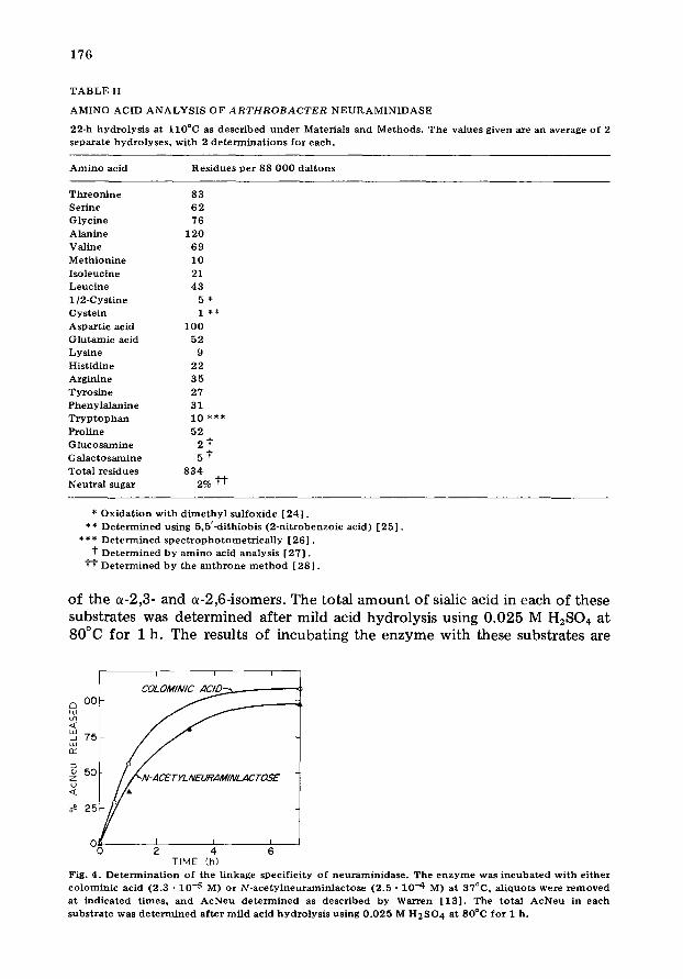
Linkage specificity. Neuraminidases isolated from other bacterial organisms hydrolyze the (a-2,3), (a-2,6) and (a-2,8) glycosidic linkage [1,2] . In order to determine the linkage specificity of the Arthrobacter enzyme, the enzyme was incubated with colominic acid or N-acetylneuraminlactose. The former is a homopolymer of sialic acid linked via a-2,8-1inkages, and the latter is a mixture
1 7 6
T A B L E II
A M I N O A C I D A N A L Y S I S O F A R T H R O B A C T E R N E U R A M I N I D A S E
22-h h y d r o l y s i s a t l l O ° C as desc r ibed u n d e r Mater ia ls and Methods . T h e values g iven are an average o f 2 separa te hyd ro ly se s , w i th 2 d e t e r m i n a t i o n s for each.
A m i n o acid R e s i d u e s pe r 88 0 0 0 da l t ons
T h r e o n i n e 83 Ser ine 62 Glyc ine 76
Alan ine 120
Val ine 69
M e t h i o n i n e 10
I so leuc ine 21 Leuc i n e 43
1 /2 -Cys t ine 5 *
Cys te in 1 * *
Aspar t i e acid 100 G l u t a m i c acid 52 L y s i n e 9
His t id ine 2 2 Arg in ine 3 5
T y r o s i n e 27
Pheny la l an ine 31 T r y p t o p h a n 10 * ** Prol ine 52
G l u c o s a m i n e 2 t G a l a c t o s a m i n e 5 t
To t a l r e s idues 834 Neu t ra l sugar 2% t t
* O x i d a t i o n wi th d i m e t h y l s u l f o x i d e [ 2 4 ] . ** D e t e r m i n e d us ing 5 ,5 ' -d i th iob i s (2 -n i t robenzo ic ac id) [ 2 5 ] .
*** D e t e r m i n e d s p e c t r o p h o t o m e t r i c a l l y [ 2 6 ] . t D e t e r m i n e d by a m i n o acid ana lys i s [ 2 7 ] .
t T D e t e r m i n e d by the a n t h r o n e m e t h o d [ 2 8 ] .
of the ~-2,3- and ~-2,6-isomers. The total amount of sialic acid in each of these substrates was determined after mild acid hydrolysis using 0.025 M H2SO4 at 80°C for I h. The results of incubating the enzyme with these substrates are
i i i
c~ O0
~ 75 w rY
~ 5O z
~ 25
O~ i i i 0 2 4 6
TIME (h) Fig. 4. D e t e r m i n a t i o n o f the l inkage spec i f i c i ty o f n e u r a m i n i d a s e . The e n z y m e was i n c u b a t e d wi th e i t he r e o l o m i n i c acid (2.3 • 10 .5 M) or N - a c e t y l n e u r a m i n l a c t o s e (2 .5 - 10 .4 M) at 37°C, a l iquo t s were r e m o v e d at i nd i ca t ed t imes , and A e N e n d e t e r m i n e d as d e s c r i b e d by Warren [ 1 3 [ . The to ta l A c N e u in each s u b s t r a t e was d e t e r m i n e d a f t e r m i l d acid h y d r o l y s i s us ing 0 .025 M H 2 S O 4 at 80°O fo r 1 h.
1 7 7
T A B L E I I I
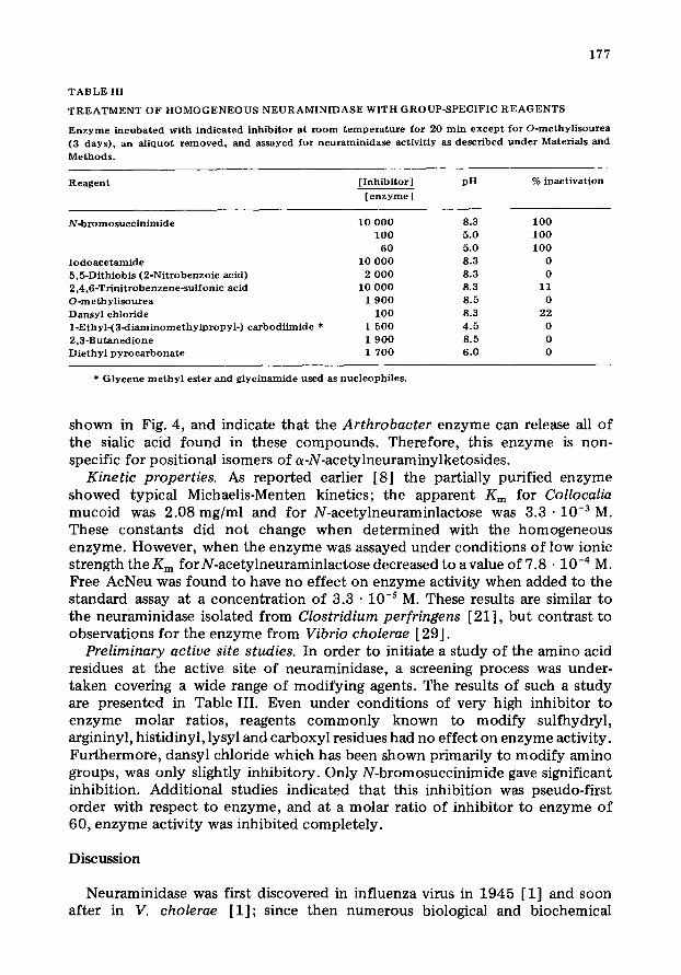
T R E A T M E N T OF H O M O G E N E O U S N E U R A M I N I D A S E W I T H G R O U P - S P E C I F I C R E A G E N T S
E n z y m e i ncuba t ed with ind icated inhibi tor at r o o m t e m p e r a t u r e for 20 rain e x c e p t for O - m e t h y l i s o u r e a (3 days ) , an a l iquot r e m o v e d , and assayed for neuramin idase act ivi t iy as desc r ibed u n d e r Materials and
M e t h o d s .
R e a g e n t [ Inh ib i to r ] pH % inact ivat ion
[ e n z y m e ]
N - b r o m o s u c c i n i m i d e
I o d o a c e t a m i d e 5,5-Di th iobis (2 -Ni t robenzo ic acid) 2 ,4 ,6 -Tr in i t robenzene-su l fonic acid O-m ethyl i so t t rea Dansy l ch lor ide 1 - E t h y l - ( 3 - d i a m i n o m e t h y l p r o p y l - ) c a r b o d i i m i d e * 2 ,3 -Bu taned ione D i e t h y l p y r o c a r b o n a t c
10 0 0 0 8.3 100 100 5.0 100
60 5.0 100 10 0 0 0 8 .3 0
2 0 0 0 8.3 0 10 0 0 0 8.3 11
1 9 0 0 8.5 0 1 0 0 8.3 22
1 500 4 .5 0 1 9 0 0 8.5 0 1 700 6 .0 0
* G l y c e n e m e t h y l ester and g lyc ina mide used as nuc leophi l e s .
shown in Fig. 4, and indicate that the Arthrobacter enzyme can release all of the sialic acid found in these compounds. Therefore, this enzyme is non- specific for positional isomers of a-N-acetylneuraminylketosides.
Kinetic properties. As reported earlier [8] the partially purified enzyme showed typical Michaelis-Menten kinetics; the apparent Km for Collocalia mucoid was 2.08 mg/ml and for N-acetylneuraminlactose was 3 . 3 . 1 0 -3 M. These constants did not change when determined with the homogeneous enzyme. However, when the enzyme was assayed under conditions of low ionic strength the Km for N-acetylneuraminlactose decreased to avalue of 7.8 • 10 -4 M. Free AcNeu was found to have no effect on enzyme activity when added to the standard assay at a concentration of 3.3 • 10 -s M. These results are similar to the neuraminidase isolated from Clostridium perfringens [21], but contrast to observations for the enzyme from Vibrio cholerae [29].
Preliminary active site studies. In order to initiate a study of the amino acid residues at the active site of neuraminidase, a screening process was under- taken covering a wide range of modifying agents. The results of such a study are presented in Table III. Even under conditions of very high inhibitor to enzyme molar ratios, reagents commonly known to modify sulfhydryl, argininyl, histidinyl, lysyl and carboxyl residues had no effect on enzyme activity. Furthermore, dansyl chloride which has been shown primarily to modify amino groups, was only slightly inhibitory. Only N-bromosuccinimide gave significant inhibition. Additional studies indicated that this inhibition was pseudo-first order with respect to enzyme, and at a molar ratio of inhibitor to enzyme of 60, enzyme activity was inhibited completely.
Discussion
Neuraminidase was first discovered in influenza virus in 1945 [1] and soon after in V. cholerae [1]; since then numerous biological and biochemical
178
studies have been carried out with partially purified enzyme preparations. Purification of neuraminidases to homogenei ty from pathogenic bacterial sources has been handicapped by low yields under growing conditions and by the complexity of cultivation media. By utilization of a saprophytic Arthrobacter isolate which secretes an inducible, extracel lular enzyme, a relatively simple procedure for producing and purifying its neuraminidase to homogeneity has now been developed. The purity of this enzyme preparation was established by polyacrylamide gel electrophoresis under denaturing and non~lenaturing conditions, and by gel electrofocusing. Following earlier studies which indicated the presence of approximately 3--5 mg of enzyme per liter of induction filtrate, and the total absence of associated protease and other glyco- hydrolase activities [8], the final yield approached 1 mg of enzyme per liter. This is in marked contrast to the recent report by Nees et al. [21] on the purification of neuraminidase from Cl. perfringens, in which the final yield of enzyme was 0.01 mg of enzyme per liter of growth filtrate. The ease of obtaining large amounts of Arthrobacter neuraminidase by the foregoing procedures makes this particular enzyme preparation ideal for biochemical modifications and for use in cell biology studies.
The availability of this homogeneous Arthrobacter neuraminidase has enabled us to determine a number of its physical and chemical properties. Our estimate of 89 000 for its molecular weight, based on SDS polyacrylamide gel electrophoresis, accords with the value (87 000) obtained earlier [8] following gel filtration of the partially purified preparation. The concordance of these determinations strongly suggests a single polypept ide chain. This value does not differ markedly from the molecular weight of a cluster of other bacterial neuraminidases [1,2]. The presence of carbohydrate in neuraminidase was first reported for the viral enzyme [1,2]. Our chemical analysis demonstrates that the Arthrobacter enzyme is also a glycoprotein containing both glucosamine and galactosamine, and is about 2% neutral carbohydrate by weight. However, gel electrofocusing of the homogeneous enzyme revealed one major and 6 minor catalytic forms. These isofunctional enzymes, in all probability, do not differ significantly in molecular weight, since only a single protein band was observed using SDS polyacrylamide gel electrophoresis. Microheterogeneity has also been observed in the partial purification of neuraminidases from a number of other bacterial sources [10,19--21]. The amino acid composit ion of the enzyme was determined. In contrast to the enzyme from Diplococcus pneumoniae purified earlier in this laboratory and which did not appear to con- tain any 1/2 cystine, the Arthrobacter enzyme contains two cystine and one free cysteinyl residue. Indeed, there appears to be little in common with respect to the amino acid composit ion of these two proteins [10]. In order to further investigate analogies in the primary sequences of microbial neuraminidases, we have prepared monospecific rabbit antibodies against the homogeneous Arthrobacter enzyme. Parenthetically, the homologous antigen- ant ibody reaction in gel diffusion studies further indicates the homogenei ty of the Arthrobacter enzyme (Huchzermeier, R., Tanenbaum, S.W. and Flashner, M., unpublished data).
Although the reactions catalyzed by neuraminidase and the substrate specificity of the enzyme have been thoroughly investigated [1,2], little is

1 7 9
known concerning the identity of those amino acids involved in substrate binding or catalysis. Our initial approach, using group-specific reagents to chemically modify essential amino acid residues, is summarized in Table III. Since no inhibition was observed with reagents known to modify sulfhydryl, lysyl, carboxyl, histidinyl, or argininyl residues, the active site of the enzyme appears inaccessible. In contrast, N-bromosuccinimide, at a low molar ratio of inhibitor to enzyme, inhibits enzyme activity completely. These results are in keeping with a preliminary report of Bachmayer [30]. Furthermore, we have observed complete protect ion of the enzyme against inactivation by N-bromo- succinimide when the assay was carried out in the presence of 2<leoxy-2,3- dehydro-N-acetylneuraminic acid, a known competit ive inhibitor of the enzyme (ref. 31 and Wang, P. and Flashner, M., unpublished data). These results suggest that a t ryptophan residue is essential for catalysis.
In catalytic properties the Arthrobacter enzyme resembles very closely the D. pneumoniae or Cl. perfringens enzymes. All three enzymes have very similar Km values for N-acetylneuraminlactose, and are insensitive to inhibition by AcNeu. In addition, neither of these enzymes require Ca 2÷ ions for maximum activity, and all are sensitive [22,23] to changes in ionic strength. This is in contrast to the enzyme from V. cholera, which responds to inhibition by AcNeu, requires Ca 2÷ ions for activity and is unaffected by ionic strength when assayed in the presence of low molecular weight substrates.
Because of its facile purification after induction in a concentrated minimal replacement medium, taken together with the convergent criteria for homogenei ty as presented here, Arthrobacter neuraminidase provides a starting point for structure-function studies of this important glycohydrolase. Our preliminary survey of side-chain reactivities implicates t ryptophan at the active center, but has ye t to reveal the nature of the proton-donating amino acid residue. However, it can be anticipated that continuing probes with this protein and group specific reagents under reaction conditions which perturb its tertiary structure, and the combined use of affinity-labeling or transition state analogs, will provide more informat ion on the nature of the amino acid residues involved in catalysis.
Acknowledgments
We wish to thank Mr. Craig Hohm for carrying out the electrofocusing experiments. The work was supported by a Public Health Service grant (AI- 12532) from the National Institute of Allergy and Infectious Disease.
References
1 G o t t s c h a l k , A . a n d D r z e n i e k , R . ( 1 9 7 2 ) in G l y c o p r o t e i n s : The i r C o m p o s i t i o n , S t r u c t u r e , a n d F u n c - t i o n ( G o t t s c h a l k , A. , ed . ) , Vol . 5 A , p p . 3 8 1 - - 4 0 2 , Elsevier , A m s t e r d a m
2 D r z e n i e k , R . ( 1 9 7 2 ) in C u r r e n t T o p i c s in M i c r o b i o l o g y a n d I m m u n o l o g y ( A r b e r , W. e t al . , eds . ) , Vol . 59 , p p . 3 5 - - 7 5 , S p r i n g e r - V e r l a g , N e w Y o r k
3 Winzler , R . J . ( 1 9 7 0 ) in I n t e r n a t i o n a l Rev . o f C y t o l o g y ( B o u r n e , G .H. a n d Daniel l i , J .F . , eds . ) , Vol . 29 , p p . 7 7 - - 1 2 5 , A c a d e m i c Press , N e w Y o r k
4 D e n , H. , M a l i n z a k , D .A. a n d R o s e n b e r g , A. ( 1 9 7 5 ) J . C h r o m a t o g r . 1 1 1 , 2 1 7 - - 2 2 2 5 H a t t e n , M.W.C. a n d R e g o e c z i , E. ( 1 9 7 3 ) B i o c h i m . B i o p h y s . A c t a 3 2 7 , 1 1 4 - - 1 2 0 6 H u a n g , C.C. a n d A m i n o f f , D. ( 1 9 7 4 ) B i o c h i m . B i o p h y s . A c t a 3 7 1 , 4 6 2 - - 4 6 9

180
7 Rood~ J.J. and Wilkinson, R.G. (1974) Biochim. Biophys. Acta 334, 168--178 8 Flashner, M., Wang, P., Hurley, J. and Tanenbaum, S.W. (1977) J. Bacteriol. 129, 1457--1465 9 Tanenbaum, S.W. and Sun, S.C. (1971) Biochim. Biophys. Acta 229 ,824- -828
10 Francus, T., Tanenbaum, S.W., Lundgren, H. and Flashner, M. (1974) in Isozymes (Markert, C.L., ed.), Vol. I, PP. 753--765, Academic Press, New York
11 Wang, P., Tanenbaum, S.W. and Flashner, M. (1977) Fed. Proc. 36, 2355 12 Howe, C., Lee, L.T. and Rose, H.M~ (1961) Arch. Biochem. Biophys. 95, 512--520 13 Warren, L. (1959) J. Biol. Chem. 234, 1971--1975 14 Lowry, O.H., Rosebrough, W.J., Farr, A.L. and Randall , R.J. (1951) J. Biol. Chem. 193, 265--275 15 Wrigley, C.W. (1971) in New Techniques in Amino Acid, Peptides and Protein Analysis (Niederweiser,
A. and Pataki, G., eds.), pp. 291--339, Ann Arbor Science Publisher, Ann Arbor, Michigan 16 Gabriel, O. (1971) Methods in Enzymology (Jakoby, W.B., ed.), Vol. 22, pp. 565---578, Academic
Press, New York 17 Tupper, H. and Palese, S. (1969) F E B S Lett . 3, 72--75 18 Weber, K. and Osborn, M. (1969) J. Biol. Chem. 224, 4406--4412 19 Moriyama, T. and Barksdale, L. (1967) J. Bacteriol. 94, 1565--1581 20 Hayano, S. and Tanaka, A. (1967) J. Bacteriol. 93, 1753--1757 21 Nees, S., Veh, R.W. and Schauer, R. (1975) Hoppe-Seyler 's Z. Physiol. Chem. 356, 1027--1042 22 Barton, N.W., Lipovac, V. and Rosenberg, A., (1975) J. Biol. Chem. 250, 8462--8466 23 Barton, N.W. and Rosenberg, A., (1973) J. Biol. Chem. 248, 7353--7358 24 Spencer, R.L. and Wold, F. (1969) Anal. Biochem. 3 2 , 1 8 5 - - 1 9 0 25 Ellman, G.L. (1959) Arch. Biochem. Biophys. 82, 70--77 26 Edelhoch, H. (1967) Biochemistry 6, 1948--1954 27 Spiro, R.G., Spiro, N.J. and Bhoyoo, V.D. (1976) J. Biol. Chem. 251, 6409--6419 28 Steifer, S., Dayton, S., Novick, B. and Muntwyler , E. (1950) Arch. Biochem. Biophys. 25, 191--200 29 Mohr, E. and Schramm, G. (1960) Z. Naturforsch. 156, 568--575 30 Bachmayer, H. (1972) FEBS Lett. 23 ,217 - -219 31 Meindl, P., Bodo, G., Palese, P., Schulman, J. and Tuppy, H. (1974) J. Virol. 58 ,457- -463