Embed Size (px)
Citation preview

Chapter 15
Purification and Partial Characterization of aThermostable Laccase from Pycnoporus sanguineus CS-2with Ability to Oxidize High Redox Potential Substratesand Recalcitrant Dyes
Sergio M. Salcedo Martínez, Guadalupe Gutiérrez-Soto,Carlos F. Rodríguez Garza, Tania J. Villarreal Galván,Juan F. Contreras Cordero and Carlos E. Hernández Luna
Additional information is available at the end of the chapter
http://dx.doi.org/10.5772/56374
1. Introduction
Laccases (benzenediol: oxygen oxidoreductases, EC 1.10.3.2) are enzymes that catalyze theoxidation of phenolic compounds and aromatic amines with the simultaneous reduction ofmolecular oxygen to water [1]. They are widely distributed in many plants and fungi, someinsects and bacteria, being particularly abundant in white-rot basidiomycetes [2]. Typicalfungal laccases are described as glycosylated multicopper proteins, which are produced asextracellular monomeric forms of around 60-80 kDa, containing four copper atoms and 15-20%carbohydrates. Operatively, they are moderately thermotolerant, showing optima activity at50-55 °C, and under acidic conditions (pH 3-5); although their maxima stability occurs in thealkaline zone (pH 8-9) [3]. Their copper atoms are distributed in three different sites bringingunique spectroscopic properties: The type 1 copper (CuT1) atom, is responsible of the intenseblue color of enzymes by light absorption around 610 nm; The type 2 copper (CuT2) atomexhibits a weak absorption in the visible region; and the two type 3 copper (CuT3) atoms arepresent as a binuclear center, which has an absorption maximum about 330 nm. Moreover,CuT2 and CuT3 copper atoms are structural and functionally arranged as a trinuclear cluster.The four copper atoms form part of the active site of enzyme contributing directly to reaction.CuT1 is involved in the initial electron subtraction from reducer substrates, while trinuclear
© 2013 Martínez et al.; licensee InTech. This is an open access article distributed under the terms of theCreative Commons Attribution License (http://creativecommons.org/licenses/by/3.0), which permitsunrestricted use, distribution, and reproduction in any medium, provided the original work is properly cited.

CuT2 and CuT3 cluster is responsible of the electron transference, from CuT1 to diatomicoxygen [4].
According to their redox potential, most of blue laccases belong to class II (-500 to -600 mV) orclass III (-700 to -800 mV) laccases [5]. This is a disadvantage when compared to the ability oflignin peroxidase (LiP) and manganese peroxidase (MnP) to attack compounds with higherredox potential, including non-phenolic lignin units. To overcome this limitation laccases haveevolutively developed a synergistic catalytic strategy, which combines a flexible ability torecognize a great variety of chemical compounds, with an extended capability to act at thedistance through the activation of diffusible low molecular substances which serve as redoxmediators. From a biological stand point this strategy let laccases to become one of the mostversatile enzymes in nature, adaptable to multiple functions in plants, insects, fungi andbacteria. Another interesting possibility arises from the properties of atypical “yellow” and“white” laccases, which have shown the ability to catalyze the direct oxidation of high redoxpotential non-phenolic lignin model substrates or polyaromatic hydrocarbons [6, 7]. It has beenproposed that the improved redox capabilities of these laccases come up either, by substitutingsome copper atoms for zinc, iron, or manganese in the metal clusters or by a change of theredox state of the CuT1(due to the interaction with a lignin-derived ligand) at the active siteof, otherwise normal laccase protein structures. So, evolution and prevalence of laccases as apart of the lignin modifying enzyme (LME) system in white-rot basidiomycetes could also bethe result of a “biochemical spring-up” mechanism acting under a short term ecophysiologicalselective pressure.
Whether directly or by mediation, laccases are able to oxidize a broad range of natural orxenobiotic compounds, including: mono, poly or methoxy- amine- and chloro-substitutedphenols as well as aromatic heterocyclic and inorganic/organometallic substances; some ofthem recognized among the most recalcitrant industrial pollutants, for example; polycyclicaromatic hydrocarbons (PAH), pentachlorophenols (PCP), polychlorinated biphenyls (PCB),1,1,1-trichloro-2,2-bis(4-chlorophenyl)ethane (DDT), trinitrotoluene (TNT), and many azo,triarylmethane, anthraquinonic, indigoid and heterocyclic textile dyes [8,9]. Therefore,laccases are considered enzymes with a great potential for the development of environmentaland industrial applications. Current and potential laccase applications include biobleachingof pulp and bioremediation of pulp and paper industries, bioremediation of olive millwastewater, bioremediation of effluents of the textile and dye manufacturing industries,biocatalytic synthesis of antibiotics and novel polymeric materials, development of biosensors,clarification and stabilization of beer, juices and wines, and panification [1, 10, 11]. Some laccaseformulations have already reached a commercial significance, but general thought is that theirbiotechnological applications and performances could be greatly improved or expanded withthe development and finding of new enzyme variants with desirable functional properties,such as higher redox potential, optimum activity at neutral or alkaline pH and thermal stability[12, 13, 14]. It has been proposed that these new laccases could be obtained by proteinengineering or through the exploration of the natural biodiversity. The importance of pro‐spective studies in natural biodiversity applying an ecophysiological approach is illustratedby reports about isolation of new thermostable laccases from fungi, either from thermophilic
Applied Bioremediation - Active and Passive Approaches354
compost [15] or tropical environments [13,16], or by the finding of novel laccases withimproved ability to oxidize substrates with a higher than normal redox potential culturingunder solid phase conditions [6, 7,17]. Northeast Mexico shelters a high diversity of white-rotbasidiomycetes as a result of its particular combination of physiography and climate, includingspecies associated to pine, oak and mixed forests, sub-mountain and semi-desert scrublands,and grass-land. In this work we first present information on the isolation, identification andselection of a northeast Mexico native strain of Pycnoporus sanguineus CS2, as a potentialproducer of thermostable laccases. Results on the purification and partial characterization ofits laccase are then exposed, stressing on its thermal stability and ability to attack high redoxpotential substrates and recalcitrant dyes without the participation of redox mediators.
2. Materials and methods
2.1. Chemicals
All chemicals used as buffers, enzyme substrates, culture media ingredients and electropho‐resis reagents, were reactive grade and commercially available through local distributors ofDifco, Sigma-Aldrich and Fluka, or BioRad products: PDB (potato dextrose broth), bacterio‐logical agar, yeast extract, malt extract, peptone and dextrose, were from Difco. Acrylamide,bis-acrylamide, TEMED (N,N,N’,N’- tetramethylethylenediamine), 2-mercaptoethanol, SDS(sodium dodecyl sulfate), trizma-base, glycine, Coomassie blue, and low range markers kit,were from Bio Rad. Enzyme substrates and dyes: 2,6-dimethoxyphenol (2,6-DMP); o-dianisi‐dine (3,3′-dimethoxybenzidine); ABTS (2,2’–azino-bis(3-ethylbenzthiazolin-6-sulphonic acid);syringaldazine (4-hydroxy-3,5-dimethoxybenzaldehyde azine); DMAB (3-dimethylamino‐benzoic acid), MBTH (3-methyl-2-benzothiazolinone hydrazone); Methyl Red (Acid Red 2; CI13020); Reactive Black 5 (RB 5; CI 20505), were from Sigma, Fluka or Aldrich. Chromatographicmatrices; DEAE-Sepharose and Q-Sepharose from Sigma-Aldrich, and Biogel P-100 fromBioRad. All other chemicals, including solvents, inorganic salts, acids and bases, were fromReactivos Químicos Monterrey, S.A. or CTR-Scientific S.A. de C.V. Solutions and culture mediawere prepared with bidistilled water from Laboratorios Monterrey, S. A.
2.2. Isolation and identification of fungal strain
The Pycnoporus strain used in our experiments was isolated from fruit bodies developing ondecayed logs that were gathered in a man-disturbed sub-mountain scrubland around Mon‐terrey, N.L. (Northeast México). Mycelia cultures were obtained by standard mycologicaltechniques, according to the procedure previously described [18]. Briefly, small flesh sectionswere aseptically removed from inside the carpophores, and transferred to YMGA (0.4% Yeastextract, 1.0% Malt extract, 0.4% Glucose, 1.5% agar) plates, supplemented with 10% TartaricAcid and 0.004% Benomyl. Plates were incubated at 28 °C, and those with extensive myceliagrowth were analyzed under the microscope to confirm a successful isolation. Stock cultureswere maintained by periodic transfers every two or three months on YMGA plates and keptrefrigerated at 4 °C. Carpophore morphologic features, measurements and photographs were
Characterization of a Thermostable Laccase from Pycnoporus sanguineus CS-2http://dx.doi.org/10.5772/56374
355

registered previous to dissections for microscopic examination, and identification was doneby following the taxonomical keys in reference [19], and in [20] for genera of polypores andthe most common macromycetes from Mexico, respectively.
2.3. Enzyme and protein assays
Laccase activity was determined by triplicate at 25 °C in 3 ml cuvettes, monitoring the increasein absorbance at A468 (ε=49,600 M-1cm-1), using a Shimadzu UV-VIS mini 1240 spectrophotom‐eter and 2,6-DMP as substrate. The assay mixture contained 0.01ml enzymatic extract, 0.1 mlof 60 mM 2.6-DMP in 2.89 ml of 200 mM citrate-phosphate buffer at pH 4.0 [21]. One unit oflaccase activity was defined as the amount of enzyme required to oxidize 1 μmol of 2, 6-DMPper minute at 25 °C. In some cases, it was necessary to assess the presence of lignin peroxidase(LiP) and manganese peroxidase (MnP). LiP activity was estimated by the H2O2-dependantveratryl alcohol oxidation to veratraldehyde as in reference [22] MnP activity was measuredby the formation of Mn3+-tartrate complex during the oxidation of MnSO4 in tartrate buffer asin [23]. The protein concentration was estimated by the Bradford assay (Protein Assay Bradfordof BioRad) with bovine serum albumin as standard.
2.4. Strain selection and enzyme production
Isolated Pycnoporus strain was selected as a potential source of thermostable laccases in apreliminary screening with crude enzyme preparations. 250 ml Erlenmeyer flask, containing50 ml of natural LME inducers containing Bran Flakes (BF) media (2% Bran Flakes® in 60 mMpotassium phosphate pH 6.0) [24], were inoculated with three 0.5 cm diameter cylinders ofmycelia taken from the border of a YMGA growing colony and incubated at 28 °C underagitation at 150 rpm. Aliquots (200 μl) were removed from cultures and the extracellular fluidwas separated by centrifugation at 14 K (Eppendorf 5415 C). Enzyme activity in supernatantswas determined with 2 mM 2, 6-DMP final concentration, as described above, after sampleincubation at 60 °C during different times in a four hour period. This phase of study includedfour different Pycnoporus sp. (CS 2, CS 20, CS 43 and LE 90) strains from the native basidio‐mycete collection of our laboratory. Among them, Pycnoporus sanguineus CS 2 was selected onthe basis of its ability to produce a thermostable 2,6-DMP oxidizing activity, and for showingapparently a single band of activity when incubated with 2,6-DMP and SGZ in a parallel nativePAGE analysis of crude supernatants.
Enzyme production was evaluated in submerged liquid cultures on the natural containinglaccase-inductors BF or a modified Kirk medium (MK) [25], with the following composition:10 g l-1 dextrose, 1.0 g l-1 yeast extract, 5.0 g l-1 peptone, 2.0 g l-1 ammonium tartrate, 1.0 g l-1
KH2PO4, 0.5 g l-1 MgSO4, 0.5 g l-1 KCl, and 1.0 ml of 100 X trace element solution (0.5 g EDTA,0.2 g FeSO4, 0.01 g ZnSO4, 0.003 g MnCl2, 0.03 g H3BO4, 0.02 g CoCl2, 0.001 g CuCl2, and 0.003g NaMoO4 in 100 ml); amended with 350 μM CuSO4 and 3% ethanol [26]. Cultures wereperformed at 28 °C and 150 rpm for 14 days. 50 μL aliquots were taken every two days todetermine the laccase activity.
Applied Bioremediation - Active and Passive Approaches356
2.5. Laccase purification
All the procedures were performed at 4 °C, unless otherwise stated. Extracellular liquid from14 day-old submerged cultures was separated from mycelium by filtration through a cotton-polyester 50:50% cloth. Then, water-soluble polysaccharides were removed from samplesolution by freezing (- 20 °C for 24 h), thawing and filtration (Whatman # 1). Culture filtratewas concentrated to approximately 200 ml by 10 kDa ultrafiltration (Millipore prep/scale TFFcartridge). The obtained fluid was further reduced to 20 ml by using a stirred ultrafiltrationsystem equipped with an YM10 membrane (Amicon, Millipore). The reddish-brown enzymeconcentrate was equilibrated by diafiltration with 20 mM potassium phosphate, pH 6.0, andapplied to a pre-equilibrated anion-exchange DEAE-Sepharose column (2.5 × 17 cm). Once onthe column, unadsorbed protein and most of the pigment were removed by washing with twovolumes of equilibrium buffer. Retained proteins were eluted with a linear gradient ofpotassium phosphate pH 6.0 from 20 to 300 mM, and the eluted fractions were assayed forlaccase activity and the A280 nm monitored. Fractions with laccase activity were pooled,concentrated, equilibrated by diafiltration with 100 mM potassium phosphate, and applied ona pre-equilibrated Biogel P-100 column (2.6 x 65 cm). The loaded proteins were eluted with thesame buffer. Active fractions were pooled, concentrated and diafiltrated against 20 mMpotassium phosphate buffer pH 6.0. Enzyme was further purified by anion-exchange on a pre-equilibrated Q-Sepharose column (2.5 x 17 cm). Once set the sample, active fractions wereeluted with a lineal gradient of potassium phosphate from 20 to 300 mM. These fractions werepooled, concentrated and diafiltrated against water, and stored at - 20 °C.
2.6. Electrophoresis analysis
Protein purity and molecular mass were evaluated by sodium dodecyl sulfate-polyacrylamidegel electrophoresis (SDS-PAGE) as in reference [27] with 4% stacking gel and 12% resolvinggel. Protein bands were stained with Coomassie brilliant blue and the molecular mass (Mr) ofpurified laccase was determined by calculating the relative mobility of standard proteinmarkers: phosphorylase b 97.4 kDa; serum albumin 66.2 kDa; ovalbumin 45 kDa; carbonic anhy‐drase 31 kDa; lysozyme 14.4 kDa; aprotinin 6.5 kDa (SDS-PAGE molecular weight standards lowrange, BioRad). Native PAGE was carried out as described at reference [28]. Activity stainingof laccase was performed by incubating with 2, 6-DMP or the pair MBTH + DMAB in 200 mMsodium acetate buffer, pH 4.5. For identification and comparing proposes, the correspondinglaccase band was removed from a parallel gel and submitted to the Proteomic Unit of IBT-UNAM at Cuernavaca, Morelos for aminoacid sequencing; resulting in the sequencing of sixinternal peptides.
2.7. UV-Vis absorbance spectra
As a part of the characterization of the physicochemical properties of the laccase, its absorbancespectrum from 200 to 800 nm was obtained in a UV-Vis Shimadzu-Mini 1240 spectrophotom‐eter. The assay was performed with 25 μM of protein diluted in 1 ml of bidistilled water.
Characterization of a Thermostable Laccase from Pycnoporus sanguineus CS-2http://dx.doi.org/10.5772/56374
357
2.8. Effect of pH on enzymatic activity
Optimum pH of activity was determined in McIlvine buffer (consisting of a combination of100 mM citrate/50 mM potassium phosphate) adjusted in a range from 3.0 to 7.0. Activitydetermination was made according to described method with 2,6-DMP using 0.2 M citratephosphate buffer, at pH 4.0.
2.9. Effect of temperature on enzyme activity and stability
The effect of temperature on reaction rate was determined using 2, 6-DMP in 0.2 M citrate/phosphate buffer, at pH 4.0. The temperature of reaction mixture was adjusted to indicatevalue and then the reaction was started by the addition of enzyme. The assays were done bytriplicate and data in graphics appear as relative activity as a function of temperature,considering as 100% the average of maxima obtained. The activation energy of the system wascalculated by the Arrhenius model, according to the expression: Log k = [-Ea / 2.303R (1/T)] +Log A, where: k is the rate constant (it depends of temperature); A is preexponential factor orfrecuency factor. Ea the activation energy (expressed in J/mol); R is the gas universal constant(8.314 J K-1 mol-1), and T the absolute temperature (°K). In the thermostability assays, theenzyme was pre-incubated at 50, 60 and 70 °C for the indicated periods of time and activitywas measured at 25 °C on 2, 6-DMP in 0.2 M citrate/phosphate buffer, at pH 4. The assays werecarried out by triplicate and data are expressed as percent of remaining activity as a functionof incubation time; taking as 100% the average value of activity at time zero for each temper‐ature. Inactivation process was adjusted to an exponential decay model, from which theconstants of heath inactivation (k) and the half-life times were calculated according to theexpression: ln (No/N) = kt, where; No is the activity at the starting of incubation; N is remainingactivity after a certain incubation time (t); k correspond to the first-order inactivation constant,and t1/2 is the half-life time, calculated as ln 2/k = 0.693/k
2.10. Determination of kinetic parameters
Kinetic analysis was performed on some common substrates of laccase: 2, 6-DMP, ABTS, o-dianisidine and SGZ. Reaction mixtures were prepared in 0.2 M citrate/phosphate buffer atpH 4.0 and the change in optical density by minute was measured by triplicate at differentsubstrate concentrations (0.05, 0.1, 0.5, 1.0, 5.0, 10.0 mM) or those indicated for each assay. Theassays were performed at 468 nm for 2, 6-DMP (ε = 49,600 M-1 cm-1), 436 nm for ABTS (29,400M-1cm-1), 460 nm for o-dianisidine (11,000 M-1 cm-1) and 525 nm for SGZ (ε= 65 000 M-1 cm-1).The values of the Michaelis constant (Km), maximum velocity (Vmax), turnover number (Kcat)and specificity constant Kcat/Km were estimated according to the Lineweaver-Burk method.
2.11. Decolorization assays
The decolorizing ability of laccase was evaluated with two recalcitrant dyes, the non-phenolicazo Methyl Red (MR), and the diazo reactive black 5 (RB 5). The reaction mixture consisted of0.890 ml of 0.2 M citrate/phosphate buffer, pH 4.0, 0.1 ml of 250 μM MR or RB5 (final concen‐tration 25 μM), and 0.01 ml of pure laccase (final concentration 5 U/ml). Assays were performed
Applied Bioremediation - Active and Passive Approaches358

at 25 °C and reaction was initiated with the addition of enzyme. Decolorization was estimatedby the decreasing of absorbance at 530 nm for MR or 597 nm for RB5. The results are expressedas the percent of remaining color as a function of incubation time according to the relationship:remaining color (%) = [(Abs final/ Abs initial)]*100, where: Abs final correspond to the absorbancevalue at the indicated incubation times, and Abs initial: is the initial (t = 0) absorbance value.
3. Results and discussion
3.1. Strain identification
In this study an autochthonous strain of Pycnoporus sp (CS 2) was selected as a potential sourceof thermostable laccases for its ability to produce a thermotolerant 2, 6-DMP oxidizing activityin preliminary assays with crude filtrates from submerged cultures. This basidiomycete wasinitially isolated from fruit bodies, growing on decayed logs in a man disturbed sub-mountainscrubland around Monterrey, N.L. México (Figure 1), and identified by its morphological andmicroscopic traits. According to their morphological and microscopic characteristics, thecarpophores corresponded to the species Pycnoporus sanguineus (L.) Murrill, for their brightorange to orange-red, red or cinnabar-red shelf-like basidiomes, which are nearly round toelongated or fan-shaped in outline, have a dry surface, smooth or finely hairy, wrinkled orwarty and attain 2-12 cm diameter and 0.2 to 0.5 cm thick. Their margins are thin and the undersurfaces are covered by small pores (3-4 per mm), bright orange to orange red or red rangingfrom 0.5-1.5 mm long. Their white spores are smooth and oblong-elliptical in shape and rangefrom 4.2 to 5.2 microns long by 2 to 3.5 microns width and the flesh is tough, red to yellowishred, staining black with KOH. On the bases of these features, we assigned the strain understudy as Pycnoporus sanguineus CS 2.
3.2. Production and purification of laccase
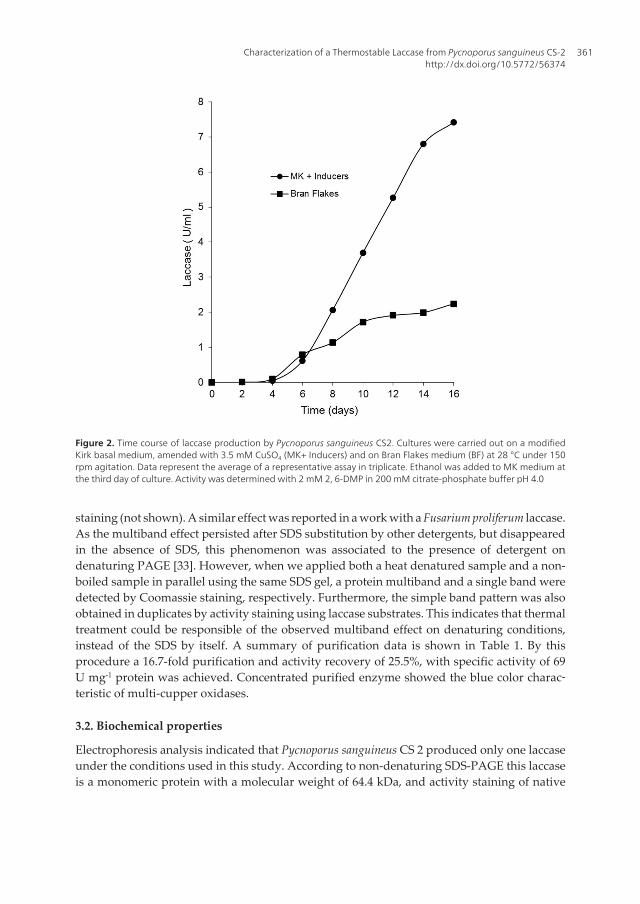
Guzmán (2003), considers that P. sanguineus is a tropical variant of the temperate zone speciesP. cinnabarinus, adapted to man disturbed sites, where it is common in fallen logs and fences,always in sunny places [29]. As its closely related species, P. cinnabarinus and P. coccineus, P.sanguineus is recognized as an efficient lignin decomposer, in spite of its relatively simple LMEsystem composed of laccases [5]. These features make Pycnoporus species an attractive groupof white-rot basidiomycetes for the production and purification of unusual laccases [16, 30].In this study, laccase production was carried out in submerged liquid cultures on a modifiedKirk basal medium (MK), amended with 3.5 mM CuSO4 and 3 % ethanol, as chemical laccaseinducers and on Bran-Flakes medium (BF), containing natural LME inducers. Under theseconditions maxima volumetric productions were reached in both media after 14-16 days(Figure 2). As laccase titers on MK media were about thrice higher than that on BF media (7.5U ml-1 vs 2.3 U min-1), it was selected for enzyme production in purification assays. Consistentlywith other reports on LME production by Pycnoporus species, LiP and MnP were not detected[5, 16, 30, 31, 32].
Characterization of a Thermostable Laccase from Pycnoporus sanguineus CS-2http://dx.doi.org/10.5772/56374
359

Laccase purification was started from about 1850 ml of mycelium-free filtrates from 14 day-old submerged cultures. After 10K ultraconcentration and sequential steps of anionic exchangechromatography on DEAE- Sepharose, gel filtration on Biogel P-100, and anionic exchange onQ-Sepahrose, laccase activity eluted as an apparently single protein peak with 100-140 mMphosphate (Figure 3). When aliquots of pooled laccase from this last chromatographic stepwere analyzed by denaturing SDS-PAGE, multiple protein bands were detected by Coomassie
Figure 1. Fruit bodies (carpophores), mycelium colony and submerged culture of Pycnoporus sanguineus CS 2. Fungusidentification was performed according to macroscopic and microscopic features. Strain isolation was done by tissuetransference from the inner flesh of carpophores using mycological standard methodologies. Develop of orange-redpigmented mycelium on the edge of the solid plate colonies, and extracellular production of a reddish pigment undersubmerged conditions, were indicative of a successful isolation. Production of extracellular mucilage was also ob‐served on submerged cultures. Isolation and identification details are given in text.
Applied Bioremediation - Active and Passive Approaches360
staining (not shown). A similar effect was reported in a work with a Fusarium proliferum laccase.As the multiband effect persisted after SDS substitution by other detergents, but disappearedin the absence of SDS, this phenomenon was associated to the presence of detergent ondenaturing PAGE [33]. However, when we applied both a heat denatured sample and a non-boiled sample in parallel using the same SDS gel, a protein multiband and a single band weredetected by Coomassie staining, respectively. Furthermore, the simple band pattern was alsoobtained in duplicates by activity staining using laccase substrates. This indicates that thermaltreatment could be responsible of the observed multiband effect on denaturing conditions,instead of the SDS by itself. A summary of purification data is shown in Table 1. By thisprocedure a 16.7-fold purification and activity recovery of 25.5%, with specific activity of 69U mg-1 protein was achieved. Concentrated purified enzyme showed the blue color charac‐teristic of multi-cupper oxidases.
3.2. Biochemical properties
Electrophoresis analysis indicated that Pycnoporus sanguineus CS 2 produced only one laccaseunder the conditions used in this study. According to non-denaturing SDS-PAGE this laccaseis a monomeric protein with a molecular weight of 64.4 kDa, and activity staining of native
Figure 2. Time course of laccase production by Pycnoporus sanguineus CS2. Cultures were carried out on a modifiedKirk basal medium, amended with 3.5 mM CuSO4 (MK+ Inducers) and on Bran Flakes medium (BF) at 28 °C under 150rpm agitation. Data represent the average of a representative assay in triplicate. Ethanol was added to MK medium atthe third day of culture. Activity was determined with 2 mM 2, 6-DMP in 200 mM citrate-phosphate buffer pH 4.0
Characterization of a Thermostable Laccase from Pycnoporus sanguineus CS-2http://dx.doi.org/10.5772/56374
361

gels exhibited a single broad band when incubated with both, 2,6-DMP and the pair MBTH +DMAB, showing the same migration as the Coomassie blue stained band (Figure 4). Molecularmass of purified laccase was very similar to those reported for different Pycnoporus sangui‐neus strains [30, 31, 32], and it was consistent with the reported for most of basidiomyceteslaccases [2, 3]. As expected for its visual appearance described above, the UV-Vis spectrum ofpurified enzyme was characteristic of the typical blue laccases, displaying the absorbance peaknear to 600 nm related to the Cu-T1 centers, and the shoulder at 330 nm of Cu-T3 binuclearcenters (Figure 5). Nonetheless, the oxidative coupling of MBTH and DMAB in the absence ofmediators was indicative that Pycnoporus sanguineus CS2 laccase has the capability to catalyzereactions requiring a higher than usual redox potential for typical laccases [15].
Figure 3. Elution profile for laccase from Pycnoporus sanguineus CS2 on anion-exchange column chromatographywith Q-Sepharose (2.5 x 17 cm). The enzyme was eluted with a potassium phosphate (pH 6.0) linear gradient from20-300 mM (dashed line) at a flow rate of 1.0 ml/min.
Purification stepProtein
(mg)Enzyme Activity (IU)
Specific Activity
(U/mg)
Recovery
(%)
Purification
(fold)
Culture filtrate 1551 6477 4.18 100.0 1.0
Ultraconcentration 10 K 422 3724 8.8 57.4 2.1
DEAE-Sepharose FF 64 2308 35.8 35.6 8.5
Biogel P-100 32 1924 59.2 29.7 14.1
Q-Sepharose 22 1522 69.8 23.5 16.7
Table 1. Purification of Pycnoporus sanguineus CS 2 laccase
Applied Bioremediation - Active and Passive Approaches362

Figure 4. Electrophoresis analyses of purified Pycnoporus sanguineus CS 2 laccase by non-denaturing SDS- PAGE (leftpanel) and Native PAGE (right panel). Lanes M and Lac correspond to the Coomassie staining of molecular weightmarkers and purified laccase, respectively. The markers were phosphorylase b (97.4 kDa), serum albumin (66.2 kDa),ovalbumin (45 kDa), carbonic anhydrase (31 kDa), trypsin inhibitor (21.5 kDa), and lysozyme (14.4). On the right, lane1 shows the Coomassie staining of purified laccase, and lanes 2 and 3, the activity staining with 2,6-DMP and the pairMBTH + DMAB, respectively.
Figure 5. UV-Vis Spectrum of Pycnoporus sanguineus CS 2 Laccase. Assay was performed with a preparation of 25 μMlaccase in bidistilled water. Insert shows the enlargement of the peak close to 610 nm.
Characterization of a Thermostable Laccase from Pycnoporus sanguineus CS-2http://dx.doi.org/10.5772/56374
363

In addition to blue laccases, other “atypical” forms of the enzyme named “yellow” laccasesand “white” laccases have been reported. In the first case, it has been proposed that a variationin the redox state of Cu-T1 centers, by the presence of endogenous ligands, decreases theabsorbance at 600 nm, without altering the spectral characteristics of the Cu-T2 and Cu-T3centers, resulting in a yellow color [6]. In white laccases, like the one produced by Pleurotusostreatus, it has been informed the presence of a single copper atom, which is accompanied bytwo of zinc and one of iron, instead of the regular four copper atoms [7]. In both, yellow andwhite laccases, the protein structure is similar to that found in blue laccases, but the changesin the redox state of the active site (whereas by the presence of the endogenous mediator orby the substitutions in the Cu centers), enables them to oxidize directly substrates of higherredox potential. According to all the above, the laccase produced by P. sanguineus CS 2corresponds to a blue laccase, most likely containing the regular composition of Cu in itscatalytic centers, but like atypical laccases is capable of acting on substrates of higher redoxpotential.
3.3. pH and temperature dependence
Enzyme was further characterized for its pH and temperature dependence. The effect of pHon laccase activity was studied using some of the most common laccase substrates, includingthe phenolic 2, 6-DMP, o-dianisidine and SGZ, as well as the non-phenolic ABTS. In general,laccase exhibited optima activity in the zone of pH between 3.0 and 4.5, depending on theparticular substrate, then it declined in a gradual way towards the neutral zone of pH, andwas completely lost at pH 6.5. Optimal pH values were 3.5, 3.5, 4.5 and 3.0 for 2, 6-DMP, o-dianisidine, SGZ and ABTS, respectively (Figure 6). These results were similar to thosereported in literature for most of the fungal laccases [2]. It is known that biphasic pH-activityprofiles with phenolic substrates (as the one showed by SGZ), are a consequence of twoopposite effects: one generated by the difference in the redox potential between the reducersubstrate and the Cu-T1 centers, when changing from acidic to neutral conditions. The otherone is directly associated to the inhibitory action of OH- ions over the activity of Cu-T2/T3centers. For non-phenolic substrates as ABTS, the first effect should be minimal and theinhibition by OH- reflects the monotonic decrease in the enzyme activity [12, 34].
The influence of temperature on P. sanguineus CS 2 laccase activity was investigated with 2, 6-DMP (2 mM), at pH 4.0, in the zone, from 20–80 °C. The profile temperature-activity showeda gradual increase from the lower limit at 20 °C to achieve an optimal value at 65 °C, anddeclined as temperature approached 80 °C. However, the enzymatic activity in these condi‐tions remained relatively high compared to the value showed under optimal conditions (witha level close to 85%) (Figure 7). Indicating that P. sanguineus CS 2 laccase is a thermotolerantenzyme [35]. These data were evaluated according to the Arrhenius model in order to estimatethe energy of activation (Ea) for the system. This parameter has been relatively little studied inthermotolerant laccases. The calculated Ea value (16.2 kJ/mol) for P. sanguineus CS 2 laccase issimilar to the values reported for other thermotolerant laccases, as that for Myceliophorathermophila (19 kJ/mol) [36] and for the recombinant laccase from Coprinus cinereus (14 kJ/mol)[37], but smaller than those calculated in this report for other laccases, which apparently did
Applied Bioremediation - Active and Passive Approaches364

not show a direct relationship between thermotolerance and the magnitude of Ea. On the otherhand, the function showed a change in slope in the high temperature zone (50-70 °C) beforethe enzyme denaturing breaking zone. This effect could correspond to a decrease in the Ea ofthe system, caused by a thermotropic transition of the protein conformation, which shouldfacilitate the limiting step of the reaction. Other possibility would be the coexistence of twoenzyme populations, one of them showing an increased activity by temperature and the otherbeing totally inactivated by thermal denaturing. These alternatives should be further explored.
Thermostability is a desirable property of industrial enzymes. Curves of temperature-stabilityof P. sanguineus CS 2 laccase showed that enzyme retained practically all of its activity afterincubation for 8 h at 50 and 60 °C. Moreover, when incubations at 60 °C were extended to 24h, the laccase retained 98% of its original activity (not shown). The enzyme also retained almost50% of its activity after 4h at 70 °C. Inactivation curve showed a first-order decaying behavior(correlation > 0.96), with a calculated half-life (t1/2) of 3.85 h [corresponding to a constant ofthermal inactivation (k) of 0.187 h-1]. To the best of our knowledge, this is one of the highestt1/2 values found in laccases from mesophilic fungi. It is known that most of typical fungallaccases lose their activity in a few minutes at 60 °C [3, 15, 30, 38].
In comparison to laccases isolated from other Pycnoporus species, t1/2 value at 70 °C heredescribed is higher than those reported for laccase I (0.13 h) and laccase II (2.06 h) fromPycnoporus sp SYBC-L1 [13, 30], and for the laccase from the thermotolerant P. sanguineusCeIBMD001 (0.21 h) [16]. Native laccase also seems to be more resistant to thermal inactivationthan P. sanguineus SCC 108 laccase (t1/2 = 3.33 h at 65 °C) reported by [31] and the P. sangui‐
Figure 6. pH versus activity profiles of Pycnoporus sanguineus CS 2 laccase on various substrates. Assays were done bytriplicate in 200 mM citrate-phosphate buffer at indicated pH, with 2, 6-DMP (2 mM), o-dianisidine (0.66 mM), SGZ(0.05 mM) and ABTS (2 mM).
Characterization of a Thermostable Laccase from Pycnoporus sanguineus CS-2http://dx.doi.org/10.5772/56374
365

Figure 7. Effect of temperature on Pycnoporus sanguineus CS 2 laccase activity. Assays were performed by triplicate in200 mM citrate-phosphate buffer at pH 4.0. Reaction rates were measured under saturating substrate concentrations(2 mM 2,6-DMP). The fitting line in lower panel shows the results of the Arrhenius analysis of data, corresponding to:LOG (Vmax) = [(-Ea /2.303 RT) + constant].
Applied Bioremediation - Active and Passive Approaches366

neus CCT- 4518 laccase studied in [39], which lost 60% of its initial activity after 2 h at 70 °C.Interestingly, three laccases from tropical or subtropical strains of Pycnoporus species (P.sanguineus BRFM 902, P. sanguineus BRFM 66, and P. coccineus BRFM 938) of different geo‐graphic regions (French Guinea, China and Australia, respectively) with remarkable thermalresistance have been recently reported by a research group in France [13]. A relationshipbetween P. sanguineus CS 2 laccase with these and other Pycnoporus laccases already describedwas established by comparing the aminoacid sequences of an internal protein fragment(peptides 2+3+4, Table 2) from the native laccase with those sequences deposited at GenBank.Aminoacid sequence of P. sanguineus CS 2 laccase showed 99 % similarity to P. sanguineusBRFM 902 laccase, 93% to P. coccineus BRFM 938 [13], P. cinnabarinus PM laccases [5], and P.sanguineus BRFM 66 laccase [13], but only 84 % to Trametes cinnabarina [40] and 71% to Psanguineus CeIBMD001 laccase [16]. These results highlight the importance of Pycnoporusspecies biodiversity for the prospection for new thermostable laccases.
3.4. Kinetic properties
The kinetic properties of enzyme were studied with some typical substrates. The values of theMichaelis constant (Km), catalytic constant (Kcat) and specificity constant (Kcat/Km), werecalculated by the Lineweaver-Burk method. Laccase showed the highest affinity and molecularactivity, on ABTS (Km = 23 mM, Kcat = 221 s-1) compared to o-dianisidine (Km = 44 mM, Kcat =197 s-1), and 2, 6-DMP (Km =41 mM, Kcat = 88 s-1). So, in terms of catalytic efficiency the bestsubstrate resulted ABTS (Kcat/Km = 9.4 x 10⁶ s-1 M-1) followed by o-dianisidine (Kcat/Km = 4.5 x10⁶ s-1 M-1) and 2, 6-DMP (Kcat/Km = 2.2 x 10⁶ s-1 M-1). These results are summarized in Table 3,comparing the values of specificity constants (Kcat/Km) for these substrates with those reported
Figure 8. Effect of temperature on Pycnoporus sanguineus CS 2 laccase stability. Incubations were performed at vari‐ous temperatures in distilled water. Aliquots were withdrawn at the indicated times and initial rates measured at 25°C in 0.2 mM citrate/phosphate buffer at pH 4.0 with 2 mM 2, 6-DMP.
Characterization of a Thermostable Laccase from Pycnoporus sanguineus CS-2http://dx.doi.org/10.5772/56374
367

for other Pycnoporus laccases, the native enzyme showed higher values for all assayedsubstrates except for the reported laccase II from Pycnoporus sp SYBC-L1 [30]. Like typicallaccases, the enzyme of P. sanguineus CS 2, showed activity on a variety of substrates, such asthe phenolic 2, 6-DMP, o-dianisidine and SGZ, as well as the non-phenolic ABTS.
Peptide Amino acid sequences
1 EAVVVNGITPAPLIAGKK
2* GPFVVYDPNDPQASLYDIDNDDTVITLADWYHLAAKVGQR
3* FPLGADATLINGLGR
4* TPGTTSADLAVIKVTQGK
5 YSFVLDASQPVDNYWIRANPPFGNVGFAGGINSAILR
6 SAGSSEYNYDNPVFR
* Contiguous peptides of the internal laccase fragment used in alignments
Table 2. Amino acid sequences corresponding to internal peptides of Pycnoporus sanguineus CS2 laccase
SubstrateKm
(μM)
Vmax
(μmol/min/ml)
kcat
(s-1)
kcat/Km
(s-1/M-1)
2,6-DMP 41 500 88 2.16 x 10⁶
o-dianisidine 44 1111 197 4.49 x 10⁶
ABTS 23 1250 221 9.38 x 10⁶
Table 3. Kinetics constants of Pycnoporus sanguineus CS 2 laccase
Among these substrates this laccase showed preference for ABTS and this characteristic wasconsistent with most of fungal laccases [32, 41, 42]. Unexpectedly the substrate saturationgraphics with SYR showed a sigmoidal-like behavior instead of the common hyperbolic one(not shown). This result could be explained considering a kinetic mechanism of positivecooperativity as that described for monomeric mnemonical enzymes [43], where a conforma‐tional change of interacting enzyme at the end of the first catalytic cycle, reacts more readilywith a second substrate molecule than other free-enzyme. Other factor contributing to thisresult could be the presence of ethanol in routinely SYR assay affecting the substrate solubilityand/or enzyme activity. Whether mechanistic on phenomenological, this observation must betaken into account in future works, considering the relevance of this substrate in laccasecharacterization.
3.5. Dye decolorization
As revealed by the activity staining of native gels shown above, P. sanguineus CS 2 laccasewas also able to promote the oxidative coupling between MBTH and DMAB. This reac‐
Applied Bioremediation - Active and Passive Approaches368

tion has been considered as indicative of the ability of some laccases to catalyze reactionsrequiring a higher redox potential, as in the case of the enzymatic decolorization of manysynthetic dyes. The non-phenolic azo MR [44, 45] and diazo RB 5 dyes [46] have been usedas models for studying the ability of laccases to degrade recalcitrant compounds (Figure 9).Although general consensus is that laccases require meditators for acting over these dyes,P. sanguineus CS 2 laccase showed the capability to decolorize directly both compounds, butwith different ability. Decolorization of MR and RB 5 reached a level of 70 %, and 15%respectively, after 4 h at 25 °C.
Figure 9. Chemical structure of the recalcitrant methyl red and reactive black 5 dyes used in this study.
While Ganoderma lucidum [38], Trametes trogii [47] and Lentinula edodes [45] laccases wereonly able to decolorize RB 5 in the presence of mediators, a recent report state that threePycnoporus laccases [13], were able to perform this decolorization in the absence ofmediators, under similar conditions used in this work, with decolorization reaching from29 to 45% after 52 h, at room temperature. The recalcitrance of RB 5 to laccase decoloriza‐tion has been explained by its high redox potential or steric hindrances limiting accessibil‐ity of enzyme to –OH and –NH2 groups in dye. As in this study native laccase attainedaround 70% decolorization after 20 h at room temperature, decolorization assays wereperformed at 60 °C taking advantage of its thermostability trying to overcome limitingfactors. As expected, decolorization process was faster under the influence of tempera‐
Characterization of a Thermostable Laccase from Pycnoporus sanguineus CS-2http://dx.doi.org/10.5772/56374
369

Figure 10. Decolorization of methyl red and reactive black 5 by Pycnoporus sanguineus CS2 laccase. Assays were per‐formed by incubating 25 μM Methyl Red (upper panel) and 25 μM Reactive Black 5 (middle and lower panels) withlaccase (5U/ml) in 0.2 mM citrate/phosphate buffer at pH 4.0 Aliquots were withdrawn from the assay mixture at theindicated times and remaining color was determined as described in text. Lines showed the best data fittings corre‐sponding to the exponential first-order (dashed) or polynomial second-order decay functions (continuous).
Applied Bioremediation - Active and Passive Approaches370

ture, reaching around 50% after 4 h, although it also seems to be limited faster (Figure 10).While MR decolorization fitted an exponential first order decay model, RB 5 decoloriza‐tion changes rapidly from this behavior to fit a polynomial second order model. This effectcould be related to several factors as an increased enzyme inactivation by endogenousgenerated reaction intermediates and/or dead-end transformation products. This relation‐ship must be investigated in future work. Nonetheless these results illustrate the poten‐tial of the thermostable Pycnoporus sanguineus CS2 laccase for practical applications.
4. Conclusion and future prospects
Its thermostability and ability for acting on high redox substrates and recalcitrant dyes, makesPycnoporus sanguineus CS 2 laccase a good prospect for its application in industrial andenvironmental processes. This laccase could also be interesting as a model in studies associ‐ating structure-function of thermotolerant proteins from mesophilic microorganisms.
Acknowledgements
Authors thank the financial support provided by the Sistema de Fondos INNOVAPYME-CONACYT (Proyecto No. 139352). We also tank to Unidad de Proteómica, IBT-UNAM forassistance in peptide sequencing.
Author details
Sergio M. Salcedo Martínez1,2, Guadalupe Gutiérrez-Soto1,3, Carlos F. Rodríguez Garza1,Tania J. Villarreal Galván1, Juan F. Contreras Cordero4 and Carlos E. Hernández Luna1*
*Address all correspondence to: [email protected]
1 Autonomous University of Nuevo León, Laboratory of Enzymology, Faculty of BiologicalSciences, San Nicolás de los Garza, N.L. México
2 Autonomous University of Nuevo León, Department of Botanic, Faculty of Biological Sci‐ences, San Nicolás de los Garza, N.L. México
3 Autonomous University of Nuevo León, Department of Biotechnology, Faculty of Agrono‐my, San Nicolás de los Garza, N.L. México
4 Autonomous University of Nuevo León, Department of Microbiology and Immunology,Faculty of Biological Sciences, San Nicolás de los Garza, N.L. México
Characterization of a Thermostable Laccase from Pycnoporus sanguineus CS-2http://dx.doi.org/10.5772/56374
371

References
[1] Mayer AM, Staples RC. Laccase: New Functions for an Old Enzyme. Phytochemistry2002;60 551-565.
[2] Baldrian P. Fungal Laccases-Occurrence and Properties. FEMS Microbiological Re‐views 2005;20 1-28.
[3] Morozova OV, Shumakovich GP, Gorbacheva MA, Shleev SV, Yaropolov AI. ‘‘Blue’’Laccases. Biochemistry (Moscow) 2007;72 1136–1412.
[4] Thurston CF. The Structure of Fungal Laccases. Microbiology 1994 ;140 19-26.
[5] Eggert C, Temp U, Eriksson K-EL. The Ligninolytic System of the White- rot FungusPycnoporus cinnabarinus: Purification and Characterization of the Laccase. AppliedEnvironmental Microbiology 1996;62 1151–1158.
[6] Leontievsky AA, Vares T, Lankinen P, Shergill JK, Pozdnyakona NN, MyasoedovaNM. Blue and Yellow Laccases of Ligninolytic Fungi. FEMS Microbiological Letters1997;156 9-14.
[7] Palmieri G, Giardina, P, Bianco C, Sacloni A, Capasso A, Sannia G.A A Novel WhiteLaccase from Pleurotus ostreatus. Journal of Biological Chemistry 1997;50 31301-31307.
[8] Pointing SB. Feasibility of Bioremediation by White–rot Fungi. Applied Microbiologyand Biotechnology 2001;57 20-33.
[9] Reddy CA. The Potential of White-rot Fungi in the Treatment of Pollutants. CurrentOpinion in Biotechnology 1995;6 320-328.
[10] Rodríguez-Couto S, Toca-Herrera JL. Laccases in the Textile Industry. Biotechnologyand Molecular Biolology Reviews 2006a;1 115-120.
[11] Rodríguez-Couto S, Toca-Herrera JL. Industrial and Biotechnological Applications ofLaccases: A review. Biotechnology Advances 2006b;24 500-513.
[12] Xu F, Berka RM, Wahleithner JA, Nelson BA, Shuster JR, Brown SH, Palmer AE, So‐lomon EI. Site-directed Mutations in Fungal Laccase: Effect on Redox Potential, Ac‐tivity and pH Profile. Biochemistry Journal 1998;334(1) 63-70.
[13] Uzan E, Nousiainen P, Balland V, Sipila J, Piumi F, Navarro D, Asther M, Record E,Lomascolo A. High Redox Potential Laccases from the Ligninolytic Fungi Pycnoporuscoccineus and Pycnoporus sanguineus Suitable for White Biotechnology: from GeneCloning to Enzyme Characterization and Applications. Journal of Applied Microbiol‐ogy 2010;108 2199-2213.
[14] Rodgers CJ, Blanford CF, Giddens SR, Skamnioti P, Armstrong FA Gurr SJ. DesignerLaccases: A Vogue for High-potential Fungal Enzymes?. Trends in Biotechnology2009;28(2) 63-72.
Applied Bioremediation - Active and Passive Approaches372

[15] Jordaan J, Leukes WD. Isolation of a Thermostable Laccase with DMAB and MBTHOxidative Coupling Activity from a Mesophilic White-rot Fungus. Enzyme and Mi‐crobial Technology 2003;33 212-219.
[16] Dantán-González E, Vite-Vallejo O, Martínez-Anaya C, Méndez-Sánchez M, Gonzá‐lez MC, Palomares LA, Folch-Mallol J. Production of Two Novel Laccase Isoforms bya Thermotolerant Strain of Pycnoporus sanguineus Isolated from an Oil-polluted Trop‐ical Habitat. International Microbiology 2008;11 163–169.
[17] Pozdnyakova NN, Turkovskaya OV, Yudina EN Rodakiewicz-Nowak Y. Yellow Lac‐case from the Fungus Pleurotus ostreatus D1: Purification and Characterization. Ap‐plied Biochemistry and Microbiology 2006;42(1) 56-61.
[18] Hernández-Luna CE, Gutiérrez-Soto G, Salcedo-Martínez SM. Screening for Decolor‐izing Basidiomycetes in Mexico. World Journal of Microbiology and Biotecnology2008;24 465-473.
[19] Ryvarden L. Genera of Polypores Nomenclature and Taxonomy. Synopsis fungorumVol. 5. Fungiflora A/S Oslo; 1991.
[20] Guzmán G. Identificación de los Hongos: Comestibles, Venenosos, Alucinantes y De‐structores de la Madera: Editorial Limusa S.A. México; 1980.
[21] Abadulla E, Tzanov T, Costa S, Robra K, Gübitz G. Decolorization and Detoxificationof Textile Dyes with a Laccase from Trametes hirsuta. Applied and Environmental Mi‐crobiology 2000;66 3357-3362.
[22] Tien M, Kirk TK. Lignin Peroxidase of Phanerochaete chrysosporium. Methods in Enzy‐mology 1988;161 238-248.
[23] Kuan C, Johnson J, Tien M. Kinetic Analysis of Manganese Peroxidase. The Reactionwith Manganese Complexes. Journal of Biological Chemistry 1993;268 20064–20070
[24] Pickard MA, Vandertol H, Roman R, Vazquez-Duhalt R. High Production of Ligni‐nolytic Enzymes from White-rot Fungi in Cereal Bran Flakes Liquid Medium. Cana‐dian Journal of Microbioliology 1999;45 627-631.
[25] Dhouib A, Hamza M, Zouari H, Mechichi T, Hmidi R, Labat M, Martinez MJ, SayadiS. Screening for Ligninolytic Enzyme Production by Diverse Fungi from Tunisia.World Journal of Microbiology and Biotechnology 2005 ;21 1415-1423.
[26] Zouari-Mechichi H, Mechichi T, Dhouib A, Sayadi S, Martínez AT, Martínez MJ. Lac‐case Purification and Characterization from Trametes trogii isolated in Tunisia: Decol‐orization of Textile Dyes by the Purified Enzyme. Enzyme and Microbial Technology2006;39 141-148.
[27] Laemmli U. Cleavage of Structural Proteins During the Assembly of the Head of theBacteriophage T4. Nature 1970;227 680-685.
[28] Garfin DE. Methods in Enzymology. Academic Press Inc. 1990.
Characterization of a Thermostable Laccase from Pycnoporus sanguineus CS-2http://dx.doi.org/10.5772/56374
373

[29] Guzmán G. Los Hongos del Edén Quintana Roo. (Introducción a la Micología Tropicalde México). INECOL y CONABIO, Xalapa ; 2003.
[30] Wang ZX, Caia YJ, Liao XR, Taoc GJ, Lia YY, Zhanga F, Zhang DB. Purification andCharacterization of Two Thermostable Laccases with High Cold Adapted Character‐istics from Pycnoporus sp. SYBC-L1. Process Biochemistry 2010;45 1720-1729.
[31] Litthauer D, Jansen van Vuuren M, van Tonder A, Wolfaardt FW. Purification andKinetics of a Thermostable Laccase from Pycnoporus sanguineus (SCC 108). EnzymeMicrobial Technology 2007;40 563–568.
[32] Lu L, Zhao M, Zhang BB, Yu SY, Bian X-J, Wang W, Wang Y. Purification and Char‐acterization of Laccase from Pycnoporus sanguineus and Decolorization of an Anthra‐quinone Dye by the Enzyme. Applied Microbiology and Biotechnology 2007;741232-1239.
[33] Hernández-Fernaud JR, Marina A, González K, Vázquez J, Falcón MA. Production,Partial Characterization and Spectroscopic Study of the Extracellular Laccase Activi‐ty from Fusarium proliferatum. Applied Microbiology and Biotechnology 2006 ;70212-221.
[34] Madzak C, Mimmi MC, Caminade E, Brault A, Baumberger S, Briozzo P, Mougin C,Jolivalt C. Shifting the Optimal pH of Activity for a Laccase from the Fungus Tra‐metes versicolor by Structure-based Mutagenesis. Protein Engineering 2006;334 63-70.
[35] Hildén K, Hakala TK, Lundell T. Thermotolerant and Thermostable Laccases. Bio‐technological Letters 2009;31 1117–1128.
[36] Holm KA, Nielsen DM, Eriksen J. Automated Colorimetric Determination of Re‐combinant Fungal Laccase Activity in Fermentation Samples Using Syringaldazineas Chromogenic Substrate. Journal of Automated Chemistry 1998;20 199 –203.
[37] Schneider K, Caspersen MB, Mondorf K, Halkier T, Skov LK, Ostergaard PR, BrownKM, Brown SH, Xu F. Characterization of a Coprinus cinereus Laccase. Enzyme andMicrobial Technology 1999;25 502–508.
[38] Murugesan K, Nam IH, Kim YM, Chang YS. Decolorization of Reactive Dyes by aThermostable Laccase Produced by Ganoderma lucidum in Solid-state Culture. En‐zyme and Microbial Technology 2007;40 1662-1672.
[39] García TA, Santiago MF, Ulhoa CJ. Studies on the Pycnoporus sanguineus CCT-4518Laccase Purified by Hydrophobic Interaction Chromatography. Applied Microbiolo‐gy and Biotechnology 2007;75 311–318.
[40] Antorini M, Herpoel-Gimbert I, Choinowski T, Sigoillot JC, Asther M, WinterhalterK, Piontek K. Purification, Crystallization and X-ray Diffraction Study of Fully Func‐tional Laccases from Two Ligninolytic Fungi. Biochemistry and Biophysics Acta2002;1594 (1) 109-114
Applied Bioremediation - Active and Passive Approaches374

[41] Ko EM, Leem YE, Choi HT. Purification and Characterization of Laccase Isoenzymesfrom the White-rot Basidiomycete Ganoderma lucidum. Applied Microbiology and Bi‐otechnology 2001;57 98-102.
[42] Zang H, Zang Y, Huang F, Gao P, Chen J. Purification and Characterization of aThermostable Laccase with Unique Oxidative Characteristics from Tramentes hirsuta.Biotechnology Letters 2009;31 837-843.
[43] Ricard J, Noat G. Kinetic Co-operativity of Monomeric Mnemonical Enzymes ‘TheSignificance of the Kinetic Hill- coefficient. European Journal of Biochemistry1985;152 557-564.
[44] Haibo Z, Yinglong Z, Feng H, Peiji G, Jiachuan C. Purification and Characterizationof a Thermostable Laccase with Unique Oxidative Characteristics from Trametes hir‐suta. Biotechnological Letters 2009;31 837-843.
[45] Nagai M, Sato T, Watanabe H, Saito K, Kawata M, Enei H. Purification and Charac‐terization of an Extracellular Laccase from the Edible Mushroom Lentinula edodes,and Decolorization of Chemically Different Dyes. Applied Microbiology and Biotech‐nology 2002;60 327-335.
[46] Camarero S, Ibarra D, Martínez MJ, Martínez AT. Lignin-Derived compounds as Effi‐cient Laccase Mediators for Decolorization of Different Types of Recalcitrant Dyes.Applied and Environmental Microbiology 2005;71(4) 1775-1784.
[47] Zeng X, Cai Y, Liao X, Zeng X, Li W, Zhang D. Decolorization of Synthetic Dyes byCrude Laccase from a Newly Isolated Trametes trogii Strain Cultivated on Solid Agro-industrial Residue. Journal of Hazardous Materials 2011;189 517-525.
Characterization of a Thermostable Laccase from Pycnoporus sanguineus CS-2http://dx.doi.org/10.5772/56374
375