Embed Size (px)
Citation preview
Plant Physiol. (1 996) 11 0: 11 67-1 175
Purification and Characterization of the Enzymes of Fructan Biosynthesis in Tubers of Helianthus fuberbsus Colombia
11. Purification of Sucrose:Sucrose 1 -Fructosyltransferase and Reconstitution of Fructan Synthesis in Vitro with Purified Sucrose:Sucrose 1 -Fructosyltransferase and Fructan:Fructan
1 -Fructosyltransferase
Andries J. Koops* and Harry H. Jonker
Department of Cell Biology, Agricultura1 Research Department, Centre for Plant Breeding and Reproduction Research, P.O. Box 16, NL-6700 AA Wageningen, The Netherlands
Sucrosesucrose 1 -fructosyltransferase (1 -SST), an enzyme in- volved in fructan biosynthesis, was purified to homogeneity from tubers of Helianfbus fuberosus that were harvested in the accumu- lation phase. Cel filtration under native conditions predicted a molecular mass of about 67 kD. Electrophoresis or gel filtration under denaturing conditions yielded a 27- and a 55-kD fragment. 1 -SST preferentially catalyzed the conversion of sucrose into the trisaccharide 1 -kestose (CF,). Other reactions catalyzed by 1 -SST at a lower rate were self-transfructosylations with CF, and 1 , l -nystose (CF,) as substrates yielding CF, and 1,1,1 -fructosylnystose, respec- tively, as products. 1 -SST also catalyzed the removal of the terminal fructosyl unit from both CF, and CF,, which resulted in the release of sucrose and CF,, respectively, and free Fru. The purified enzyme did not display P-fructosidase activity. An enzyme mixture of puri- fied 1 -SST and fructan:fructan 1 -fructosyltransferase, both isolated from tubers, was able to synthesize fructans up to a degree of polymerization of at least 13 with sucrose as a sole substrate.
Fructans consisting of up to 50 P(Z+l)-linked fructosyl units are the main storage carbohydrates in tubers of Heli- anthus tuberosus. Enzymes that are proposed to be involved in fructan synthesis in H. tuberosus are 1-SST (EC 2.4.1.99) and 1-FFT (EC 2.4.1.100) (Edelman and Jefford, 1968). Ac- cording to the model of fructan biosynthesis devised by those authors, 1-SST catalyzes the synthesis of the trisac- charide GF, from two molecules of SUC, whereas 1-FFT mediates the redistribution of fructosyl units between GF, and larger fructans (GF,, y1 > 2; for full chemical names, see Lewis, 1993). Although it is stated that the current enzy- mological evidence is not sufficient to sustain the above- described model (Cairns, 1993), this two-step model of fructan biosynthesis is still feasible as a working hypothe- sis in studies on fructan metabolism.
A fructosyltransferase with a molecular mass of about 70 kD has recently been purified from tubers of H. tuberosus
mum DP increases, by transferring fructosyl units from oligofructans (GF,, n 2 2) onto fructan acceptors with a similar or higher DP (Koops and Jonker, 1994). This en- zyme also mediates depolymerization reactions, by which the maximum DP decreases, by transferring fructosyl units from higher-molecular-mass fructans onto Suc (Liischer et al., 1993; Koops and Jonker, 1994). Since the enzyme rec- ognized GF, as the smallest fructosyl donor substrate, it was designated as 1-FFT in both studies.
The existence of SST, an enzyme that predominantly or exclusively catalyzes the synthesis of the trisaccharide GF, from SUC, has, at least in Asteraceae, not been dem- onstrated unequivocally. An enzyme purified from chic- ory roots (Van der Ende and Van Laere, 1993) could accomplish the synthesis of GF, from Suc at nonphysi- ological SUC levels, but preferentially catalyzed the hy- drolysis of Suc at concentrations below 400 mo1 m-'. The chicory enzyme was therefore designated as invertase. 1-SST was claimed to have been purified to homogeneity from tubers of H. tuberosus (Praznik et al., 1990). In this study, dormant tubers, which do not actively accumulate fructans but do contain invertase (Venuat et al., 1993), were used as a source for protein extraction. Enzyme activity in the most pure fractions was estimated by hexose release, which does not allow discrimination be- tween invertase and SST activity. SST may have been purified successfully from members of Liliaceae, such as onion (Shiomi et al., 1985; Angenent et al., 1993) and asparagus (Shiomi and Izawa, 1980), although the crude protein extract used in the latter study may have been contaminated with bacteria (Cairns, 1992). A multifunc- tional fructosyltransferase was very recently purified from barley (Duchateau et al., 1995). This enzyme, be- cause of its ability to synthesize 6-kestose from SUC, was initially designated as Suc:Suc 6-fructosyltransferase
(Liischer et a1.,-1993; Koops and Jonker, 1994). This enzyme Abbreviations: Chaps, 3-[(cholamidopropyl)dimethylammo-
fructan:fructan 1-fructosyltransferase; GF,, 1-kestose; GF,, 1,l-nys- tose; GF,, 1,1,1 fructosylnystose; 6-SFT, Suc:fructan 6-fructosyl-
catalyzes polymerization reactionsr by which the nio]-l-propanesulfonic acid; DP, degree of polymerization; I-FFT,
* Corresponding author; e-mail [email protected]; fax 31- 317-418054. transferase; 1-SST, Suc:Suc 1-fructosyltransferase.
1167 www.plantphysiol.orgon July 7, 2018 - Published by Downloaded from
Copyright © 1996 American Society of Plant Biologists. All rights reserved.
1168 Koops and Jonker Plant Physiol. Vol. 110, 1996
(Simmen et al., 1993). A later study (Duchateau et al., 1995) revealed that the enzyme could also catalyze the transfer of fructosyl units onto Glc, oligofructans, and water (invertase activity), which rationalized renaming the enzyme 6-SFT.
The present paper describes the purification of a fructo- syltransferase from tubers of H. tuberosus, which has an activity profile distinctly different from that of the earlier- purified 1-FFT (Koops and Jonker, 1994) and which has no invertase activity. In contrast to 1-FFT from H. tuberosus (Koops and Jonker, 1994), this enzyme can use Suc as the smallest donor substrate in synthetic transfructosylation reactions, of which the synthesis of GF, is the predominant reaction. This enzyme is therefore designated 1-SST. The present paper also describes experiments that verify the two-enzyme model of fructan synthesis in H. tuberosus (Edelman and Jefford, -1968). Purified 1-SST and 1-FFT were recombined to simulate the fructan-synthesizing po- tential of the original crude material. The ability of this enzyme mixture to synthesize fructans was evaluated with Suc as the only substrate.
MATERIALS A N D METHODS
Plant Material
Heliantkus tuberosus cv Colombia was grown on the trial fields of the Agricultura1 Research Department, Center for Plant Breeding and Reproductive Research, location De Haaff. Tubers were harvested in July and August, 1992, and stored at -80°C.
Preparation of Crude Protein Extract
Extraction of proteins from the frozen tubers (-8OOC) was according to the method described for 1-FFT (Koops and Jonker, 1994). The protein extract from about 800 g of tubers was adjusted to 45% (w/v) saturation with (NH,),SO,. The insoluble proteins were pelleted by cen- trifugation (lO,OOOg, 30 min) and discarded. The 45% supernatant was brought to 70% (w/v) saturation by further addition of (NH,),SO,. The pellet, obtained after a second centrifugation (lO,OOOg, 30 min), was redis- solved in 65 mL of a 50 mM phosphate buffer, pH 6.5, with 1 mM DTT and 1 mM PMSF (Sigma-Aldrich, Bornem, Belgium), and desalted by dialysis against 10 miv phosphate buffer, pH 6.5, with 1 mM DTT and 1 mM PMSF for 16 h. After buffer replacement, dialysis was continued for another 3 h. The whole procedure was performed at temperatures between O and 4°C. The buffer components were obtained from Acros Chimica (Geel, Belgium) unless indicated otherwise.
Purification of 1 -SST by Liquid Chromatography
A n ion- Exch a nge Ch roma tograph y
A11 columns and column packings were obtained from Pharmacia unless indicated otherwise. The centrifuged (30,00Og, 30 min) dialysate was applied to a 25 X 120 mm Q Sepharose Phast Flow column (4"C), prewashed with 10
mM bis-Tris, pH 6.5, 1 mM DTT, 1 mM PMSF, and 5 mM EDTA in Milli Q water (Millipore). Bound proteins were eluted with a NaCl gradient (0-300 mM) in the same buffer at a flow rate of 5 mL min-l. Fractions of 10 mL were collected and frozen in liquid N,. For chromatography on Mono Q (final purification step), a 5 X 50 mm column was pre-equilibrated with 10 mM phosphate buffer, pH 6.5, 1 mM DTT, 1 mM EDTA, and 0.1% (w/v) Chaps. Bound proteins were eluted with a NaCl gradient (0-500 mM) at a flow rate of 0.5 mL min-I. Fraction size was O.!j mL.
Hydrophobic lnteraction Chromatography
The Q Sepharose fractions with 1-SST activity were ad- justed to 400 mM with solid (NH,),SO,. Fractions of 20 mL were loaded onto a 50 X 15 mm column of Phenyl Sepha- rose High Performance or Phenyl Sepharose High Substi- tution, each pre-equilibrated with 10 mM bis-Tris buffer, pH 6.5, containing 500 mM (NH,),SO,, 1 mM DTT, 1 mM PMSF, 2 mM EDTA, and 0.1% (w/v) Chaps (buffer A) at 12°C. Elution of bound proteins was carried out using a linear gradient (O-100%) of pre-equilibration buffer with- out (NH,),SO,, containing 25% (v/v) 2-ethoxyethanol, at a flow rate of 1 mL min-l. Fraction size was 2 mL.
Con A Chromatography
Phenyl Sepharose fractions up to 10 mL were injected onto a 5 X 50 mm Con A Sepharose column, which had been prewashed in 20 mM bis-Tris, pH 6.5, 250 mM NaC1, 0.5 mM CaCI,, 0.5 mM MnCI,, 1 mM DTT, and 1 mM PMSF. Bound 1-SST was eluted with 500 mM a-CH,-mannopyr- anoside in the same buffer. Flow rate was 1 mL min-', and fraction size was 2 mL.
Hydroxylapatite Chromatograph y
1-SST containing fractions of one Con A run were pooled and applied to a 5 X 200 mm column packed with spherical (15 pm) hydroxylapatite (Merck Nederland, Amsterdam, The Netherlands). The column was pre-equilibrated in 2 miv CaCI,, 10 mM NaC1,l mM DTT, 1 mM PMSF, and 0.1% (w/v) Chaps. Proteins bound to the column were eluted with a stepped gradient ( O - l O O ~ o ) of pre-equilibration so- lution containing 500 mM phosphate buffer, pH 6.5, at a flow rate of 0.5 mL min-'. Fraction volume WEIS 1.5 mL.
Gel-Permeation Chromatography
Mono Q fractions (2 mL) were loaded onto a Hiload 16 X 600 mm Superdex 75 prep grade column that was pre- washed in 10 mM phosphate buffer, pH 6.5, and 1 mM DTT. Proteins were eluted in the same buffer at a flovlr rate of 0.5 mL min-' at 20°C. Fractions of 0.5 mL were collected. To analyze the 27- and 55-kD proteins of SST, either 2% DTT or 8 M urea was added to the phosphate buffer, and gel filtration was performed under the same conditions.
www.plantphysiol.orgon July 7, 2018 - Published by Downloaded from Copyright © 1996 American Society of Plant Biologists. All rights reserved.
Fructan Biosynthesis in Helianthus tuberosus 1169
1 -SST Assay
1-SST activity of column fractions was routinely assayed at 35°C. Aliquots of 15 pL were mixed with 15 pL of 500 mM Suc in 200 mM citrate/phosphate buffer, pH 5.0. After 3 h of incubation the reaction was stopped by boiling the incubation mixture in a water bath for 5 min. The assay mixtures were analyzed by HPLC. GF, synthesis was taken as a measure of 1-SST activity.
Protein concentration was determined by the Bradford protein microassay (Bio-Rad) using BSA as a standard.
Electrophoresis
SDS-PAGE was performed on Excel Gel 8-18 (Pharma- cia) as described by Koops and Jonker (1994). Nondenatur- ing PAGE was performed on a Phastsystem using precast Phastgels (homogenous 20) according to the recommenda- tions of the manufacturer (Pharmacia). For nondenaturing PAGE protein fractions were desalted and concentrated by centrifugation through Microcon-30 ultrafiltration tubes (Grace BV, Amicon division, Capelle, The Netherlands). For sequential analysis of 1-SST by native PAGE and SDS- PAGE, two identical Mono Q fractions were prerun in a nondenaturing Phastgel; the location of the protein band in one lane was determined by silver staining and the corre- sponding band was excised from the nonstained lane. The 1-SST-containing gel slice was briefly washed in SDS sam- ple buffer and directly applied onto the Excel Gel 8-18 for analysis by SDS-PAGE.
Analysis of Sugars and Fructans
Glc, Fru, and Suc were quantified on a 7.8 X 300 Rezex RCM monosaccharide column (Phenomenex, Torrance, CA) run with Milli Q water at 0.75 mL min-’ at 85°C. The oligofructans GF,, GF,, GF,, and GF, were analyzed by reversed-phase HPLC according to Koops and Jonker (1994). Analyses of oligofructans and fructans with a higher DP were performed by high-pressure anion-ex- change chromatography on a Dionex (Breda, The Nether- lands) series 4000 ion chromatograph equipped with a 250 X 4 mm CarboPac PA1 anion-exchange column (Dionex) and a 25 X 3 mm CarboPac PA guard column. Fructans were separated with a 60-min linear gradient of 25 to 40 mM NaAc in 100 mM NaOH at a flow rate of 1 mL min-’ (Timmermans et al., 1994). Detection was by pulsed am- perometry with a gold-working electrode. Fructans were
identified by comparison of their retention times with those of fructan standards isolated and purified from H . tuberosus (Sigma) according to the method of Heinze and Praznik (1991). The fructan oligomers were quantified ac- cording to the method of Timmermans et al. (1994), using rhamnose as an interna1 standard.
Kinetic Analysis of Purified 1 -SST
Mono Q fractions with 1-SST activity were used for kinetic analyses. Buffer and substrate solutions were pipet- ted into 5-mm-diameter, 0.2-mL tapered glass via1 inserts (Chromacol, London, UK) and freeze-dried overnight. The reaction was started by adding 10 pL of 1-SST-containing fractions to the freeze-dried reaction mixtures. The 1-SST assay mixture was incubated for 3 h at 25°C in a water bath, unless indicated otherwise. The time course chosen was a compromise between the incubation time required to ob- tain sufficient product for analysis and limitation of trans- fer rate by substrate depletion. Maximum substrate deple- tion was 30%. The reactions were terminated by boiling the incubation mixtures in a water bath for 5 min. 1-SST activ- ity as a function of temperature was studied according to the procedure described earlier (Koops and Jonker, 1994). Each measurement was performed in duplicate.
Fructan Synthesis with 1 -SST and 1 -FFT
Purified 1-SST and 1-FFT were used to investigate whether fructans could be synthesized from Suc in vitro. 1-FFT was purified according to Koops and Jonker (1994). The incubations were performed in 1 mL of 100 mM SUC, 2 mM DTT, 20 mM citrate/phosphate buffer, pH 5.5, and 0.01% (w/v) sodium azide at 25°C in a water bath. Reaction time was 80 h. Reaction was stopped by boiling the reaction mixture for 5 min. Reaction samples were deproteinized by centrifugation through Microcon-3 utrafiltration devices (Grace). The filtrate was analyzed by high-pressure anion- exchange chromatography.
RESULTS
Purification of 1 -SST
Small tubers harvested in early summer were found to contain the highest 1-SST activity (data not shown) and were used for 1-SST purification. The enrichment of 1-SST during the purification procedure (Table I) was evaluated
Table 1. Purification protocol of I-SST from tubers o f H. tuberosus
Purification Step Total Activity Protein Content Specific Activity Purification Factor
uni& Crude extract 135 45-70% (NH,)SO, ppt and dialysis 50.1
Phenyl Sepharose 23.3 Con A 14.8 Hydroxylapatite 5.0 Mono Q 4.7
Q Sepharose 39.7
mg mg-’ protein 1430 0.09 469 0.1 1 146 0.27 14 1.7 4.1 3.6 0.2 25 0.08 59
fold a -
1.2 3.0
18.8 40
278 655
a -, N o purification.
www.plantphysiol.orgon July 7, 2018 - Published by Downloaded from Copyright © 1996 American Society of Plant Biologists. All rights reserved.
1170 Koops and Jonker Plant Physiol. Vol. 110, 1996
by incubating fractions with Sue and measuring GF2 andGlc formation by HPLC. The first fractionation step, i.e.precipitation by (NH4)2SO4, was performed in two stages.Approximately one-third of the total amount of proteins,including about 75% of 1-FFT, was removed by the firstprecipitation with 45% (w/v) (NH4)2SO4. 1-SST, togetherwith about 30% of the total proteins, was concentrated byaddition of (NH4)2SO4 to a final concentration of 70%(w/v) and subsequent centrifugation. The proteins precip-itated by 70% (NH4)2SO4 were redissolved and dialyzedagainst phosphate buffer, pH 6.5. This step inevitably re-sulted in a substantial loss of 1-SST activity (80% at pH 4.5;30% at pH 6.5). At pH 6.5,1-SST bound quantitatively to QSepharose and was eluted between 200 and 250 mM NaCl.The enzyme bound to Phenyl Sepharose when pre-equili-brated at 500 mM (NH4)2SO4. The release of 1-SST activityfrom Phenyl Sepharose by a negative (NH4)2SO4 gradientwas marginal. The application of parallel 500 to 0 mM(NH4)2SO4 and 0 to 25% (v/v) 2-ethoxyethanol gradientswas needed to obtain higher 1-SST recoveries; 1-SST elu-tion was achieved at 250 to 150 mM (NH4)2SO4 and 12.5 to17.5% (w/v) 2-ethoxyethanol. Binding of 1-SST to Con Aand the reversibility of this binding by a-CH3-mannopyr-anoside was indicative of 1-SST being a glycoprotein. Al-most 65% of 1-SST activity was recovered by a batch-wiseelution with 500 mM a-CH3-mannopyranoside. Hydroxyl-apatite chromatography perfectly discriminated between1-SST and residual 1-FFT activity, since 1-FFT did not bindto the hydroxylapatite matrix. 1-SST was eluted from thehydroxylapatite by 70 mM phosphate buffer. A secondanion-exchange chromatography step, using the high-per-formance Mono Q packing, was applied as a final step toremove impurities.
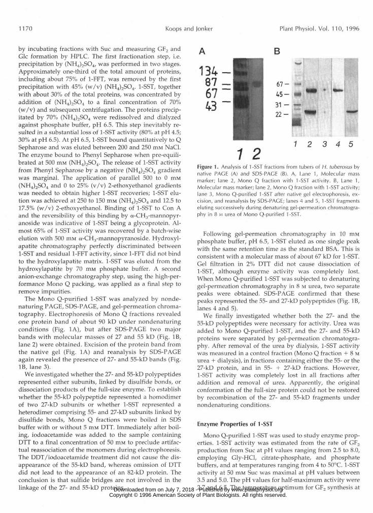
The Mono Q-purified 1-SST was analyzed by nonde-naturing PAGE, SDS-PAGE, and gel-permeation chroma-tography. Electrophoresis of Mono Q fractions revealedone protein band of about 90 kD under nondenaturingconditions (Fig. 1A), but after SDS-PAGE two majorbands with molecular masses of 27 and 55 kD (Fig. IB,lane 2) were obtained. Excision of the protein band fromthe native gel (Fig. 1A) and reanalysis by SDS-PAGEagain revealed the presence of 27- and 55-kD bands (Fig.IB, lane 3).
We investigated whether the 27- and 55-kD polypeptidesrepresented either subunits, linked by disulfide bonds, ordissociation products of the full-size enzyme. To establishwhether the 55-kD polypeptide represented a homodimerof two 27-kD subunits or whether 1-SST represented aheterodimer comprising 55- and 27-kD subunits linked bydisulfide bonds, Mono Q fractions were boiled in SDSbuffer with or without 5 mM DTT. Immediately after boil-ing, iodoacetamide was added to the sample containingDTT to a final concentration of 50 mM to preclude artifac-tual reassociation of the monomers during electrophoresis.The DDT/iodoacetamide treatment did not cause the dis-appearance of the 55-kD band, whereas omission of DTTdid not lead to the appearance of an 82-kD protein. Theconclusion is that sulfide bridges are not involved in thelinkage of the 27- and 55-kD proteins.
1 27 2 3 4 5
Figure 1. Analysis of 1-SST fractions from tubers of H. tuberosus bynative PAGE (A) and SDS-PAGE (B). A, Lane 1, Molecular massmarker; lane 2, Mono Q fraction with 1-SST activity. B, Lane 1,Molecular mass marker; lane 2, Mono Q fraction with 1 -SST activity;lane 3, Mono Q-purified 1-SST after native gel electrophoresis, ex-cision, and reanalysis by SDS-PAGE; lanes 4 and 5, 1-SST fragmentseluting successively during denaturing gel-permeation chromatogra-phy in 8 M urea of Mono Q-purified 1-SST.
Following gel-permeation chromatography in 10 mMphosphate buffer, pH 6.5, 1-SST eluted as one single peakwith the same retention time as the standard BSA. This isconsistent with a molecular mass of about 67 kD for 1-SST.Gel filtration in 2% DTT did not cause dissociation of1-SST, although enzyme activity was completely lost.When Mono Q-purified 1-SST was subjected to denaturinggel-permeation chromatography in 8 M urea, two separatepeaks were obtained. SDS-PAGE confirmed that thesepeaks represented the 55- and 27-kD polypeptides (Fig. IB,lanes 4 and 5).
We finally investigated whether both the 27- and the55-kD polypeptides were necessary for activity. Urea wasadded to Mono Q-purified 1-SST, and the 27- and 55-kDproteins were separated by gel-permeation chromatogra-phy. After removal of the urea by dialysis, 1-SST activitywas measured in a control fraction (Mono Q fraction + 8 Murea + dialysis), in fractions containing either the 55- or the27-kD protein, and in 55- + 27-kD fractions. However,1-SST activity was completely lost in all fractions afteraddition and removal of urea. Apparently, the originalconformation of the full-size protein could not be restoredby recombination of the 27- and 55-kD fragments undernondenaturing conditions.
Enzyme Properties of 1 -SST
Mono Q-purified 1-SST was used to study enzyme prop-erties. 1-SST activity was estimated from the rate of GF2production from Sue at pH values ranging from 2.5 to 8.0,employing Gly-HCl, citrate-phosphate, and phosphatebuffers, and at temperatures ranging from 4 to 50°C. 1-SSTactivity at 50 mM Sue was maximal at pH values between3.5 and 5.0. The pH values for half-maximum activity were2.7 and 6.5. The temperature optimum for GF2 synthesis at www.plantphysiol.orgon July 7, 2018 - Published by Downloaded from
Copyright © 1996 American Society of Plant Biologists. All rights reserved.

Fructan Biosynthesis in Helianthus tuberosus 1171
50 mM SUC was 20 to 25°C. 1-SST activity at 5°C was 50% of that at the temperature optimum, which is equal to a Q,, of 1.3 in the range of 20 to 5°C. The other value for half- maximum activity was 37°C.
1-SST activity at pH 6.5 was not significantly affected by 5 mM Ca2+ or Mg2'; however, 5 mM Mn2+ reduced the activity about 20 ? 4%. Enzyme activity was inhibited by 1 mM of either CuSO, (to 53 ? 4% of control rate) or AgNO, (85 2 8%). The SH-reagents N-ethylmaleimide and iodoac- etamide (each 1 mM) reduced 1-SST activity by about 10%. 1-SST activity was stimulated by 20 mM pyridoxine (120 t 3% of control rate) or pyridoxal-HC1 (147 2 5%). Stimula- tion of polymerizing fructosyltransferase activity by these compounds may be attributed to inhibition of invertase (Cairns, 1989) or to fructan exohydrolase activity (Wagner and Wiemken, 1986). However, no detectable amount of Fru was present when either pyridoxine or pyridoxal-HC1 was omitted, suggesting that the increase in the GF, syn- thesis was due to a direct stimulatory effect on synthetic activity.
The rate of GF, production by 1-SST as a function of the [SUC] is presented in Figure 2. Maximum activity was attained only at [SUC] higher than 1 M. The ability of 1-SST to metabolize Suc was studied in a long-term incubation (80 h at 25°C). To prevent microbial growth during incubation, NaN, was included in the medium. To ensure that the 1-SST fractions used in this experi- ment did not contain 1-FFT impurities, the hydroxylapa- tite chromatography step was performed twice. The 80-h incubation with 1-SST confirmed that 1-SST converted SUC into GF, efficiently (Fig. 3) , since about 90% of the Suc supplied was consumed by 1-SST. 1-SST was also
300
h C a, .- e
200 - o)
a Y 1
x
c
v
e
'5 .- 100 c o a
O O 200 400 600 800
sucrose (mM)
I
1 O00
Figure 2. Suc-concentration-dependent rate of GF, formation by 1-SST from tubers of H. tuberosus. Transfer rates are the means of duplicates 5 SD; error bars are omitted when they are smaller than the symbols.
h
4- v)
C 3
.-
2
-e cci L 4- .-
tu o) v) S O CL v) o)
v
L
n < Q
O 5 10 15 20
time (min) Figure 3. High-performance anion-exchange separations of Glc (G; 9.9 mg at 80 h after incubation), Fru (F; 6.3 mg), GF, (9.0 mg) GF, (7.2 mg), and GF, (0.9 mg) synthesized from Suc (GF; 34 mg at time O, 3.6 mg at 80 h) by 1 -SST purified from tubers of H. tuberosus, after 80 h of incubation at 25°C. Rhamnose (R) was used as an interna1 standard. The labeled oligofructans were identified by comparison with the p(2-1 )-linked fructan standards isolated and purified from H. tuberosus. PAD, Pulsed amperometry.
able to mediate the synthesis of GF, and, although to a much lesser extent, the formation of GF, (Fig. 3). Besides these oligofructans, substantial amounts of Glc and Fru were present at the end of the 80-h incubation period. Glc release was stoichiometrically related to GF, and GF, synthesis. The presence of Fru, however, indicates that the 1-SST preparation has hydrolytic activity as well. In short-term incubations with 1-SST (e.g. Fig. 2) using Suc as the only substrate, we did not find detectable amounts of Fru. This may indicate that 1-SST has no hydrolytic activity against SUC but can possibly remove fructosyl units from GF, and/or GF, only. To investigate in which reaction free Fru is released, equal amounts of 1-SST were mixed with 10 pmol of either SUC, GF,, or GF,. Samples were taken after 2, 4, and 8 h, and the sugar compositions were evaluated by HPLC (Fig. 4). Of the three substrates, Suc (Fig. 4A) was most efficiently con- sumed by 1-SST. In the first 2 h the synthesis of equimo- lar amounts of Glc and GF, was the predominant reac- tion. Fru increased only after a substantial amount of GF, had accumulated. The 1-to-1 stoichiometry between Glc and GF, was lost after 2 h, partly because of some GF, synthesis at the cost of GF, and probably also be- cause of the release of a terminal Fru from GF, (Fig. 4A). The results in Figure 48 confirm that 1-SST can use GF,
www.plantphysiol.orgon July 7, 2018 - Published by Downloaded from Copyright © 1996 American Society of Plant Biologists. All rights reserved.
1172 Koops and Jonker Plant Physiol. Vol. 11 O, 1996
C
o 4 8 0 4 8 0 4 8
time (h)
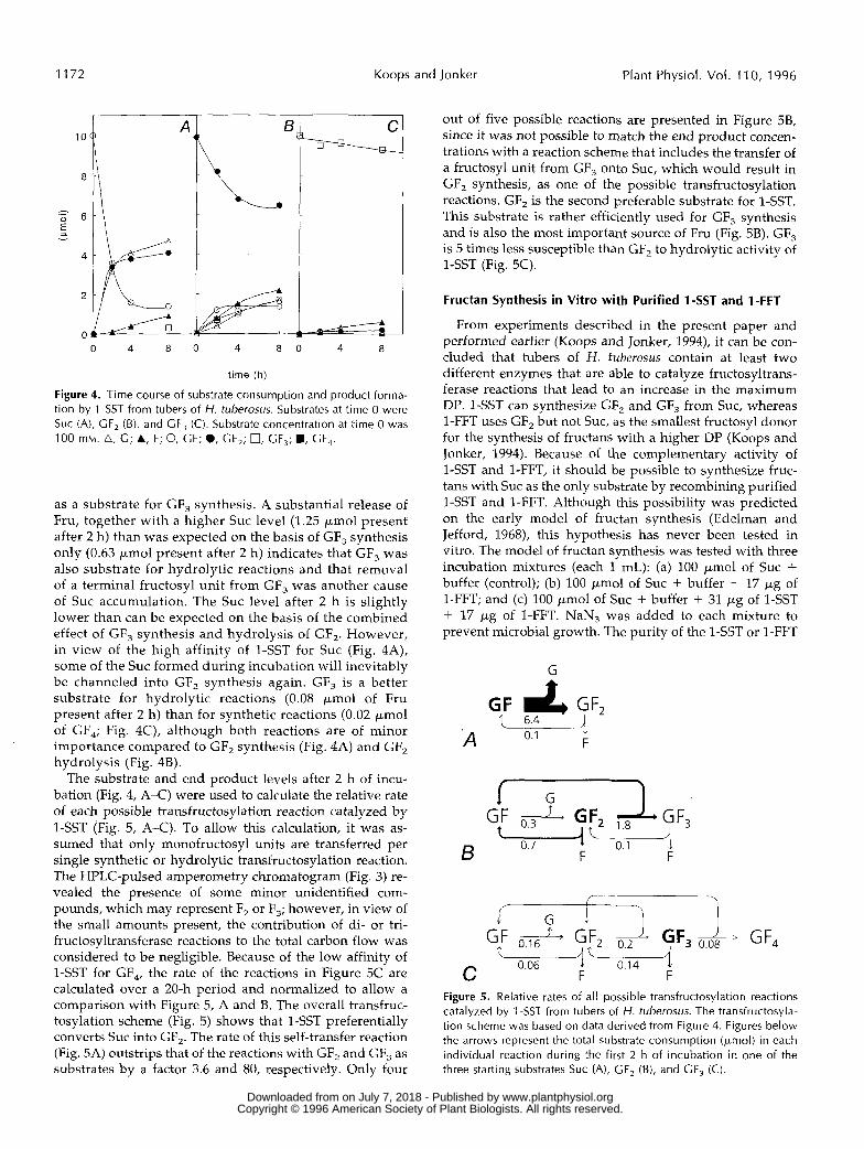
Figure 4. Time course of substrate consumption and product forma- tion by l -SST from tubers of H. tuberosus. Substrates at time O were Suc (A), GF, (B), and GF, (C). Substrate concentration at time O was 100 mM. A, G; A, F; O, GF; O, GF,; 0, GF,; ., GF,.
as a substrate for GF, synthesis. A substantial release of Fru, together with a higher Suc level (1.25 pmol present after 2 h) than was expected on the basis of GF, synthesis only (0.63 pmol present after 2 h) indicates that GF, was also substrate for hydrolytic reactions and that remova1 of a terminal fructosyl unit from GF, was another cause of Suc accumulation. The Suc level after 2 h is slightly lower than can be expected on the basis of the combined effect of GF, synthesis and hydrolysis of GF,. However, in view of the high affinity of 1-SST for Suc (Fig. 4A), some of the SUC formed during incubation will inevitably be channeled into GF, synthesis again. GF, is a better substrate for hydrolytic reactions (0.08 pmol of Fru present after 2 h) than for synthetic reactions (0.02 pmol of GF,; Fig. 4C), although both reactions are of minor importance compared to GF, synthesis (Fig. 4A) and GF, hydrolysis (Fig. 4B).
The substrate and end product levels after 2 h of incu- bation (Fig. 4, A-C) were used to calculate the relative rate of each possible transfructosylation reaction catalyzed by 1-SST (Fig. 5, A-C). To allow this calculation, it was as- sumed that only monofructosyl units are transferred per single synthetic or hydrolytic transfructosylation reaction. The HPLC-pulsed amperometry chromatogram (Fig. 3) re- vealed the presence of some minor unidentified com- pounds, which may represent F, or F,; however, in view of the small amounts present, the contribution of di- or tri- fructosyltransferase reactions to the total carbon flow was considered to be negligible. Because of the low affinity of 1-SST for GF,, the rate of the reactions in Figure 5C are calculated over a 20-h period and normalized to allow a comparison with Figure 5 , A and B. The overall transfruc- tosylation scheme (Fig. 5) shows that 1-SST preferentially converts Suc into GF,. The rate of this self-transfer reaction (Fig. 5A) outstrips that of the reactions with GF, and GF, as substrates by a factor 3.6 and 80, respectively. Only four
out of five possible reactions are presented in Figure 5B, since it was not possible to match the end product concen- trations with a reaction scheme that includes the transfer of a fructosyl unit from GF, onto SUC, which would result in GF, synthesis, as one of the possible transfructosylation reactions. GF, is the second preferable substrate for 1-SST. This substrate is rather efficiently used for GF, synthesis and is also the most important source of Fru (Fig. 5B). GF, is 5 times less susceptible than GF, to hydrolytic activity of 1-SST (Fig. 5C).
Fructan Synthesis in Vitro with Purified 1-SST anld 1-FFT
From experiments described in the present paper and performed earlier (Koops and Jonker, 1994), it can be con- cluded that tubers of H. tuberosus contain at least two different enzymes that are able to catalyze fructosyltrans- ferase reactions that lead to an increase in the maximum DP. 1-SST can synthesize GF, and GF, from SUC, whereas 1-FFT uses GF, but not SUC, as the smallest fructosyl donor for the synthesis of fructans with a higher DP (Koops and Jonker, 1994). Because of the complementary activity of 1-SST and 1-FFT, it should be possible to synthesize fruc- tans with Suc as the only substrate by recombiniing purified 1-SST and 1-FFT. Although this possibility wa:j predicted on the early model of fructan synthesis (Edelman and Jefford, 1968), this hypothesis has never been tested in vitro. The model of fructan synthesis was tested with three incubation mixtures (each 1 mL): (a) 100 pmol of Suc + buffer (control); (b) 100 pmol of Suc + buffer + 17 pg of 1-FFT; and (c) 100 pmol of Suc + buffer + 31 pg of 1-SST + 17 pg of 1-FFT. NaN, was added to each mixture to prevent microbial growth. The purity of the l-S!$T or 1-FFT
G
-. .
F 0.1 A
7
0.06 C F F Figure 5. Relative rates of all possible transfructosylation reactions catalyzed by 1 -SST from tubers of H. tuberosus. The transfructosyla- tion scheme was based on data derived from Figure 4. Figures below the arrows represent the total substrate consumption (pmol) in each individual reaction during the first 2 h of incubation in one of the three starting substrates Suc (A), GF, (B), and GF, (C).
www.plantphysiol.orgon July 7, 2018 - Published by Downloaded from Copyright © 1996 American Society of Plant Biologists. All rights reserved.
Fructan Biosynthesis in Helianthus tuberosus 1173
c: L r r
I
- u) c C =i
2
e L .- c
v
W
C O a u) e a a O GF, GF,
1 A
: x
B
I I I I I I
O 5 10 15 20 25
time (min)
fractions used for this experiment was verified by SDS- PAGE and silver staining.
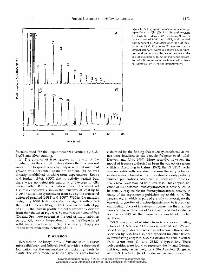
(a) The absence of free hexoses at the end of the incubation in the control mixture shows that Suc was not susceptible to spontaneous hydrolysis and that microbial growth was prevented (data not shown). (b) As was already established in short-term experiments (Koops and Jonker, 1994), 1-FFT has no activity against SUC; there were no detectable amounts of hexoses or GF, present after 80 h of incubation (data not shown). (c) Figure 6 conclusively shows that fructans, at least up to a DP of 13, can be synthesized from Suc by the concerted action of purified 1-SST and 1-FFT. Within the margins tested, the 1-SST:l-FFT ratio did not significantly affect the final DP. When 10 p g of 1-SST was mixed with 24 p g of 1-FFT, the fructan profile did not significantly deviate from that shown in Figure 6. Substantial amounts of free Glc and Fru were present at the end of the incubation period. Glc was a by-product of the 1-SST-mediated self-transfer reaction with SUC. Fru most probably re- sulted from hydrolytic activity of 1-SST.
DI SC U SSI ON
Research on the biosynthesis of fructans in H. tuberosus tubers (Edelman and Jefford, 1968) provided a theoretical foundation for the enzymology of fructan synthesis in plants. The early model of fructan synthesis was further
30 35
Figure 6. A, High-performance anion-exchange separations of Clc (C) , Fru (F), and fructans (GF,) synthesized from Suc (GF; 34 mg at time O) by a mixture of 1 -SST and 1 -FFT, both purified from tubers of H. tuberosus, after 80 h of incu- bation at 25°C. Rhamnose (R) was used as an interna1 standard. Numerals above peaks repre- sent total amount of substrate or product at the end of incubation. 6 , Anion-exchange separa- tion of a linear series of fructans (inuline) from H. tuberosus. PAD, Pulsed amperometry.
elaborated by the finding that fructosyltransferase activi- ties were localized in the vacuole (Wagner et al., 1983; Darwen and John, 1989). More recently, however, the model of fructan synthesis has been the subject of serious criticism. According to Cairns (1993), the SST/FFT model was not sufficiently sustained because the enzymological evidence was obtained with crude extracts or only partially purified preparations. Moreover, in many cases these ex- tracts were contaminated with invertase. This enzyme, be- cause of its artifactual fructosyltransferase activity, could be equally responsible for fructosyltransferase activity in many of the experiments published up to this time. The present work, which is part of a study to investigate the enzymic properties of fructosyltransferases in fructan-ac- cumulating tubers of H. tuberosus, focused on the purifica- tion and characterization of 1-SST and provided evidence for the validity of the two-enzyme model of fructan synthesis.
1-SST was purified 655-fold from fructan-accumulating tubers of H. tuberosus. SDS dissociates 1-SST into 27- and 55-kD polypeptides. The reason is unknown, although dis- sociation by SDS has also been reported for other fructo- syltransferring enzymes. SDS dissociates the acid invertase from carrot into 43- and 25-kD polypeptides. These polypeptides were found to represent the N- and C-termi- na1 fragments, respectively, of a 68-kD protein (Unger et al., 1992). The 6-SFT (69 kD under native conditions) puri-
www.plantphysiol.orgon July 7, 2018 - Published by Downloaded from Copyright © 1996 American Society of Plant Biologists. All rights reserved.

1174 Koops and Jonker Plant Physiol. Vol. 11 O, 1996
fied from barley is unstable under denaturing conditions. SDS-PAGE of 6-SFT yielded 20- and 50-kD fragments (Du- chateau et al., 1995). For 1-SST from H. tuberosus, recent data obtained by amino acid sequencing of the purified 1-SST and DNA sequencing of the corresponding cDNA from a cDNA library of H. tuberosus tubers reveals that the 27- and 55-kD proteins represent the C- and N-terminal parts, respectively, of 1-SST (with an estimated molecular mass of 75 kD for the unprocessed translation product; I.M. van der Meer and A.J. Koops, unpublished observations). These data conclusively show that the 27- and 55-kD frag- ments are derived from one protein.
The most recent study on the purification of 1-SST from Asteraceae describes a procedure to obtain the enzyme from dormant tubers of H. tuberosus in two chromato- graphic steps (Praznik et al., 1990). Gel filtration predicted a molecular mass of 69 kD for this protein, which corre- sponds well with 1-SST from fructan-accumulating tubers (gel filtration data in this report). However, the tempera- ture optimum (34"C), the pH optimum (5.4), and the activ- ity profile as a function of the [SUC] ( K , of 42 mM) of the enzyme from dormant tubers (Praznik et al., 1990) differ from those of the enzyme described in the present paper. The enzyme purified from dormant tubers may therefore represent an SST isoform that is not active or present in fructan-accumulating tubers.
As shown for many other SST-containing preparations (for an overview, see Pollock, 1986), the rate of GF, forma- tion from Suc is barely saturable (Fig. 2). Often, the rate versus concentration curves of this reaction are interpreted as first-order Michaelis-Menten equations with K , values usually higher than 100 mM. However, a full description of the kinetics of a self-transfer or any bisubstrate reaction needs four kinetic parameters. Determination of the kinetic parameters requires a set of experiments in which the substrate concentrations at the donor and acceptor site are varied independently (Mahler and Cordes, 1971). As this requirement is impossible to meet for any self-transfer reaction, the K, values reported in the literature for SST (and FFT) do not have much significance. In many studies with partially purified SST or FFT preparations, Fru release is interpreted as an indicator of contamination by invertase and is used for assessment of residual invertase activity. The present investigations, however, show that Fru release may also result from SST-mediated hydrolysis of oligofruc- tans and that synthetic and hydrolytic activity may reside on the same protein. The hydrolytic activity of SST resem- bles that of fructan exohydrolases, which hydrolyzes fruc- tans by stepwise remova1 of terminal nonreducing fructo- syl residues. In Asteraceae, fructan exohydrolases also have negligible activity against Suc (Simpson and Bonnet, 1993).
The present work shows that two enzymes are needed to synthesize fructans from Suc and that these enzymes are essentially different proteins. 1-SST and 1-FFT differ in their chromatographic, electrophoretic, and enzymic prop- erties. For example, 1-FFT is not able to catalyze the initial step of fructan synthesis, whereas 1-SST is not able to catalyze the formation of fructan polymers with a DP
higher than 5 [GF,, y1 > 41.1-SST is able to transfer fructosyl units between Suc as efficiently as 1-FFT betweeri GF,, GF,, or GF, molecules. The net number of fructosyl units trans- ferred by 1-SST during GF, formation from SUC, expressed per unit of time and protein (Fig. 2), compares to the number of fructosyl units transferred by 1-FFT during GF,, GF,, or GF, synthesis (Koops and Jonker, 1994). Although 1-SST and 1-FFT have some overlapping activity (both enzymes can catalyze the formation of GF, and GF,), GF, and GF, synthesis is much more efficiently catalyzed by 1-FFT (Koops and Jonker, 1994).
GF, formation from two molecules of Suc is the reaction most favorably catalyzed by 1-SST (Fig. 5 ) . However, some futile cycling of carbon may occur, since 1-SST can also mediate the removal of a terminal fructosyl unit from GF,, which results in one molecule of Suc and, for fructan enzymes, nonutilizable Fru (Fig. 5). 1-SST activity is poorly coordinated with that of FFT. The 1-SST-mediated GF, synthesis from Suc is favored by high [Suc] (Fig. 2), whereas the second phase of fructan synthesis, the 1-FFT- mediated conversion of GF, into fructans with a higher DE', is competitively inhibited by Suc (Koops and Jonker, 1994). The experiment described in Figure 6 shows that despite the poorly balanced enzymic properties of 1-SST and 1-FFT, it is possible to synthesize fructans frorn Suc after recombining purified 1-SST and 1-FFT. Although we were able to demonstrate the synthesis of fructans with a DP of up to 15 after 80 h of incubation, the molecular mass distribution of the fructans obtained still does not resemble the molecular mass profile in a tuber homogenate. How- ever, fructans appeared to be synthesized in a cascade-like manner. Fructosyl units are probably transferred between fructans with the same DP, as can be concluded from the finding that GF,,, is synthesized only after some GF, has accumulated (Koops and Jonker, 1994). Therefore, the DE' of fructans in a tuber homogenate might be a function of tuber age and possibly of GF, supply, which in turn, de- pends on the Suc supply at the phase of tuber filling and 1-SST activity.
On the basis of present investigations it can be concluded that the basic concept of the early model of fructan synthe- sis in H. tuberosus, one enzyme for the trisaccharide syn- thesis and one enzyme for the synthesis of the higher fructans, is still supported by experimental evidence.
ACKNOWLEDCMENTS
Dr. I.M. van der Meer, Dr. A.J. van Tunen, and Dr. C.H.R. de Vos are thanked for useful discussions and for critically reading the manuscript. Ms. M.B. van Leeuwen and Dr. T. Slaghek from Agricultural Research Department, Agricultural Research Institute (Wageningen, The Netherlands) are thanked for performing the high-pressure anion-exchange chromatography analyses.
Received September 25, 1995; accepted January 3, 1996. Copyright Clearance Center: 0032-0889 /96 / 110 / 1167/ 09.
LITERATURE ClTED
Angenent GC, Ebskamp MJM, Weisbeek PJ, Smeekens SCM (1993) Purification and properties of sucrose-sucrose fructosyl-
www.plantphysiol.orgon July 7, 2018 - Published by Downloaded from Copyright © 1996 American Society of Plant Biologists. All rights reserved.
Fructan Biosynthesis in Helianthus tuberosus 1175
transferases in barley leaves and onion seeds. In A Fuchs, ed, Inulin and Inulin Containing Crops. Elsevier, Amsterdam, pp
Cairns AJ (1989) Fructan biosynthesis in excised leaves of Lolium temulentum L. IV. Cell-free 14C labelling of specific oligofructans at low sucrose concentration. New Phytol 112: 465-473
Cairns AJ (1992) A reconsideration of fructan biosynthesis in storage roots of Asparagus officinalis L. New Phytol 120 463-473
Caims AJ (1993) Evidence for the de novo synthesis of fructan by enzymes from higher plants: a reappraisal of the SST/FFT model. New Phytol 123: 15-24
Danven CWE, John P (1989) Localization of the enzymes of fruc- tan metabolism in vacuoles isolated by a mechanical method from tubers of Jerusalem artichoke (Heliantkus tuberosus L.). Plant Physiol 89: 658-663
Duchateau N, Bortlik K, Simmen U, Wiemken A, Banca1 P (1995) Sucrose:fructan 6-fructosyltransferase, a key enzyme for divert- ing carbon from sucrose to fructan in barley leaves. Plant Physiol 107: 1249-1255
Edelman J, Jefford TG (1968) The mechanism of fructosan metab- olism in higher plants as exemplified in Heliantkus tuberosus. New Phytol 67: 517-531
Heinze B, Praznik W (1991) Separation and purification of inulin oligomers and polymers by reversed-phase high-performance liquid chromatography. Journal of Applied Polymer Science: Applied Polymer Symposium 48: 207-225
Koops AJ, Jonker HH (1994) Purification and characterization of the enzymes of fructan biosynthesis in tubers of Heliantkus tuberosus 'Colombia.' I. Fructan:fructan fructosyltransferase.
Lewis DH (1993) Nomenclature and diagrammatic representation of oligomeric fructans-a paper for discussion. New Phytol 124
Liischer M, Frehner M, Nosberger J (1993) Purification and char- acterization of fructan:fructan fructosyltransferase from Jerusa- lem artichoke (Heliantkus tuberosus L.) New Phytol 123: 717-724
Mahler HR, Cordes EH (1971) Biological Chemistry. Harper & Row, London
173-184
J EXP Bot 45: 1623-1631
583-594
Pollock CJ (1986) Fructans and the metabolism of sucrose in vascular plants. New Phytol 104: 1-24
Praznik W, Beck RHF, Spies T (1990) Isolation and character- ization of sucr0se:sucrose lF-P-D-fructosyltransferase from tubers of Helianthus tuberosus L. Agric Biol Chem 54: 2429- 2431
Shiomi N, Izawa M (1980). Purification and characterization of sucrose:sucrose 1-fructosyltransferase from roots of asparagus (Asparagus officinalis L.). Agric Biol Chem 44: 603-614
Shiomi N, Kido H, Kiriyama S (1985) Purification and properties of sucr0se:sucrose IF-P-D-fructosyltransferase in onion seeds. Phytochemistry 24: 695-698
Simmen U, Obenland D, Boller T, Wiemken A (1993) Fructan synthesis in excised barley leaves. Plant Physiol 101: 459-468
Simpson R, Bonnet GD (1993) Fructan exohydrolase from grasses. New Phytol 123: 453469
Timmermans JW, Van Leeuwen MB, Tournois H, De Wit D, Vliegenthart JFG (1994) Quantitative analysis of the molecular weight distribution of inulin by means of anion exchange HPLC with pulsed amperometric detection. J Carbohydr Chem 13:
Unger C, Hofsteenge J, Sturm A (1992) Purification and charac- terization of a soluble P-fructofuranosidase from Daucus carota. Eur J Biochem 204: 915-921
Van den Ende W, Van Laere A (1993) Purification and properties of an invertase with sucrose:sucrose fructosyltransferase (SST) activity from the roots of Cickorium intybus L. New Phytol 123:
Venuat B, Goupil P, Ledoigt G (1993) Molecular cloning and physiological analysis of an invertase isozyme in Heliantkus tissues. Biochemistry and Molecular Biology International 5
Wagner W, Keller F, Wiemken (1983) Fructan metabolism in cereals: induction in leaves and compartmentation in proto- plasts and vacuoles. 2 Pflanzenphysiol 112: 359-372
Wagner W, Wiemken A (1986) Properties and subcellular local- ization of fructan exohydrolase in leaves of barley (Hordeum vulgare cv Gerbel). J Plant Physiol 123: 429-439
881-888
31-37
955-966
www.plantphysiol.orgon July 7, 2018 - Published by Downloaded from Copyright © 1996 American Society of Plant Biologists. All rights reserved.


![A Dimensions: [mm] B Recommended land pattern: [mm] D ... · 2005-12-16 DATE SSt SSt SSt SSt SSt SSt SSt BY SSt SSt SMu SMu SSt ... RDC Value 600 800 1000 0.20 High Cur rent ... 350](https://img.dokumen.tips/doc/110x75/5c61318009d3f21c6d8cb002/a-dimensions-mm-b-recommended-land-pattern-mm-d-2005-12-16-date-sst.jpg)


![A Dimensions: [mm] B Recommended land pattern: [mm] D ... · 2013-03-12 2013-01-13 2012-12-10 2012-10-29 2012-08-27 2006-05-05 DATE SSt SSt SSt SSt SSt SSt SSt BY SSt COt COt SSt](https://img.dokumen.tips/doc/110x75/604b228bc93c005c75431c51/a-dimensions-mm-b-recommended-land-pattern-mm-d-2013-03-12-2013-01-13.jpg)

![A Dimensions: [mm] B Recommended land pattern: [mm] D ...2012-12-06 2012-10-24 2012-08-08 2012-06-28 2012-03-12 DATE SSt SSt SSt SSt SSt SSt BY SSt SSt BD BD SSt DDe CHECKED Würth](https://img.dokumen.tips/doc/110x75/60f984e176666848374d15c0/a-dimensions-mm-b-recommended-land-pattern-mm-d-2012-12-06-2012-10-24.jpg)


![A Dimensions: [mm] B Recommended land pattern: [mm] · 2020. 8. 11. · 2014-03-11 2013-12-19 2013-12-04 2013-04-10 2013-03-06 2013-02-14 2012-12-10 DATE SSt SSt SSt SSt SSt SSt SSt](https://img.dokumen.tips/doc/110x75/6145e75a8f9ff812541fec6f/a-dimensions-mm-b-recommended-land-pattern-mm-2020-8-11-2014-03-11-2013-12-19.jpg)