Embed Size (px)
Citation preview
2521
Pure Appl. Chem., Vol. 80, No. 11, pp. 2521–2536, 2008.doi:10.1351/pac200880112521© 2008 IUPAC
Synthesis and electrochemical studies onsurface-modified LiCoVO4 with La2O3 andmalonic acid for cathode material of Li-ioncells*
George Ting-Kuo Fey‡, Yung-Da Cho, andPandurangan Muralidharan
Department of Chemical and Materials Engineering, National Central University,Chung-Li, Taiwan 32054 R.O.C.
Abstract: An inverse spinel LiCoVO4 cathode material was synthesized by a citric acid-ureapolymeric method, calcined at 773 K for 5 h. The synthesized LiCoVO4 sample was surface-modified with La2O3 and/or malonic acid. The composite materials were comprehensivelycharacterized with the aid of various spectroscopic and analytical techniques. X-ray diffrac-tion (XRD) patterns revealed that single-phase crystallinity occurred when they were heatedat 773 K for 5 h in air. For the La2O3-coated samples, there was no evident signal corre-sponding to secondary-phase peaks. Fourier transform infrared (FTIR) spectra showed thecomplete elimination of organic residues, nitrates, and the formation of pure LiCoVO4 at773 K. The two strong peaks due to sp2 and sp3 carbon bonding in the Raman spectra clearlyconfirm the presence of carbon coating at the surface of LiCoVO4 particles. Transmissionelectron microscopy (TEM) images showed that the 0.5 wt % La2O3-coated LiCoVO4 mate-rial calcined with 60 wt % malonic acid was compact with an average thickness of about15 nm. This composite cathode material also demonstrated the best cell performance with aninitial capacity of 71 mAhg–1 and better thermal stability with lower heat evolution of35 Jg–1, and a higher onset temperature of thermal decomposition at 475 K, vs. 176 Jg–1 and452 K for the bare LiCoVO4 sample.
Keywords: citric acid-urea; La2O3; LiCoVO4; Li-ion cells; surface modification.
INTRODUCTION
Since the discovery of both LiNiVO4 and LiCoVO4 as new cathode materials for secondary Li batter-ies in 1993 [1], interest in inverse spinel materials has arisen due to their high-voltage behavior. A fam-ily of inverse spinel vanadate cathode materials such as LiCoVO4 (4.3 V), LiNiyCo1–yVO4 (y = 0.1–0.9)and LiNiVO4 (4.8 V) were investigated by Fey et al. [2–11]. In this structure, Li+ ions reside in octa-hedral sites and Co2+ and Ni2+ ions and V5+ ions were distributed both in octahedrally and tetrahedrallycoordinated sites.
Many attempts have been made to enhance the electrochemical properties of LiNiVO4 andLiCoVO4 materials through different synthesis techniques, such as the glycine-nitrate combustion
*Paper based on a presentation at the 3rd International Symposium on Novel Materials and Their Synthesis (NMS-III) and the17th International Symposium on Fine Chemistry and Functional Polymers (FCFP-XVII), 17–21 October 2007, Shanghai, China.Other presentations are published in this issue, pp. 2231–2563.‡Corresponding author: Tel./Fax: +886-3-425-7325; E-mail: [email protected]
process [12], citric complex method [13], and starch-assisted combustion process [14]. The particlesizes of powders synthesized by these methods are normally in the micrometer range. Vivekanandhanet al. [15] synthesized crystalline 39-nm LiNiVO4 by using a 1:1 ratio of metal ions to glycerol.Thongtem et al. [16] also successfully prepared nanocrystalline 31-nm LiCoVO4 by a solvothermal re-action at 423 K and subsequent calcination at 573 K. Landschoot et al. [17] synthesized Fe-dopedLiCoVO4 with Al2O3 coating by the citric complex method, and showed that the first discharge capac-ity was 76 mAhg–1 with good capacity retention due to the ability of the Al2O3 layer to neutralize theHF component in the liquid electrolyte. Recently, Fey et al. [18] synthesized Al2O3-coated LiCoVO4by a polymeric process, and reported that the 0.5 wt % Al2O3-coated LiCoVO4 delivered an initial dis-charge capacity of 68 mAh g–1. The Al2O3 coating technique was also applied to another inverse spinelmaterials of LiNiVO4 [19].
The major limitations associated with the inverse spinel vanadates were low discharge capacitieswith respect to their theoretical capacity (148 mAhg–1), initial drastic drops in capacity and large ca-pacity fades during continuous cycling [1,3,7,9,11]. These problems may be related to various factors,such as low electronic conductivity [6], cobalt dissolution into the HF-containing electrolyte in the caseof cobalt-based cathodes [20,21], and a disproportionate reaction at the surface of the cathode materi-als resulting in structural changes of the host material [22]. The very high cell voltage of 4.8 V obtainedusing LiNiVO4 as a cathode has not been advantageous, because most electrolytes can be oxidized bythe cathode material at voltages higher than 4.5 V [11,23], leading to a significant deterioration in cycleperformance [24,25]. However, the LiCoVO4 cathode exhibits 4.2 V, which is more suitable for someoxidation-resistant electrolytes. Furthermore, the weight percentages of cobalt in LiCoVO4 and LiCoO2are 32.6 and 60.2, respectively. The lower weight percentage of cobalt in LiCoVO4 does not have animmediately obvious cost advantage over LiCoO2, since there are differences in capacity. In the case ofLiCoO2, it is 140 mAhg–1, and in the case of LiCoVO4, it is 71 mAhg–1
. The cost advantage ofLiCoVO4 only becomes significant when its capacity is greatly increased.
Recently, LiFePO4 has emerged as a new high-power cathode material for large-size Li-ion bat-teries in applications related to power tools, electric vehicles (EVs), and hybrid electric vehicles(HEVs). This rapid advancement in high rate capability was achieved by significantly improving elec-tronic conductivity and Li+ diffusion kinetics through the use of non-lattice doping with carbon [26–30]and lattice doping with supervalent elements [31–35]. The bare LiFePO4 sample without carbon coat-ing or metal doping shows very low initial capacity and poor electronic conductivity. Both pristineLiNiVO4 and LiCoVO4 samples display similar behavior of low capacity and conductivity. Therefore,we attempt to adopt a similar LiFePO4 approach to improve the LiCoVO4 cathode system.
To obtain higher capacities and decrease the capacity fades, in this work, we prepared the pristineLiCoVO4 powder and La2O3-coated LiCoVO4 materials by a citric acid-urea polymeric method, andLa2O3-coated LiCoVO4/carbon composite cathode materials by a solid-state high-temperature method.The prepared samples were characterized by various techniques and their cell performance and coatingeffect was evaluated in terms of discharge capacity, cycle life, and thermal stability.
EXPERIMENTAL
Synthesis of LiCoVO4
According to the stoichiometric composition, proper amounts of LiNO3, Co(NO3)2�6H2O (Merck,>99 %), and NH4VO3 (Acros Organics, >98 %) to obtain LiCoVO4 were dissolved in deionized waterunder constant magnetic stirring at 353 K. To the above solution, 0.06 mole of citric acid in 20 ml ofdeionized water was added in dropwise, followed by 0.1 mole urea with continuous stirring. In theprocess, citric acid was employed as a chelating agent, and it facilitated the mixing of cations at the mo-lecular level. The addition of urea to citric acid played a vital role in the formation of tridentated com-
G. T.-K. FEY et al.
© 2008 IUPAC, Pure and Applied Chemistry 80, 2521–2536
2522
plexes between the metallic cations and citric acid that facilitated the formation of homogeneous, nano-sized particles. The resultant transparent dark bluish solution was heated gently at 363 K with continu-ous stirring for 6 h to remove the excess water. The solution turned into a transparent dark bluish soland was allowed to form a polymeric gel, which was heated at 393 K in an oven for 24 h. The driedpolymeric precursor was heated at a ramping rate of 4 K min–1 and maintained at 423 K for 3 h and573 K for 3 h, with intermediate grinding. A dark brown LiCoVO4 powder (product A) was obtainedafter further heating at 773 K for 5 h in air.
Synthesis of La2O3-coated LiCoVO4
A polymeric process was employed to prepare the 0.1, 0.5, and 1.0 wt % of La2O3-coated LiCoVO4cathode particles. Chemicals used were LiCoVO4 powders prepared by the polymeric method in theprevious section 2.1, La(NO3)3�6H2O (Aldrich) and polyvinyl alcohol (PVA, degree of polymerizationis 1500). Freshly prepared LiCoVO4 (product A) was dispersed in deionized water by a 1 h sonication,followed by 3 h stirring. The calculated 0.1, 0.5, and 1.0 wt % of La(NO3)3�6H2O to form La2O3 andPVA were dissolved in warm deionized water and added dropwise to the dispersed LiCoVO4 solutionafter sonication for 0.5 h. The mixture was stirred for 6 h at 298 K and heated at 363 K with continu-ous stirring. After removal of excess water for 7–8 h, a thick polymeric gel was formed. The gel wasfurther dried in an oven at 393 K for 12 h to form a dry brown powder, which was heated at 873 K for2 h in air with a heating ramp rate of 4 K min–1 to form a thin layer of La2O3 coating on the LiCoVO4cathode particles (product B) with the weight ratios of 99.9:0.1, 99.5:0.5, and 99.0:1.0, respectively.
Synthesis of La2O3-coated LiCoVO4/C composite materials
The La2O3-coated LiCoVO4 product (product B) was mixed with malonic acid in acetone, and ball-milled under argon for 30 min. The mixture was then calcined at 873 K for 10 h under a purified at-mosphere (5 % H2 in Ar) and a black powder (product C) was obtained for La2O3-coated LiCoVO4/Ccomposite material.
Product characterization
A powder X-ray diffractometer (PXRD), Siemens D-5000, Mac Science MXP18, equipped with aNi-filtered Cu-Kα radiation source (λ = 1.5405 Å) was used to study the structure and phase purity. Thediffraction patterns were recorded between scattering angles of 15° and 80° in steps of 0.05°. Fouriertransform infrared (FTIR) spectra were recorded at 298 K on powdered samples using the KBr wafertechnique in a Jasco-410 FTIR instrument. The spectra were recorded with a resolution of 2 cm–1 intransmittance mode from 400 to 4000 cm–1, corrected for background. The thermal analysis(TG/DTGA) measurements were carried out on a Perkin-Elmer TGA-7 series thermal analysis systemin air at a heating rate of 20 K min–1 from 298 K. The surface morphological studies were carried outon a Hitachi model S-3500V scanning electron microscope (SEM). The microstructures of the particleswere examined by a JEOL JEM-200FXII transmission electron microscope (TEM) equipped with aLaB6 gun. The sample for TEM study was prepared by dispersing the cathode powder in ethanol, plac-ing a drop of the clear solution on a carbon-coated copper grid, and subsequent drying. X-ray photonspectroscopy (XPS) and the depth profiles of lanthanum, cobalt, vanadium, and oxygen were recordedby an electron spectroscopy for chemical analysis (ESCA) instrument (VG Scientific ESCALAB 250)with monochromatic Al Kα radiation 1486.6 eV. The survey spectra were scanned in the range0.00–1400.00 eV binding energy (BE) in 1.00 eV steps.
Carbon content analyses of the products were investigated on an OIA Model 1010 TOC Analyzerwith an oxygen gas rate at 200 ml/min and a nitrogen gas rate at 350 ml/min. Raman spectroscopy was
© 2008 IUPAC, Pure and Applied Chemistry 80, 2521–2536
Cathode material of Li-ion cells 2523
performed on samples of powders using an ISA T64000 double beam pass spectrometer equipped witha microscope stage for analyzing small samples utilizing 180° incident geometry. A Spectra Physicsargon-ion laser was employed to excite laser Raman spectra using a 515-nm laser line at an incidentpower of ca. 10 mW along with a water-cooled photomultiplier tube. The scanning rate used to collectthe spectra was kept at 10 cm–1 min–1. The electronic conductivity of samples was measured by four-point conductivity measurements of Keithley Model 2400S source meter.
The cathodes for electrochemical studies were prepared by a doctor-blade coating method with aslurry of 85 wt % coated active material, 10 wt % conductive carbon black, and 5 wt % poly(vinylidenefluoride) as a binder, in N-methyl-2-pyrrolidone (NMP), as the solvent for the mixture, which was thenapplied onto a surface-cleaned aluminum foil current collector and dried at 393 K for 12 h in an oven.The coated cathode foil was then smoothed by pressing it through stainless-steel twin rollers and cutinto circular disks 13 mm in diameter.
The electrochemical performance of the above discs was carried out with coin-type cells of the2032 configuration and were assembled in an argon-filled VAC MO40-1 glove box in which the oxy-gen and water contents were maintained below 2 ppm. The above-prepared circular disk was used asthe cathode, Li metal (Foote Mineral) as the anode and a 1 M LiPF6 in 1:1 by volume ethylene car-bonate (EC)/diethyl carbonate (DEC) (Tomiyama Chemicals) as the electrolyte with a Celgard mem-brane as the separator. The cells charge–discharge cycles were performed at a 0.1 C-rate between3.0~4.5 V in a multichannel battery tester (Maccor 4000) at 298 K.
RESULTS AND DISCUSSION
X-ray diffraction
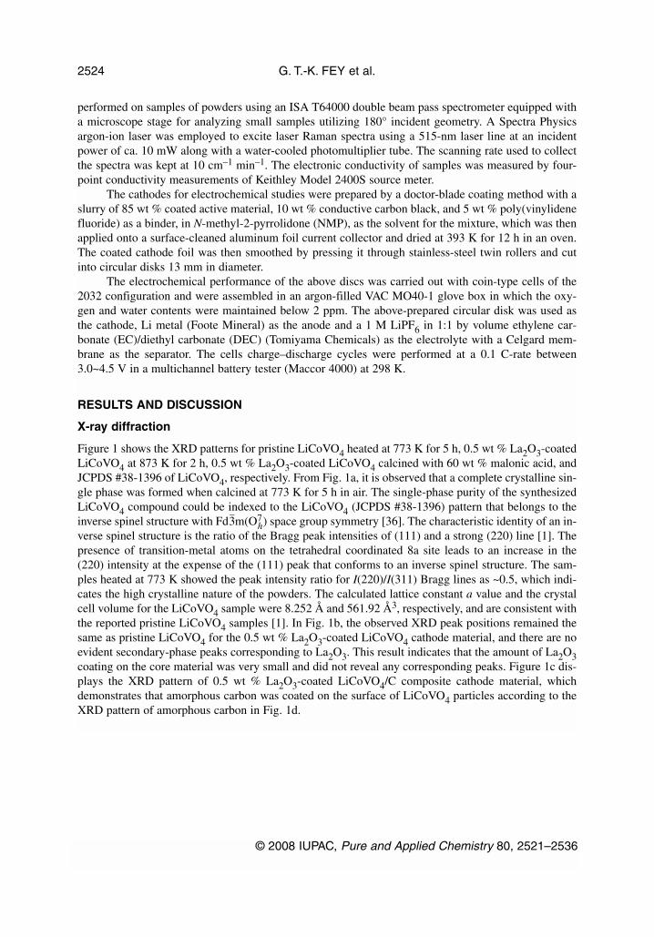
Figure 1 shows the XRD patterns for pristine LiCoVO4 heated at 773 K for 5 h, 0.5 wt % La2O3-coatedLiCoVO4 at 873 K for 2 h, 0.5 wt % La2O3-coated LiCoVO4 calcined with 60 wt % malonic acid, andJCPDS #38-1396 of LiCoVO4, respectively. From Fig. 1a, it is observed that a complete crystalline sin-gle phase was formed when calcined at 773 K for 5 h in air. The single-phase purity of the synthesizedLiCoVO4 compound could be indexed to the LiCoVO4 (JCPDS #38-1396) pattern that belongs to theinverse spinel structure with Fd3
–m(O7
h) space group symmetry [36]. The characteristic identity of an in-verse spinel structure is the ratio of the Bragg peak intensities of (111) and a strong (220) line [1]. Thepresence of transition-metal atoms on the tetrahedral coordinated 8a site leads to an increase in the(220) intensity at the expense of the (111) peak that conforms to an inverse spinel structure. The sam-ples heated at 773 K showed the peak intensity ratio for I(220)/I(311) Bragg lines as ~0.5, which indi-cates the high crystalline nature of the powders. The calculated lattice constant a value and the crystalcell volume for the LiCoVO4 sample were 8.252 Å and 561.92 Å3, respectively, and are consistent withthe reported pristine LiCoVO4 samples [1]. In Fig. 1b, the observed XRD peak positions remained thesame as pristine LiCoVO4 for the 0.5 wt % La2O3-coated LiCoVO4 cathode material, and there are noevident secondary-phase peaks corresponding to La2O3. This result indicates that the amount of La2O3coating on the core material was very small and did not reveal any corresponding peaks. Figure 1c dis-plays the XRD pattern of 0.5 wt % La2O3-coated LiCoVO4/C composite cathode material, whichdemonstrates that amorphous carbon was coated on the surface of LiCoVO4 particles according to theXRD pattern of amorphous carbon in Fig. 1d.
G. T.-K. FEY et al.
© 2008 IUPAC, Pure and Applied Chemistry 80, 2521–2536
2524

TG/DTA analysis
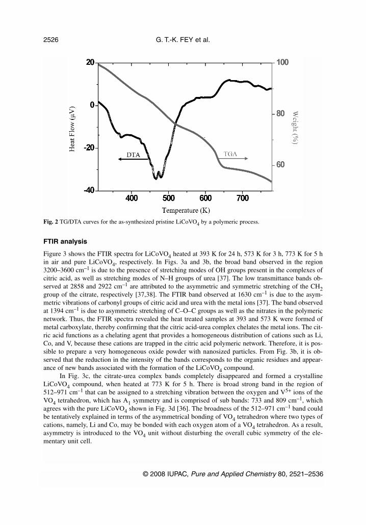
Figure 2 shows the TGA and DTA data curves for the LiCoVO4 polymeric precursor. The TGA curveshows a step-wise weight loss in the temperature ranges 313–423, 423–516, 516–646, and 646–750 K.The initial weight loss of 8 % may correspond to the loss of water and an excess of free citric acid andother organic residues. In the temperature range 423–516 K, the decomposition of the complex intointermediate and the formation of crystalline LiCoVO4 compound, corresponds to the second stage ofweight loss and is evidenced by the larger DTA curves. From 516–646 and 646–750 K, complete de-composition of the intermediate occurs and phase-pure LiCoVO4 is formed. The major combustionprocess was initiated at 423–623 K, and the weight loss slowed down after 750 K. These results weresupported by XRD and FTIR results, where the complete formation of crystalline LiCoVO4 at 773 Kwas confirmed, which agree with the results obtained from the TGA/DTA.
© 2008 IUPAC, Pure and Applied Chemistry 80, 2521–2536
Cathode material of Li-ion cells 2525
Fig. 1 XRD patterns for (a) pristine LiCoVO4; (b) 0.5 wt % La2O3-coated LiCoVO4; (c) 0.5 wt % La2O3-coatedLiCoVO4 calcined with 60 wt % malonic acid; (d) amorphous carbon; and (e) JCPDS (# 38-1396) LiCoVO4.
FTIR analysis
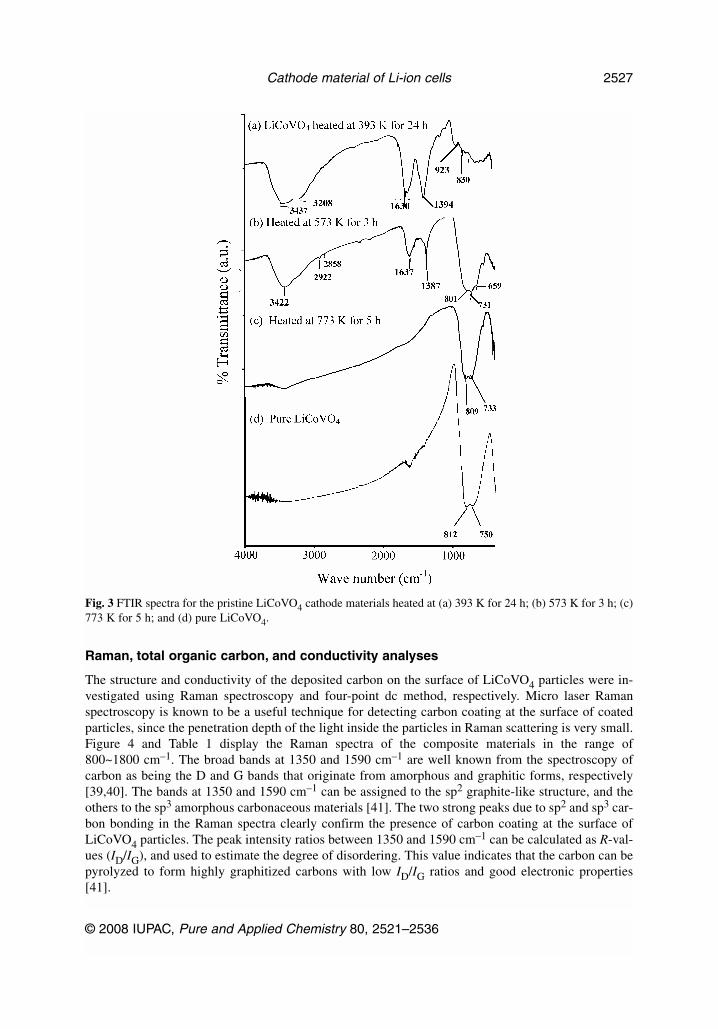
Figure 3 shows the FTIR spectra for LiCoVO4 heated at 393 K for 24 h, 573 K for 3 h, 773 K for 5 hin air and pure LiCoVO4, respectively. In Figs. 3a and 3b, the broad band observed in the region3200–3600 cm–1 is due to the presence of stretching modes of OH groups present in the complexes ofcitric acid, as well as stretching modes of N–H groups of urea [37]. The low transmittance bands ob-served at 2858 and 2922 cm–1 are attributed to the asymmetric and symmetric stretching of the CH2group of the citrate, respectively [37,38]. The FTIR band observed at 1630 cm–1 is due to the asym-metric vibrations of carbonyl groups of citric acid and urea with the metal ions [37]. The band observedat 1394 cm–1 is due to asymmetric stretching of C–O–C groups as well as the nitrates in the polymericnetwork. Thus, the FTIR spectra revealed the heat treated samples at 393 and 573 K were formed ofmetal carboxylate, thereby confirming that the citric acid-urea complex chelates the metal ions. The cit-ric acid functions as a chelating agent that provides a homogeneous distribution of cations such as Li,Co, and V, because these cations are trapped in the citric acid polymeric network. Therefore, it is pos-sible to prepare a very homogeneous oxide powder with nanosized particles. From Fig. 3b, it is ob-served that the reduction in the intensity of the bands corresponds to the organic residues and appear-ance of new bands associated with the formation of the LiCoVO4 compound.
In Fig. 3c, the citrate-urea complex bands completely disappeared and formed a crystallineLiCoVO4 compound, when heated at 773 K for 5 h. There is broad strong band in the region of512–971 cm–1 that can be assigned to a stretching vibration between the oxygen and V5+ ions of theVO4 tetrahedron, which has A1 symmetry and is comprised of sub bands: 733 and 809 cm–1, whichagrees with the pure LiCoVO4 shown in Fig. 3d [36]. The broadness of the 512–971 cm–1 band couldbe tentatively explained in terms of the asymmetrical bonding of VO4 tetrahedron where two types ofcations, namely, Li and Co, may be bonded with each oxygen atom of a VO4 tetrahedron. As a result,asymmetry is introduced to the VO4 unit without disturbing the overall cubic symmetry of the ele-mentary unit cell.
G. T.-K. FEY et al.
© 2008 IUPAC, Pure and Applied Chemistry 80, 2521–2536
2526
Fig. 2 TG/DTA curves for the as-synthesized pristine LiCoVO4 by a polymeric process.
Raman, total organic carbon, and conductivity analyses
The structure and conductivity of the deposited carbon on the surface of LiCoVO4 particles were in-vestigated using Raman spectroscopy and four-point dc method, respectively. Micro laser Ramanspectroscopy is known to be a useful technique for detecting carbon coating at the surface of coatedparticles, since the penetration depth of the light inside the particles in Raman scattering is very small.Figure 4 and Table 1 display the Raman spectra of the composite materials in the range of800~1800 cm–1. The broad bands at 1350 and 1590 cm–1 are well known from the spectroscopy ofcarbon as being the D and G bands that originate from amorphous and graphitic forms, respectively[39,40]. The bands at 1350 and 1590 cm–1 can be assigned to the sp2 graphite-like structure, and theothers to the sp3 amorphous carbonaceous materials [41]. The two strong peaks due to sp2 and sp3 car-bon bonding in the Raman spectra clearly confirm the presence of carbon coating at the surface ofLiCoVO4 particles. The peak intensity ratios between 1350 and 1590 cm–1 can be calculated as R-val-ues (ID/IG), and used to estimate the degree of disordering. This value indicates that the carbon can bepyrolyzed to form highly graphitized carbons with low ID/IG ratios and good electronic properties[41].
© 2008 IUPAC, Pure and Applied Chemistry 80, 2521–2536
Cathode material of Li-ion cells 2527
Fig. 3 FTIR spectra for the pristine LiCoVO4 cathode materials heated at (a) 393 K for 24 h; (b) 573 K for 3 h; (c)773 K for 5 h; and (d) pure LiCoVO4.
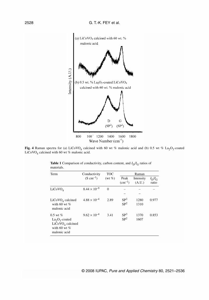
Table 1 Comparison of conductivity, carbon content, and ID/IG ratios ofmaterials.
Term Conductivity TOC Raman(S cm–1) (wt %) Peak Intensity ID/IG
(cm–1) (A.U.) ratio
LiCoVO4 8.44 × 10⁻⁹ 0 – – –– –
LiCoVO4 calcined 4.88 × 10⁻⁴ 2.89 SP3 1280 0.977with 60 wt % SP2 1310malonic acid
0.5 wt % 9.62 × 10⁻⁴ 3.41 SP3 1370 0.853La2O3-coated SP2 1607LiCoVO4 calcinedwith 60 wt %malonic acid
G. T.-K. FEY et al.
© 2008 IUPAC, Pure and Applied Chemistry 80, 2521–2536
2528
Fig. 4 Raman spectra for (a) LiCoVO4 calcined with 60 wt % malonic acid and (b) 0.5 wt % La2O3-coatedLiCoVO4 calcined with 60 wt % malonic acid.
The carbon content was determined by total organic carbon (TOC) analysis. The carbon coatingtreatment of La2O3-coated LiCoVO4 particles with malonic acid greatly improved the electronic con-ductivity of LiCoVO4 by a factor of five, as shown in Table 1. Conductivity increased along with thecarbon content of composite samples that was between 2.8 and 3.5 wt %. The individual LiCoVO4 crys-tals were wired together by the coated carbon layer, so the carbon layer provides a conductivity networkand might increase the conductivity of the materials, which would be beneficial to electrochemicalproperties.
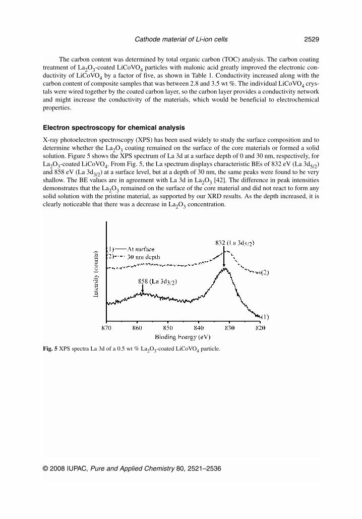
Electron spectroscopy for chemical analysis
X-ray photoelectron spectroscopy (XPS) has been used widely to study the surface composition and todetermine whether the La2O3 coating remained on the surface of the core materials or formed a solidsolution. Figure 5 shows the XPS spectrum of La 3d at a surface depth of 0 and 30 nm, respectively, forLa2O3-coated LiCoVO4. From Fig. 5, the La spectrum displays characteristic BEs of 832 eV (La 3d5/2)and 858 eV (La 3d3/2) at a surface level, but at a depth of 30 nm, the same peaks were found to be veryshallow. The BE values are in agreement with La 3d in La2O3 [42]. The difference in peak intensitiesdemonstrates that the La2O3 remained on the surface of the core material and did not react to form anysolid solution with the pristine material, as supported by our XRD results. As the depth increased, it isclearly noticeable that there was a decrease in La2O3 concentration.
© 2008 IUPAC, Pure and Applied Chemistry 80, 2521–2536
Cathode material of Li-ion cells 2529
Fig. 5 XPS spectra La 3d of a 0.5 wt % La2O3-coated LiCoVO4 particle.
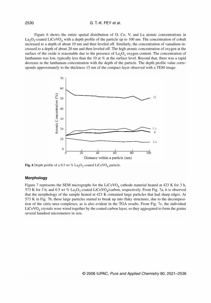
Figure 6 shows the entire spatial distribution of O, Co, V, and La atomic concentrations inLa2O3-coated LiCoVO4 with a depth profile of the particle up to 100 nm. The concentration of cobaltincreased to a depth of about 10 nm and then leveled off. Similarly, the concentration of vanadium in-creased to a depth of about 20 nm and then leveled off. The high atomic concentration of oxygen at thesurface of the oxide is reasonable due to the presence of La2O3 oxygen content. The concentration oflanthanum was low, typically less than the 10 at % at the surface level. Beyond that, there was a rapiddecrease in the lanthanum concentration with the depth of the particle. The depth profile value corre-sponds approximately to the thickness 15 nm of the compact layer observed with a TEM image.
Morphology
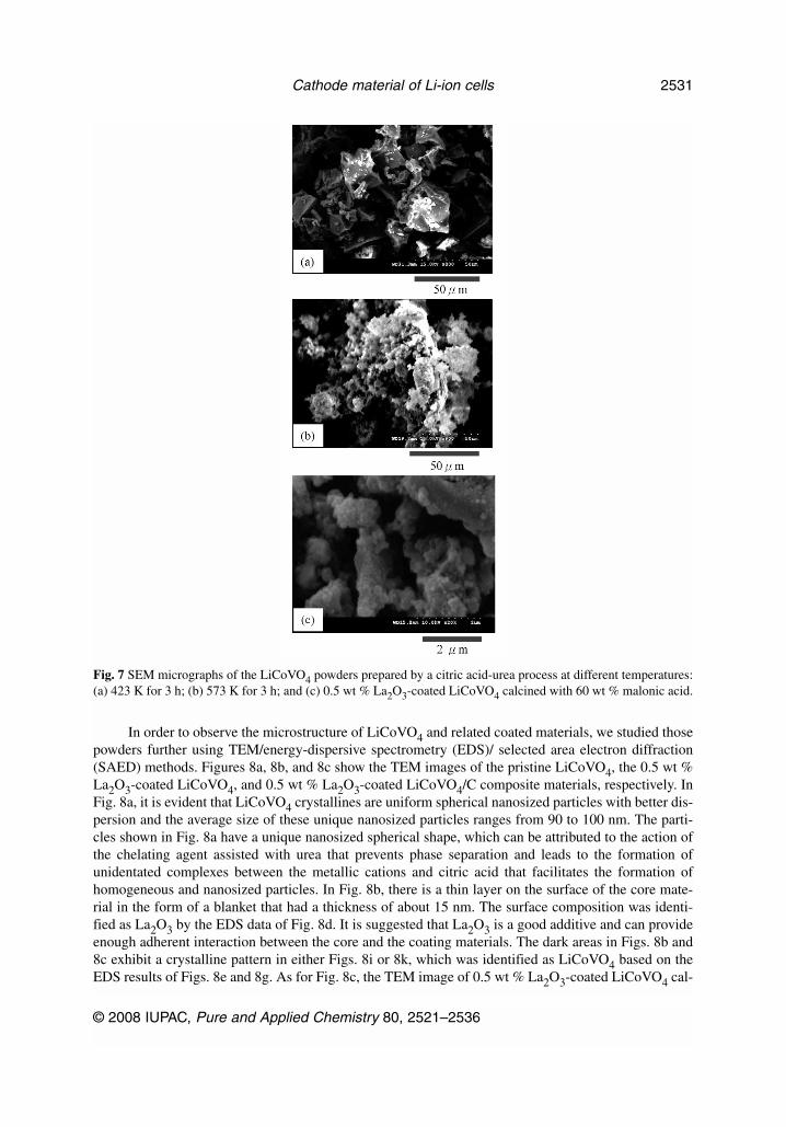
Figure 7 represents the SEM micrographs for the LiCoVO4 cathode material heated at 423 K for 3 h,573 K for 3 h, and 0.5 wt % La2O3-coated LiCoVO4/carbon, respectively. From Fig. 7a, it is observedthat the morphology of the sample heated at 423 K contained large particles that had sharp edges. At573 K in Fig. 7b, these large particles started to break up into flaky structures, due to the decomposi-tion of the citric-urea complexes, as is also evident in the TGA results. From Fig. 7c, the individualLiCoVO4 crystals were wired together by the coated carbon layer, so they aggregated to form the grainsseveral hundred micrometers in size.
G. T.-K. FEY et al.
© 2008 IUPAC, Pure and Applied Chemistry 80, 2521–2536
2530
Fig. 6 Depth profile of a 0.5 wt % La2O3-coated LiCoVO4 particle.
In order to observe the microstructure of LiCoVO4 and related coated materials, we studied thosepowders further using TEM/energy-dispersive spectrometry (EDS)/ selected area electron diffraction(SAED) methods. Figures 8a, 8b, and 8c show the TEM images of the pristine LiCoVO4, the 0.5 wt %La2O3-coated LiCoVO4, and 0.5 wt % La2O3-coated LiCoVO4/C composite materials, respectively. InFig. 8a, it is evident that LiCoVO4 crystallines are uniform spherical nanosized particles with better dis-persion and the average size of these unique nanosized particles ranges from 90 to 100 nm. The parti-cles shown in Fig. 8a have a unique nanosized spherical shape, which can be attributed to the action ofthe chelating agent assisted with urea that prevents phase separation and leads to the formation ofunidentated complexes between the metallic cations and citric acid that facilitates the formation ofhomogeneous and nanosized particles. In Fig. 8b, there is a thin layer on the surface of the core mate-rial in the form of a blanket that had a thickness of about 15 nm. The surface composition was identi-fied as La2O3 by the EDS data of Fig. 8d. It is suggested that La2O3 is a good additive and can provideenough adherent interaction between the core and the coating materials. The dark areas in Figs. 8b and8c exhibit a crystalline pattern in either Figs. 8i or 8k, which was identified as LiCoVO4 based on theEDS results of Figs. 8e and 8g. As for Fig. 8c, the TEM image of 0.5 wt % La2O3-coated LiCoVO4 cal-
© 2008 IUPAC, Pure and Applied Chemistry 80, 2521–2536
Cathode material of Li-ion cells 2531
Fig. 7 SEM micrographs of the LiCoVO4 powders prepared by a citric acid-urea process at different temperatures:(a) 423 K for 3 h; (b) 573 K for 3 h; and (c) 0.5 wt % La2O3-coated LiCoVO4 calcined with 60 wt % malonic acid.
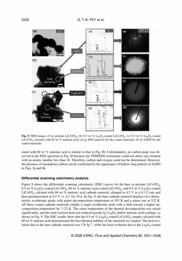
cined with 60 wt % malonic acid is similar to that in Fig. 8b. Unfortunately, no carbon peak was ob-served in the EDS spectrum in Fig. 8f because our TEM/EDS instrument could not detect any elementwith an atomic number less than 16. Therefore, carbon and oxygen could not be determined. However,the presence of amorphous carbon can be confirmed by the appearance of hollow ring patterns in SAEDin Figs. 8j and 8k.
Differential scanning calorimetry analysis
Figure 9 shows the differential scanning calorimetry (DSC) curves for the bare or pristine LiCoVO4,0.5 wt % La2O3-coated LiCoVO4, 60 wt % malonic acid-coated LiCoVO4, and 0.5 wt % La2O3-coatedLiCoVO4 calcined with 60 wt % malonic acid cathode material, charged to 4.5 V at a 0.1 C-rate andthen potentiostatted at 4.5 V vs. Li+ for 10 h. In Fig. 9, the bare cathode material displays two charac-teristic exothermic peaks with major decomposition temperature at 453 K and a minor one at 522 K.All three coated cathode materials exhibit a single exothermic peak with a shift toward a higher de-composition temperature by 7–23 K. The onset temperature of the thermal decomposition was raisedsignificantly, and the total reaction heat was reduced greatly by La2O3 and/or malonic acid-coatings, asshown in Fig. 9. The DSC results show that the 0.5 wt % La2O3-coated LiCoVO4 sample calcined with60 wt % malonic acid demonstrated the best thermal stability of the materials we studied. The heat evo-lution due to the bare cathode material was 176 Jg–1, while the heat evolution due to the La2O3-coated
G. T.-K. FEY et al.
© 2008 IUPAC, Pure and Applied Chemistry 80, 2521–2536
2532
Fig. 8 TEM images of (a) pristine LiCoVO4; (b) 0.5 wt % La2O3-coated LiCoVO4; (c) 0.5 wt % La2O3-coatedLiCoVO4 calcined with 60 wt % malonic acid; (d~g) EDS analysis for the coated materials; (h~k) SAED for thecoated materials.
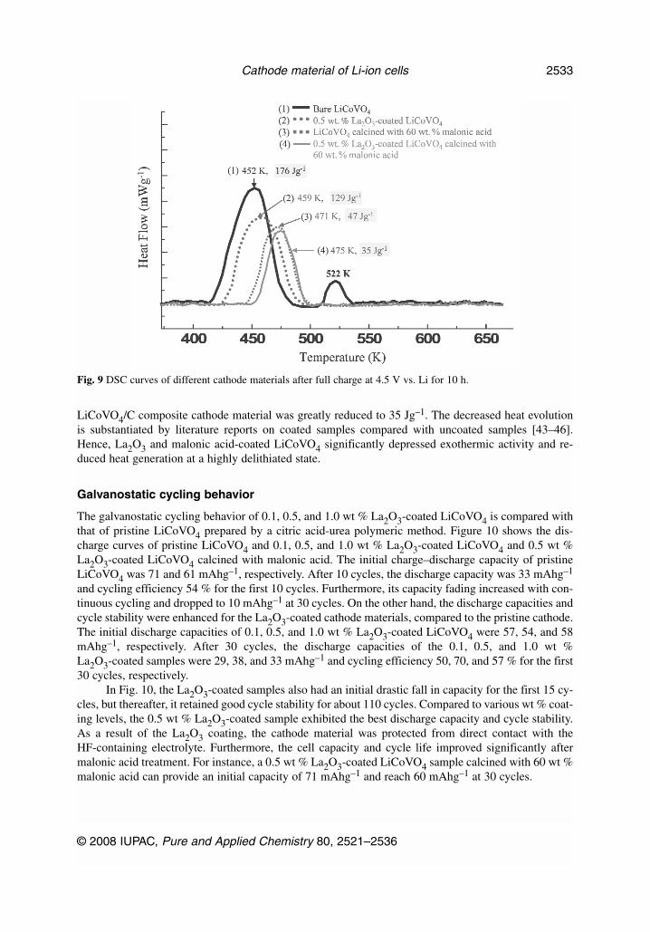
LiCoVO4/C composite cathode material was greatly reduced to 35 Jg–1. The decreased heat evolutionis substantiated by literature reports on coated samples compared with uncoated samples [43–46].Hence, La2O3 and malonic acid-coated LiCoVO4 significantly depressed exothermic activity and re-duced heat generation at a highly delithiated state.
Galvanostatic cycling behavior
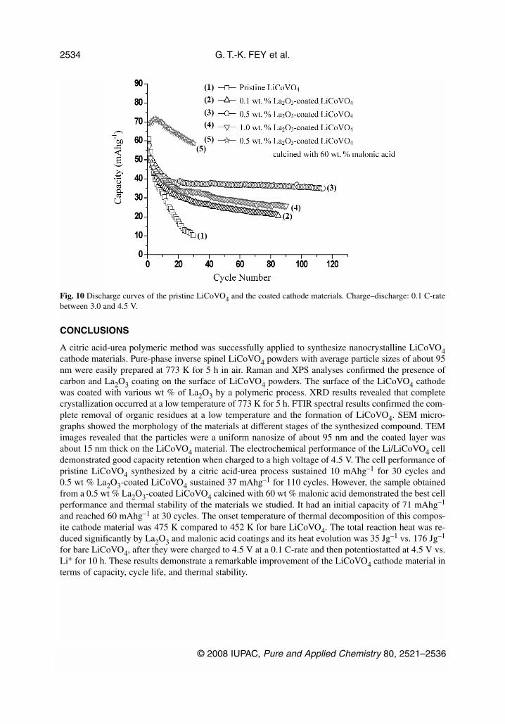
The galvanostatic cycling behavior of 0.1, 0.5, and 1.0 wt % La2O3-coated LiCoVO4 is compared withthat of pristine LiCoVO4 prepared by a citric acid-urea polymeric method. Figure 10 shows the dis-charge curves of pristine LiCoVO4 and 0.1, 0.5, and 1.0 wt % La2O3-coated LiCoVO4 and 0.5 wt %La2O3-coated LiCoVO4 calcined with malonic acid. The initial charge–discharge capacity of pristineLiCoVO4 was 71 and 61 mAhg–1, respectively. After 10 cycles, the discharge capacity was 33 mAhg–1
and cycling efficiency 54 % for the first 10 cycles. Furthermore, its capacity fading increased with con-tinuous cycling and dropped to 10 mAhg–1 at 30 cycles. On the other hand, the discharge capacities andcycle stability were enhanced for the La2O3-coated cathode materials, compared to the pristine cathode.The initial discharge capacities of 0.1, 0.5, and 1.0 wt % La2O3-coated LiCoVO4 were 57, 54, and 58mAhg–1, respectively. After 30 cycles, the discharge capacities of the 0.1, 0.5, and 1.0 wt %La2O3-coated samples were 29, 38, and 33 mAhg–1 and cycling efficiency 50, 70, and 57 % for the first30 cycles, respectively.
In Fig. 10, the La2O3-coated samples also had an initial drastic fall in capacity for the first 15 cy-cles, but thereafter, it retained good cycle stability for about 110 cycles. Compared to various wt % coat-ing levels, the 0.5 wt % La2O3-coated sample exhibited the best discharge capacity and cycle stability.As a result of the La2O3 coating, the cathode material was protected from direct contact with theHF-containing electrolyte. Furthermore, the cell capacity and cycle life improved significantly aftermalonic acid treatment. For instance, a 0.5 wt % La2O3-coated LiCoVO4 sample calcined with 60 wt %malonic acid can provide an initial capacity of 71 mAhg–1 and reach 60 mAhg–1 at 30 cycles.
© 2008 IUPAC, Pure and Applied Chemistry 80, 2521–2536
Cathode material of Li-ion cells 2533
Fig. 9 DSC curves of different cathode materials after full charge at 4.5 V vs. Li for 10 h.
CONCLUSIONS
A citric acid-urea polymeric method was successfully applied to synthesize nanocrystalline LiCoVO4cathode materials. Pure-phase inverse spinel LiCoVO4 powders with average particle sizes of about 95nm were easily prepared at 773 K for 5 h in air. Raman and XPS analyses confirmed the presence ofcarbon and La2O3 coating on the surface of LiCoVO4 powders. The surface of the LiCoVO4 cathodewas coated with various wt % of La2O3 by a polymeric process. XRD results revealed that completecrystallization occurred at a low temperature of 773 K for 5 h. FTIR spectral results confirmed the com-plete removal of organic residues at a low temperature and the formation of LiCoVO4. SEM micro-graphs showed the morphology of the materials at different stages of the synthesized compound. TEMimages revealed that the particles were a uniform nanosize of about 95 nm and the coated layer wasabout 15 nm thick on the LiCoVO4 material. The electrochemical performance of the Li/LiCoVO4 celldemonstrated good capacity retention when charged to a high voltage of 4.5 V. The cell performance ofpristine LiCoVO4 synthesized by a citric acid-urea process sustained 10 mAhg–1 for 30 cycles and0.5 wt % La2O3-coated LiCoVO4 sustained 37 mAhg–1 for 110 cycles. However, the sample obtainedfrom a 0.5 wt % La2O3-coated LiCoVO4 calcined with 60 wt % malonic acid demonstrated the best cellperformance and thermal stability of the materials we studied. It had an initial capacity of 71 mAhg–1
and reached 60 mAhg–1 at 30 cycles. The onset temperature of thermal decomposition of this compos-ite cathode material was 475 K compared to 452 K for bare LiCoVO4. The total reaction heat was re-duced significantly by La2O3 and malonic acid coatings and its heat evolution was 35 Jg–1 vs. 176 Jg–1
for bare LiCoVO4, after they were charged to 4.5 V at a 0.1 C-rate and then potentiostatted at 4.5 V vs.Li+ for 10 h. These results demonstrate a remarkable improvement of the LiCoVO4 cathode material interms of capacity, cycle life, and thermal stability.
G. T.-K. FEY et al.
© 2008 IUPAC, Pure and Applied Chemistry 80, 2521–2536
2534
Fig. 10 Discharge curves of the pristine LiCoVO4 and the coated cathode materials. Charge–discharge: 0.1 C-ratebetween 3.0 and 4.5 V.
ACKNOWLEDGMENTS
Financial support for this work was provided by the National Science Council of the Republic of Chinaunder contract No. NSC 93-2214-E-008-004. PMD thanks the NSC for the award of a postdoctoral fel-lowship.
REFERENCES
1. G. T. K. Fey, W. Li, J. R. Dahn. J. Electrochem. Soc. 141, 227 (1994).2. G. T. K. Fey. J. Active Passive Electronic Components 18, 11 (1995).3. G. T. K. Fey, W. B. Perng. Mater. Chem. Phys. 47, 279 (1997).4. G. T. K. Fey, K. S. Wang, S. M. Yang. J. Power Sources 68, 159 (1997).5. G. T. K. Fey, J. R. Dahn, M. J. Zhang. W. Li. J. Power Sources 68, 549 (1997).6. G. T. K. Fey, C. S. Wu. Pure Appl. Chem. 69, 2329 (1997).7. G. T. K. Fey, K. S. Chen. J. Power Sources 81/82, 467 (1999).8. C. H. Lu, W. C. Lee, S. J. Liou, G. T. K. Fey. J. Power Sources 81/82, 696 (1999).9. G. T. K. Fey, D. L. Huang. Electrochim. Acta 45, 295 (1999).
10. P. P. Chu, D. L. Huang, G. T. K. Fey. J. Power Sources 90, 95 (2000).11. B. J. Hwang, Y. W. Tsai, G. T. K. Fey, J. F. Lee. J. Power Sources 97/98, 551 (2001).12. S. R. S. Prabaharan, M. S. Michael, S. Radhakrishna, C. Julien. J. Mater. Chem. 7, 1791 (1997).13. J. R. Liu, M. Wang, X. Lin, D. C. Yin, W. D. Huang. J. Power Sources 108, 113 (2002).14. P. Kalyani, N. Kalaiselvi, N. Muniyandi. Mater. Chem. Phys. 77, 662 (2002).15. S. Vivekanandhan, M. Venkateswarlu, N. Satyanarayana. Mater. Lett. 58, 1218 (2004).16. T. Thongtem, A. Phuruangrat, S. Thongtem. Mater. Lett. 60, 3776 (2006). 17. N. V. Landschoot, E. M. Kelder, P. J. Kooyman, C. Kwakernaak, J. Schoonman. J. Power Sources
138, 262 (2004). 18. G. T. K. Fey, P. Muralidharan, Y. D. Cho. J. Power Sources 174, 1152 (2007). 19. G. T. K. Fey, P. Muralidharan, C. Z. Lu, Y. D. Cho. Solid State Ionics 177, 877 (2006). 20. G. G. Amatucci, J. M. Tarascon, L. C. Klein. Solid State Ionics 83, 167 (1996).21. S. T. Myung, K. Izumi, S. Komaba, Y. K. Sun, H. Yashiro, N. Kumagai. Chem. Mater. 17, 3695
(2005).22. H. Wang, Y. I. Jang, B. Huang, D. R. Sadoway, Y. M. Chiang. J. Electrochem. Soc. 146, 473
(1999).23. L. F. Wang, C. C. Ou, K. A. Striebel, J. S. Chen. J. Electrochem. Soc. 150, A905 (2003).24. K. M. Shaju, G. V. Subba Rao, B. V. R. Chowdari. Electrochim. Acta 48, 145 (2002).25. G. H. Kim, S. T. Myung, H. J. Bang, J. Prakash, Y. K. Sun. Electrochem. Solid-State Lett. 7, A477
(2004).26. D. Zane, M. Carewska, S. Scaccia, F. Cardellini, P. P. Prosini. Electrochim. Acta 49, 4259 (2004).27. K. Konstantinov, S. Bewlay, G. X. Wang, M. Lindsay, J. Z. Wang, H. K. Liu, S. X. Dou, J. H.
Ahn. Electrochim. Acta 50, 421 (2004).28. M. Gabersceka, R. Dominkoa, M. Belea, M. Remskarb, D. Hanzelb, J. Jamnika. Solid State Ionics
176, 1801 (2005).29. M. R. Yang, T. H. Teng, S. H. Wu. J. Power Sources 159, 307 (2006).30. T. Nakamura, Y. Miwa, M. Tabuchi, Y. Yamada. J. Electrochem. Soc. 153, A1108 (2006).31. P. S. Herle, B. Ellis, N. Coombs, L. F. Nazar. Nat. Mater. 3, 147 (2004).32. S. Y. Chung, J. T. Blocking, Y. M. Chiang. Nat. Mater. 2, 123 (2002).33. J. F. Ni, H. H. Zhou, J. T. Chen, X. X. Zhang. Mater. Lett. 59, 2361 (2005).34. M. Abbate, S. M. Lala, L. A. Montoro, J. M. Rosolen. Electrochem. Solid-State Lett. 8, A288
(2005).
© 2008 IUPAC, Pure and Applied Chemistry 80, 2521–2536
Cathode material of Li-ion cells 2535

35. G. X. Wang, S. Bewlay, J. Yao, J. H. Ahn, S. X. Dou, H. K. Liu. Electrochem. Solid-State Lett. 7,A503 (2004).
36. C. Julien, M. Massot, C. Pérez-Vicente. Mater. Sci. Eng. B 75, 6 (2000).37. W. Kemp. Organic Spectroscopy, Palgrave, New York (2002).38. Y. M. Hon, K. Z. Fung, S. P. Lin, M. H. Hon. J. Solid State Chem. 163, 231 (2002).39. M. M. Doeff, Y. Hu, F. McLarnon, R. Kostecki. Electrochem Solid-State Lett. 6, A207 (2003).40. Y. Hu, M. M. Doeff, R. Kostecki, R. Finones. J. Electrochem. Soc. 151, A1279 (2004).41. M. M. Doeff, Y. Hu, F. McLarnon, R. Kostecki. Electrochem. Solid-State Lett. 6, A207 (2003).42. <http://www.xpsdata.com/>: Fundamental XPS data from pure elements, pure oxides, and chem-
ical compounds.43. D. D. MacNeil, Z. Lu, Z. Chen, J. R. Dahn. J. Power Sources 108, 8 (2002).44. Y. Baba, S. Okada, J.-I. Yamaki. Solid State Ionics 148, 311 (2002).45. J. Cho. Electrochem. Commun. 5, 146 (2003).46. G. T. K. Fey, P. Muralidharan, C. Z. Lu, Y. D. Cho. Electrochim. Acta 51, 4850 (2006).
G. T.-K. FEY et al.
© 2008 IUPAC, Pure and Applied Chemistry 80, 2521–2536
2536