Embed Size (px)
Citation preview
Pulse sequencesPulse sequences
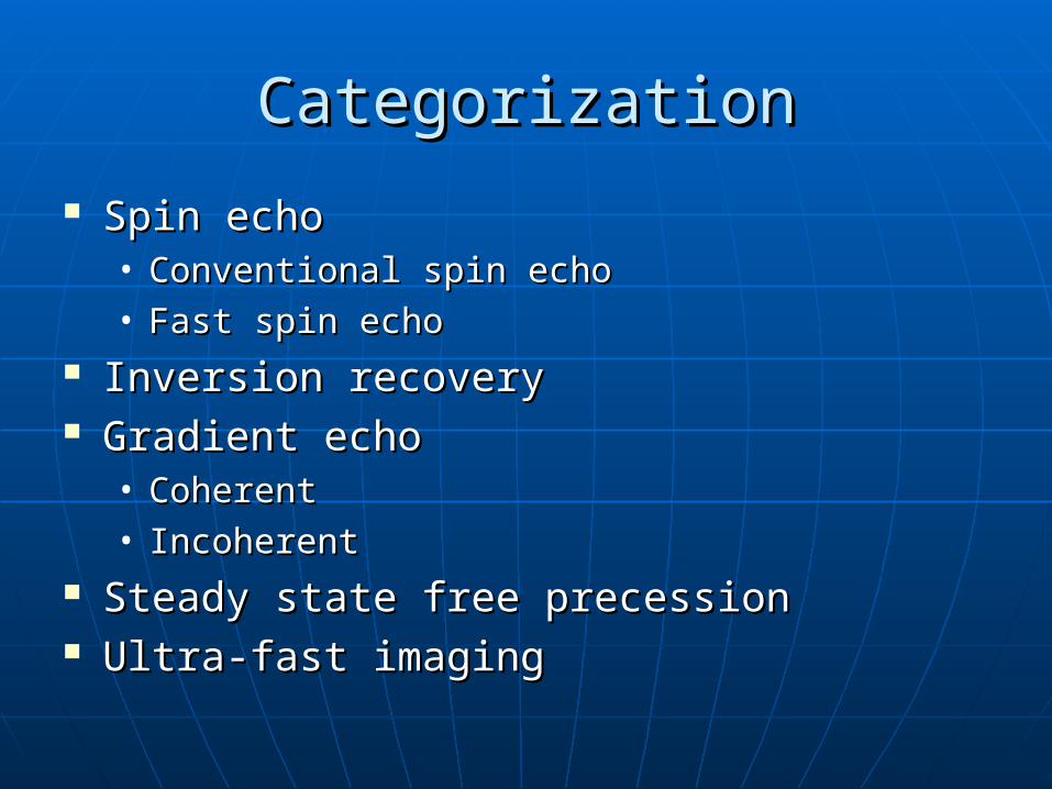
CategorizationCategorization
Spin echoSpin echo• Conventional spin echoConventional spin echo• Fast spin echoFast spin echo
Inversion recoveryInversion recovery Gradient echoGradient echo
• CoherentCoherent• IncoherentIncoherent
Steady state free precessionSteady state free precession Ultra-fast imagingUltra-fast imaging
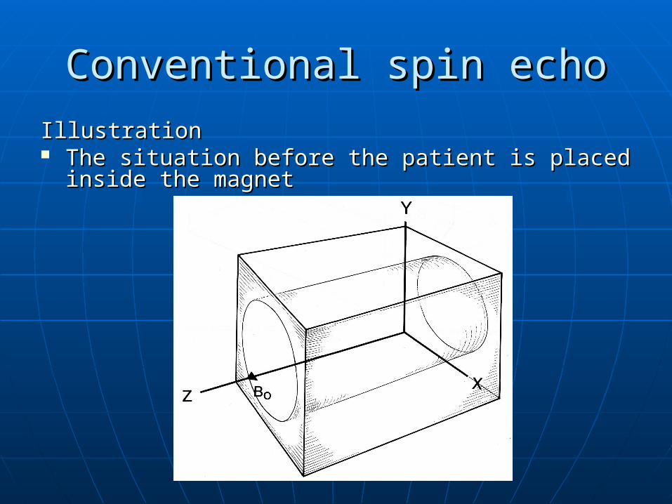
Conventional spin echoConventional spin echo
IllustrationIllustration The situation before the patient is placed inside The situation before the patient is placed inside
the magnetthe magnet
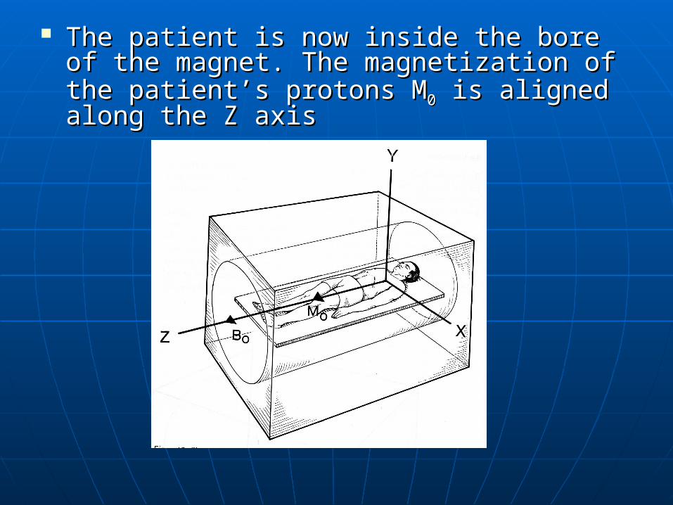
The patient is now inside the bore of the The patient is now inside the bore of the magnet. The magnetization of the magnet. The magnetization of the patient’s protons Mpatient’s protons M00 is aligned along the Z is aligned along the Z axisaxis
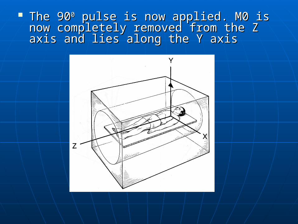
The 90The 9000 pulse is now applied. M0 is now pulse is now applied. M0 is now completely removed from the Z axis and completely removed from the Z axis and lies along the Y axis lies along the Y axis
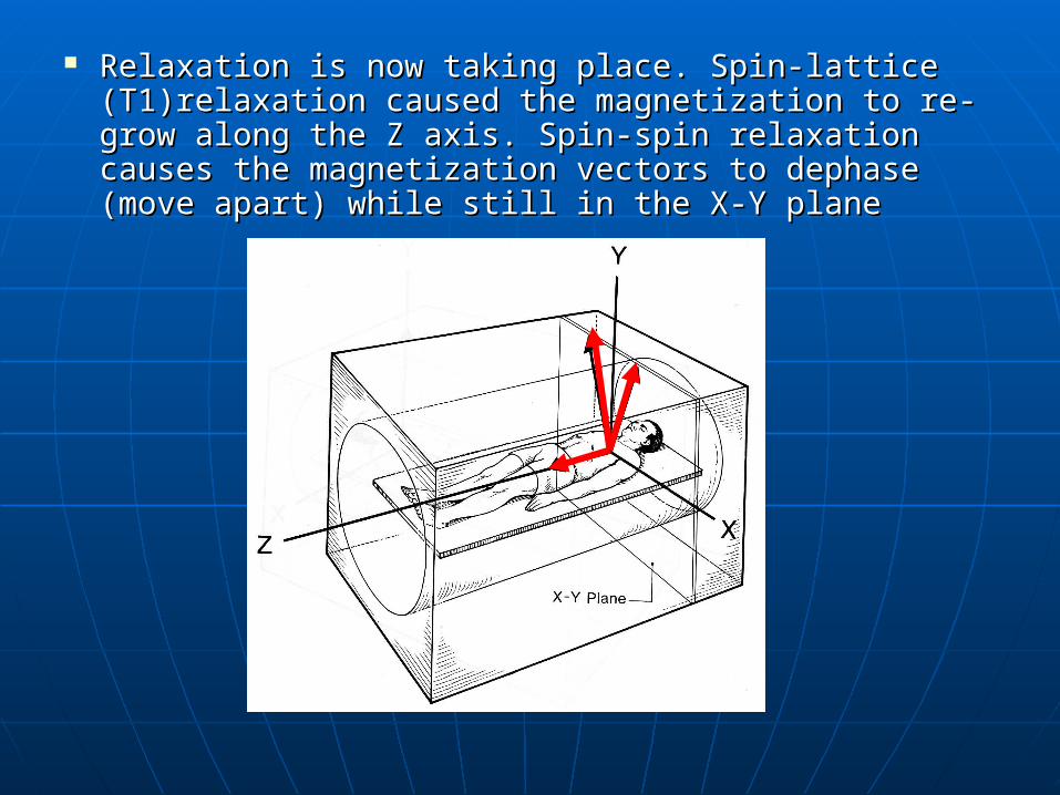
Relaxation is now taking place. Spin-lattice (T1)relaxation Relaxation is now taking place. Spin-lattice (T1)relaxation caused the magnetization to re-grow along the Z axis. Spin-caused the magnetization to re-grow along the Z axis. Spin-spin relaxation causes the magnetization vectors to dephase spin relaxation causes the magnetization vectors to dephase (move apart) while still in the X-Y plane(move apart) while still in the X-Y plane
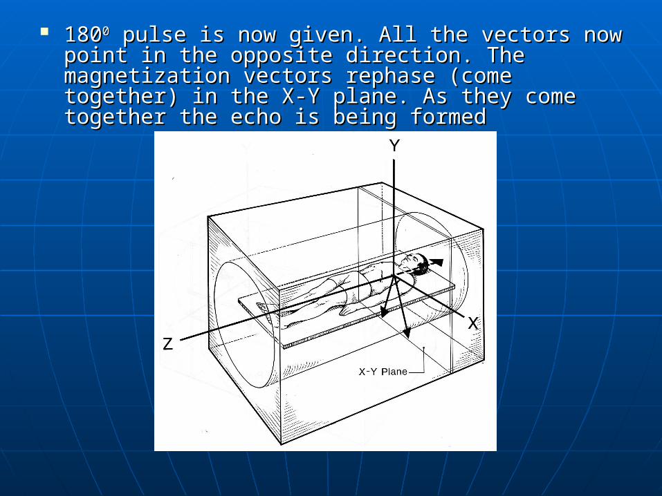
18018000 pulse is now given. All the vectors now point pulse is now given. All the vectors now point in the opposite direction. The magnetization in the opposite direction. The magnetization vectors rephase (come together) in the X-Y plane. vectors rephase (come together) in the X-Y plane. As they come together the echo is being formedAs they come together the echo is being formed
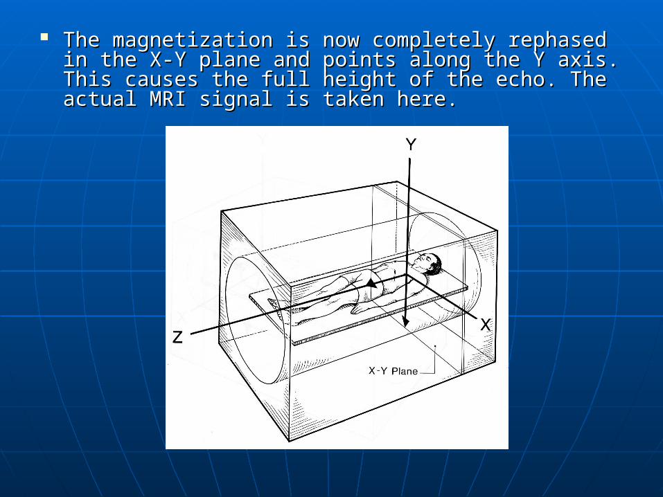
The magnetization is now completely rephased in The magnetization is now completely rephased in the X-Y plane and points along the Y axis. This the X-Y plane and points along the Y axis. This causes the full height of the echo. The actual MRI causes the full height of the echo. The actual MRI signal is taken here.signal is taken here.
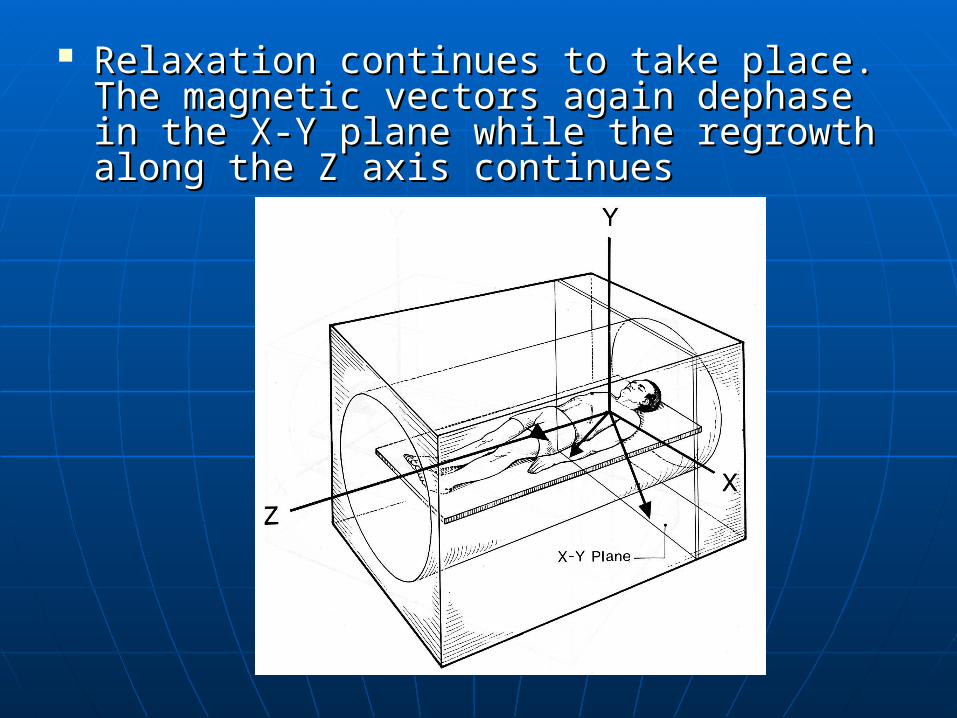
Relaxation continues to take place. The Relaxation continues to take place. The magnetic vectors again dephase in the X-Y magnetic vectors again dephase in the X-Y plane while the regrowth along the Z axis plane while the regrowth along the Z axis continuescontinues
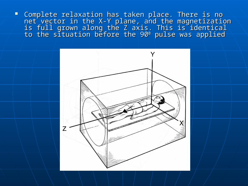
Complete relaxation has taken place. There is no net vector Complete relaxation has taken place. There is no net vector in the X-Y plane, and the magnetization is full grown along in the X-Y plane, and the magnetization is full grown along the Z axis. This is identical to the situation before the 90the Z axis. This is identical to the situation before the 9000 pulse was appliedpulse was applied
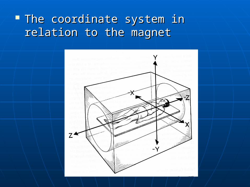
The coordinate system in relation to The coordinate system in relation to the magnetthe magnet
Short TE and short TR gives T1 Short TE and short TR gives T1 weighted image weighted image
Use two RF rephasing pulses Use two RF rephasing pulses generating two spin echoes to generating two spin echoes to produce T2 and proton density produce T2 and proton density weightingweighting
First echo has short TE and long TR – First echo has short TE and long TR – produce proton density weightingproduce proton density weighting
Second echo has a long TE and a Second echo has a long TE and a long TR – produce T2 weightinglong TR – produce T2 weighting
usesuses Gold standard for most imagingGold standard for most imaging May be used for every examintionMay be used for every examintion T1 images useful for demonstrating T1 images useful for demonstrating
anatomy – because high SNRanatomy – because high SNR With contrast enhancement T1 images With contrast enhancement T1 images
show pathologyshow pathology T2 images also demonstrate pathologyT2 images also demonstrate pathology Diseased tissues are generally more Diseased tissues are generally more
oedematous and/or vascular. They have oedematous and/or vascular. They have increased water content and, have a high increased water content and, have a high signal on T2 imagessignal on T2 images
Parameters Parameters
T1 weightingT1 weighting• Short TE 10-20 msShort TE 10-20 ms• Short TR 300 – 600 msShort TR 300 – 600 ms• Typical scan time 4-6 minTypical scan time 4-6 min
Proton density/T2Proton density/T2• Short TE 20 ms/long TE 80 ms+Short TE 20 ms/long TE 80 ms+• Long TR 2000 ms+Long TR 2000 ms+• Typical scan time 7-15 minTypical scan time 7-15 min

AdvantagesAdvantages• Good image qualityGood image quality• Very versatileVery versatile• True T2 weighting sensitive to pathologyTrue T2 weighting sensitive to pathology
DisadvantagesDisadvantages• Scan times relatively longScan times relatively long
Fast spin echoFast spin echo
In contrast to conventional spin echo, fast In contrast to conventional spin echo, fast spin echo applies a train of 180spin echo applies a train of 1800 0 pulses per pulses per TR and different phase encoding steps are TR and different phase encoding steps are used.used.
Each 180Each 1800 0 pulse produce an echo (proton pulse produce an echo (proton density & T2)density & T2)
This drastically reduce the scan timeThis drastically reduce the scan time The number of 180The number of 1800 0 pulses in the train pulses in the train
called the called the turbo factorturbo factor or or train lengthtrain length
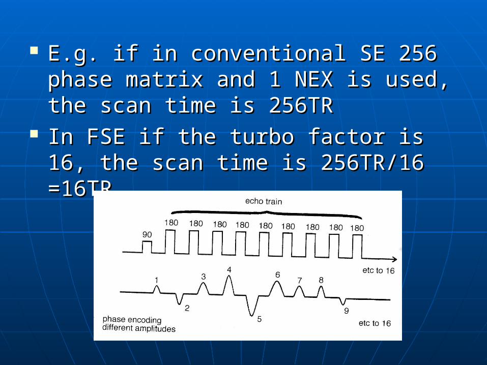
E.g. if in conventional SE 256 phase E.g. if in conventional SE 256 phase matrix and 1 NEX is used, the scan matrix and 1 NEX is used, the scan time is 256TRtime is 256TR
In FSE if the turbo factor is 16, the In FSE if the turbo factor is 16, the scan time is 256TR/16 =16TRscan time is 256TR/16 =16TR
Uses Uses
Useful in most clinical applicationsUseful in most clinical applications Central nervous system, pelvis, Central nervous system, pelvis,
musculoskeletal regionsmusculoskeletal regions NoteNote
• Fat remains bright on T2Fat remains bright on T2 Unless fat saturation techniques are usedUnless fat saturation techniques are used
• Muscles appear darker in FSE images Muscles appear darker in FSE images • Artefacts from metal implants is Artefacts from metal implants is
significantly reducedsignificantly reduced
Parameters Parameters
T1 weightingT1 weighting• Short effective TE less than 20msShort effective TE less than 20ms• Short TR 300 – 600 msShort TR 300 – 600 ms• Turbo factor 2-6Turbo factor 2-6• Typical scan time 30s to 1 minTypical scan time 30s to 1 min
T2 weightingT2 weighting• Long effective TE 100 msLong effective TE 100 ms• Long TR 4000 ms+Long TR 4000 ms+• Turbo factor 8-20Turbo factor 8-20• Typical scan time 2 minTypical scan time 2 min
Advantages & DisadvantagesAdvantages & Disadvantages
Reduced scan timeReduced scan time High resolution High resolution
matrices and multiple matrices and multiple NEX can be usedNEX can be used
Image quality Image quality improvedimproved
Increase T2 Increase T2 informationinformation
Some flow and motion Some flow and motion effects increasedeffects increased
Incompatible with Incompatible with some imaging optionssome imaging options
Fat bright on T2 Fat bright on T2 Reduces magnetic Reduces magnetic
susceptibility effect, susceptibility effect, so so should not be used should not be used when haemorrhage is when haemorrhage is suspectedsuspected
Inversion RecoveryInversion Recovery
Starts with a 180Starts with a 1800 0 inversion pulse.inversion pulse. This inverts NMV through 180This inverts NMV through 18000 into into
full saturation.full saturation. When inverting pulse removed NMV When inverting pulse removed NMV
begins to relax back to Bbegins to relax back to B00 A 90A 9000 excitation pulse is then applied excitation pulse is then applied
at a time at a time TI (Time from Inversion) TI (Time from Inversion) from the 180from the 1800 0 inversion pulseinversion pulse
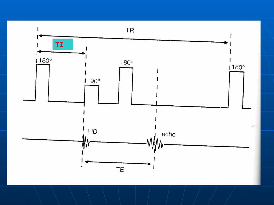
TI
TI determines the weighting & TI determines the weighting & contrastcontrast
Short TI gives T1 contrastShort TI gives T1 contrast Long TI gives proto density contrastLong TI gives proto density contrast After the 90After the 900 0 excitation pulse 180excitation pulse 18000
rephasing pulse is applied at a time rephasing pulse is applied at a time TETE
This produces the spin echoThis produces the spin echo TR is the time between each 180TR is the time between each 1800 0
inverting pulseinverting pulse
Uses Uses
conventionally used to produce conventionally used to produce heavily T1 weighted images to heavily T1 weighted images to demonstrate anatomy & in contrast demonstrate anatomy & in contrast enhanced imagingenhanced imaging
Now more widely used in conjunction Now more widely used in conjunction with fast spin echo to produce T2 with fast spin echo to produce T2 weighted imagesweighted images
ParametersParameters
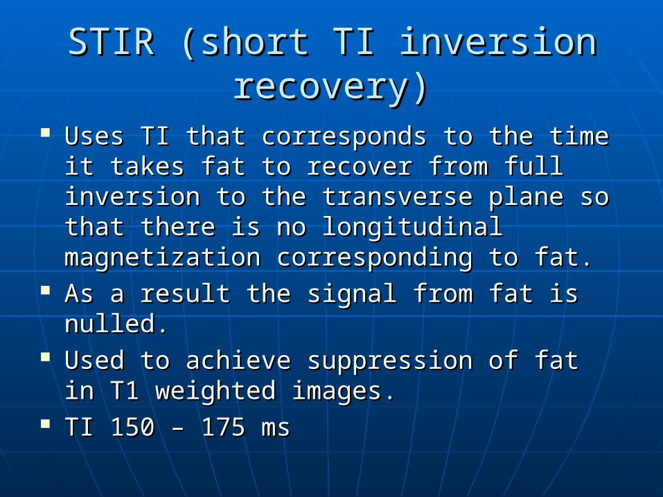
STIR (short TI inversion recovery)STIR (short TI inversion recovery)
Uses TI that corresponds to the time it Uses TI that corresponds to the time it takes fat to recover from full inversion to takes fat to recover from full inversion to the transverse plane so that there is no the transverse plane so that there is no longitudinal magnetization corresponding longitudinal magnetization corresponding to fat.to fat.
As a result the signal from fat is nulled.As a result the signal from fat is nulled. Used to achieve suppression of fat in T1 Used to achieve suppression of fat in T1
weighted images.weighted images. TI 150 – 175 msTI 150 – 175 ms

FLAIR (Fluid attenuated inversion FLAIR (Fluid attenuated inversion Recovery)Recovery)
The signal from CSF is nulled by The signal from CSF is nulled by selecting a TI corresponding to the selecting a TI corresponding to the time of recovery of CSF from 180 to time of recovery of CSF from 180 to the transverse plane and there is no the transverse plane and there is no longitudinal magnetization present.longitudinal magnetization present.
Used to suppress signal from CSF in Used to suppress signal from CSF in T2 weighted imagesT2 weighted images
TI - 1700 -2200 msTI - 1700 -2200 ms


![Concepts and Engineering Aspects of a Neutron Resonance Spin … · 2014. 4. 23. · Neutron Resonance Spin-Echo (NRSE) [1,2] is an alternative to the conventional Neutron Spin-Echo](https://img.dokumen.tips/doc/110x75/610964ceb9a53a05954102e6/concepts-and-engineering-aspects-of-a-neutron-resonance-spin-2014-4-23-neutron.jpg)