Embed Size (px)
Citation preview

THE PULMONARY AND SYSTEMIC CIRCULATIONS INCONGENITAL HEART DISEASE
BY
D. C. DEUCHAR AND R. KNEBEL *
From the Cardiac Department, Guy's Hospital
Received August 2, 1951
The application of cardiac catheterization to the study of the human circulation (Forssmann,1929; Cournand and Ranges, 1941) provided a new technique for obtaining information about thepulmonary circulation and also for the study of the physiological effects of congenital heart disease(Bing et al., 1947a; Dexter et al., 1947b; Cournand et al., 1949). Extensive studies have beenmade in which the pulmonary circulation in congenital heart disease has been investigated andin particular the pulmonary blood flow and peripheral resistance have been evaluated (for instanceBing et al., 1947b.; Handelsman et al., 1948; Hickam, 1949; Wood, 1950). It has long beenknown, however, for the systemic circulation that the elasticity of the arterial walls in relation totheir Windkessel (pressure reservoir) function is of great importance as well as the peripheral resis-tance (Frank, 1899 and 1926; Broemser and Ranke, 1930; Wezler and Boger, 1939). Cournand(1947) has recently pointed out that the same consideration applies to the pulmonary circulationbut so far no quantitative estimations of the " elasticity resistance " in the pulmonary circulationhave been presented.
Cardiac catheterization used for the investigation of congenital heart disease yields data that maybe used for the study of the systemic circulation as well as the pulmonary, so that it is possible tocompare the effect of the cardiac abnormality on the two circulations although hitherto attentionhas usually been concentrated on the pulmonary circulation.
Formulae that give mathematical expression to the arterial Windkessel function are availableand some are applicable to the data obtained by catheterization studies in normal subjects or inthose with many forms of acquired or congenital heart disease. These or other formulae appliedto the analysis of the changes occurring in the two circulations are capable of yielding valuableresults enabling these changes to be studied with greater precision. From cardiac catheterizationstudies made in patients with suspected or proven congenital heart disease we have been able tomake such an analysis in a variety of conditions and have confirmed the importance of the elasticityresistance in the pulmonary arterial system and of the interrelationship of the various componentsthat determine the arterial blood pressure.
METHODSThe data used have been obtained by the catheterization technique described by Holling and Zak
(1950). Resting oxygen consumptions have been measured in a Benedict spirometer on the fasting patientsimmediately before catheterization; blood samples have normally been taken from each of the superiorvena cava, inferior vena cava, right atrium, right ventricle, and pulmonary artery, from other chambers orvessels if entered and from a systemic artery by puncture; blood oxygen contents have been estimated bythe Haldane apparatus (Douglas and Priestley, 1948). Blood pressure records were obtained by a modified
* R. K. was working in the Cardiac Dept., Guy's Hospital, with a British Council Fellowship for German UniversityTeachers.
225
on March 9, 2020 by guest. P
rotected by copyright.http://heart.bm
j.com/
Br H
eart J: first published as 10.1136/hrt.14.2.225 on 1 April 1952. D
ownloaded from

DEUCHAR AND KNEBEL
Hamilton manometer (Hamilton et al., 1934) or by the Southern Instruments blood pressure recorder *using a Kelvin-Hughes pen recorder, both instruments having a resonant frequency of over 15 c.p.s. with aNo. 8 cardiac catheter attached. Zero pressure levels have been taken approximating to the patient'smid-axillary line. Patients, if not anesthetized, were sedated with phenobarbitone, grains 1-2 according toage and size. Anesthesia, when used, was induced by rectal thiopentone supplemented by intravenousthiopentone if required, care being taken as far as possible to avoid undue fluctuations in the patient'scondition.
The blood flows were calculated according to the Fick principle. Whenever possible the pulmonaryarterial oxygen content has been used for mixed venous blood, but where left to right shunts were presentsamples proximal to the site of the shunt have had to be used. Where normal, the systemic arterial oxygensaturation has been substituted for the pulmonary venous oxygen saturation in calculating the pulmonaryflow; where reduced on account of a right to left shunt a value of 96 per cent has been assumed for the pul-monary venous oxygen saturation. Where it was reduced and a pulmonary factor was suspected the arterialoxygen saturation while breathing oxygen has been determined; if this was then found to be normal theresting arterial oxygen saturation has been substituted for the pulmonary venous saturation.
All patients submitted to cardiac catheterization in whom pressures were measured by the Hamilton orS.I. manometer were considered, but strict criteria were applied in the selection of patients for analysis.The majority were necessarily rejected as unsuitable on various grounds; thus, all patients with unknown,possibly raised pulmonary venous pressure, those with shunts into the pulmonary artery or with suspectedpulmonary regurgitation (for reasons given later), all patients in whom the pulmonary artery was not enteredso that no pressure record was obtained, and all those in whom the record obtained was not sufficientlyclear to indicate the incisura marking the division between systole and diastole (mostly those with pulmonarystenosis) and all patients where for any reason the results seemed doubtful or unreliable have been excluded.Finally, 24 patients were selected as suitable for study.
The following symbols have been used to define the various characteristics of the pulmonary or systemiccirculations, co-existing in time, as obtained from the pressure curves and cardiac output data:
Ps = Systolic blood pressure in mm. Hg.Pd = Diastolic blood pressure in mm. Hg.Pp = Pulse pressure (=Pspd) in mm. Hg.Pm = Integrated mean pressure in mm. Hg.Vm = Minute volume of pulmonary or systemic blood flow in cm.3/min.W = Peripheral flow resistance in dynes. sec./cm.5El = Effective volume elasticity coefficient of the arterial system (=" elasticity resistance ") in dynes./
cm.5The quotient E'/W, which is without dimensions.S, D, and S+D = The systolic, diastolic, and total pulse times respectively in a (= 10-3 sec.).In obtaining the data from the pressure curves the mean values obtained throughout a respiratory cycle
have been used. The systemic arterial blood pressures were measured by a sphygmomanometer in the earlierinvestigations and by arterial puncture (brachial or femoral) in all the later.
Applying Poisseuille's law (analogous to Ohm's law and the basis of the so-called Aperia's formula) tothe circulatory conditions in the pulmonary and systemic circuits, the total flow resistance is given by theequation,
W=(Pm-Pv)/C . . . . . . . . . . . . . (1)wherepm is the mean arterial pressure, pv the mean venous pressure in dynes./cm.2, and c the mean blood flowper second (VmI6O) in cm.3/sec. In the systemic circulation, in normal conditions, the mean venous pressuredoes not exceed about 3-6 mm. Hg so that compared with the magnitude of the arterial pressure it may beneglected (Wezler and Boger, 1939) and Pm may be substituted for (pm-p,,) in the above equation. Wherethe mean venous pressure exceeds 6 mm. Hg this approximation is not permissible and in some of our casesit has been necessary on this account to use the right ventricular diastolic pressure as a measure of the meanvenous pressure, as pressures in the systemic veins have not been recorded. For the pulmonary circulation,in some cases of atrial septal defect or anomalous pulmonary venous drainage it is possible to make directmeasurements of the mean venous pressure (Cournand et al., 1947; Hickam, 1949), and in others the dias-tolic pressure in the left atrium can be determined or the right atrial diastolic pressure may be substituted as
* An instrument operating on the electrical capacitance change principle manufactured by Southem Instru-ments, Ltd., Camberley, Surrey.
226
on March 9, 2020 by guest. P
rotected by copyright.http://heart.bm
j.com/
Br H
eart J: first published as 10.1136/hrt.14.2.225 on 1 April 1952. D
ownloaded from

CONGENITAL HEART DISEASE
an approximation; where, however, there is no such abnormality it is necessary to assume that the mean pul-monary venous pressure approximates to zero. In the presence of any condition, notably mitral stenosis,where the pulmonary venous pressure may be abnormally high such an approximation is not of coursepermissible.
The term W is expressed in absolute units in dynes. seconds/cm.5, and represents the total effective resis-tance of the small arteries, arterioles, and capillaries. To obtain W in these units it is necessary tomultiply the pressures measured in mm. Hg by the factor 1332 (representing the product of the sp. gr. ofmercury and the acceleration due to gravity).
The mean pulmonary arterial blood pressure has been determined by integration of the pulse curve, ashas been done by Coumand and his co-workers (Riley et al., 1948) and by Hickam (1949). The basis of thismethod is illustrated in Fig. 1, and is that described by Wezler and Boger (1939), who used it to analyse
12Smm
H9
100
75
k- S -S-- D -*
OI I I I I I I I I
-Ia sec.
I I
FIG. 1.-Diagram illustrating the method used for obtaining the meanarterial blood pressure by integration of the pulse curve. For fullexplanation see text.
systemic arterial pulse curves. The area of the pulse curve is measured by planimetry and the value soobtained is divided by the length S+D to gain the value hp, the height of the shaded rectangle the area ofwhich represents the area measured. The mean pressure of the pulse curve is then obtained from the relationhp x (ps-pd)/H, where H is the height of the pulse curve and (p3-pd) is the pulse pressure (i.e. the ampli-tude of the pulse curve in mm. Hg). The mean blood pressure in mm. Hg is the sum of this value and thediastolic pressure.
The quotient hp/H gives the fraction of the pulse pressure that equals the mean pressure of the pulsecurve. From values obtained by the above method in a series of pulse tracings we have found this quotientfor the pulmonary arterial pulse curves to have a value of 0-398 with a standard deviation ±0-08 (or ex-pressed as a percentage about 40% 4 8%). It will be seen that the coefficient of variation is rather high, soit is clearly preferable, for the pulmonary artery, to perform planimetry in each case rather than to use thisvalue to calculate the mean pressure from the pulse pressure.
227
-
-
on March 9, 2020 by guest. P
rotected by copyright.http://heart.bm
j.com/
Br H
eart J: first published as 10.1136/hrt.14.2.225 on 1 April 1952. D
ownloaded from
DEUCHAR AND KNEBEL
In many of our cases systemic arterial pulse curves were not available and in them the mean systemicpressures have been calculated from the sphygmomanometric figures, using the value of 42 per cent for theabove quotient as found by Wezler and Boger (1939) from a large series of subclavian arterial pulse curves.
Otto Frank (1899) and Wezler (1938) in their theory of blood pressure, derived from consideration ofa Windkessel model, have given an equation, applicable to steady conditions, relating the diastolic pres-sure to the various components determining it. These components are the mean blood flow per second(c), the effective volume elasticity coefficient (E') of the arterial system, the peripheral flow resistance (W)and the systolic and diastolic times.
cW(l-eSxE./w)P le(S+D) xE'IW.. .(2)
This equation is very difficult to solve for E', but Broemser and Ranke (1930) have obtained an approxi-mation based upon the same considerations,
Pp. WPd El. D.. . . .(3)
In this equation it is possible to substitute for W, using equation (1), the expression (Pm-pv)Ic, and asC= VmI60 the last equation can be written,
El= (Pmpv)x(ps-pd)x6O (4)E PdXDXVm . . . . . . . . . .
It is possible to use this equation to obtain values for E' by the use of pressure measurements and estima-tions of the blood flows obtained by independent methods such as are used in cardiac catheterization.The value of E', like W, is expressed in absolute units of the C.G.S. system; in calculating it, therefore, thepressures must be expressed in dynes./cm.2 and the diastolic time in seconds.
These formulae are based upon considerations of normal circulatory dynamics beyond the semilunarvalves and are not therefore applicable in cases where there is either incompetence of these valves or anadditional flow of blood through some abnormal channel into the arterial system. It is for this reason thatwe have had to exclude from analysis many patients in whom there was evidence or suspicion of pulmonaryregurgitation or of a shunt through a patent ductus arteriosus or similar channel into the pulmonary artery.
NORMAL VALUESIn most cases of congenital heart disease there are abnormal features present affecting the
pulmonary circulation. In order to analyse and assess the significance of the changes in the variousfactors upon which the pulmonary arterial pressure depends, it is necessary to have some knowledgeof their normal values. It is known from the observations made by Riley et al. (1948), Stead et al.(1947) and Hickam and Cargill (1948) that large changes can occur in the volume of the pulmonaryflow in various circumstances with only slight changes of pulmonary arterial pressure, which impliesthat the other factors controlling this pressure are themselves subject to large variations. Normalvalues must therefore be related to standard conditions and the resting or basal state is used through-out our study.
Adequate data concerning these normal values for the pulmonary circulation are not yet availableand figures for the pulmonary blood flow and arterial pressure in normal subjects have been obtainedonly in a comparatively small number of persons, nearly all of whom have been adults. It is knownfrom studies of the systemic circulation (Wezler, 1942) that the values of the factors involved varywith the age of the subject, and knowledge of the values in children is particularly scant. In orderto assess the significance of our observations in congenital heart disease we had first to obtain someapproximations for the normal values related to age. The figures we have adopted are given inTable I; values for intermediate ages have been obtained by interpolation.
Systolic and diastolic pressures in normal subjects over twenty years of age have been reportedby Cournand (1947), Werko (1947), Dexter et al. (1947a) and Lagerlof and Werko (1948). Thefigures we have accepted for subjects of twenty years and over represent the average of these, andusing our observation on the relationship of the mean pressure of the pulse curve to the pulse
* e is the base of the natural or Napierian logarithm.
228
on March 9, 2020 by guest. P
rotected by copyright.http://heart.bm
j.com/
Br H
eart J: first published as 10.1136/hrt.14.2.225 on 1 April 1952. D
ownloaded from

CONGENITAL HEART DISEASE
TABLE INoRMAL VALuEs FOR THE PULMONARY CItCULATION RELATED TO THE AGE OF THE SUBJECT
Age in Years 5 10 20 and over
Systolic pressure (Ps) mm. Hg .. .. .. 23
Diastolic pressure (Pd) mm. Hg .. .. .. 8
Pulse pressure (Pp) mm. Hg .. .. .. 15
Mean pressure (Pm) mm. Hg .. .. .. 14
Pulse rate per minute .. .. .. .. 96 82 74
Diastolic time (D) a .. .. .. .. 390 460 500
Pulmonary blood flow (Vm) cm.3/min. .. 3000 4000 6000
Peripheral resistance (W) DyCns-sc. 400 300 200
Elasticity resistance (El) cDm5 1800 1200 700
Elasticity resistance /E'\Peripheral resistance f * 45 ! 40 35
pressure (i.e. mean pressure of pulse curve=40% of pulse pressure) we have calculated the value forthe mean pressure given. As no published figures are available for children it has been necessaryto assume the same values for all ages.
Cardiac outputs in normal resting adult subjects have been estimated using cardiac catheteriza-tion techniques by Cournand (1945), Cournand et al. (1945), McMichael and Sharpey-Schafer(1944), Stead et al. (1945), Werko (1947) and Lagerlof and Werko (1948) in a total of over eightysubjects. The average value obtained from these figures is 6 1./min. which is 30 per cent higher thanthe average value for normal adults obtained by Wezler and Boger (1939) using physical methods.The latter have obtained average values for subjects aged 5 and 10 years and we have added 30 percent to these to obtain comparable approximations for the normal outputs in children. As somejustification for this procedure it may be noted that the values so obtained agree with the averagevalues we have found in children for the systemic blood flow.
Using these basic figures we have calculated a normal value for the pulmonary peripheral resis-tance in adults=200 dynes. sec./cm5. Lagerlof and Werko (1948) have recorded systemic intra-arterial pressures in adults by arterial puncture and using their average figures (130/80 mm.) we havecalculated the corresponding value for the systemic peripheral resistance= 1400 dynes. sec./cm5.In normal subjects the systemic and pulmonary blood flows are equal so these values are comparableand it will be seen that the systemic and pulmonary peripheral resistances are in the relation 7 :1.
In order to calculate the elasticity resistance (E') by the method used here it is necessary to knowthe diastolic time. Lagerl6f and Werko (1948) give 74 as the resting pulse rate in their normaladults during catheterization; from this figure, using the relation S/S+D=0-375 (an average valueobtained from our pulse tracings in 21 subjects), we have calculated an approximate normal diastolictime (D)= 500cr. Using this a normal value for the elasticity resistance (E') in the pulmonary arterialsystem a figure of 700 dynes/cm.5 has been obtained. Correspondingly, again using Lagerlof andWerko's figure for the systemic arterial pressure, the normal value obtained for the elasticity resistancein the systemic arterial system is 1700 dynes/cm.5 By their physical methods in normal adultsWezler and Boger (1939) obtained a value for this factor of 1800 dynes./cm.5 which agrees wellwith our figure. The systemic and pulmonary elasticity resistances are in the relation 2.4: 1. It
229
on March 9, 2020 by guest. P
rotected by copyright.http://heart.bm
j.com/
Br H
eart J: first published as 10.1136/hrt.14.2.225 on 1 April 1952. D
ownloaded from

DEUCHAR AND KNEBEL
follows from the different relations between the peripheral and elasticity resistances in the systemicand pulmonary circulations that the quotient E'/W (system.)= 1 -2 and E'/W (pulm.)=3 5.
Using the pulse rates given by Wezler (1942) for children of 5 and 10 years as a basis for calcu-lating the diastolic time as above we have obtained the values given in Table I for the above-mentionedfactors at those ages. For the systemic circulations we have used the blood pressure figures forchildren given by Fleisch (1927) as a basis for our calculations.
It is appreciated that many of these values are rather artificial but so long as the necessaryobservations on normal children are not available some such approximations are inevitable. Whendirect estimations have been obtained it will obviously be necessary to substitute these. Meanwhilethe departures from normal that we shall consider are in most cases so large that even considerableerrors in the normal values adopted would not affect the validity of our conclusion-s. In all caseswe have elected, in accordance with the views of Rossier et al. (1950), to present the absolutevalues rather than to attempt a relation to body surface.
RESULTS IN NORMAL HEARTSAll our patients have been studied on account of suspected congenital heart disease so that we
are able to present results from only one subject where the findings seemed to indicate that the heartwas normal.
Case 1. H. T. (No. P268), 16, m. Suspicion of congenital heart disease was raised on a soft systolicmurmur in the pulmonary area, with a normal pulmonary second sound, found after a mass radiograph wassaid to show an abnormal shape of heart with a prominent pulmonary conus. There were no symptomsand no disability. Investigations here showed that the heart was displaced to the left by a sternal depres-sion; the cardiogram showed right axis deviation with normal chest leads; cardiac catheterization did notreveal any evidence of intracardiac abnormality but the cardiac output at rest was increased with a normaloxygen consumption.
The values calculated for the pulmonary and systemic haemodynamics are given in Fig. 2. Inthis and all subsequent similar figures the pulmonary circulation is represented on the left and thesystemic on the right; the height of the columns represents the values of the various factors in termsof the percentage of the normal values for the age of the subject. In this figure absolute values aregiven above the appropriate column in each case. The cardiac output is increased (+65%), but inboth circulations the peripheral and elasticity resistances are diminished so that the pressures areall within normal limits. Both the resistances in each circulation are equally affected so that thequotient E'/Wis within normal limits in both. This case serves as an example of the alteration of theresistances in the normal subject, already referred to, which results in the accommodation ofa changein the cardiac output without changes of the blood pressures, shown here to be occurring similarlyin both circulations.
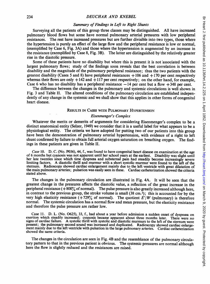
RESULTS IN LEFT TO RIGHT SHUNTSWe have been able to analyse the findings in eight patients with left to right shunts leading ta
an increased pulmonary blood flow. The results are given in Table II. There are two patientswith atrial septal defect, two with anomalous pulmonary veins, and four with ventricular septaldefect, one of these last possibly having an atrial septal defect also.
Atrial Septal DefectCase 2. B. W. (No. 0626), 7, m, came under observation for symptoms associated with a paroxysmal
arrhythmia (? atrial flutter). His exercise tolerance was scarcely limited. Systolic and faint diastolicmurmurs were heard at the apex; the pulmonary second sound was duplicated and increased. Radioscopyshowed moderate cardiac enlargement due to the right ventricle with enlarged, pulsatile pulmonary arteries.The cardiogram showed no axis deviation in the standard leads but a notched R wave in VI Cardiaccatheterization demonstrated a shunt into the right atrium, proved due to an atrial septal defect by passageof the catheter into the left atrium.
230
on March 9, 2020 by guest. P
rotected by copyright.http://heart.bm
j.com/
Br H
eart J: first published as 10.1136/hrt.14.2.225 on 1 April 1952. D
ownloaded from

CONGENITAL HEART DISEASE 231
CASE1
E
0) fJ) i,
0 0 0 'm 0 0 0 0 0 03
oo o0 0oo o o o o -
.toNORMAL 1 - 1* * |* --50~~~~~~~~~~~~~~~~
-500
Ps Pd Pp Pm Vm W E E/W Ps Pd Pp Pm Vm W E EWPULMONARY CIRCULATION SYSTEMIC CIRCULATION
FIG. 2.-Diagrammatic representation of the hiemodynamic analysis in a patient with a normal heart. In this andsubsequent similar diagrams the height of each column represents the observed value of the factor indicated bythe symbol at its foot (for list of symbols see page 226) in terms of its percentage change from normal; in thiscase the absolute figures are also given above each column. The pulmonary circulation is shown on the left andthe systemic on the right. Similar changes are present in both circulations; the minute volume (Vm) is increasedbut the peripheral (W) and elasticity (E) resistances are so reduced that the blood pressures are within normallimits. Case 1.
In this patient the shunt is 7 3 1./min., leading to a pulmonary blood flow of 11*3 1./min.: theright atrial pressure is within normal limits, but the right ventricular systolic pressure is slightlyelevated. An interesting feature is the small systolic pressure drop on passing into the pulmonaryartery from the right ventricle (34 mm. dropping to 21 mm. Hg), which may be due to a " relativestenosis " produced by the dilatation of the pulmonary artery (Burchell et al., 1950). Taking intoaccount the patient's age the pulmonary arterial pressures are probably within normal limitsalthough below our " normal " values; this, despite the increased blood flow, results from theextremely low peripheral resistance (W (pulm.)= 60 units which is - 83% of normal and the lowestvalue we have found in our series). The elasticity resistance is also greatly reduced (E'(pulm.)=500 units or -68% of normal) so that the pulse pressure is not increased despite the high strokevolume (110 cm.3). The quotient E'l W(pulmonary) is high. In the systemic circulation the bloodflow, pressures, and peripheral resistance are essentially normal with the elasticity resistance onlyslightly increased.
Case 3. J. R. (No. 0291), 7, m., had negligible disability but examination had revealed a cardiac ab-normality. There was a soft systolic murmur maximal at the apex; a diastolic murmur was not certainlyheard. The pulmonary second sound was split but not loud. Radioscopy showed moderate cardiacenlargement mostly due to the right ventricle with enlargement of the pulmonary arteries and some pulsationnear the hila of the.lungs. The cardiogram showed some right ventricular preponderance (R.V.P.) with anotched R in VI. Cardiac catheterization revealed a shunt into the right atrium.
The systemic flow in this patient could not be satisfactorily estimated owing to a large differencebetween the oxygen contents of the two venwe cave and the impossibility of obtaining a true mixedvenous blood sample in the presence of the shunt into the right atrium, so data are only available forthe pulmonary circulation in which the blood flow is 12 1./min. As in the previous patient the rightatrial pressure is normal, the right ventricular systolic slightly elevated and there is a small pressuregradient between the right ventricle and the pulmonary artery (37-30 mm. Hg systolic). The mean
on March 9, 2020 by guest. P
rotected by copyright.http://heart.bm
j.com/
Br H
eart J: first published as 10.1136/hrt.14.2.225 on 1 April 1952. D
ownloaded from

DEUCHAR AND KNEBEL
pulmonary arterial pressure is normal, but the pulse pressure (25 mm. Hg or +67% of normal) isincreased with a slight rise of the systolic and a considerable reduction of the diastolic pressure.These changes are accounted for by the low peripheral resistance and normal elasticity resistance,the quotient E'/W (pulmonary) being very high. The high pulse pressure reflects the normalelasticity resistance occurring with the large stroke volume of 128cm.3
Anomalous Pulmonary Venous DrainageCase 4. W. B. (No. 0392), 12, m. Congenital heart disease was diagnosed at 6 years. Investigation
had revealed some bronchiectatic changes in the left lower lobe, presumed the sequel of acute respiratoryillness in infancy. There had been dyspnoea on exertion since early childhood. Clinical examinationrevealed dextrocardia, a systolic murmur and thrill over the pr=cordium (no diastolic murmur) and a loudpulmonary second sound. Radioscopy showed dextrocardia with situs inversus, no ventricular enlargement,but pulmonary arteries enlarged with moderate pulsation. Cardiac catheterization demonstrated a shuntinto the venous atrium, and pulmonary veins were intubated entering this directly from the left lung.
The pulmonary blood flow in this patient is of the same order as in the preceeding two cases buthere, in contrast, the pressures are increased. The systolic, diastolic, mean, and pulse pressures areall about double the normal, whereas the peripheral and elasticity resistances and the quotientE'/W(pulmonary) are not significantly changed from normal, so that the pressure changes areeffected by the increased blood flow only. The systemic circulation is essentially normal in allrespects.
Case 5. M.G. (No. H288), 8, f., had been listless as an infant and subsequently had some limitation ofexercise tolerance. Congenital heart disease was diagnosed at 5 years. There was a loud systolic murmurand thrill at the apex and in the pulmonary area where the second sound was duplicated and loud. Radio-scopy revealed enlargement of the right ventricle and also probably of the right atrium, with enlarged'pulsatile pulmonary arteries. The cardiogram showed R.V.P. Cardiac catheterization demonstrated ashunt into the right atrium and pulmonary veins draining into or near the lower end of the superior venacava were intubated
Allowing for the age difference, the increase of the pulmonary flow in this patient relative to thenormal value is similar to that in the last, but the mean pressure is much higher (Pm= +465% ofnormal) due to the high peripheral resistance (+ 106% of normal). Proportionately the diastolicpressure is more increased than the systolic so that the pulse pressure is less changed. This reflectsthe low elasticity resistance (-31% of normal), the significance of which is all the greater in thepresence of the high pressure. The relation of this to the enlargement of the pulmonary arterywill be considered later. Again the systemic circulation shows no significant changes from normal,apart from a rather high elasticity resistance.
Ventricular Septal DefectCqse 6. R. F. (No. 0142), 12, m., had no disability and congenital heart disease was diagnosed on exami-
nation at 4 years. There was a systolic thrill and murmur in the pulmonary area with a diastolic murmurat the apex, and a loud pulmonary second sound. Radioscopy showed both ventricles to be enlarged withpulmonary arteries also enlarged and showing pulsation to the periphery of the lung fields. The cardiogramshowed no definite preponderance. Cardiac catheterization demonstrated a shunt into the right ventricle.
The very -large pulmonary flow is here associated with high values for the pulmonary systolic,diastolic, and mean pressures, these being due to the volume of the flow as the peripheral resistanceis on the low side of normal. The greatly increased pulse pressure (+220%) similarly results fromthe high stroke volume as the elasticity resistance also is low. These relationships are well shownin Fig. 3A. In the systemic circulation the blood flow is normal and the other features are on thelow side of normal.
Case 7. D. O'D. (No. 0634), 11, f. Congenital heart disease had been diagnosed at 2 years on routineexamination but later there was some dyspnoea on exertion. There were systolic and diastolic murmurs atthe apex and the pulmonary second sound was greatly increased. Radioscopy showed cardiac enlargement
232
on March 9, 2020 by guest. P
rotected by copyright.http://heart.bm
j.com/
Br H
eart J: first published as 10.1136/hrt.14.2.225 on 1 April 1952. D
ownloaded from

CONGENITAL HEART DISEASE
due to the right ventricle, great enlargement of the main pulmonary artery, a hilar dance, and pulsation tothe periphery of the lung fields. The cardiogram showed no definite preponderance. Cardiac catheteriza-tion demonstrated a shunt into the right ventricle.
The pulmonary flow is even larger than in the last patient, being the largest we have found:the general pattern of the changes is the same except that the elasticity resistance is more nearlynormal. Again the systemic circulation is essentially normal in all respects.
+700 +700
+600C +600_CCASEb6 I CASES8
+500 +500
+400 +400
+300 - +300
+200 i- +200
+100 +100
NORMAL .fl
-100 .-- -. - - - - . . 4 . -100Ps Pd Pp PR Vm W E' qW Ps Pd Pp Pm Vm W E w Ps Pd Pp Pm Ym W tE rs ra rp rm Vm Wiv y/W
PULMONARY CIRC. SYSTEMIC CIRC. PULMONARY CIRC. SYSTEMIC CIRC.
A BFIG. 3.-(A) Ventricular Septal Defect. The diagram illustrates the pattern of the himodynamic changes and shows
that the pulmonary hypertension arises entirely from the increased blood flow (Vm), the resistances beingreduced (Case 6). (B) Ventricular Septal Defect. In contrast to Case 6 the diagram shows that in this patientthe pulmonary hypertension is due to an increase of the resistances (W and E) as well as to an increased bloodflow (Case 8). The systemic circulation is relatively unaffected in both cases.
Case 8. V. H. (No. 0467), 48, f., had had recurrent attacks of bronchitis from childhood with somewinter asthmatic attacks; dyspnoea on exertion had increased during the last three years. There wasslight cyanosis; systolic and diastolic murmurs were widely heard over the precordium with an increasedpulmonary second sound. Radioscopy showed cardiac enlargement chiefly due to the right ventricle; thepulmonary arteries were greatly enlarged with considerable pulsation. The cardiogram showed someR.V.P. Cardiac catheterization demonstrated a shunt into the right ventricle; the arterial oxygen satura-tion at rest was 87 but rose to 100 per cent on breathing oxygen.
This patient shows a great increase in the pulmonary arterial pressures, the diastolic beingrelatively the most affected; these changes are the result of several factors as the blood flow, peri-pheral and elasticity resistances are all increased. The changes in the two resistances are of thesame order so that the quotient E'/W (pulmonary) is normal. The systemic circulation yieldsnormal figures. These relationships are illustrated in Fig. 3B.
Case 9. D. H. (No. 0163), 3, m., was unwell from infancy having a persistent cough and dyspncea oneffort, although this seems to be decreasing as he grows. A systolic thrill was palpable all over the prncor-dium with systolic and diastolic murmurs best heard at the apex. Radioscopy showed considerable cardiacenlargement and enlarged pulmonary arteries with pulsation to the periphery of the lung fields. Cardiaccatheterization demonstrated a moderately large shunt into the right ventricle and the oxygen contentdifferences were suggestive but not decisive of a small shunt into the right atrium.
The pattern of the changes in the pulmonary circulation closely resembles that in Case 4 in typeand degree, the increases in all the pressures being solely a reflection of the high blood flow. Thesystemic circulation is essentially normal although the elasticity resistance is slightly increased.R
I
233
II. . . - .- 1,1- SAI el lel.8 91- ft.1 ft- 0- W- W
on March 9, 2020 by guest. P
rotected by copyright.http://heart.bm
j.com/
Br H
eart J: first published as 10.1136/hrt.14.2.225 on 1 April 1952. D
ownloaded from
DEUCHAR AND KNEBEL
Summary of Findings in Left to Right ShuntsSurveying all the patients of this group three classes may be distinguished. All have increased
pulmonary blood flows but some have normal pulmonary arterial pressures with low peripheralresistances. The rest have increased pressures but are further divisible into two types, those wherethe hypertension is purely an effect of the large flow and the peripheral resistance is low or normal,(exemplified by Case 6, Fig. 3A) and those where the hypertension is augmented by an increase inthe resistance (exemplified by Case 8, Fig. 3B). The latter are distinguished by the relatively greaterrise in the diastolic pressure.
Some of these patients have no disability but where this is present it is not associated with thelargest pulmonary flows; study of the findings soon reveals that the best correlation is betweendisability and the magnitude of the pulmonary peripheral resistance; thus the two patients with thegreatest disability (Cases 5 and 8) have peripheral resistances + 106 and + 170 per cent respectivelywhereas their flows are only + 142 and + 117 per cent respectively; on the other hand, for example,Case 6 who has no disability has a peripheral resistance -14 per cent but a flow +348 per cent.
The difference between the changes in the pulmonary and systemic circulations is well shown inFig. 3 and Table II. The altered conditions of the pulmonary circulation are established indepen-dently of any change in the systemic and we shall show that this applies in other forms of congenitalheart disease.
RESULTS IN CASES WITH PULMONARY HYPERTENSIONEisenmenger's Complex
Whatever the merits or demerits of arguments for considering Eisenmenger's complex to be adistinct anatomical entity (Selzer, 1949) we consider that it is a useful label for what appears to be aphysiological entity. The criteria we have adopted for putting two of our patients into this grouphave been the demonstration of pulmonary arterial hypertension, with evidence of a right to leftshunt confirmed by failure to obtain full arterial oxygen saturation on breathing oxygen. The find-ings in these patients are given in Table II.
Case 10. D. C. (No. P036); 44, f., was found to have congenital heart disease on examination at the ageof 6 months but cyanosis was not apparent until her school years at the earliest. Disability was slight untilher late twenties since which time dyspnoea and substernal pain had steadily become increasingly severelimiting factors. A diastolic thrill and murmur with a short systolic murmur were found to the left of thesternum. Radioscopy showed cardiac enlargement mainly due to the left ventricle with great dilatation ofthe main pulmonary arteries; pulsation was easily seen in these. Cardiac catheterization showed the criteriastated above.
The changes in the pulmonary circulation are illustrated in Fig. 4A. It will be seen that thegreatest change in the pressures affects the diastolic value, a reflection of the great increase in theperipheral resistance (+ 800% ofnormal). The pulse pressure is also greatly increased although here,in contrast to the previous group, the stroke volume is small (38 cm.3); this is accounted for by thevery high elasticity resistance (+729% of normal). The quotient E'/W (pulmonary) is thereforenormal. The systemic circulation has a normal flow and mean pressure, but the elasticity resistanceand therefore the pulse pressure are rather low.
Case 11. D. L. (No. 0625), 33, f., had about a year before admission a sudden onset of dyspncea onexertion which steadily increased; cyanosis became apparent about three months later. There were nosigns of cardiac failure. A systolic thrill with systolic and diastolic murmurs to the left of the sternum werepresent; the pulmonary second sound was increased and duplicated. Radioscopy showed cardiac enlarge-ment mainly due to the left ventricle with pulsation in the large pulmonary arteries. Cardiac catheterizationshowed the same criteria.
The changes in the circulation are seen in Fig. 4B and the resemblance of the pulmonary circula-tory pattern to that in the previous patient is obvious. The systemic pressures are normal althoughhere the flow is slightly reduced and the resistances are raised.
234
on March 9, 2020 by guest. P
rotected by copyright.http://heart.bm
j.com/
Br H
eart J: first published as 10.1136/hrt.14.2.225 on 1 April 1952. D
ownloaded from

CONGENITAL HEART DISEASE 235
+800 +800S+ 700 CASE 10 +700 CASE 11
4-600. +600
+ 5O0 +500
+ 400, +400
+ 300 0
+ 200 i0
+ 100 +0
NORMALL
-10Ps Pd Pp PMVm W E' E' PS ftip PPMVmW E'00
PiVfd Pp Pm Vm W E' EIW Ps Pd Pp PMVm W E'EYWPULMONARY CIRC. SYSTEMIC CIRC. PULMONARY CIRC. SYSTEMIC CIRC.
A BFIG. 4.-(A) Eisenmenger's Complex. The pattern of the changes in the pulmonary circulation shows clearly how
the hypertension is due entirely to the increased resistances (Wand E') while the blood flow is greatly reduced.The normal mean systemic arterial pressure (Pm) and blood flow (Vm) are in marked contrast (Case 10). (B)Eisenmenger's Complex. The pulmonary pattern in this patient shows a striking resemblance to that in Fig. 4A.In the systemic circulation the reduced blood flow has been compensated for by changes in the resistances so thatthe pressures are normal (Case 11).
These patients show a remarkable resemblance to each other but stand in marked contrast withthe previous group in that the pulmonary blood flows are reduced; the mechanism producinghypertension here is solely the increase in the resistances. In keeping with the previous group thehigh peripheral resistances are associated with greater disability and again the brunt of the physio-logical disturbance is borne by the pulmonary circulation.
Transposition of the Great VesselsIn only one patient, with partial transposition and a ventricular septal defect, were full data
available for analysis. We have elected to present the results at this point for, as will be seen, thehemodynamic changes lie intermediate between those of the preceding two groups.
Case 12. H. K. (No. 0109), 13, m., had been cyanosed from birth and had suffered moderate disabilitydue to dyspncea on exertion. There was a loud systolic and a rumbling diastolic murmur to the left of thesternum; the pulmonary second sound was greatly increased. Radioscopy showed some cardiac enlarge-,ment mostly affecting the right ventricle; the pulmonary arteries were enlarged with a hilar dance. Thecardiogram showed R.V.P. During cardiac catheterization both the aorta and pulmonary artery wereentered, the oxygen saturation of blood in the latter being found the higher.
The pattern of the changes is shown in Fig. 5A. Like the two patients above the greatest changein the pulmonary arterial pressures affects the diastolic value (+563% of normal) and correspond-ingly the chief factor in producing the hypertension is the high peripheral resistance. The elasticityresistance also is increased but proportionately less so; so that E'/W (pulmonary) is low. Unlikethe two patients above, but resembling the group with left to right shunts, there is an increase of thepulmonary blood flow, so that all the three possible factors are here involved in producing thehypertension. In common with both groups the increased pulmonary peripheral resistance isassociated with some disability and the systemic circulation shows only minor changes from normal.
on March 9, 2020 by guest. P
rotected by copyright.http://heart.bm
j.com/
Br H
eart J: first published as 10.1136/hrt.14.2.225 on 1 April 1952. D
ownloaded from
236 DEUCHAR AND KNEBJJL
2Io1oNII ° I ° - o4 .Z IX -
0~~~~~08T1+ewt+T $+
t6 2 _ _aX 82\
Il0 nIII~~~~~~~ -~~~~~~~~~-
1+~~
| R E; I | W V VX-Zo8 8_\|\oOX|N3 |\O |1-4I *0 %O - CRo R
n .-c
00o 00rnC l
N ~~~~~~~~~~~~~00c~-r00 000
o xatex | | w " I +'WI R v°I -HI I °, I+++
ooirmn-N80 0\
.~~~*_
X . ;o -% ;ot eQ t- 8c42R"0
+ +< > i S+ ++ IO
\0 __\
\0\.\\o; 0
}SS~~~~~~S rf S X- 1n
.!z~~~~~~~-' co, m t.. m cX i oI
II +~~~~~~+
0000000N~~Ne ~0f0 00N0~~+ +
o000 00N0qe VNe~~~~N 00~~~~~~~~~~~e0o
0 O 00c t-
OXX0X'01-!XO OX~~~~~~~~~~~'CMN V N *~ 0 0 V00
00% 00 N~~m C000 0000nW'- I
SoSo~~~~~~c
N' -ro~~~~~MV .*-en0
C.a
Z~~O N f'I ~~~~' hI IC~W No00 0%0 N
I
- -. , - A .r Y% rpikr " "
on March 9, 2020 by guest. P
rotected by copyright.http://heart.bm
j.com/
Br H
eart J: first published as 10.1136/hrt.14.2.225 on 1 April 1952. D
ownloaded from

CONGENITAL HEART DISEASE 237
41~~~~~~~~~~:-A18I.o.o" o° -. t-* - °Ht14l1Sap8,, l _t1 8X8+8xRe8>8toXRCo+
X+C~~~~
[email protected] g\ \o Oo\00\ \\o nX E IXt8++8o Xoog-mX I.,
mX
u~~~~~~~~~~~~~~VEI>o o 8 48 oot 8*t0~~~~~~~~~
,u|8E0E |'o\X 0\0 X+ f0 00 |0
2o o 08o 10,80%08841Ne -o 0v=&*^fiF\ t\ V N- en,2F622o 3E + ++ '+
. o OX O\ 02 02\0 ure0 01p X\-01Q6 EZ t-0o 00 00O _ 2cFA-a_ > N _ 7> _ oie0r0r> .0 % ffi - - O - * -=0
I I X f-
~~we| | -E|~E 8080 o°3R5e<Y. g O\ \ X 0O\ O \°\ \° o°\° \\\
0 0\0NX 0 N 0 'OO 0 O 00 O )N N ~~~~~~~~~ N~~; %O§, - 0 0
-0~~~ ~ ~ ~ ~~~~r- ~~~~(-en w qNo.s * NO I
UQ 000\0@\00rE 00t0°° co V\en -- n°\en 0NN 0%o a.~1~N - ,rI 0 *~
o o~O + + aa++
___- _ ____ _ _i _ _l¢ E
_E~~~~~~~~~x.0 '.1 I. '.o u^FI C000000r00000,e'1
IZcI 1 - I 1- - ---Ise
+ -H +++-H+
23 £v--2+ 3 ° g 3+m*A 4300I-,* 4
~++ + ++ ++++ o
0 3 00 ggu0o
§ -N ~0r- 7 0N V
VYv- %0 0000In t0
o 0. t- 0.m Mti O
0) In 0 0 IInenNt
ID 0 - " -r t O enN ~o 0% 0 ro~
d 00 ~~ ~~~~o0% N -000 HN
en ON.4 "'O 0 (.1 0-, 0 N in N .10\-wv -. 00 NIn00)a - n%ON-t
0% 0% 0% 0 0% 0000 0
00 w~~~~~~~~~~~~~~~~~~0
.0.0~~~~~0 ~~~~~~~~~~~
In0%N ~~~~~~~~~~nNId 00 0% 00
NcoN N - o
N eq - - - - N N
on March 9, 2020 by guest. P
rotected by copyright.http://heart.bm
j.com/
Br H
eart J: first published as 10.1136/hrt.14.2.225 on 1 April 1952. D
ownloaded from
4-600 +600
+500 - CASE 12 4500SCASE 14
+ 400-+400
+300 +300
+200 -4200-
+400 -4100
NORMALW
Ps Pd Pp PmVm W E' EN Ps Pd Pp Pm Vm W E' I Ps Pd Pp Pm Vm W E' E)W Ps Pd Pp Pm Vm W EEYWPULMONARY CIRC. SYSTEMIC CIRC. PULMONARY CIRC. SYSTEMIC CIRC.
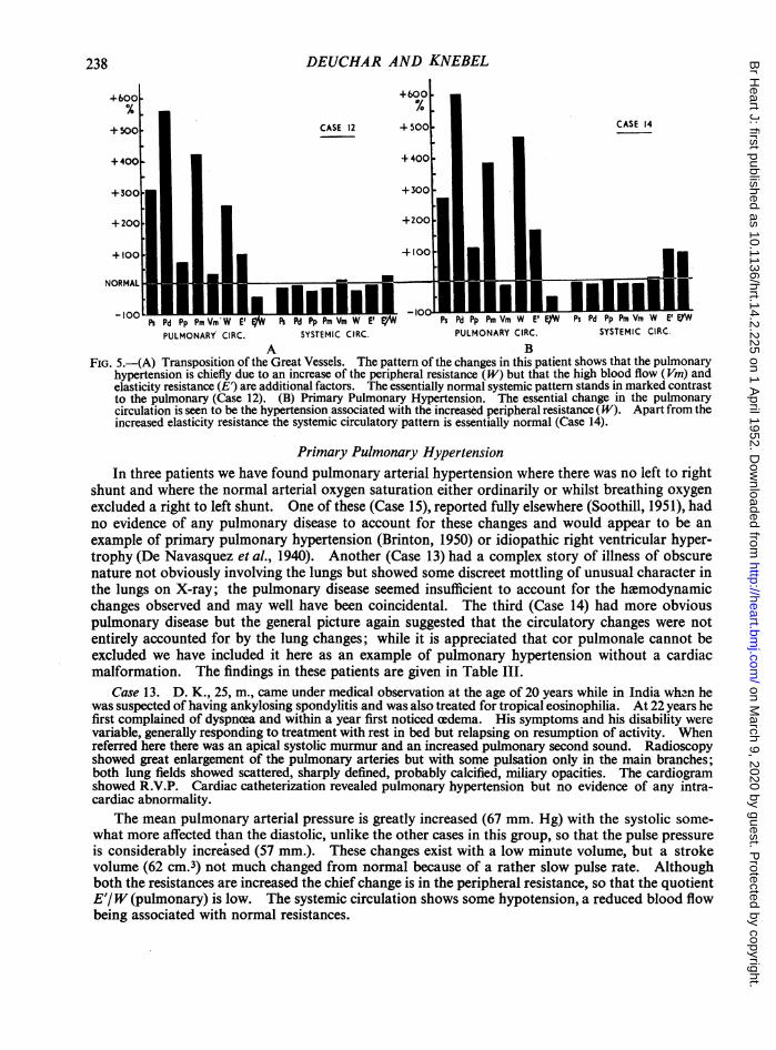
A BFIG. 5.-(A) Transposition of the Great Vessels. The pattern of the changes in this patient shows that the pulmonary
hypertension is chiefly due to an increase of the peripheral resistance (W) but that the high blood flow (Vm) andelasticity resistance (E) are additional factors. The essentially normal systemic pattern stands in marked contrastto the pulmonary (Case 12). (B) Primary Pulmonary Hypertension. The essential change in the pulmonarycirculation is seen to be the hypertension associated with the increased peripheral resistance (W). Apart from theincreased elasticity resistance the systemic circulatory pattern is essentially normal (Case 14).
Primary Pulmonary HypertensionIn three patients we have found pulmonary arterial hypertension where there was no left to right
shunt and where the normal arterial oxygen saturation either ordinarily or whilst breathing oxygenexcluded a right to left shunt. One of these (Case 15), reported fully elsewhere (Soothill, 1951), hadno evidence of any pulmonary disease to account for these changes and would appear to be anexample of primary pulmonary hypertension (Brinton, 1950) or idiopathic right ventricular hyper-trophy (De Navasquez et al., 1940). Another (Case 13) had a complex story of illness of obscurenature not obviously involving the lungs but showed some discreet mottling of unusual character inthe lungs on X-ray; the pulmonary disease seemed insufficient to account for the hemodynamicchanges observed and may well have been coincidental. The third (Case 14) had more obviouspulmonary disease but the general picture again suggested that the circulatory changes were notentirely accounted for by the lung changes; while it is appreciated that cor pulmonale cannot beexcluded we have included it here as an example of pulmonary hypertension without a cardiacmalformation. The findings in these patients are given in Table III.
Case 13. D. K., 25, m., came under medical observation at the age of 20 years while in India when hewas suspected of having ankylosing spondylitis and was also treated for tropical eosinophilia. At 22 years hefirst complained of dyspnoea and within a year first noticed cedema. His symptoms and his disability werevariable, generally responding to treatment with rest in bed but relapsing on resumption of activity. Whenreferred here there was an apical systolic murmur and an increased pulmonary second sound. Radioscopyshowed great enlargement of the pulmonary arteries but with some pulsation only in the main branches;both lung fields showed scattered, sharply defined, probably calcified, miliary opacities. The cardiogramshowed R.V.P. Cardiac catheterization revealed pulmonary hypertension but no evidence of any intra-cardiac abnormality.
The mean pulmonary arterial pressure is greatly increased (67 mm. Hg) with the systolic some-what more affected than the diastolic, unlike the other cases in this group, so that the pulse pressureis considerably increased (57 mm.). These changes exist with a low minute volume, but a strokevolume (62 cm.3) not much changed from normal because of a rather slow pulse rate. Althoughboth the resistances are increased the chief change is in the peripheral resistance, so that the quotientE'/W (pulmonary) is low. The systemic circulation shows some hypotension, a reduced blood flowbeing associated with normal resistances.
238 DEUCHAR AND KNEBEL
on March 9, 2020 by guest. P
rotected by copyright.http://heart.bm
j.com/
Br H
eart J: first published as 10.1136/hrt.14.2.225 on 1 April 1952. D
ownloaded from

CONGENITAL HEART DISEASE
Case 14. M. L., 19, f., had suffered from bronchitis and asthma from early childhood but at an earlyage had already shown cyanosis and dyspnoea on exertion and had had several attacks of right-sided heartfailure before being investigated. No murmurs were heard and the pulmonary second sound was thoughtto be normal. Radioscopy showed moderate cardiac enlargement due to the right ventricle with greatenlargement of both pulmonary arteries but pulsation only just seen in the main vessels. The cardiogramshowed R.V.P. Cardiac catheterization revealed pulmonary hypertension and no evidence of a left to rightshunt; the arterial oxygen saturation while breathing oxygen was normal.
The pattern of changes in the circulation is shown in Fig. 5B where will be seen the great increaseof the pulmonary diastolic pressure (56 mm. Hg) reflecting the high peripheral resistance (+471%of normal), the blood flow being rather low. The elasticity resistance is less affected (+ 167% ofnormal), so that again the quotient E'/W (pulmonary) is low. The changes in the systemic circula-tion are similar to, but less than, those in the last patient, and the pressures are probably withinnormal limits.
Case 15. V. B., 22, m. (Soothill, 1951), was well until a year before his investigation when he began tocomplain of dyspnoea on exertion which increased rapidly and was followed nine months later by congestivefailure. After treatment he improved and cardiac catheterization was performed but he died a few monthslater in a second attack of cardiac failure. No murmur was heard but the pulmonary second sound was in-creased and duplicated. Radioscopy showed considerable cardiac enlargement due to the right ventricleand large pulmonary arteries confined, as was the pulsation, to the more proximal branches. l The cardio-gram showed R.V.P. with S-T depression and inversion of T waves from V1-V6. Cardiac catheterizationyielded results similar to those in the preceding patient. Post mortem, the thoracic viscera were examined byDr. de Navasquez who reported that the findings, apart from some secondary changes in the larger pul-monary vessels, were those of idiopathic right ventricular hypertrophy (De Navasquez et al., 1940).
The changes in the pulmonary circulation are the same as in the previous case but here are seenin extreme form, the great hypertension existing with a much reduced cardiac output (-68% ofnormal) so that the peripheral resistance is + 1900 per cent of normal, the highest value found inour series. In the systemic circulation the low cardiac output is compensated for by increases inthe resistances so that the pressures are nearly normal.
The three patients in this group have in common great pulmonary hypertension with reducedpulmonary blood flows, the major factor in the production of the hypertension being the greatincrease of the peripheral resistance. Two of them had evidence of associated parenchymatouslung changes in the form of central cyanosis abolished on breathing oxygen. There is some resem-blance between this group and Eisenmenger's complex group, but here the elasticity resistance isrelatively less increased and some at least of the increase observed is almost certainly secondary tothe stretching of the vessel walls by the hypertension. The predominant change here is the increasedperipheral resistance. This is associated in all the cases with disability and in Case 15 the observa-tion of an extremely high resistance was shortly followed by death. In all three the cardiac outputwas reduced and this has led to some changes in the systemic circulation, but these are largelydirected to maintaining normal pressures and are all trivial compared with the pulmonary changes.
RESULTS IN PULMONARY STENOSISIn nine patients with pulmonary stenosis the pulmonary arterial curves and other results were
satisfactory for analysis. Six were examples of uncomplicated pulmonary valvular stenosis, i.e.patients in whom there was no evidence of a shunt at any site; only two of our many cases of Fallot'stetralogy yielded suitable curves. The remaining patient (reported in detail elsewhere, Deucharand Zak, 1951) was an example of the combination of pulmonary stenosis with an increased pulmon-ary blood flow. The findings in these patients are given in Table III.
Pulmonary Stenosis with a Left to Right ShuntCase 16. D. A. (No. P259), 13, m., was diagnosed as having congenital heart disease at 8 months but
was very little disabled. There was a systolic thrill and murmur. On radioscopy the lung fields did notappear light but there was no abnormal pulsation. The cardiogram showed R.V.P. Cardiac catheterization
239
on March 9, 2020 by guest. P
rotected by copyright.http://heart.bm
j.com/
Br H
eart J: first published as 10.1136/hrt.14.2.225 on 1 April 1952. D
ownloaded from

240 DEUCHAR AND KNEBEL
demonstrated pulmonary stenosis and also a left to right shunt (8-6 1./min.) which was thought probablyto be due to anomalous pulmonary venous drainage into the right atrium.
The pattern of the changes in the circulation is shown in Fig. 6A, and apart from the pressuredrop across the pulmonary valve (represented by the shaded column showing the right ventricularsystolic pressure) it will be seen that it most closely resembles in form that shown in Fig. 3A illus-trating a patient with a left to right shunt through a ventricular septal defect, although here thechanges are smaller. The pressures in the pulmonary artery are slightly raised and this is duesolely to the increased blood flow as the resistances are much reduced, especially so the elasticityresistance which is only 240 units (-77% of normal), giving a low value for the quotient E'/W(pulmonary). The systemic circulation is essentially normal in all respects.
4400 +400
+300 +300CASE 16 CASE 17
+200 ±200
-1-100 + 100~
NORMAL
Ps Pd Pp Pm Vm W E' E7W Ps Pd Pp Pm Vm W E' EW Ps PdPp Pm Vm W E' EIW Ps Pd Pp Pm Vm W E'E,WPULMONARY CIRC. SYSTEMIC CIRC. PULMONARY CIRC. SYSTEMIC CIRC.
A BFIG. 6.-In this and the succeeding diagrams the dotted column above the pulmonary Ps column represents the right
ventricular systolic pressure in terms of its change from normal; it gives some indication of the severity of thestenosis. (A) Pulmonary Stenosis with a left to right shunt. The increased pulmonary blood flow is here respons-ible for the high normal pulmonary arterial pressure despite the great reduction in the resistances. The systemiccirculation is essentially normal. Comparison with Fig. 3A shows that the total pattern ofthe changes is the same,though less in degree in this case (Case 16). (B) Uncomplicated Pulmonary Stenosis. The pulmonary circu-latory pattern shows how a normal blood flow is associated with low pressures and low resistances in thispatient; again the systemic pattern is virtually normnal (Case 17).
Uncomplicated Pulmonary StenosisCase 17. R. R. (No. P273), 23, m., had no symptoms until 14 years of age; dyspnoea on exertion was
then noticed and had since increased slightly but when investigated he was still able to work as a farmlabourer. There was a systolic thrill and murmur in the pulmonary area with a diminished pulmonarysecond sound. Radioscopy showed moderate cardiac enlargement due to the right ventricle and grossdilatation of the pulmonary trunk with pulsation visible in the dilated portion but not beyond. The cardio-gram showed R.V.P. with T inversion from VI-V4. Cardiac catheterization demonstrated pulmonarystenosis with no evidence of any shunt.
In this patient there is a normal blood flow but definite pulmonary hypotension due to the lowresistances, of which the peripheral resistance is the more reduced so that the quotient E'/W (pul-monary) is greater than normal. This pattern of changes is shown in Fig. 6B. The systemic circu-lation is again essentially normal.
Case 18. I. P. (No. H302), 22, f., was diagnosed in childhood as having congenital heart disease but hadno disability even after an illness which seems to have been bacterial endocarditis. There was a systolicthrill and murmur in the pulmonary area. Radioscopy showed no general enlargement of the heart but alittle fullness of the right ventricle with slightly dilated pulmonary arteries and some pulsation there. Thecardiogram showed no definite evidence of ventricular preponderance. Cardiac catheterization revealed amild degree of pulmonary stenosis with no evidence of a shunt.
The cardiac output is slightly increased in this patient but in both circulations the pressures arewithin normal limits as there is a compensatory reduction in the peripheral resistances. The rather
on March 9, 2020 by guest. P
rotected by copyright.http://heart.bm
j.com/
Br H
eart J: first published as 10.1136/hrt.14.2.225 on 1 April 1952. D
ownloaded from
CONGENITAL HEART DISEASE
large stroke volume (95cm.3) is associated in the two circulations, however, with different changesin the pulse pressure; this is slightly reduced in the pulmonary but increased in the systemicreflecting correspondingly slight, contrasted changes in the elasticity resistances. The pattern inthe pulmonary circulation closely resembles that illustrated in our normal case (Fig. 2).
Case 19. C. A. (No. P247), 12,m., had been known to have congenital heart disease for many years andcomplained of some tiredness on exertion but had little disability. There was a systolic thrill and murmurto the left of the sternum with a diminished pulmonary second sound. Radioscopy showed a normal sizeheart but gross dilatation of the left pulmonary artery. The cardiogram showed R.V.P. with T inversionfrom Vl-V5. Cardiac catheterization demonstrated a severe degree of pulmonary valvular stenosis (ps inthe R.V. = 180 mm. Hg, the highest we have recorded) with no shunt.
The cardiac output is within normal limits and the pulmonary circulatory pattern exactlyresembles Case 17 above in type though the changes are slightly smaller. In the systemic circulationthe only deviation from normal is a slight increase in the pulse pressure associated with increase ofthe elasticity resistance. It is interesting that a repeat study after pulmonary valvulotomy showed aconsiderable reduction of the right ventricular pressure but that in all respects the pulmonary andsystemic circulation figures were unchanged.
Case 20. P. N. (No. 0511), 15, f., first noticed some dyspncea on exertion at the age of 10 years; thishad not changed and she suffered little disability. There was a systolic thrill and murmur to the left of thesternum with a diminished pulmonary1 second sound. Radioscopy showed considerable enlargement ofthe right ventricle and prominence of the main pulmonary artery, its branches being small. The cardiogramwas normal in the standard leads but the chest leads showed R.V.P. Cardiac catheterization revealed amoderate degree of pulmonary stenosis with no evidence of a shunt.
The cardiac output and the pulmonary arterial pressure although higher than the "normal"values are probably within normal limits. The peripheral resistance is on the low side of normal,but the elasticity resistance is somewhat more reduced giving a low value for the quotient E'/W(pulmonary). The systemic circulation is normal.
Case 21. J. F. (No. P206), 16, m., was diagnosed as having congenital heart diseise at the age of 3 yearswhen the murmur was heard but had no disability until about 8-10 years, when he got tired easily but wasable to walk two miles. There was a systolic murmur and thrill to the left of the sternum with diminutionof the pulmonary second sound. Radioscopy showed some enlargement of the right ventricle with pulmon-ary arteries within normal limits. The cardiogram showed R.V.P. Cardiac catheterization revealed severepulmonary stenosis with no shunt. Two years later at the age of 18 years there was some suggestion ofincreasing disability and he had an attack of unconsciousness. There was no change in the physical signs.Cardiac catheterization was repeated (Table III) and showed only some change in the pulmonary arterialpressure.
At both catheterizations a degree of pulmonary hypertension was found to accompany thestenosis; the pressure tracing from the first catheterization was not satisfactory for analysis. Thecardiac output, the mean pulmonary arterial pressure, and the peripheral resistance were essentiallythe same on each occasion, but the first time the diastolic pressure was relatively more elevatedthan the systolic, whereas the second time the reverse is the case, due to the increased elasticityresistance on this occasion. The pattern of changes seen at the second catheterization is illustratedin Fig. 7A. The systemic circulation was essentially normal on both occasions.
Case 22. C. H. (No. 0468), 9, f., was found to have a murmur at six years but the time of onset of herdisability was uncertain; dyspnaea on exertion had gradually increased and was moderately severe at thetime of investigation. She also suffered from a chronic cough. There was a systolic thrill and murmurassociated with a diminished second sound in the pulmonary area; rhonchi were heard in both lungs.Radioscopy showed considerable cardiac enlargement mainly due to the right ventricle; the pulmonaryconus area was prominent but the branches of the pulmonary artery were small. The cardiogram showedR.V.P. Cardiac catheterization revealed a severe degree of pulmonary stenosis; the arterial oxygen wasonly just below normal and this was thought to reflect her pulmonary disease rather than to indicate a shunt.
241
on March 9, 2020 by guest. P
rotected by copyright.http://heart.bm
j.com/
Br H
eart J: first published as 10.1136/hrt.14.2.225 on 1 April 1952. D
ownloaded from
242 DEUCHAR A4-Soo0
+400 CASE 21
+ 300
+4200
± 100
NORMAL
Ps Pd Pp Pm Vm W E' EN Ps Pd Pp Pm Vm W E E;WPULMONARY CIRC SYSTEMIC CIRC.
A
IND KNEBEL+ 500
+ 400
+300
-4- 200
+ 100 -
Ps Pd Pp Pm Vm W E' i/WPULMONARY CIRC.
B
CASE 23
Ps Pd Pp Pm Vm W El s;
SYSTEMIC CIRC.
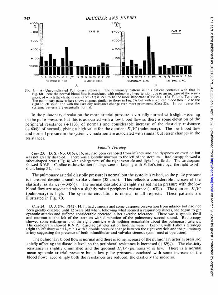
FIG. 7. (A) Uncomplicated Pulmonary Stenosis. The pulmonary pattern in this patient contrasts with that inFig. 6B; here the normal blood flow is associated with pulmonary hypertension due to an increase of the resist-ances, of which the elasticity resistance (E') is seen to be the more important (Case 21). (B) Fallot's Tetralogy.The pulmonary pattern here shows changes similar to those in Fig. 7A but with a reduced blood flow due to theright to left shunt and with the elasticity resistance change even more prominent (Case 23). In both cases thesystemic patterns are essentially normal.
In the pulmonary circulation the mean arterial pressure is virtually normal with slight wvideningof the pulse pressure, but this is associated with a low blood flow so there is some elevation of theperipheral resistance (+ 113% of normal) and considerable increase of the elasticity resistance(+604% of normal), giving a high value for the quotient E'/W (pulmonary). The low blood flowand normal pressure in the systemic circulation are associated with similar but lesser changes in theresistances.
Fallot's Tetlralogv,Case 23. D. S. (No. 0168), 16, m., had been cyanosed from infancy and had dyspnoea on exerttoll but
was not greatly disabled. There was a systolic murmur to the left of the sternum. Radioscopy showed asabot-shaped heart (Fig. 8) with enlargement of the right ventricle and light lung fields. The cardiogramishowed R.V.P. Cardiac catheterization findings were in keeping with Fallot's tetralogy, the right to leftshunt being 3 1./min.
The pulmonary arterial diastolic pressure is normal but the systolic is raised, so the pulse pressureis increased despite a small stroke volume (38 cm.3). This reflects a considerable increase of theelasticity resistance (+345%). The normal diastolic and slightly raised mean pressure with the lowblood flow are associated with a slightly raised peripheral resistance (+63%). The quotient E'/W(pulmonary) is high. The systemic circulation is normal in all respects. These patterns areillustrated in Fig. 7B.
Case 24. D. J. (No. P142), 14, f., had cyanosis and some dyspnzea on exertion from infancy but had notbeen greatly disabled until 12 years old when, following what seemed a respiratory illness, she began to getcyanotic attacks and suffered considerable decrease in her exercise tolerance. There was a systolic thrilland murmur to the left of the sternum with diminution of the pulmonary second sound. Radioscopyshowed some enlargement of the right ventricle with nothing remarkable about the pulmonary arteries.The cardiogram showed R.V.P. Cardiac catheterization findings were in keeping with Fallot's tetralogy(right to left shunt= 25 ./min.) with a double pressure change between the right ventricle and the pulmonaryartery suggesting the presence of both infundibular and valvular stenosis (confirmed at operation).
The pulmonary blood flow is normal and there is some increase of the pulmonary arteriat pressure,chiefly affecting the diastolic level, so the peripheral resistance is increased (+89%). The elasticityresistance is slightly diminished and the quotient E'/W (pulmonary) is low. There is a normalmean systemic arterial pressure but a low pulse pressure associated with some increase of theblood flow: accordingly both the resistances are reduced, the elasticity the more so.
- c-
on March 9, 2020 by guest. P
rotected by copyright.http://heart.bm
j.com/
Br H
eart J: first published as 10.1136/hrt.14.2.225 on 1 April 1952. D
ownloaded from

CONGENITAL HEART DISEASE
Summary of Cases with Pulmonary StenosisIn this group the only feature common to all cases is a pulmonary stenosis, ranging in degree
as can be seen from the right ventricular pressures in Table III, from moderate to severe. It isclear that there is no typical circulatory pattern associated with this owing to the large number ofother variables in these patients. Despite the small number in this series and the complexity ofthe inter-relationships involved it seems to us that some correlations are suggested by our findings.The first case is unusual in having a left to right shunt'associated with the stenosis but apart from thatdoes not differ in any fundamental way from the rest of the group in whom the blood flows rangefrom slightly raised to considerably diminished; so there seems no reason for not including thispatient in our consideration of the group as a whole.
FIG. 9 -Pulmonary Stenosis with left to right~iG. 8.-Fallot's Tetralogy. Plain radiograph of the chest shulmntrAgoardeieasograph dshwingthea larg
showing the sabot-shaped heart and small pulmonary ploayatre soitdwt oarteries associated with a high elasticity resistance (Table elastiitreitne(Tbe6Ian.i AIII and Fig 7B). Case 23.
The pulmonary arterial pressures in the group range from the moderately increased (Case 21)to the considerably diminished (Case 17). In those with a high pressure, this is due in Case 16 to ahigh blood flow with low resistances, in Case 23 predominantly to high elasticity resistance, and inCase 24 to a high peripheral resistance; Cases 21 and 23 show some increase of both resistances.In all the other patients the resistances are low.
There is a range of pulmonary elasticity resistances from -77 per cent (Case 16) to +607 percent (Case 22) of normal and comparing these values with the size of the pulmonary arteries asjudged on the radiographs, radioscopy and the angiocardiographs we have observed that there issome correlation between dilatation of the pulmonary artery and a low elasticity resistance and viceversa. Thus in Case 16 the large pulmonary arteries shown on angiocardiography (Fig. 9) areassociated with the lowest elasticity resistance; in Case 17 dilatation is again found with a low elas-ticity. Conversely in Case 23 (Fig. 8) the typical sabot shape due to small pulmonary arteries isassociated with a high elasticity resistance. In Case 22, where the elasticity resistance was the highestin this group, altho'ugh there is some prominence in the pulmonary conus area on the plain radio-graph, the angiocardiograph shows that much of this is due to the ventricular enlargement and that,the pulmonary arteries are small, especially in the lung fields.
There seems to be no simple relation between the degree of stenosis and the disability (e.g. Case19 appears to be one of the most severe stenoses but his disability is slight) which is scarcely sur-
243
e
on March 9, 2020 by guest. P
rotected by copyright.http://heart.bm
j.com/
Br H
eart J: first published as 10.1136/hrt.14.2.225 on 1 April 1952. D
ownloaded from
DEUCHAR AND KNEBEL
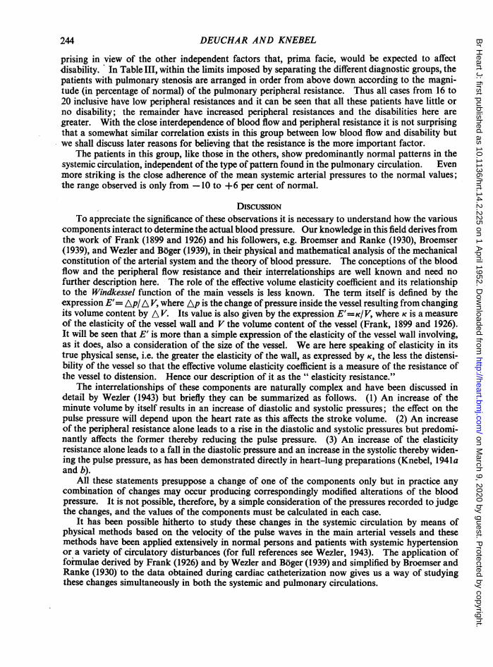
prising in view of the other independent factors that, prima facie, would be expected to affectdisability. In Table- III, within the limits imposed by separating the different diagnostic groups, thepatients with pulmonary stenosis are arranged in order from above down according to the magni-tude (in percentage of normal) of the pulmonary peripheral resistance. Thus all cases from 16 to20 inclusive have low peripheral resistances and it can be seen that all these patients have little orno disability; the remainder have increased peripheral resistances and the disabilities here aregreater. With the close interdependence of blood flow and peripheral resistance it is not surprisingthat a somewhat similar correlation exists in this group between low blood flow and disability butwe shall discuss later reasons for believing that the resistance is the more important factor.
The patients in this group, like those in the others, show predominantly normal patterns in thesystemic circulation, independent of the type of pattern found in the pulmonary circulation. Evenmore striking is the close adherence of the mean systemic arterial pressures to the normal values;the range observed is only from -10 to +6 per cent of normal.
DISCUSSIONTo appreciate the significance of these observations it is necessary to understand how the various
components interact to determine the actual blood pressure. Our knowledge in this field derives fromthe work of Frank (1899 and 1926) and his followers, e.g. Broemser and Ranke (1930), Broemser(1939), and Wezler and Boger (1939), in their physical and mathematical analysis of the mechanicalconstitution of the arterial system and the theory of blood pressure. The conceptions of the bloodflow and the peripheral flow resistance and their interrelationships are well known and need nofurther description here. The role of the effective volume elasticity coefficient and its relationshipto the Windkessel function of the main vessels is less known. The term itself is defined by theexpression E'= Ap/A V, where Ap is the change of pressure inside the vessel resulting from changingits volume content by A V. Its value is also given by the expression E'=K/V, where K is a measureof the elasticity of the vessel wall and V the volume content of the vessel (Frank, 1899 and 1926).It will be seen that E' is more than a simple expression of the elasticity of the vessel wall involving,as it does, also a consideration of the size of the vessel. We are here speaking of elasticity in itstrue physical sense, i.e. the greater the elasticity of the wall, as expressed by K, the less the distensi-bility of the vessel so that the effective volume elasticity coefficient is a measure of the resistance ofthe vessel to distension. Hence our description of it as the " elasticity resistance."
The interrelationships of these components are naturally complex and have been discussed indetail by Wezler (1943) but briefly they can be summarized as follows. (1) An increase of theminute volume by itself results in an increase of diastolic and systolic pressures; the effect on thepulse pressure will depend upon the heart rate as this affects the stroke volume. (2) An increaseof the peripheral resistance alone leads to a rise in the diastolic and systolic pressures but predomi-nantly affects the former thereby reducing the pulse pressure. (3) An increase of the elasticityresistance alone leads to a fall in the diastolic pressure and an increase in the systolic thereby widen-ing the pulse pressure, as has been demonstrated directly in heart-lung preparations (Knebel, 1941aand b).
All these statements presuppose a change of one of the components only but in practice anycombination of changes may occur producing correspondingly modified alterations of the bloodpressure. It is not possible, therefore, by a simple consideration of the pressures recorded to judgethe changes, and the values of the components must be calculated in each case.
It has been possible hitherto to study these changes in the systemic circulation by means ofphysical methods based on the velocity of the pulse waves in the main arterial vessels and thesemethods have been applied extensively in normal persons and patients with systemic hypertensionor a variety of circulatory disturbances (for full references see Wezler, 1943). The application offormulae derived by Frank (1926) and by Wezler and Boger (1939) and simplified by Broemser andRanke (1930) to the data obtained during cardiac catheterization now gives us a way of studyingthese changes simultaneously in both the systemic and pulmonary circulations.
244
on March 9, 2020 by guest. P
rotected by copyright.http://heart.bm
j.com/
Br H
eart J: first published as 10.1136/hrt.14.2.225 on 1 April 1952. D
ownloaded from

CONGENITAL HEART DISEASE
It is known from the work on the systemic circulation just referred to that the normal values forthese components vary with age. At present factual data are only available for calculation of thenormal pulmonary values in adults, so, as described, we have had to construct from a variety ofsources some approximations for the values of childhood. From these calculations it is possibleto give some quantitative expression to the well known differences between the two circulations(Cournand, 1947 and 1950). Normally, the minute volume of the two circulations is the same; itfollows that the pressure differences between them must arise from differences of resistances. Fromour figures it can be seen that the greater difference is in the peripheral resistance (which for thesystemic circulation is 7 times as large as for the pulmonary in adults), the difference in the elasticityresistances being only about one-third of this. This is shown in the important quotient E'/W witha value 3 5 for the pulmonary circulation as opposed to 1 2 for the systemic in normal adults.These resistances are not, however, fixed quantities and we are discussing values observed underresting or basal conditions; this is well illustrated by the figures obtained in our normal case (Fig. 2)where the high cardiac output has been accommodated in both circulations by equal changes in thetwo resistances so that the pressures are maintained within normal limits.
In our patients we have observed a wide range of variations from the normal circulatory patternsand many have shown pulmonary hypertension. In the patients with left to right shunts we haveobserved examples of this hypertension due solely to high minute volume (Fig. 3A) and othersdue partly to an increase of the resistances (Fig. 3B), but in all the increased flow is the chief factor.Other members of this group must not be overlooked who, like our normal case above, have accom-modated the increased flow by reducing the resistances and so maintained normal pressures. Bycontrast the two patients with Eisenmenger's complex have pulmonary hypertension with smallminute volumes and therefore due solely to changes in the resistances (Fig. 4A and B). The groupwith primary pulmonary hypertension similarly have hypertension due to an increase of resistancesalthough here the elasticity is less affected (Fig. 5B). Our case of transposition of the great vessels(Fig. 5A) occupies a somewhat intermediate position. We have also observed slight pulmonaryhypertension in a few patients with pulmonary stenosis and in these the responsible componentsare again the resistances, but from Fig. 7 and the values in Table III for Case 22 it is clear that theelasticity resistance is of greater importance here. Pulmonary hypertension in these patients maytherefore result from a considerable variety of mechanisms.
In the pulmonary stenotic group we have already drawn attention to the inverse relation betweenthe size of the pulmonary artery and the observed elasticity resistance; thus dilated pulmonaryarteries have been observed in cases with very low values and, conversely, small arteries with highvalues. This important observed relationship agrees with the relationship expected from the defini-tion of the elasticity resistance which, as already noted, is concerned with the vessel size in such away that, other things being equal, the larger the vessel the less the elasticity resistance. The im-portance of the elasticity resistance in relation to the pulmonary arterial pressure in this group hasjust been noted, hence the importance of the vessel size. The qualification " other things beingequal" in the statement concerning elasticity resistance and vessel size is important. If the vesselbe distended by a high pressure the size will be large but the elasticity resistance may be large alsoowing to the increase of the factor K when the vessel wall is stretched to near its elastic limit. Thisaccounts for the significance we have attached in some patients to relatively low elasticity resistancesoccurring with considerable hypertension, and also for the absence of the simpler relation betweenelasticity resistance and vessel size in many of the earlier cases with pulmonary hypertension,although it commonly holds in the first group when hypertension is slight or absent.
The elasticity resistance must clearly play an important role also in the matter of pulsation of thepulmonary vessels as seen on radioscopy. We have endeavoured to investigate this in some selectedcases that have already been discussed elsewhere by Campbell (1951), adding a few others from thepulmonary stenotic group for contrast. Campbell has concluded that increased pulmonary bloodflow is the important factor and that raised arterial pressure although having some effect in producingvisible pulsation is less important.
245
on March 9, 2020 by guest. P
rotected by copyright.http://heart.bm
j.com/
Br H
eart J: first published as 10.1136/hrt.14.2.225 on 1 April 1952. D
ownloaded from

DEUCHAR AND KNEBEL
It is possible from our data to analyse the relationship more exactly. During systole, part ofthe volume of blood ejected from the ventricle flows directly into the peripheral circulatory bed, therest serving to dilate the arterial tree from its diastolic volume; during diastole this latter portion isalso driven into the periphery by the elastic recoil of the arterial wall; this is the essence of theWindkessel function of the arterial system. If the volume A V in the expression E'= Ap/A V isthe volume just mentioned that dilates the arteries, then Ap is the pulse pressure, available from thepressure records. E' is calculable and therefore it is possible to obtain A V. This volume is ameasure of the " storing capacity" of the Windkessel function of the arteries and is spoken of asthe pulse volume, as opposed to the stroke volume of which it forms a part. The stroke volume(Vs) being also known it is possible to determine from the simple function A V/Vs the portion ofit which is stored in the arterial tree in the way described. This value is related closely to thequotient E'/W and we shall discuss this elsewhere (Deuchar and Knebel, 1952).
The values we have obtained in 17 patients are given in Table IV together with the size of the pul-monary artery and the degree of pulsation seen on radioscopy as reported by Dr. Campbell.
The two examples of atrial septal defect (Cases 2 and 3) provide an instructive contrast; bothhave large and nearly equal stroke volumes yet in the former there was striking pulsation whereasin the latter it was seen only in the larger pulmonary vessels. From the table it can be seen that thisis associated with a much larger pulse volume and " stored portion " in the former than in the latter(A V/Vs=036 and 017, respectively). This suggests that a large pulse volume or" stored portion"may be in some patients an important determining factor in producing visible pulsation. Furtherstudy of the table shows that all the patients in the anomalous pulmonary venous drainage andventricular septal defect group have large pulse volumes and stored portions (A V/Vs rangingfrom 030 to 056) associated with considerable pulsation. (Case 9 has a large pulse volumewith Vs=21 cm.3 as the age of the patient was only 3 years). In the pulmonary stenotic groupthere was one patient (Case 18) who showed some, pulsation in the large pulmonary vessels.This patient stands in striking contrast to the other two with pulmonary stenosis in respect especiallyof the pulse volume, which is several times greater in the case with pulsation than in the others(Table IV); interestingly the pulse pressure in this patient was lower than in the others (Pp= 13 and26 or 23 mm. Hg, respectively).
Less easy to explain on the basis of the pulse volume is the pulsation seen in the Eisenmenger,transposition, or primary pulmonary hypertension groups for although the pulsation is generallysomewhat less obvious it is none the less present with pulse volumes that are on the average con-siderably less than those we have so far considered. In all these cases, however, on account of thehigh peripheral resistance they have, the " stored portion " is high. It may be that this being so therate of dilatation of the arteries in systole is greater and therefore more obvious on radioscopyalthough the amplitude may not be so great. It would be interesting to investigate this by means ofelectrokvmographic studies.
It seems, therefore, that in many cases a large pulse volume is the chief factor in producingvisible pulsation, as can be readily understood, but even when this is small a high " stored portion"appears by some less obvious mechanism to be important; as already noted a high " storedportion" often occurs when the peripheral resistance and hence the arterial pressure is high, sothat this latter mechanism is probably the one concerned in patients showing visible pulsation inwhom there is hypertension without an increased blood flow.
In all the diagnostic groups we have considered there are some patients who are disabled to agreater or lesser degree but the reasons why one patient should be disabled and another not are byno means clear. It is easy to visualize the mechanical effect of a pulmonary stenosis restricting theblood flow and producing disability, but, from our cases it is clear that the disability is not relatedto the severity of the'stenosis alone (see Case 19 in Table III). The cause of disability in othergroups such as those with left to right shunts is less apparent. We have noticed in the course ofthis study, and have already commented upon, a relationship that seems to exist between an increasedpulmonary peripheral resistance and the presence of disability in all of these groups, even those with
246
on March 9, 2020 by guest. P
rotected by copyright.http://heart.bm
j.com/
Br H
eart J: first published as 10.1136/hrt.14.2.225 on 1 April 1952. D
ownloaded from

CONGENITAL HEART DISEASE
TABLE IVPULMONARY CIRCULATORY HAEMODYNAMICS AND APPEARANCES OF THE PULMONARY ARTERIES ON RADIOSCOPY *
r~~~~ ~~~ ~ ~
.-_.______________
Elasticity Peripheral Pulse Stroke Pulse PulsationResistance Resistance Pres- -olueStoredsize OfCase ResEn W Et/W sure Volume Volume Portion Right Peri-Dynes Dynes. sec. (mm. A V,/Vs P.A. Right Middle Pena HilarCM.5 CM.5 Hg) (cm.3) (cm.3) P.A. Third pheral Dance
Third
ATRIAL SEPTAL DEFECT
2 7 500 60 83 15 110 40 0364 ++ ++ ++ (+) (+)3 7 1500 100 150 25 128 22 0.172 ++ ++ (+) - -
ANOMALOUS PULMONARY VENOUS DRAINAGE
4 19 800 170 4-8 35 177 58 0328 ++ ++ + (+) -5 8 1100 1 700 1-6 32 69 39 0566 ++ _++ + + -
VENTRICULAR SEPTAL DEFECT
6 12 740 240 3-1 48 234 87 0372 ++ ++ ++ + +7 11 1000 200 50 53 236 71 0-301 ++ ++ + + +8 48 2000 540 3-8 66 138 44 0-319 +(+) +++ ..++ + +9 3 2000 410 4*9 32 69 21 0-304 ++(+) ++ ++ ++ -
EISENMENGER'S COMPLEX
10 44 5800 1800 3 2 67 38 15 0 395 ++ ++ ++ + +
11 33 4900 1900 2-5 54 32 15 0468 ++ + - - -TRANsPOSMON OF THE GREAT VESSELS
12 1l31 2100 960 2-2 41 58 26 0448 +++ ++ + (+) -
PRIMARY PULMONARY HYPERTENSION
13 25 2600 1300 20 57 62 29 0468 +++ + (+) - -14 19 2000 1100 1 8 32 39 21 0538 ++ - - - -15 22 6800 4000 1-7 48 21 9 0-428 ++(+) ++ + - -
PULMONARY STENOSIS
18 22 470 120 3-9 13 95 37 0-390 +(+) + (+) - -22 9 9300 660 14*1 23 14 3 0-214 - - - - -23 16 4000 390 10-4 26 37 9 0-243 - - - - -
* The data relating to radioscopic appearances were kindly supplied by Dr. Maurice Campbell.
pulmonary stenosis. With these it is difficult to dissociate the possible influence of the pulmonaryblood flow and of the peripheral resistance as these tend to be inversely related and prima faciein this group a reduction of pulmonary blood flow seems a likely cause of disability. In the groupof left to right shunts, however, the position is somewhat different as here the blood flow is increasedin all cases, but disability here also correlated best with high values of the peripheral resistance, andin the other groups the high peripheral resistance is probably the determining factor in producinga low blood flow.
We would suggest that a high peripheral resistance may reflect a loss of flexibility in the circula-
247
on March 9, 2020 by guest. P
rotected by copyright.http://heart.bm
j.com/
Br H
eart J: first published as 10.1136/hrt.14.2.225 on 1 April 1952. D
ownloaded from

tory adjustments, which we have already seen (vide Case 1) are normally able to accommodatechanges in blood flow such as are required on exercise, and that such loss of flexibility is an essentialmechanism by which disability may arise whatever the anatomical abnormality present. Althoughthe correlation we have observed has involved the peripheral resistance, this is probably only becauseit plays the predominant role in the regulating mechanisms and elasticity resistance changes mayalso affect the flexibility of adjustment. Our series has not, however, been sufficiently large to allowof investigation of this point which, in the light of our observations regarding the relation betweenvessel size and elasticity resistance, may be of importance in understanding the significance of post-stenotic dilatation of the pulmonary arteries in pulmonary stenosis.
Another feature that we regard as of fundamental physiological importance has been noted tobe common to all groups and that is the difference between the changes observed in the two circu-
lations. The nature of these differences has already been stressed in each group and is well shown inthe figures illustrating the circulatory dynamics. In all cases the systemic circulation is essentiallynormal or any adjustments found are directed towards maintaining normality of arterial pressureespecially and also of blood flow where possible. In contrast the pulmonary circulation may sufferlarge changes from normal and these can sometimes be interpreted as being necessary, in view ofthe cardiac abnormality present, to assist the maintenance of normal conditions within the systemiccirculation. Our findings, therefore, would suggest that the regulatory mechanisms of the systemiccirculation are predominant and that such regulation as exists in the pulmonary circulation is sub-ordinate to the needs of the systemic.
SUMMARYA method has been devised, using data obtained by cardiac catheterization studies and formulk
relating to the Windkessel (pressure reservoir) function of the arterial system, that enables btheelasticity resistance as well as the peripheral resistance to be calculated for both systemic andpulmonary circulations.
Sets of normal values according to age have been calculated for both circulations. These figuresgive a quantitative expression to the differences between the systemic and pulmonary circulations.
The data obtained from cardiac catheterization studies on 24 subjects with congenital heartdisease have been used and the findings are presented in groups according to the anatomicaldiagnosis; the patterns of the circulatory changes in these groups have been illustrated and described.
Pulmonary hypertension has been observed in many patients and the different mechanisms of itsproduction have been discussed with reference to the observed findings. It has been shown that incongenital heart disease it may result from a variety of circulatory changes requiring full analysisfor their differentiation.
In cases with pulmonary stenosis a relation has been observed between vessel size and theelasticity resistance in the pulmonary arterial system which is in agreement with the theoreticallypredicted relation and affects the circulatory dynamics.
The relation between the elasticity resistance and the occurrence of visible pulsation in thepulmonary arteries on radioscopy has been examined, and it has been shown that these are probablyconnected by the influence of the former on the pulse volume or "stored portion" of the strokevolume in conditions either of increased flow or high pressure.A relationship has been observed in all groups between a high pulmonary peripheral resistance
and disability; the significance of this has been discussed briefly.In all groups the circulatory patterns in the two circulations have shown striking differences;
the systemic circulation is always adjusted towards normal whatever the changes in the pulmonarycirculation.
We wish to express our great indebtedness and thanks to Dr. Maurice Campbell for his encouragement and assist-ance in producing this paper and also for permission to report details of several of his patients. We also wish to thankMr. R. C. Brock for the use of the case records of some of his patients and Dr. G. A. Zak for the catheterizationresults in Cases 8,20 and 21. We are greatly indebted also to the Department of Medical Photography and Illustration,Guy's Hospital, for preparing the figures for publication.
DEUCHAR AND KNEBEL248
on March 9, 2020 by guest. P
rotected by copyright.http://heart.bm
j.com/
Br H
eart J: first published as 10.1136/hrt.14.2.225 on 1 April 1952. D
ownloaded from

CONGENITAL HEART DISEASE
REFERENCES
Bing, R. J., Vandam, L. D., and Gray, F. D., Jr. (1947a). Bull. Johns Hopkins Hosp., 80, 107 and 121.D
(1947b). Bull. Johns Hopkins Hosp., 80, 323.Brinton, W. D. (1950). Brit. Heart J., 12, 305.Broemser, P. (1939). Verh. dtsch. Ges. Kreislaufforsch., 289.- , and Ranke, 0. F. (1930). Z. Biol., 90, 467.Burchell, H. B., and Wood, E. H. (1950). Proc. Staff Meet. Mayo Clin., 25, 41.Campbell, M. (1948). Guy's Hosp. Rep., 97, 1.- , (1951). Brit. Heart J., 13, 438.Cournand, A. (1945). Fed. Proc., 4, 207.
(1947. Bull. New York Acad. Med., 23, 27.(1950). Circulation, 2, 641.and Ranges, H. A. (1941). Proc. Soc. Exper. Biol. Med., 46, 462.
, Baldwin, J., and Himmelstein, A. (1949). Cardiac Catheterization in Congenital Heart Disease. New York:Commonwealth Fund., Motley, H. L., Himelstein, A., Dresdale, D., and Baldwin, J. (1947). Amer. J. Phys., 150, 267., Riley, R. L., Breed, E. S., Baldwin, E. de F., and Richards, D. W. (1945). J. Clin. Invest., 24, 106.
De Navasquez, S. J., Forbes, J. R., and Holling, H. E. (1940). Brit. Heart J., 2, 177.Deuchar, D. C., and Knebel, R. (1952). Z. Ges. exp. Med. In the press.- , and Zak, G. A. (1952). Guy's Hosp. Rep., 101, 1.Dexter, L., Haynes, F. W., Burwell, C. S., Eppinger, E. C., Sagerson, R. P., and Evans, J. M. (1947a). J. Clin.
Invest., 26, 554.Sosman, M. C., and Evans, J. M. (1947b). J. Clin. Invest., 26, 561.
Douglas, C. G., and Priestley, J. G. (1948). Human Physiology. 3rd Edition. Oxford: Oxford Univ. Press.Fleisch, A. (1927). Handbuch der normalen und pathologischen Physiologie, 7, part 2, 1273. Edited by Bethe and
Emsden. Berlin: Springer.Forssman, W. (1929). Klin. Wschr., 8, 2085 and 2287.Frank, 0 (1899). Z. Biol., 37, 483.
(1926). Z. Biol., 85, 91.Hamilton, W. F., Brewer, G., and Brotman, I. (1934). Amer. J. Phys., 107, 427.Handelsman, J. C., Bing, R. J., Campbell, J. A., and Griswold, H. E. (1948). Bull. Johns Hopkins Hosp., 82, 615.Hickam, J. B. (1949). Amer. Heart J., 38, 801.-, and Cargill, W. H. (1948). J. Clin. Invest., 27, 10.Holling, H. E., and Zak, G. A. (1950). Brit. Heart J., 12, 153.Knebel, R. (1941a and b). Arch. exp. Path. Pharmak., 197, 485 and 496.Lagerlof, H., and Werko, L. (1948). Acta. Phys. Scand., 16, 75.McMichael, J., and Sharpey-Schafer, E. P. (1944). Brit. Heart J., 6, 33.Riley, R. L., Himmelstein, A., Motley, H. L., Weiner, H. M., and Cournand, A. (1948). Amer. J. Phys., 152, 372.Rossier, P. H., Maier, C., and Volkmann, M. (1950). Cardiologia, 15, 147.Selzer, A. (1949). Arch. intern. Med., 84, 798.Soothill, J. F. (1951). Guy's Hosp. Rep., 100, 232.Stead, E. A., Jr. Hickam, J. B., and Warren, J. V. (1947). Tr. Ass. Amer. Physicians, 60, 74.
Warren, J. V., Merrill, A. J., and Brannon, E. S. (1945). J. Clin. Invest., 24, 326.Werko, L. (1947). Acta. Med. Scand. Suppl., 193, 1.Wezler, K. (1938) Z. Biol., 98, 438.- (1942). Z. Altersforschung, 3, 199, and 4, 1.
(1943). Wein Arch. Inn. Med., 37, 226.and Boger, A. (1939). Ergebn. Physiol., 41, 292.
Wood, P. (1950). Brit. med. J., 2, 639 and 693.
S
249
on March 9, 2020 by guest. P
rotected by copyright.http://heart.bm
j.com/
Br H
eart J: first published as 10.1136/hrt.14.2.225 on 1 April 1952. D
ownloaded from