Embed Size (px)
Citation preview
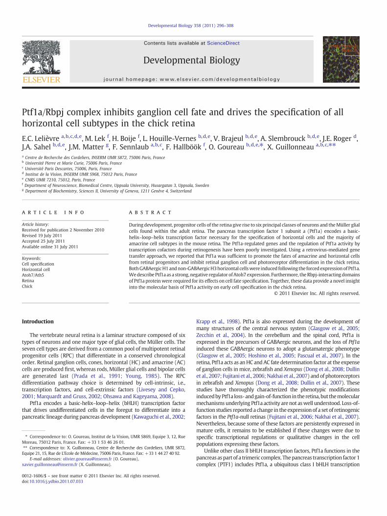
Developmental Biology 358 (2011) 296–308
Contents lists available at ScienceDirect
Developmental Biology
j ourna l homepage: www.e lsev ie r.com/deve lopmenta lb io logy
Ptf1a/Rbpj complex inhibits ganglion cell fate and drives the specification of allhorizontal cell subtypes in the chick retina
E.C. Lelièvre a,b,c,d,e, M. Lek f, H. Boije f, L. Houille-Vernes b,d,e, V. Brajeul b,d,e, A. Slembrouck b,d,e, J.E. Roger d,J.A. Sahel b,d,e, J.M. Matter g, F. Sennlaub a,b,c, F. Hallböök f, O. Goureau b,d,e,⁎, X. Guillonneau a,b,c,⁎⁎a Centre de Recherche des Cordeliers, INSERM UMR S872, 75006 Paris, Franceb Université Pierre et Marie Curie, 75006 Paris, Francec Université Paris Descartes, 75006, Paris, Franced Institut de la Vision, INSERM UMR S968, 75012 Paris, Francee CNRS UMR 7210, 75012, Paris, Francef Department of Neuroscience, Biomedical Centre, Uppsala University, Husargatan 3, Uppsala, Swedeng Department of Biochemistry, Sciences II, University of Geneva, 1211 Genève 4, Switzerland
⁎ Correspondence to: O. Goureau, Institut de la VisionMoreau, 75012 Paris, France. Fax: +33 1 53 46 26 01.⁎⁎ Correspondence to: X. Guillonneau, Centre de RecheEquipe 21, 15, Rue de L'Ecole de Médecine, 75006 Paris, Fra
E-mail addresses: [email protected] (O. [email protected] (X. Guillonneau).
0012-1606/$ – see front matter © 2011 Elsevier Inc. Aldoi:10.1016/j.ydbio.2011.07.033
a b s t r a c t
a r t i c l e i n f oArticle history:Received for publication 2 November 2010Revised 19 July 2011Accepted 25 July 2011Available online 31 July 2011
Keywords:Cell specificationHorizontal cellAtoh7/Ath5RetinaChick
During development, progenitor cells of the retina give rise to six principal classes of neurons and theMüller glialcells found within the adult retina. The pancreas transcription factor 1 subunit a (Ptf1a) encodes a basic-helix–loop–helix transcription factor necessary for the specification of horizontal cells and the majority ofamacrine cell subtypes in the mouse retina. The Ptf1a-regulated genes and the regulation of Ptf1a activity bytranscription cofactors during retinogenesis have been poorly investigated. Using a retrovirus-mediated genetransfer approach, we reported that Ptf1a was sufficient to promote the fates of amacrine and horizontal cellsfrom retinal progenitors and inhibit retinal ganglion cell and photoreceptor differentiation in the chick retina.BothGABAergicH1andnon-GABAergicH3horizontal cellswere induced following the forcedexpressionof Ptf1a.Wedescribe Ptf1a as a strong, negative regulator ofAtoh7 expression. Furthermore, the Rbpj-interacting domainsof Ptf1a proteinwere required for its effects on cell fate specification. Together, these data provide a novel insightinto the molecular basis of Ptf1a activity on early cell specification in the chick retina.
, UMR S869, Equipe 3, 12, Rue
rche des Cordeliers, UMR S872,nce. Fax: +33 1 44 27 40 92.reau),
l rights reserved.
© 2011 Elsevier Inc. All rights reserved.
Introduction
The vertebrate neural retina is a laminar structure composed of sixtypes of neurons and one major type of glial cells, the Müller cells. Theseven cell types are derived from a common pool of multipotent retinalprogenitor cells (RPC) that differentiate in a conserved chronologicalorder. Retinal ganglion cells, cones, horizontal (HC) and amacrine (AC)cells are produced first, whereas rods, Müller glial cells and bipolar cellsare generated last (Prada et al., 1991; Young, 1985). The RPCdifferentiation pathway choice is determined by cell-intrinsic, i.e.,transcription factors, and cell-extrinsic factors (Livesey and Cepko,2001; Marquardt and Gruss, 2002; Ohsawa and Kageyama, 2008).
Ptf1a encodes a basic-helix–loop–helix (bHLH) transcription factorthat drives undifferentiated cells in the foregut to differentiate into apancreatic lineage during pancreas development (Kawaguchi et al., 2002;
Krapp et al., 1998). Ptf1a is also expressed during the development ofmany structures of the central nervous system (Glasgow et al., 2005;Zecchin et al., 2004). In the cerebellum and the spinal cord, Ptf1a isexpressed in the precursors of GABAergic neurons, and the loss of Ptf1ainduced these GABAergic neurons to adopt a glutamatergic phenotype(Glasgow et al., 2005; Hoshino et al., 2005; Pascual et al., 2007). In theretina, Ptf1a acts as anHC andAC fate determination factor at the expenseof ganglion cells in mice, zebrafish and Xenopus (Dong et al., 2008; Dullinet al., 2007; Fujitani et al., 2006;Nakhai et al., 2007) andof photoreceptorsin zebrafish and Xenopus (Dong et al., 2008; Dullin et al., 2007). Thesestudies have thoroughly characterized the phenotypic modificationsinducedbyPtf1a loss- andgain-of-function in the retina, but themolecularmechanisms underlying Ptf1a activity are not aswell understood. Loss-of-function studies reported a change in the expressionof a set of retinogenicfactors in the Ptf1a-null retinas (Fujitani et al., 2006; Nakhai et al., 2007).Nevertheless, because some of these factors are persistently expressed inmature cells, it remains to be established if these changes were due tospecific transcriptional regulations or qualitative changes in the cellpopulations expressing these factors.
Unlike other class II bHLH transcription factors, Ptf1a functions in thepancreas aspart of a trimeric complex. Thepancreas transcription factor 1complex (PTF1) includes Ptf1a, a ubiquitous class I bHLH transcription

297E.C. Lelièvre et al. / Developmental Biology 358 (2011) 296–308
factor (E-protein) and the mammalian Suppressor of Hairless protein,Rbpj (PTF1-J complex) or its paralog, Rbpjl (PTF1-L complex) (Beres et al.,2006;Masui et al., 2007; Obata et al., 2001). The Ptf1a–Rbpj interaction isalso required for Ptf1a to specify GABAergic cells in the dorsal spinal cordand the cerebellum (Hori et al., 2008). However, the requirement of thePtf1a–Rbpj interaction in the specification of retinal cell types remains tobe elucidated.
The chick retina has proven to be a powerful model to study retinaldifferentiation and its genetic regulation. Moreover, in contrast to micepossessing only one axon-bearing HC subtype (Peichl and Gonzalez-Soriano, 1994), the chick retina has both axon-less and axon-bearingHCsubtypes, as found in the majority of vertebrate retinas (Gallego, 1986;Genis-Galvez et al., 1979). The chick retina contains H1 axon-bearingHCs, H2 axon-less “stellate” HCs and H3 axon-less “candelabrum” HCs(Edqvist et al., 2008; Tanabe et al., 2006). Therefore, this model is morerepresentative of Ptf1a activity on HC subtype determination invertebrates.
This study aimed to gain a better understanding of the molecularregulation underlying the Ptf1a activity during retinal developmentusing the chick retina model. In this study, we showed that the forcedexpression of Ptf1a leads to a massive disorganization of thedifferentiated retina and changes in retinal cell representation thatcomplement the retinal phenotypeof Ptf1a-nullmice: an increase ofACsand allHC subtypes and a decrease of ganglion and photoreceptor cells.Using this model, we identified several retinogenic factors that wererapidly regulated by Ptf1a overexpression and reported that ectopicPtf1a strongly downregulated Atoh7 expression in the chick retina.Finally, our study demonstrated that the interaction between Ptf1a andRbpj cofactors was required for Ptf1a activity in the developing retina.
Materials and methods
Animals
Gallus gallus white leghorn embryos were obtained from Haas(France). The animal experimentation was conducted in accordancewith the Association for Research in Vision and Ophthalmology (ARVO)statement on the use of animals in Ophthalmic and Vision Research anda protocol approved by our local animal care committee.
DNA sequences
Chick genome sequences (V2.0 and V3.0) were obtained fromThe Genome Institute at Washington University (http://genome.wustl.edu/). The expressed sequence tags (accession numbers: BU487258 andBU347629) were ordered from Source Bioscience and sequenced(MWG-Operon). The alignments were performed using Basic LocalAlignment Search Tool available atNCBI (http://blast.ncbi.nlm.nih.gov/)and VNTI software (Invitrogen).
Retroviral stock and plasmid production
The mouse Ptf1awas subcloned using specific CDS primers into thepDONR221 vector (Invitrogen) using BP clonase (Invitrogen). The Ptf1amutants were generated using Polymerase Chain Reaction (PCR)-basedmutagenesis. The pDONR221-Ptf1a plasmid was recombined in thepresence of LR recombinase (Invitrogen) into either RCAS-BP(A)-NHY,which allowed for the fusion of a HA-tagwith the N-terminal part of thePtf1a protein (gift from Dr. Loftus) or the pCIG gateway vectors (Rogeret al., 2006). The RCAS viral stocks with titersN1×108 colony formingunits permilliliter (cfu/ml)wereprepared by transfectingDF1 cellswithviral DNA constructs using FuGENE6 Reagent (Roche Diagnostics)(Yang, 2002). Viruses were concentrated by centrifugation using 100 KCentrifugal Filters Amicon Ultra (Millipore). All embryos were injectedinto the right optic vesicle at embryonic day 2 (E2). The openings in theeggs were sealed with scotch tape and further incubated at 37.5 °C.
Parental RCAS-BP(A) viruses (Hughes et al., 1987) served as controls inthe viral infection experiments.
Cryosection
The eyes were fixed in 4% paraformaldehyde (PFA) and incubatedin 30% sucrose (Sigma) in phosphate-buffered saline (PBS) overnightfollowed by 1 h incubation at 37 °C in PGS (PBS, 7.5% gelatin (Sigma),and 10% sucrose). Eyes were embedded in PGS, frozen at −50 °C inisopentane and stored at −80 °C. Ten micrometer-thick cryosectionswere collected.
Cell dissociation
The retinas were collected in HBSS without Ca2+/Mg2+, trypsinized(1 mg/ml) (Sigma) and incubated for 10 min at 37 °C. The digestionwasstopped with HBSS with Ca2+/Mg2+ containing a trypsin inhibitor(Sigma) (1 mg/ml), and the cells were mechanically dissociated in thepresence of DNaseI (Sigma) (0.1 mg/ml). For flow cytometry, thesuspended cells were washed with PBS, fixed for 15 minwith 2% PFA atroom temperature (RT) and washed in PBS before immunostaining.For the manual counting, 1×10⁵ cells/ml were seeded on poly-L-lysine(Sigma) coated plates. After 2 h, adherent cells were fixed 10 min with2% PFA at RT before immunostaining.
Antibodies
Mouse anti-gag (3c2), anti-Ap2α (3B5), anti-Islet1 (39.4D5), anti-Lim1/2 (4 F2), and anti-Visinin (7 G4) antibodies were purchasedfrom the Developmental Studies Hybridoma Bank. Mouse anti-Brn3a(MAB1585), mouse anti-Glutamine Synthetase (MAB302), mouseanti-Prox1 (MAB5652), and rabbit anti-PhosphoHistone3 (07–145)antibodies were obtained from Millipore, rabbit anti-gag (p27) fromCharles River, rabbit anti-Protein Kinase Cα (PKCα) (Sc-208) fromSanta Cruz, and rabbit anti-Prox1 (DP3501P) from Acris. The rabbitanti-Ptf1a antibody was a gift from Dr. Edlund (Umeå University), andthe rabbit anti-TrkA was a gift from Pr. Lefcort (Montana StateUniversity). Primary antibodies were detected using AlexaFluor488-,AlexaFluor594-, AlexaFluor633- (Invitrogen) or Phycoerythrine(PE)- (Beckman Coulter) conjugated goat antibodies.
Immunostaining and in situ hybridization
Immunostaining was performed as previously described (Rogeret al., 2006). Retinal section counterstaining was performed with 4′,6′-diamidino-2-phenylindole (DAPI) (1:1200). Apoptotic cells weredetected by terminal deoxynucleotidyl transferase (TdT)-mediateddUTP nick end labeling (TUNEL) labeling using the In Situ Cell DeathDetection Kit (Roche Diagnostics). For S-phase cell labeling, 5 μg (E7embryos) or 10 μg (E9 embryos) of 5-ethynyl-2′-deoxyuridine (EdU)were injected into the intravitreal space 2 h before being sacrificed.The Click iTTM EdU Imaging kit (Invitrogen) was used to visualizeEdU-positive cells.
Digoxigenin-labeled Rbpj and cAtoh7 probes were generated bycloning template DNA (the full length coding sequence of cAtoh7 andthe cRbpj coding sequence from 123 to 873 bp) into pCRII-TOPOvector (Invitrogen). In situ hybridization was performed as previouslydescribed (Roger et al., 2006).
Images were captured with a DM5500microscope (Leica) equippedwith an ORCA ER Hamamatsu camera or a LSM710 confocalmicroscope(Zeiss) and analyzed with MetaMorph software (Molecular Devices).
Flow cytometry and cell sorting
Dissociated cells were incubated in blocking buffer (PBS, 10% fetalcalf serum (FCS), 2% goat serum, and 0.1% saponin) 1 h at RT. Primary

298 E.C. Lelièvre et al. / Developmental Biology 358 (2011) 296–308
antibodies were diluted in blocking buffer, applied 2 h at RT, andwashed. The cell pellets were then incubated for 1 h with 5 μl ofPE- and/or AlexaFluor633-conjugated secondary antibodies (1:10) atRT. The data were collected using an LSRII cytometer (BD Biosciences)and analyzed using BD FACS DIVA software. For cell sorting, a total of1×105 GFP-positive cells were collected in lysis buffer for RNAextraction using Vantage Sorter (BD Biosciences).
Electroporation
The eyes were collected at E5, and the pigmented epithelium wasremoved from the retina. Whole retinas were positioned in anelectroporation chamber (CUY520P5, Sonidel, Napagene, Japan) filledwith PBS with Ca2+/Mg2+ containing plasmids (0.5 μg/μl). Electropo-ration was performed using a CUY215C square wave generator(Sonidel) and consisted of 5 pulses at 30 V with 50 ms duration, 1 sinterval and repeated twice. Whole retinas were then cultured asfloating explants at 37 °C in DMEM, 10% FCS, and 1% penicillin/streptomycin. For confocal microscopy, the retinas were fixed 20 minin 4%PFA and rinsed for 24 h in PBS. For inclusion, the fixed retinaswerecryoprotected and embedded in PGS.
RNA isolation, reverse-transcription and quantitative PCR
Total RNA was extracted using the Nucleospin RNAII Kit (MachereyNagel). Retrotranscription was performed as described previously(Roger et al., 2006). Real-time PCR was performed using 7300 Real-Time PCR System (Applied Biosystems) in a 20 μl final volume withPower SYBR Green PCR Master Mix (Applied Biosystems) and 0.25 μMprimers. All samples were run in triplicate. Primers used for real-timePCR analysis are listed in Table S1 (see supplementary materials).
Cell fractionation
Protein extracts were collected in hypotonic buffer (20 mMHEPESpH 7.9, 1 mM Na3VO4, 1 mM NaGlycerophoshate, 5 mM EDTA pH 7.5,1 mM EGTA pH 7.5, 1 mM DTT, and protease inhibitors (Calbiochem,Merck4 Biosciences)) plus 0.2% Nonidet P-40, incubated on ice for15 min and centrifuged (20 s at 16,000 g). For nuclear extracts, pelletswere resuspended in saline buffer (120 mM NaCl and 20% glycerol inhypotonic buffer) supplemented with protease inhibitor cocktail,incubated for 30 min at 4 °C and centrifuged for 20 min at 16,000 g tocollect the supernatant. For cytosolic extracts, NaCl (120 mM) wasadded to the supernatant of the first centrifugation, and the extractwas centrifuged for 20 min at 16,000 g to remove debris. Glycerol(10%) was then added to the supernatant.
Western blotting
Western blotting was conducted as described previously (Rogeret al., 2006) using the following primary antibodies: mouse anti-HA(MMS 101R, Covance), goat anti-LaminB (sc-6216, Santa Cruz), mouseanti-Actin (Sigma), goat anti-Ptf1a (ab62818, Abcam), and rat anti-Rbpj(T6719, Institute of Immunology, Tokyo, Japan).
Co-immunoprecipitation
InfectedDF1 cellswere lysed in immunoprecipitation buffer (50 mMTris/HCl pH 8.0, 120 mM NaCl, 0.5% Nonidet P-40, and proteaseinhibitors) (Rodolosse et al., 2009). The lysates were incubatedovernight and centrifuged for 20 min at 16,000 g. Protein extracts(250 μg) were incubated with anti-HA antibody (Covance, 1/100) for2 h at RT to immunoprecipitate the HA-tagged proteins. The immunecomplex was then captured by incubating for 2 h at RT with 40 μl ofProtein-G Sepharose beads (Fast Flow, GE Healthcare). The complexeswere pelleted by gentle centrifugation, washed four times with
immunoprecipitation buffer and eluted in the loading buffer beforewestern blotting.
Statistical analyses
The quantifications are expressed as the mean±standard error ofthe mean (s.e.m.). Analyses were conducted using the Student's t-testassuming equal variances (two groups) or one-way analysis of variancefollowed by Tukey's multiple comparison tests (three or more groups)using Prism 5.0 (Graphpad software). (*) pb0.05, (**) pb0.01, (***)pb0.001, (ns) not significant. panova, p-value of the one-way analysis ofvariance; pTukey, p-value of the Tukey post-hoc test.
Results
Forced expression of Ptf1a results in a loss of retinal lamination
The Ptf1a spatiotemporal expression pattern has been studiedduring retinogenesis (Boije et al., 2008; Fujitani et al., 2006; Nakhaiet al., 2007).Ptf1amRNAwasdetected fromembryonic day3 (E3) to E15in the developing chick retina. Early, it is expressed in the innerneuroblastic layer (NBL) and then becomes restricted to the innernuclear layer (INL) (Boije et al., 2008). To gain insight into the functionof Ptf1a during chick retinal development, a replication-competentretrovirus, RCAS, was used to drive ectopic and overexpression of Ptf1ain the retina. The chick Ptf1a gene sequence is not fully known.We havecharacterized its C-terminal using the available information at TheGenome Institute and two ESTs. It contains the highly conserved bHLHDNA-binding and Rbpj/Rbpjl-interaction domains (Beres et al., 2006)(Fig. S1). However, the N-terminal of the chick Ptf1a gene has remaineduncharacterized in recent chicken genome assemblies. In contrast, themouse Ptf1a sequence is fully known, and the Rbpj/Rbpjl-interactiondomains have been extensively studied (Beres et al., 2006). Moreover,previous studies have reported that mouse Ptf1a blocks the differenti-ation of dILB excitatory neurons and promotes dILA inhibitory neuronspecification in the chick spinal cord, a phenotype that was consistentwith Ptf1a loss of function studies in mouse (Glasgow et al., 2005; Horiet al., 2008; Mizuguchi et al., 2006; Wildner et al., 2006). These resultssuggest a conserved activity of mouse Ptf1a in avian and rodentmodels.Therefore, themouse Ptf1a cDNAwas chosen to allow formisexpressionexperiments of Ptf1a in the chick retina and for geneticmanipulations ofthe Ptf1a coding sequence. Effective wild type Ptf1a protein mis/overexpression from RCAS-HA-Ptf1a (RCAS-Ptf1a) was verified bywestern blotting for the HA-tagged proteins in DF1 cells (Fig. 1A) andby immunohistochemistry in E9 retinas (Fig. 1D). In the chick retina,viral injections resulted in a widespread infection. Retinal patchesinfected with the different RCAS viruses and infected retinal dissociatedcells could be detected using either the p27 or 3c2 anti-gag antibodies(Figs. 1C and E).
During any developmental stage, the plexiform and nuclear layershad formed properly in the empty-RCAS (RCAS) control infectedpatches (Figs. 1F–G, J–K and N–O). At E7, no major structural defectswere observed in the RCAS-Ptf1a-infected patches other than thepresence of a thicker retina and a decreased cell density (Figs. 1H and I).In contrast, at E9 (Figs. 1L andM) and,more extensively, at E12 (Figs. 1Pand Q), the lamination of the RCAS-Ptf1a-infected patches was severelydisturbedwithdisorganizedplexiformandnuclear layers. No changes inthe retinal structure were observed outside of the RCAS-Ptf1a-infectedareas (data not shown).
Forced expression of Ptf1a affects progenitor cell proliferation and migration
These defects in retinal structure and lamination might arise fromdisturbedmigration ormodification of the cell cycle. Therefore, S-phasecells were pulse-labeled with 5-ethynyl-2′-deoxyuridine (EdU), andmitotically active progenitors were immunostained using a Phospho-
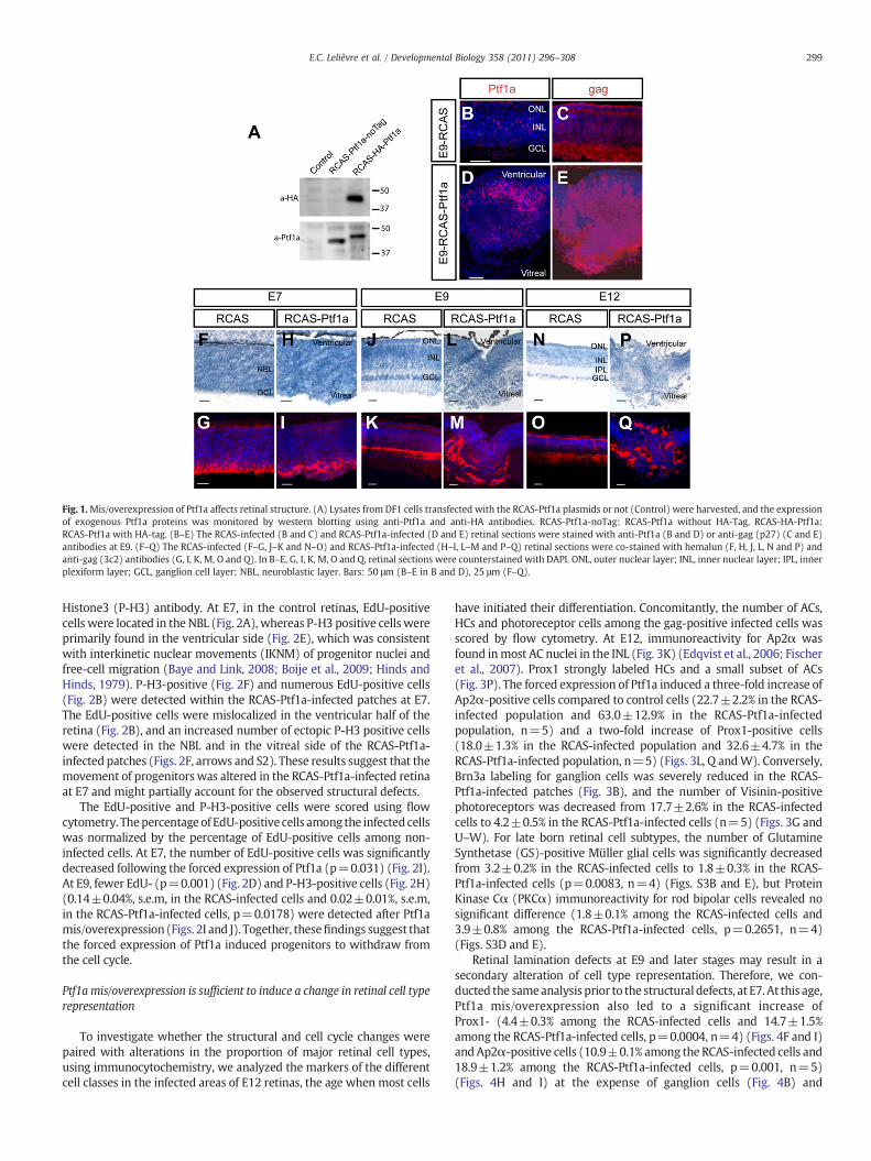
Fig. 1.Mis/overexpression of Ptf1a affects retinal structure. (A) Lysates from DF1 cells transfected with the RCAS-Ptf1a plasmids or not (Control) were harvested, and the expressionof exogenous Ptf1a proteins was monitored by western blotting using anti-Ptf1a and anti-HA antibodies. RCAS-Ptf1a-noTag: RCAS-Ptf1a without HA-Tag, RCAS-HA-Ptf1a:RCAS-Ptf1a with HA-tag. (B–E) The RCAS-infected (B and C) and RCAS-Ptf1a-infected (D and E) retinal sections were stained with anti-Ptf1a (B and D) or anti-gag (p27) (C and E)antibodies at E9. (F–Q) The RCAS-infected (F–G, J–K and N–O) and RCAS-Ptf1a-infected (H–I, L–M and P–Q) retinal sections were co-stained with hemalun (F, H, J, L, N and P) andanti-gag (3c2) antibodies (G, I, K, M, O and Q). In B–E, G, I, K, M, O and Q, retinal sections were counterstained with DAPI. ONL, outer nuclear layer; INL, inner nuclear layer; IPL, innerplexiform layer; GCL, ganglion cell layer; NBL, neuroblastic layer. Bars: 50 μm (B–E in B and D), 25 μm (F–Q).
299E.C. Lelièvre et al. / Developmental Biology 358 (2011) 296–308
Histone3 (P-H3) antibody. At E7, in the control retinas, EdU-positivecells were located in the NBL (Fig. 2A), whereas P-H3 positive cells wereprimarily found in the ventricular side (Fig. 2E), which was consistentwith interkinetic nuclear movements (IKNM) of progenitor nuclei andfree-cell migration (Baye and Link, 2008; Boije et al., 2009; Hinds andHinds, 1979). P-H3-positive (Fig. 2F) and numerous EdU-positive cells(Fig. 2B) were detected within the RCAS-Ptf1a-infected patches at E7.The EdU-positive cells were mislocalized in the ventricular half of theretina (Fig. 2B), and an increased number of ectopic P-H3 positive cellswere detected in the NBL and in the vitreal side of the RCAS-Ptf1a-infected patches (Figs. 2F, arrows and S2). These results suggest that themovement of progenitors was altered in the RCAS-Ptf1a-infected retinaat E7 and might partially account for the observed structural defects.
The EdU-positive and P-H3-positive cells were scored using flowcytometry. Thepercentage of EdU-positive cells among the infected cellswas normalized by the percentage of EdU-positive cells among non-infected cells. At E7, the number of EdU-positive cells was significantlydecreased following the forced expression of Ptf1a (p=0.031) (Fig. 2I).At E9, fewer EdU- (p=0.001) (Fig. 2D) and P-H3-positive cells (Fig. 2H)(0.14±0.04%, s.e.m, in the RCAS-infected cells and 0.02±0.01%, s.e.m,in the RCAS-Ptf1a-infected cells, p=0.0178) were detected after Ptf1amis/overexpression (Figs. 2I and J). Together, thesefindings suggest thatthe forced expression of Ptf1a induced progenitors to withdraw fromthe cell cycle.
Ptf1a mis/overexpression is sufficient to induce a change in retinal cell typerepresentation
To investigate whether the structural and cell cycle changes werepaired with alterations in the proportion of major retinal cell types,using immunocytochemistry, we analyzed the markers of the differentcell classes in the infected areas of E12 retinas, the age when most cells
have initiated their differentiation. Concomitantly, the number of ACs,HCs and photoreceptor cells among the gag-positive infected cells wasscored by flow cytometry. At E12, immunoreactivity for Ap2α wasfound inmost AC nuclei in the INL (Fig. 3K) (Edqvist et al., 2006; Fischeret al., 2007). Prox1 strongly labeled HCs and a small subset of ACs(Fig. 3P). The forced expression of Ptf1a induced a three-fold increase ofAp2α-positive cells compared to control cells (22.7±2.2% in the RCAS-infected population and 63.0±12.9% in the RCAS-Ptf1a-infectedpopulation, n=5) and a two-fold increase of Prox1-positive cells(18.0±1.3% in the RCAS-infected population and 32.6±4.7% in theRCAS-Ptf1a-infected population, n=5) (Figs. 3L, Q andW). Conversely,Brn3a labeling for ganglion cells was severely reduced in the RCAS-Ptf1a-infected patches (Fig. 3B), and the number of Visinin-positivephotoreceptors was decreased from 17.7±2.6% in the RCAS-infectedcells to 4.2±0.5% in the RCAS-Ptf1a-infected cells (n=5) (Figs. 3G andU–W). For late born retinal cell subtypes, the number of GlutamineSynthetase (GS)-positive Müller glial cells was significantly decreasedfrom 3.2±0.2% in the RCAS-infected cells to 1.8±0.3% in the RCAS-Ptf1a-infected cells (p=0.0083, n=4) (Figs. S3B and E), but ProteinKinase Cα (PKCα) immunoreactivity for rod bipolar cells revealed nosignificant difference (1.8±0.1% among the RCAS-infected cells and3.9±0.8% among the RCAS-Ptf1a-infected cells, p=0.2651, n=4)(Figs. S3D and E).
Retinal lamination defects at E9 and later stages may result in asecondary alteration of cell type representation. Therefore, we con-ducted the sameanalysis prior to the structural defects, at E7.At this age,Ptf1a mis/overexpression also led to a significant increase ofProx1- (4.4±0.3% among the RCAS-infected cells and 14.7±1.5%among the RCAS-Ptf1a-infected cells, p=0.0004, n=4) (Figs. 4F and I)andAp2α-positive cells (10.9±0.1% among the RCAS-infected cells and18.9±1.2% among the RCAS-Ptf1a-infected cells, p=0.001, n=5)(Figs. 4H and I) at the expense of ganglion cells (Fig. 4B) and

300 E.C. Lelièvre et al. / Developmental Biology 358 (2011) 296–308
photoreceptor cells (10.2±0.7% in the RCAS-infected retinas and6.5±0.4% in the RCAS-Ptf1a-infected retinas, p=0.0014, n=5)(Figs. 4D and I). These results indicated that the Ptf1a mis/over-expression effects on early-born retinal cell types occurred prior to thedefects in retinal lamination and argued against the notion that the lossof photoreceptors and ganglion cells was secondary to the retinallaminationdefects. Interestingly, a fewAp2α-positive cells (Fig. 4H)andthe majority of Prox1-positive cells (Fig. 4F) were ectopically located inthe vitreal side of the retina.
We performed TUNEL labeling to assess if the reduction of some cellsubtypes could be due to early cell death. A significantly higher numberof apoptotic cells, located throughout the entire thickness of the NBL,was detected in the RCAS-Ptf1a patches (43.3±9.8 cells per area)compared to the patches infected with the control virus (8.5±0.2 cellsper area, n=3, p=0.0305) (Figs. 4J–L).
Ptf1a mis/overexpression is sufficient to induce all HC subtype specification
Our results showed that Ptf1amis/overexpression increased the sizeof the HC population. The chick HC population is composed of at leastthree HC subtypes that can be molecularly distinguished by theexpression of Prox1, Lim1, Islet1, TrkA and GABA (Edqvist et al., 2008;Fischer et al., 2007). All three subtypes express Prox1. The H1 subtype(50% of all HCs) has GABA, is Lim1-positive, Islet1-negative, and TrkA-negative. The H2 (approximately 10% of all HCs) also has GABA, is Lim1-negative, Islet1-positive but TrkA-negative, and the H3 (approximately40% of all HCs) does not have GABA, is Lim1-negative and Islet1/TrkAdouble-positive. We first investigated the normal Ptf1a expression inthe HC subtypes in the developing chick retina. The retinawas analyzedwith respect to Ptf1a, Prox1, Lim1 and Islet1 immunoreactivity in E6.5retinas when HCs migrated bi-directionally and, in E9, E12 and E16
Fig. 2. Effects of Ptf1a mis/overexpression on retinal progenitor cell proliferation. (A–H) Sectretinas were stained with EdU and anti-gag (3c2) antibodies (A–D) or with anti-P-H3 andectopic location in the RCAS-Ptf1a-infected retinas. (I and J) Quantitative analysis of EdU-poand E9. Values represent the mean±s.e.m. The percentage of EdU-positive cells among theinfected cells for each embryo. NBL, neuroblastic layer; GCL, ganglion cell layer; INL, inner
retinas when the HC layer (HCL) was established (Boije et al., 2009;Edqvist andHallbook, 2004). At E6.5, after the onset of Prox1 expressionin HCs, 31±5% (n=3) of the Ptf1a-immunoreactive cells were alsoProx1-positive (Fig. 5A). The Ptf1a/Prox1 double-positive cells werelocalized on the vitreal side of the retina but also scattered in theprospective INL, which was consistent with the location of migratingHCs at this age. At E9, all Ptf1a/Prox1 double-positive cells were locatedin the developing HCL. Of the Prox1-positive cells in the HCL, 64±5%(n=3)were Ptf1a-positive HCs (Fig. 5B). This result implies that not allHCs expressed Ptf1a at this age. The Lim1-positive cells were Ptf1a-positive, and this overlap was found at both E9 (96±1%, n=3) in theHCL and at earlier ages on the vitreal side (Figs. 5E and F). In contrast,only a few Ptf1a/Islet1 double-positive cells could be seen at E6.5 and atE9 (5±0.6%, n=3) (Figs. 5I and J).
To assess whether Ptf1awas able to direct the development of all HCsubtypes, we next analyzed the presence of HC subtype markers in theRCAS-Ptf1a-infected patches. At E7, an accumulation of Prox1/Lim1double-positive and Prox1/Islet1 double-positive cells (Figs. 6B and D)were found in the normal location of the GCL in the RCAS-Ptf1a-infectedpatches compared to the control patches, suggesting that both the H1and H3 subtypes had been generated following Ptf1a mis/overexpres-sion. These results were strengthened by the TrkA immunoreactivity ofIslet1-positive cells in the RCAS-Ptf1a-infected areas (Fig. 6F). Further-more, the accumulation of some HCs in the vitreal side suggested thatHC migration was either arrested or delayed by Ptf1a mis/over-expression. These results were similar at E9 when all HCs had normallymigrated back to the HCL. Supernumerary Prox1/Lim1 double-positive(Fig. 6H), Prox1/Islet1 double-positive (Fig. 6J), and Islet1/TrkA double-positive cells (Fig. 6L)were observed in the RCAS-Ptf1a-infected retinas.Islet1-positive and TrkA-negative cellswere foundwithin the clusters ofProx1-positive cells of the RCAS-Ptf1a-infected patches (Fig. 6l, arrows),
ions from the RCAS-infected (A, C, E and G) and the RCAS-Ptf1a-infected (B, D, F and H)anti-gag antibodies (E–H) at E7 and E9. In F, arrows point to P-H3 positive cells in ansitive (I) or P-H3-positive cells (J) among the RCAS- and RCAS-Ptf1a-infected cells at E7infected cells was normalized by the percentage of EdU-positive cells among the non-nuclear layer. Bars: 25 μm.

Fig. 3. Effects of wild-type and mutant forms of Ptf1a on retinal cell differentiation. (A–T) The RCAS-infected (A, F, K and P), RCAS-Ptf1a-infected (B, G, L and Q), RCAS-Ptf1a-ΔC2-infected (C, H, M and R), RCAS-Ptf1a-ΔC12-infected (D, I, N and S) and RCAS-Ptf1a-Δbasic-infected (E, J, O and T) patches from E12 retinas were immunostained usinganti-Brn3a (A–E), anti-Visinin (F–J), anti-Ap2α (K–O), and anti-Prox1 (P–T) antibodies. For clarity, the gag labeling is not represented. Retinal sections were counterstained withDAPI. (U and V) Representative flow cytometry analysis after staining the RCAS-infected (U) and RCAS-Ptf1a-infected (V) dissociated cells with anti-gag (p27) and anti-Visininantibodies at E12. (W) Quantitative analysis of Visinin-, Ap2α- and Prox1-positive cells among the infected cells at E12. Values represent the mean±s.e.m. of at least four separateeye counts. ONL, outer nuclear layer; INL, inner nuclear layer; GCL, ganglion cell layer; PE, Phycoerythrin; Alexa633, AlexaFluor633. Bars: 50 μm.
301E.C. Lelièvre et al. / Developmental Biology 358 (2011) 296–308
suggesting thepresence ofH2HCs.Moreover, TrkAand Lim1wereneverexpressed in the same cell, indicating thatH1andH3HCswere correctlyspecified (Fig. 6N). We found that the proportion of non-GABAergicTrkA-positive cells among the Prox1-positive HCs decreased signifi-cantly from 43.3±1.9% in the RCAS-infected patches to 27.1±2.8% inthe RCAS-Ptf1a-infected areas (p=0.0007), whereas a slight, but notsignificant, increase of Lim1-positive HCs was observed (51.9±2.2% inthe RCAS-infected patches and 58.9±2.1% in the RCAS-Ptf1a-infectedpatches, p=0.0898) (Fig. 6O). Moreover, the back-migration of ectopicHCs to the HCL was inhibited, and ectopic HCs remained on the vitrealside of the E9 RCAS-Ptf1a-infected retinas.
Ptf1a overexpression induces changes in retinogenic factor expression
To gain knowledge about themolecularmechanisms underlying thechanges in retinal cell specification, we studied early changes in the
expression of a selected set of genes involved in retinal differentiation.The E5whole chick retinaswere electroporated ex vivowith pCIG-Ptf1a-GFP plasmids, which drive the green fluorescent protein (GFP)expression (Roger et al., 2006) (Fig. 7A). The GFP-positive cells wereisolated by fluorescence activated cell sorting as early as 36 h afterelectroporation to study changes in gene expression that were asindependent as possible from cell differentiation. The empty pCIG-GFPplasmids were used as controls. First, our quantification demonstratedthat electroporation of pCIG-Ptf1a in the chick retinas resulted in astrong expression of mouse Ptf1a mRNA, compared to the referencelevel (the non-specific amplification in pCIG-electroporated retinalcells) (Fig. 7B). Surprisingly, endogenous chick Ptf1amRNA levels weresignificantly decreased in the mouse Ptf1a-overexpressing cells(p=0.0002) (Fig. 7B). No effects were observed on either Ngn2, adirect target of Ptf1a in the spinal cord and the cerebellum (Henke et al.,2009), or Sox2. We found that Pax6 and Ap2α, coding for two

Fig. 4. Retinal cell type representation is modified in E7 retinas prior to lamination defects. (A–H) The E7 RCAS-infected (A, C, E and G) and RCAS-Ptf1a-infected (B, D, F and H) retinalpatches were immunostained using an anti-gag antibody and either anti-Brn3a (A and B), anti-Visinin (C and D), anti-Prox1 (E and F) or anti-Ap2α (G–H) antibodies. Celltype-specific labeling in panels A–H was represented without gag labeling in panels a–h, respectively. (I) Quantitative analysis of Visinin-, Ap2α- or Prox1-positive cells among theinfected cells. The values represent the mean±s.e.m of at least four separate retinal counts and are representative of two independent injections. (J–L) Cells undergoing apoptosiswere TUNEL-labeled at E7 in the RCAS-infected (J) and RCAS-Ptf1a-infected (K) patches detected using anti-gag (p27) antibody. Sections in J and K were counterstained with DAPI.(L) Quantitative analysis of the number of apoptotic cells per area in the RCAS- and RCAS-Ptf1a-infected patches at E7. The values represent the mean±s.e.m. of at least threeseparate retinas. NBL, neuroblastic layer; GCL, ganglion cell layer. Bars: 50 μm (A, C, E, and G in A and B, D, F, and H in B), 25 μm (J and K).
302 E.C. Lelièvre et al. / Developmental Biology 358 (2011) 296–308
transcription factors highly expressed in ACs (Belecky-Adams et al.,1997; de Melo et al., 2003; Edqvist et al., 2006), were upregulated inPtf1a-overexpressing cells (p=0.009) (Fig. 7B). A significant 2.6-foldincrease of NeuroAB mRNA, a transcription factor suggested to beinvolved in GABAergic AC development (Ohkawara et al., 2004), wasalso observed in Ptf1a-overexpressing cells (p=0.0167). Interestingly,the expression levels of Ascl1, a pro-amacrine transcription factor inchick retina (Mao et al., 2009), was downregulated (p=0.0037),suggesting that Ptf1a might act downstream of Ascl1 and inhibit itsexpression in the retina asdescribed in the spinal cord (Hori et al., 2008).The expression level of Otx2, a key regulatory gene for photoreceptordevelopment (Nishida et al., 2003), was decreased by 2.5-fold in Ptf1a-overexpressing cells (p=0.0076), and Atoh7, a transcription factorinvolved in ganglion cell specification (Liu et al., 2001;Matter-Sadzinskiet al., 2005), was strongly repressed by 6.4-fold by Ptf1a overexpression(p=0.0071) (Fig. 7B). Furthermore, Atoh7mRNAwas nearly undetect-able by in situ hybridization in RCAS-Ptf1a-infected patches (Fig. S5C).Consistently, ex vivo, ectopic Ptf1a downregulated the activity of thechick Atoh7 promoter. Indeed, a significant decrease in the number ofGFP-positive cells was observed in retinas co-electroporated withRCAS-Ptf1a and pAtoh7-GFP, a plasmid that drives GFP expressionunder the control of the chick Atoh7 promoter, compared to RCAS andpAtoh7-GFPco-electroporated retinas (Figs. S5E, F andH,panova=0.001,ptukeyb0.01).
We further focused our analysis on transcription factors involved inHC genesis. The mRNA levels of Neurod4 (Fig. 7B) and Prox1 (Fig. 7C),two factors involved in HC development (Dyer et al., 2003; Inoue et al.,2002), were not altered, whereas Lim1, which is necessary for H1specification in the chick retina (Suga et al., 2009), was inducedfollowing Ptf1a overexpression (Fig. 7C). Foxn4 is upstream of Ptf1a
duringAC/HC specification inmouse retina (Fujitani et al., 2006; Li et al.,2004). Interestingly, Ptf1a overexpression induced a two-fold decreaseof Foxn4 expression levels (p=0.0030) (Fig. 7C). To investigatewhetherFoxn4 conversely regulates Ptf1a in the chick retina, we electroporatedE5 retinaswithpCIG-Foxn4-GFPvectors (Fig. 7A). Foxn4overexpressionresulted in a significant increase in Ptf1a expression (p=0.0498) andLim1 and Prox1 mRNA expression levels (Fig. 7C).
Ectopic Ptf1a requires its interaction with endogenous RBP-J
Ptf1a forms the PTF1 complex with any one of the E-proteins andRbpj (PTF1-J) or Rbpjl (PTF1-L) (Beres et al., 2006). Ptf1a interacts withRbpjl through its C1 domain and with Rbpj through its C2 and C1domains (Fig. S4K). The C2 domain deletionwas sufficient to abolish theformationof the PTF1-J complex (Beres et al., 2006). In situhybridizationrevealed that Rbpj mRNA was expressed throughout the NBL at earlystages and in the INL at later stages up to E12 (see Figs. S6A–J).Conversely, Rbpjl mRNA was neither detected during chick retinaldevelopment (data not shown) nor in mature retinas by quantitativePCR (Fig. S6K). To test the requirement for Rbpj proteins for Ptf1aactivity in the chick retina, we generated Ptf1a-ΔC2 and Ptf1a-ΔC1ΔC2(Ptf1a-ΔC12)mutant formsof Ptf1a that interactwithE-proteinsbut areunable to interactwithRbpj (Beres et al., 2006) (Fig. S4L). The Ptf1a-ΔC2and Ptf1a-ΔC12 proteins were expressed at levels equivalent to Ptf1afull-length proteins (Fig. S4N). Moreover, subcellular fractionationrevealed that deletion of the C1 and C2 domains did not preventPtf1a-ΔC12 nuclear importation (Fig. S4O). The injection of RCAS-Ptf1a-ΔC2 and RCAS-Ptf1a-ΔC12 in the optic cup did not lead to grossstructural defects of the retina (Figs. 3 and S4E–H). However, the outerplexiformandnuclear layersweredisruptedwith rosette-like structures

Fig. 5. Ptf1a expression in subtypes of developing horizontal cells. Epifluorescence micrographs show Ptf1a, Prox1 (A–D), Ptf1a, Lim1 (E–H), and Ptf1a, Islet1 (I–L) double-labeling ofE6.5, E9, E12 and E16 chick retinas, and the corresponding split fluorescence images are to the right of each panel. Insets in b, f, and j are higher magnifications of the boxes in B, F andJ. Arrows point at double-labeled cells. GCL, ganglion cell layer; INL, inner nuclear layer; ONL, outer nuclear layer. Bars: 25 μm.
303E.C. Lelièvre et al. / Developmental Biology 358 (2011) 296–308
(Figs. 3H and I). Early-born cell types were analyzed by immunochem-istry andflowcytometry in the RCAS-Ptf1a-ΔC2- andRCAS-Ptf1a-ΔC12-infected population of E12 retinas, but no alteration of the cell typedistribution was observed following the forced expression of these twoPtf1amutant forms (Figs. 3C–D,H–I,M–N,R–S andW). The requirementof Rbpj–Ptf1a interaction for gene regulation downstream of ectopicPtf1a was assessed by evaluating the ability of ectopic Ptf1a-ΔC12 toregulate the Atoh7 promoter activity. Co-electroporation of pAtoh7-GFPwith the RCAS-Ptf1a-ΔC12 plasmids did not decrease the number ofGFP-positive cells compared to co-electroporation with the controlRCAS vectors (Figs. S5G and H).
Ectopic Ptf1a activity is independent from Rbpj pool depletion
Rbpj is a major downstream effector of the Notch pathway. Previousstudies have suggested that the Ptf1a activity might be downregulatedby sequestering Rbpj from Ptf1a (Cras-Meneur et al., 2009) followingNotchactivation. Therefore,we examined if ectopic Ptf1amight functionin the chick retina by conversely sequestering Rbpj. We generated aPtf1a-Δbasic mutant where the highly conserved RER-R amino acids(168–172) (Figs. S4L and M) in the basic domain were replaced byAVA-A. These mutations were shown to abolish MyoD and bHLHa15/Mist1 bHLH transcription factor binding to DNA (Lemercier et al., 1998;Skowronska-Krawczyk et al., 2005). The Ptf1a-Δbasic protein was stillimported into the nucleus (see Fig. S4O) and retained its ability tointeract with Rbpj (see Fig. S4P). The in vitro reporter assay confirmedthat the transcriptional activity of Ptf1a-Δbasic was strongly decreased(data not shown). No alteration of retinal structure and cell typerepresentation (Figs. 3E, J, O, T and S4I and J) was detected within theE12 RCAS-Ptf1a-Δbasic-infected patches. This result argues against the
hypothesis that the activity of wild-type ectopic Ptf1a might becompletely mediated by a disruption of Rbpj function.
Discussion
Our study demonstrated that Ptf1a was sufficient to affect the chickretinal cell composition in an Rbpj-dependent manner. Our “candidate-gene” approach showed that Ptf1a overexpression selectively upregu-lated NeuroAB, Ap2α and Lim1 while it downregulated Otx2 and Atoh7.This study provides a newmolecular basis for Ptf1a activity in specifyingthe ACs and all HC subtypes and inhibiting photoreceptor and ganglioncell genesis.
Cellular and molecular mechanisms underlying ectopic Ptf1a activityduring chick retinal development
Several hypotheses may explain the changes in cell typerepresentation induced by Ptf1a.
First, cell proliferation defects may change the final retinal cellcomposition. Indeed, early cell cycle exit has been shown to favorearly-born retinal cell types. Conversely, late cell cycle exit biasesprogenitor cells toward later neuronal fates (Austin et al., 1995;Dorsky et al., 1997; Henrique et al., 1997; Jadhav et al., 2006; Liveseyand Cepko, 2001; Nelson et al., 2007; Ohnuma et al., 2002; Silva et al.,2003; Yaron et al., 2006).We found that the forced expression of Ptf1ain the chick retina induced a premature cell-cycle exit, but led to adramatic decrease of early-born ganglion and cone photoreceptorcells. Thus, it is unlikely that cell proliferation defects alone accountfor the effect of ectopic Ptf1a activity in chick retinogenesis. How Ptf1ainduced cell cycle withdrawal remains to be defined. The cell cycleexit could be linked to the defects in the IKNM of retinal progenitors

Fig. 6. Ptf1amis/overexpression is sufficient to increase thenumber of all horizontal cell subtypes. (A–F) The RCAS-infected (A, C and E) and RCAS-Ptf1a-infected (B, D and F) patchesweredouble immunostained at E7with either anti-Lim1 and anti-Prox1 antibodies for H1 cells (A and B), anti-Islet1 and anti-Prox1 antibodies for H2 and H3 cells (C and D) or anti-Islet1 andanti-TrkA antibodies for H3 cells (E and F). a–f are highermagnifications of the boxes in A–F, respectively. a’–f’ and a”–f” are splitfluorescence images of a–f. Arrows point to some double-positive cells. (G–N) The RCAS-infected (G, I, K andM) and RCAS-Ptf1a-infected (H, J, L and N) patches at E9were double immunostainedwith either anti-Lim1 and anti-Prox1 (G and H),anti-Islet1 and anti-Prox1 (I and J), anti-Islet1 and anti-TrkA (K and L) or anti-TrkA and anti-Lim1 antibodies (M and N). g–n are highermagnifications of boxes in G–N, respectively. g’–n’and g”–n” are split fluorescent images of g–n, respectively. Arrows point to some Islet1-positive/TrkA-negative cells. (O) The quantification of Lim1- (H1), Islet1- (H2–H3) andTrkA-positive (H3) cells among the Prox1-positive HCs in the RCAS- and RCAS-Ptf1a-infected patches at E9. The values represent the mean±s.e.m. NBL, neuroblastic layer; ONL, outernuclear layer; INL, inner nuclear layer; GCL, ganglion cell layer; pHCL, prospective horizontal cell layer. Bars: 25 μm (A–F), 5 μm (a–f), 50 μm (G–N), 10 μm (g–n).
304 E.C. Lelièvre et al. / Developmental Biology 358 (2011) 296–308
observed following Ptf1a mis/overexpression. Indeed, in the chickretina, pharmacological perturbation of IKNM was shown to promotepremature neurogenesis (Murciano et al., 2002) through a reductionof Notch-mediated lateral inhibition (Del Bene et al., 2008; Murciano
et al., 2002). Consistent with this model, the expression of Hes5, atarget of the Notch pathway in the retina (Nelson et al., 2006), wassignificantly decreased in electroporated Ptf1a-overexpressing cells(data not shown). Moreover, the expression level of cyclin D1 that is

Fig. 7. Changes in retinogenic factor mRNA expression following Ptf1a overexpression. (A) Schematic structure of pCIG expression vectors. IRES, internal ribosomal entry site.(B) The expression level of candidate genes was analyzed by quantitative PCR (qPCR) on mRNA from GFP-positive sorted retinal cells electroporated at E5 with either pCIG-GFP(empty pCIG) or pCIG-Ptf1a-GFP (pCIG-Ptf1a). mmPtf1a, mouse Ptf1a; ggPtf1a, chick Ptf1a. (C) The expression level of a subset of genes involved in HC development was assessed byqPCR on mRNA isolated from GFP-positive sorted retinal cells electroporated at E5 with empty pCIG, pCIG-Ptf1a or pCIG-Foxn4-GFP (pCIG-Foxn4). Data represent the mean±s.e.m.of a least four retinas (Hayes et al., 2007; Matter-Sadzinski et al., 2001; Skowronska-Krawczyk et al., 2009).
305E.C. Lelièvre et al. / Developmental Biology 358 (2011) 296–308
required for RPC proliferation (Fantl et al., 1995; Godbout andAndison, 1996; Sicinski et al., 1995) was also significantly down-regulated in Ptf1a-overexpressing cells (data not shown), suggestingthat ectopic Ptf1a might regulate the core of the cell cycle machinery.
Second, Ptf1a might bias progenitor cells toward the AC and HCfates. We found that the expression of Atoh7 (Brown et al., 2001;Wang et al., 2001) and Otx2 (Nishida et al., 2003), which specifyganglion cells and photoreceptors, respectively, were inhibited in cellsthat overexpressed Ptf1a. In contrast, Lim1, involved in HC specifica-tion (Suga et al., 2009), and NeuroAB, which is presumably involved inAC genesis (Ohkawara et al., 2004), were upregulated. Thesetranscriptional regulations support a mechanism where ectopicPtf1a might act cell-autonomously to drive retinal progenitors towardthe AC and HC fates at the expense of ganglion cells andphotoreceptors by regulating their transcriptional program. Theeffects of ectopic Ptf1a on cell fate might also be influenced by cellproliferation regulation. Indeed, early cell cycle exit was shown toenhance the activity of proneural factors (Atoh7, Ngn1, and Neurod4)that promote early cell fates (Ohnuma et al., 2002). Recently, themajority of Ptf1a-expressing cells were found to originate fromAtoh7-positive progenitor cells, and Ptf1a expression drove their
differentiation toward ACs/HCs at the expense of ganglion cells in thezebrafish retina (Jusuf et al., 2011). Our overexpression study showedthat ectopic Ptf1a was a negative regulator of Atoh7. Therefore, itwould be interesting to assess whether endogenous Ptf1a, which isturned on after Atoh7 in the Atoh7-positive lineage, is able todownregulate Atoh7 expression during retinogenesis, how it mightregulate Atoh7, and if this regulation is involved in the fate switchtoward ACs/HCs.
Third, Ptf1amight also selectively induce the death of ganglion andphotoreceptor cells, modifying the cell representation in favor of ACs/HCs. The number of apoptotic cells was increased in Ptf1a-infectedretinas at E7, but the precise determination of their identity wasdifficult to assess because many apoptotic cells have lost theirdifferentiation markers. To our knowledge, no report has demon-strated a direct induction of cell death either by Ptf1a or by themisexpression of transcription factors without apoptotic regulatoryproperties. Dullin et al.(2007) did not detect any apoptosis inductionfollowing Ptf1a overexpression in Xenopus retinas, suggesting thatPtf1a has no pro-apoptotic function per se. Secondary cell death is acommon feature of transcription factor gain- and loss-of-functionsand has often been associated with defects in cell trans-commitment

306 E.C. Lelièvre et al. / Developmental Biology 358 (2011) 296–308
in the retina (Fujitani et al., 2006; Mao et al., 2009; Qiu et al., 2008;Zheng et al., 2009). Therefore, the increase in cell death is likely asecondary event that occurs following the late re-specification ofalready committed precursors. Alternatively, a lack of crucial trophicsupport and inappropriate cell–cell interactions may lead to theelimination of prematurely differentiated cells.
Transcriptional regulation upstream and downstream of Ptf1a
Our results showed that Foxn4 upregulated Prox1, Lim1 and Ptf1ain the chick retina, suggesting that Foxn4might be sufficient to induceHC fate in the developing chick retina, which is in contrast to mice(Li et al., 2004). Together with the upregulation of endogenous Ptf1aby Foxn4, these data confirm that Ptf1a functions downstream ofFoxn4 in the transcriptional cascade leading to HC specification asproposed in mice (Fujitani et al., 2006). Interestingly, we discoveredthat Ptf1a downregulated Foxn4, indicating that Ptf1a would berequired to finely regulate Foxn4 expression in HC and AC precursors.In line with this hypothesis, Li et al. reported that in Foxn4LacZ
knock-in mice, β-galactosidase (LacZ) expression was upregulatedand persisted longer than in wild type retinas (Li et al., 2004). ThisFoxn4 locus deregulation could result, at least partially, from the lossof Ptf1a expression in Foxn4−/− mice (Fujitani et al., 2006).
An increase in photoreceptor cells was reported in addition to thedecrease of ACs/HCs following Foxn4 and Ptf1a inactivation (Donget al., 2008; Dullin et al., 2007; Fujitani et al., 2006; Li et al., 2004;Nakhai et al., 2007). Consistently, our misexpression study demon-strated that Ptf1a inhibited photoreceptor cell production anddecreased Otx2 expression. Inversely, the loss of Otx2 in the mouseretina decreased the number of photoreceptors in favor of ACs(Nishida et al., 2003). These gain- and loss-of-function studieshighlight a possible connection between the production of HCs/ACson the one hand and photoreceptors and ganglion cells on the other.This balance may rely, to some extent, on Notch-mediated lateralinhibition. Notch signaling was shown to specifically inhibit mousephotoreceptor specification (Jadhav et al., 2006; Yaron et al., 2006). Invarious structures of the central nervous system, Ascl1 and Foxn4have been implicated in the regulation of the Delta-like/Notchsignaling pathway. Notably, in the ventral spinal cord, Foxn4 functionsupstream of Ascl1, Delta-like and Notch expression to regulate thediversification of excitatory and inhibitory V2 cells by lateralinhibition (Del Barrio et al., 2007). In the retina (Nelson et al., 2009;Nelson and Reh, 2008) andmouse dorsal spinal cord (Mizuguchi et al.,2006), Ascl1 activates Delta-like and Ptf1a expression. Thus, similar tothe spinal cord, Foxn4 and/or Ascl1 might initiate lateral inhibitionamong a subset of retinal progenitors. These factors would then berequired to sustain Notch signaling in AC/HC precursors that wereactivated by Delta-expressing neighboring cells that were prone todifferentiate as photoreceptor cells. Ascl1 and Foxn4 expressionwould also contribute to turning off the photoreceptor and ganglioncell specification programs in AC/HC precursors through Ptf1aactivation. Further studies will be needed to determine to whatextent Foxn4 and Ascl1 function together to regulate Notch signalingand Ptf1a expression in the chick retina. Ptf1a alone might not sustainNotch signaling because Hes5 was downregulated in Ptf1a-over-expressing cells (data not shown).
Surprisingly, endogenous chick Ptf1a mRNA levels were signifi-cantly decreased in mouse Ptf1a-overexpressing cells. These resultswere in contrast with the positive autoregulation of Ptf1a that occursin the pancreas, spinal cord and cerebellum (Masui et al., 2008;Meredith et al., 2009). Recently, Jusuf et al.(2011) reported that afeedback loop originating from HCs/ACs operated to limit the numberof cells initiating Ptf1a expression in the zebrafish retina. Thus,supernumerary ACs/HCs might regulate the expression of pro-amacrine factors, such as Foxn4 and Ptf1a, in autocrine or paracrinefeedback loops. Alternatively, our results demonstrated that Foxn4
was downregulated in mouse Ptf1a-overexpressing cells and thatFoxn4 activated endogenous chick Ptf1a. Chick Ptf1a expressionwouldnot be triggered in mouse Ptf1a-overexpressing cells as a result of theabsence of its upstream activator, Foxn4.
PTF1-J complex during retinogenesis
Previous studies have demonstrated that the Ptf1a/Rbpj/E-protein(PTF1-J) complex is the functional endogenous PTF1 complexrequired for GABAergic cell specification in mouse cerebellum andspinal cord (Hori et al., 2008). Nonetheless, the formation of thisfunctional trimeric complex during retinogenesis has not yet beenassessed. In this study, we showed that the loss of the Rbpj-bindingdomains within the Ptf1a protein was sufficient to abolish the Ptf1aeffects on chick retinal cell differentiation, indicating that ectopic Ptf1awas dependent on the Rbpj cofactors in the chick retina.
Does the endogenous Ptf1a factor require Rbpj for the specificationof ACs and HCs? Several points suggest that this functional interactionoccurs. First, we showed that Rbpj was expressed in the retina duringretinogenesis, notably in the INL (Figs. S6A–J). Second, recentconditional loss-of-function studies (Riesenberg et al., 2009; Zhenget al., 2009) reported that retinas null for Rbpj exhibited a phenotypethat mimics some features of the Ptf1a-null retinas (Fujitani et al.,2006; Nakhai et al., 2007) and complement our gain-of-functionstudy. Indeed, the loss of Rbpj in the retina induced an increase ofganglion cells together with an upregulation of Atoh7 (Riesenberget al., 2009; Zheng et al., 2009). Moreover, the loss of Rbpj in Chx10-positive retinal cells induced a significant decrease of ACs and HCs inP21 mouse retinas (Zheng et al., 2009). The ganglion cell increase wasattributed to Notch3 signaling inhibition (Riesenberg et al., 2009). Wefound that the Ptf1a–Rbpj interaction was necessary to inhibitganglion cell specification following Ptf1a overexpression. Based onour study, the increase in ganglion cell number in Rbpj-null micemight result from either Notch3 signaling inhibition or fromendogenous PTF1-J complex inactivation. The increase of ACs/HCswas not observed following Rbpj inactivation in the alpha-positiveperipheral retinal cells (Riesenberg et al., 2009).The discrepanciesbetween the two conditional Rbpj knock-out mouse models mightresult from the inactivation of Rbpj in different progenitor pools or agenetic compensation by Rbpjl because Rbpj was shown to compen-sate for the loss of Rbpjl in the Rbpjl−/− mouse pancreas (Masui et al.,2010). They could also be due to the use of different amacrine cellmarkers.
It is noteworthy that, in humans, mutations of the Rbpj-interactiondomain of Ptf1a protein caused optic nerve hypoplasy in addition to acerebellar and pancreatic agenesis (Sellick et al., 2004). The samephenotype was reported in Rbpj-null mice where ectopic ganglioncells underwent cell death during late developmental stages. Even ifthe late specific cell death of the supernumerary ganglion cellsremained to be assessed in Ptf1a-deficient retina, this supports ahighly conserved Ptf1a–Rbpj functional interaction during vertebrateretinal development.
Horizontal cell subtype specification by Ptf1a
It has been shown that Ptf1a is required to specify GABAergicneurons over glutamatergic neurons in the mouse dorsal spinal cord(Glasgow et al., 2005) and cerebellum (Hoshino et al., 2005). However,Ptf1a controls the specification of glutamatergic climbing fiber neuronsof the inferior olivary nucleus (Yamada et al., 2007), suggesting thatPtf1a is a neuronal fate determinant in this structure. In the retina, twohypotheses were formulated: either Ptf1a is involved in all AC/HCspecifications (Fujitani et al., 2006; Jusuf and Harris, 2009), or Ptf1afavorsonly theGABAergic cell subtypes (Dullin et al., 2007).We found inthis study that the number of bothH1GABAergic cells (andprobably H2cells) and H3 non-GABAergic HCs were increased in the RCAS-Ptf1a-

307E.C. Lelièvre et al. / Developmental Biology 358 (2011) 296–308
infected patches. Thus, Ptf1a is likely to act primarily as a general HCdetermination factor.
We detected only a few Islet1-positive HCs (H2 or H3 cells) thatexpressed endogenous Ptf1a during chick retinogenesis, which arguesagainst a direct exclusion between Islet1 and Ptf1a. One hypothesis isthat H3 HCs, which are the majority of the Lim1-negative and Islet1-positive, may not express Ptf1a and that the few Ptf1a/Islet1 double-positive cells in the HCL were H2 HCs. Alternatively, Ptf1a expressioncould be transiently required for the specification of H2/H3 cells andturned off at the onset of expression of H2 and H3markers in the late-born Islet1-positive HCs (Edqvist et al., 2008). Consistent with thishypothesis in the zebrafish retina containing both Lim1-positive andIslet1-positive HC subtypes (Hallböök, unpublished), the knock-downof Ptf1a expression caused a decrease of Prox1/Islet1-positive HCs(Dong et al., 2008).
We found that Lim1-positive HCs expressed Ptf1a and that Ptf1aremained during bidirectional migration and once the cells stoppedtheir migration in the HCL, indicating that Ptf1a might be involved inseveral steps of H1 cell specification/differentiation beyond itsimplication in all HC fate. In contrast, we found that Ptf1a down-regulated Islet1 expression, suggesting that Ptf1a should be turned offfor Islet1 to be expressed (data not shown). Therefore, secondary tothe specification for all HCs, differential regulation of Ptf1a expressionin the HC subtypes (sustained expression in H1 and downregulationin H2/H3) might be necessary to achieve their proper differentiation.In the context of forced and persistent Ptf1a expression, thisregulation might fail and result in a decrease of the H3 proportionamong HCs.
Supplementarymaterials related to this article can be found onlineat doi:10.1016/j.ydbio.2011.07.033.
Acknowledgments
We thank Pr. Lefcort for the anti-TrkA antibody, Pr. Edlund for theanti-Ptf1a antibody, Pr. Real for the Ptf1aK200R construct and Pr. Loftusfor the RCAS-BP(A)-NHY plasmid. We are grateful to Prs. Martinerie,Laurent and Chiodini for their helpful support. We acknowledge Drs.Sadzinski-Matter, Thomasseau and Sandlung for technical assistance.This work was financed by INSERM, Retina France, EU (LSHG-CT-2005-512036, ERC-StG-210345), French ANR (ANR-Geno-031-03, ANR-08-MNPS-003), Swedish Research Council, Swedish SSMF and SSF.
References
Austin, C.P., Feldman, D.E., Ida Jr., J.A., Cepko, C.L., 1995. Vertebrate retinal ganglion cellsare selected from competent progenitors by the action of Notch. Development 121,3637–3650.
Baye, L.M., Link, B.A., 2008. Nuclear migration during retinal development. Brain Res.1192, 29–36.
Belecky-Adams, T., Tomarev, S., Li, H.S., Ploder, L., McInnes, R.R., Sundin, O., Adler, R., 1997.Pax-6, Prox 1, and Chx10 homeobox gene expression correlates with phenotypic fateof retinal precursor cells. Invest. Ophthalmol. Vis. Sci. 38, 1293–1303.
Beres, T.M., Masui, T., Swift, G.H., Shi, L., Henke, R.M., MacDonald, R.J., 2006. PTF1 is anorgan-specific and Notch-independent basic helix–loop–helix complex containingthe mammalian Suppressor of Hairless (RBP-J) or its paralogue, RBP-L. Mol. Cell.Biol. 26, 117–130.
Boije, H., Edqvist, P.H., Hallbook, F., 2008. Temporal and spatial expression oftranscription factors FoxN4, Ptf1a, Prox1, Isl1 and Lim1 mRNA in the developingchick retina. Gene Expr. Patterns. 8, 117–123.
Boije, H., Edqvist, P.H., Hallbook, F., 2009. Horizontal cell progenitors arrest in G2-phaseand undergo terminal mitosis on the vitreal side of the chick retina. Dev. Biol. 330,105–113.
Brown, N.L., Patel, S., Brzezinski, J., Glaser, T., 2001. Math5 is required for retinalganglion cell and optic nerve formation. Development 128, 2497–2508.
Cras-Meneur, C., Li, L., Kopan, R., Permutt, M.A., 2009. Presenilins, Notch dose controlthe fate of pancreatic endocrine progenitors during a narrow developmentalwindow. Genes Dev. 23, 2088–2101.
deMelo, J., Qiu, X., Du, G., Cristante, L., Eisenstat, D.D., 2003. Dlx1, Dlx2, Pax6, Brn3b, andChx10 homeobox gene expression defines the retinal ganglion and inner nuclearlayers of the developing and adult mouse retina. J. Comp. Neurol. 461, 187–204.
Del Barrio, M.G., Taveira-Marques, R., Muroyama, Y., Yuk, D.I., Li, S., Wines-Samuelson, M.,Shen, J., Smith, H.K., Xiang, M., Rowitch, D., Richardson, W.D., 2007. A regulatory
network involving Foxn4, Mash1 and delta-like 4/Notch1 generates V2a and V2bspinal interneurons from a common progenitor pool. Development 134, 3427–3436.
Del Bene, F., Wehman, A.M., Link, B.A., Baier, H., 2008. Regulation of neurogenesis byinterkinetic nuclear migration through an apical–basal notch gradient. Cell 134,1055–1065.
Dong, P.D., Provost, E., Leach, S.D., Stainier, D.Y., 2008. Graded levels of Ptf1adifferentially regulate endocrine and exocrine fates in the developing pancreas.Genes Dev. 22, 1445–1450.
Dorsky, R.I., Chang, W.S., Rapaport, D.H., Harris, W.A., 1997. Regulation of neuronaldiversity in the Xenopus retina by Delta signalling. Nature 385, 67–70.
Dullin, J.P., Locker, M., Robach, M., Henningfeld, K.A., Parain, K., Afelik, S., Pieler, T.,Perron, M., 2007. Ptf1a triggers GABAergic neuronal cell fates in the retina. BMCDev. Biol. 7, 110.
Dyer,M.A., Livesey, F.J., Cepko, C.L., Oliver, G., 2003. Prox1 function controls progenitor cellproliferation and horizontal cell genesis in the mammalian retina. Nat. Genet. 34,53–58.
Edqvist, P.H., Hallbook, F., 2004. Newborn horizontal cells migrate bi-directionally acrossthe neuroepithelium during retinal development. Development 131, 1343–1351.
Edqvist, P.H., Myers, S.M., Hallbook, F., 2006. Early identification of retinal subtypes inthe developing, pre-laminated chick retina using the transcription factors Prox1,Lim1, Ap2alpha, Pax6, Isl1, Isl2, Lim3 and Chx10. Eur. J. Histochem. 50, 147–154.
Edqvist, P.H., Lek, M., Boije, H., Lindback, S.M., Hallbook, F., 2008. Axon-bearing andaxon-less horizontal cell subtypes are generated consecutively during chick retinaldevelopment from progenitors that are sensitive to follistatin. BMC Dev. Biol. 8, 46.
Fantl, V., Stamp, G., Andrews, A., Rosewell, I., Dickson, C., 1995. Mice lacking cyclin D1are small and show defects in eye and mammary gland development. Genes Dev. 9,2364–2372.
Fischer, A.J., Stanke, J.J., Aloisio, G., Hoy, H., Stell, W.K., 2007. Heterogeneity of horizontalcells in the chicken retina. J. Comp. Neurol. 500, 1154–1171.
Fujitani, Y., Fujitani, S., Luo, H., Qiu, F., Burlison, J., Long, Q., Kawaguchi, Y., Edlund, H.,Macdonald, R.J., Furukawa, T., Fujikado, T., Magnuson, M.A., Xiang, M., Wright, C.V.,2006. Ptf1a determines horizontal and amacrine cell fates during mouse retinaldevelopment. Development 133, 4439–4450.
Gallego, A., 1986. Chapter 7. Comparative studies on horizontal cells and a note onmicroglial cells. Prog. Retin. Res. 5, 165–206.
Genis-Galvez, J.M., Prada, F., Armengol, J.A., 1979. Evidence of three types of horizontalcells in the chick retina. Jpn. J.Ophtamol. 23, 378–387.
Glasgow, S.M., Henke, R.M., Macdonald, R.J., Wright, C.V., Johnson, J.E., 2005. Ptf1adetermines GABAergic over glutamatergic neuronal cell fate in the spinal cord dorsalhorn. Development 132, 5461–5469.
Godbout, R., Andison, R., 1996. Elevated levels of cyclin D1 mRNA in theundifferentiated chick retina. Gene 182, 111–115.
Hayes, S., Nelson, B.R., Buckingham, B., Reh, T.A., 2007. Notch signaling regulatesregeneration in the avian retina. Dev. Biol. 312, 300–311.
Henke, R.M., Savage, T.K., Meredith, D.M., Glasgow, S.M., Hori, K., Dumas, J., MacDonald,R.J., Johnson, J.E., 2009. Neurog2 is a direct downstream target of the Ptf1a–Rbpjtranscription complex in dorsal spinal cord. Development 136, 2945–2954.
Henrique, D., Hirsinger, E., Adam, J., Le Roux, I., Pourquie, O., Ish-Horowicz, D., Lewis, J.,1997. Maintenance of neuroepithelial progenitor cells by Delta–Notch signalling inthe embryonic chick retina. Curr. Biol. 7, 661–670.
Hinds, J.W., Hinds, P.L., 1979. Differentiation of photoreceptors and horizontal cells inthe embryonic mouse retina: an electron microscopic, serial section analysis.J. Comp. Neurol. 187, 495–511.
Hori, K., Cholewa-Waclaw, J., Nakada, Y., Glasgow, S.M., Masui, T., Henke, R.M., Wildner,H., Martarelli, B., Beres, T.M., Epstein, J.A., Magnuson, M.A., Macdonald, R.J.,Birchmeier, C., Johnson, J.E., 2008. A nonclassical bHLH Rbpj transcription factorcomplex is required for specification of GABAergic neurons independent of Notchsignaling. Genes Dev. 22, 166–178.
Hoshino, M., Nakamura, S., Mori, K., Kawauchi, T., Terao, M., Nishimura, Y.V., Fukuda, A.,Fuse, T., Matsuo, N., Sone, M., Watanabe, M., Bito, H., Terashima, T., Wright, C.V.,Kawaguchi, Y., Nakao, K., Nabeshima, Y., 2005. Ptf1a, a bHLH transcriptional gene,defines GABAergic neuronal fates in cerebellum. Neuron 47, 201–213.
Hughes, S.H., Greenhouse, J.J., Petropoulos, C.J., Sutrave, P., 1987. Adaptor plasmidssimplify the insertion of foreign DNA into helper-independent retroviral vectors.J. Virol. 61, 3004–3012.
Inoue, T., Hojo, M., Bessho, Y., Tano, Y., Lee, J.E., Kageyama, R., 2002. Math3 and NeuroDregulate amacrine cell fate specification in the retina. Development 129, 831–842.
Jadhav, A.P., Mason, H.A., Cepko, C.L., 2006. Notch 1 inhibits photoreceptor productionin the developing mammalian retina. Development 133, 913–923.
Jusuf, P.R., Harris, W.A., 2009. Ptf1a is expressed transiently in all types of amacrine cellsin the embryonic zebrafish retina. Neural Dev. 4, 34.
Jusuf, P.R., Almeida, A.D., Randlett, O., Joubin, K., Poggi, L., Harris, W.A., 2011. Origin anddetermination of inhibitory cell lineages in the vertebrate retina. J. Neurosci. 31,2549–2562.
Kawaguchi, Y., Cooper, B., Gannon, M., Ray, M., MacDonald, R.J., Wright, C.V., 2002. Therole of the transcriptional regulator Ptf1a in converting intestinal to pancreaticprogenitors. Nat. Genet. 32, 128–134.
Krapp, A., Knofler, M., Ledermann, B., Burki, K., Berney, C., Zoerkler, N., Hagenbuchle, O.,Wellauer, P.K., 1998. The bHLH protein PTF1-p48 is essential for the formation ofthe exocrine and the correct spatial organization of the endocrine pancreas. GenesDev. 12, 3752–3763.
Lemercier, C., To, R.Q., Carrasco, R.A., Konieczny, S.F., 1998. The basic helix–loop–helixtranscription factorMist1 functions as a transcriptional repressor ofmyoD. EMBO J. 17,1412–1422.
Li, S., Mo, Z., Yang, X., Price, S.M., Shen,M.M., Xiang,M., 2004. Foxn4 controls the genesis ofamacrine and horizontal cells by retinal progenitors. Neuron 43, 795–807.

308 E.C. Lelièvre et al. / Developmental Biology 358 (2011) 296–308
Liu, W., Mo, Z., Xiang, M., 2001. The Ath5 proneural genes function upstream of Brn3POU domain transcription factor genes to promote retinal ganglion celldevelopment. Proc. Natl. Acad. Sci. U. S. A. 98, 1649–1654.
Livesey, F.J., Cepko, C.L., 2001. Vertebrate neural cell-fate determination: lessons fromthe retina. Nat. Rev. Neurosci. 2, 109–118.
Mao, W., Yan, R.T., Wang, S.Z., 2009. Proneural gene ash1 promotes amacrine cellproduction in the chick retina. Dev. Neurobiol. 69, 88–104.
Marquardt, T., Gruss, P., 2002. Generating neuronal diversity in the retina: one fornearly all. Trends Neurosci. 25, 32–38.
Masui, T., Long, Q., Beres, T.M., Magnuson, M.A., MacDonald, R.J., 2007. Early pancreaticdevelopment requires the vertebrate Suppressor of Hairless (RBPJ) in the PTF1bHLH complex. Genes Dev. 21, 2629–2643.
Masui, T., Swift, G.H., Hale, M.A., Meredith, D.M., Johnson, J.E., Macdonald, R.J., 2008.Transcriptional autoregulation controls pancreatic Ptf1a expression during devel-opment and adulthood. Mol. Cell. Biol. 28, 5458–5468.
Masui, T., Swift, G.H., Deering, T., Shen, C., Coats,W.S., Long,Q., Elsasser, H.P.,Magnuson,M.A.,MacDonald, R.J., 2010. Replacement of Rbpjwith Rbpjl in the PTF1 complex controls thefinal maturation of pancreatic acinar cells. Gastroenterology 139, 270–280.
Matter-Sadzinski, L., Matter, J.M., Ong, M.T., Hernandez, J., Ballivet, M., 2001.Specification of neurotransmitter receptor identity in developing retina: thechick ATH5 promoter integrates the positive and negative effects of several bHLHproteins. Development 128, 217–231.
Matter-Sadzinski, L., Puzianowska-Kuznicka, M., Hernandez, J., Ballivet, M., Matter, J.M.,2005. A bHLH transcriptional network regulating the specification of retinalganglion cells. Development 132, 3907–3921.
Meredith, D.M., Masui, T., Swift, G.H., MacDonald, R.J., Johnson, J.E., 2009. Multipletranscriptional mechanisms control Ptf1a levels during neural developmentincluding autoregulation by the PTF1-J complex. J. Neurosci. 29, 11139–11148.
Mizuguchi, R., Kriks, S., Cordes, R., Gossler, A., Ma, Q., Goulding, M., 2006. Ascl1 andGsh1/2 control inhibitory and excitatory cell fate in spinal sensory interneurons.Nat. Neurosci. 9, 770–778.
Murciano, A., Zamora, J., Lopez-Sanchez, J., Frade, J.M., 2002. Interkinetic nuclearmovement may provide spatial clues to the regulation of neurogenesis. Mol. Cell.Neurosci. 21, 285–300.
Nakhai, H., Sel, S., Favor, J., Mendoza-Torres, L., Paulsen, F., Duncker, G.I., Schmid, R.M.,2007. Ptf1a is essential for the differentiation of GABAergic and glycinergic amacrinecells and horizontal cells in the mouse retina. Development 134, 1151–1160.
Nelson, B.R., Reh, T.A., 2008. Relationship between Delta-like and proneural bHLH genesduring chick retinal development. Dev. Dyn. 237, 1565–1580.
Nelson, B.R., Gumuscu, B., Hartman, B.H., Reh, T.A., 2006. Notch activity is downregulatedjust prior to retinal ganglion cell differentiation. Dev. Neurosci. 28, 128–141.
Nelson, B.R., Hartman, B.H., Georgi, S.A., Lan, M.S., Reh, T.A., 2007. Transient inactivationof Notch signaling synchronizes differentiation of neural progenitor cells. Dev. Biol.304, 479–498.
Nelson, B.R., Hartman, B.H., Ray, C.A., Hayashi, T., Bermingham-McDonogh, O., Reh, T.A.,2009. Acheate-scute like 1 (Ascl1) is required for normal delta-like (Dll) geneexpression and notch signaling during retinal development. Dev. Dyn. 238,2163–2178.
Nishida, A., Furukawa, A., Koike, C., Tano, Y., Aizawa, S., Matsuo, I., Furukawa, T., 2003.Otx2 homeobox gene controls retinal photoreceptor cell fate and pineal glanddevelopment. Nat. Neurosci. 6, 1255–1263.
Obata, J., Yano, M., Mimura, H., Goto, T., Nakayama, R., Mibu, Y., Oka, C., Kawaichi, M.,2001. p48 subunit of mouse PTF1 binds to RBP-Jkappa/CBF-1, the intracellularmediator of Notch signalling, and is expressed in the neural tube of early stageembryos. Genes Cells 6, 345–360.
Ohkawara, T., Shintani, T., Saegusa, C., Yuasa-Kawada, J., Takahashi, M., Noda, M., 2004. Anovel basic helix–loop–helix (bHLH) transcriptional repressor, NeuroAB, expressed inbipolar and amacrine cells in the chick retina. Brain Res. Mol. Brain Res. 128, 58–74.
Ohnuma, S., Hopper, S., Wang, K.C., Philpott, A., Harris, W.A., 2002. Co-ordinating retinalhistogenesis: early cell cycle exit enhances early cell fate determination in theXenopus retina. Development 129, 2435–2446.
Ohsawa, R., Kageyama, R., 2008. Regulation of retinal cell fate specification by multipletranscription factors. Brain Res. 1192, 90–98.
Pascual, M., Abasolo, I., Mingorance-Le Meur, A., Martinez, A., Del Rio, J.A., Wright, C.V.,Real, F.X., Soriano, E., 2007. Cerebellar GABAergic progenitors adopt an externalgranule cell-like phenotype in the absence of Ptf1a transcription factor expression.Proc. Natl. Acad. Sci. U. S. A. 104, 5193–5198.
Peichl, L., Gonzalez-Soriano, J., 1994. Morphological types of horizontal cell in rodentretinae: a comparison of rat, mouse, gerbil, and guinea pig. Vis. Neurosci. 11, 501–517.
Prada, C., Puga, J., Perez-Mendez, L., Lopez, R., Ramirez, G., 1991. Spatial and temporalpatterns of neurogenesis in the chick retina. Eur. J. Neurosci. 3, 559–569.
Qiu, F., Jiang, H., Xiang, M., 2008. A comprehensive negative regulatory programcontrolled by Brn3b to ensure ganglion cell specification from multipotentialretinal precursors. J. Neurosci. 28, 3392–3403.
Riesenberg, A.N., Liu, Z., Kopan, R., Brown, N.L., 2009. Rbpj cell autonomous regulationof retinal ganglion cell and cone photoreceptor fates in the mouse retina.J. Neurosci. 29, 12865–12877.
Rodolosse, A., Campos, M.L., Rooman, I., Lichtenstein, M., Real, F.X., 2009. p/CAFmodulates the activity of the transcription factor p48/Ptf1a involved in pancreaticacinar differentiation. Biochem. J. 418, 463–473.
Roger, J., Brajeul, V., Thomasseau, S., Hienola, A., Sahel, J.A., Guillonneau, X., Goureau, O.,2006. Involvement of Pleiotrophin in CNTF-mediated differentiation of the lateretinal progenitor cells. Dev. Biol. 298, 527–539.
Sellick,G.S., Barker, K.T., Stolte-Dijkstra, I., Fleischmann,C., Coleman,R.J., Garrett, C.,Gloyn,A.L.,Edghill, E.L., Hattersley, A.T., Wellauer, P.K., Goodwin, G., Houlston, R.S., 2004. Mutationsin PTF1A cause pancreatic and cerebellar agenesis. Nat. Genet. 36, 1301–1305.
Sicinski, P., Donaher, J.L., Parker, S.B., Li, T., Fazeli, A., Gardner, H., Haslam, S.Z., Bronson,R.T., Elledge, S.J., Weinberg, R.A., 1995. Cyclin D1 provides a link betweendevelopment and oncogenesis in the retina and breast. Cell 82, 621–630.
Silva, A.O., Ercole, C.E., McLoon, S.C., 2003. Regulation of ganglion cell production byNotch signaling during retinal development. J. Neurobiol. 54, 511–524.
Skowronska-Krawczyk, D., Matter-Sadzinski, L., Ballivet, M., Matter, J.M., 2005. Thebasic domain of ATH5 mediates neuron-specific promoter activity during retinadevelopment. Mol. Cell. Biol. 25, 10029–10039.
Skowronska-Krawczyk, D., Chiodini, F., Ebeling, M., Alliod, C., Kundzewicz, A., Castro, D.,Ballivet, M., Guillemot, F., Matter-Sadzinski, L., Matter, J.M., 2009. Conservedregulatory sequences in Atoh7 mediate non-conserved regulatory responses inretina ontogenesis. Development 136, 3767–3777.
Suga, A., Taira, M., Nakagawa, S., 2009. LIM family transcription factors regulate thesubtype-specific morphogenesis of retinal horizontal cells at post-migratory stages.Dev. Biol. 330, 318–328.
Tanabe, K., Takahashi, Y., Sato, Y., Kawakami, K., Takeichi,M., Nakagawa, S., 2006. Cadherinis required for dendritic morphogenesis and synaptic terminal organization of retinalhorizontal cells. Development 133, 4085–4096.
Wang, S.W., Kim, B.S., Ding, K., Wang, H., Sun, D., Johnson, R.L., Klein, W.H., Gan, L., 2001.Requirement for math5 in the development of retinal ganglion cells. Genes Dev. 15,24–29.
Wildner, H., Muller, T., Cho, S.H., Brohl, D., Cepko, C.L., Guillemot, F., Birchmeier, C.,2006. dILA neurons in the dorsal spinal cord are the product of terminal and non-terminal asymmetric progenitor cell divisions, and require Mash1 for theirdevelopment. Development 133, 2105–2113.
Yamada, M., Terao, M., Terashima, T., Fujiyama, T., Kawaguchi, Y., Nabeshima, Y.,Hoshino, M., 2007. Origin of climbing fiber neurons and their developmentaldependence on Ptf1a. J. Neurosci. 27, 10924–10934.
Yang, X.J., 2002. Retrovirus-mediated gene expression during chick visual systemdevelopment. Methods 28, 396–401.
Yaron, O., Farhy, C., Marquardt, T., Applebury, M., Ashery-Padan, R., 2006. Notch1functions to suppress cone-photoreceptor fate specification in the developingmouse retina. Development 133, 1367–1378.
Young, R.W., 1985. Cell differentiation in the retina of the mouse. Anat. Rec. 212,199–205.
Zecchin, E., Mavropoulos, A., Devos, N., Filippi, A., Tiso, N., Meyer, D., Peers, B.,Bortolussi, M., Argenton, F., 2004. Evolutionary conserved role of ptf1a in thespecification of exocrine pancreatic fates. Dev. Biol. 268, 174–184.
Zheng, M.H., Shi, M., Pei, Z., Gao, F., Han, H., Ding, Y.Q., 2009. The transcription factorRBP-J is essential for retinal cell differentiation and lamination. Mol. Brain 2, 38.
![Glioma Induction by Intracerebral Retrovirus Injection [Abstract] · Keywords: Glioma, Retrovirus, Intracerebral injection, Murine model, PDGFBB, PTEN, P53 [Background] Glioblastoma](https://img.dokumen.tips/doc/110x75/5cac603b88c993d4278c23f8/glioma-induction-by-intracerebral-retrovirus-injection-abstract-keywords.jpg)