Embed Size (px)
Citation preview

PTA 130Fundamentals of
Treatment I
The Shoulder and Shoulder Girdle

Lesson Objectives
Identify key anatomical muscles and structures of the shoulder and arm.
Identify common tissue injuries, conditions and surgical interventions.
Analyze restorative interventions for common injuries.
Identify soft tissue specific mobilizations for the shoulder and arm.
Identify flexibility and ROM exercises.

Shoulder Factors
The shoulder girdle allows for mobility of the upper extremity in multiplanar directions
One of the primary functions of the shoulder is to position the hand
The shoulder girdle only has one bony attachment to the axial skeletonCan you name the joint?
High injury risk because major shoulder stabilization comes from muscle strength and coordination

Shoulder Anatomy Review

Joints of the Shoulder Girdle Complex
The clavicle articulates with the sternum at the sternoclavicular joint
Stability is provided by muscles and jointsThree synovial joints:
Glenohumeral AcromioclavicularSternoclavicular
Two functional articulations:ScapulothoracicSuprahumeral (subacromial space)

Concorde Career College
Shoulder Stability
Structural stability provided by: LigamentsCapsuleGlenoid labrum
Dynamic stability provided by:Muscular strengthNeuromuscular controlProprioceptive input Skilled motor response


Scapulothoracic Articulation
Motions of the Scapula: Elevation and depressionProtraction and retractionUpward and downward rotation
• What motion happens with flexion of the humerus? Winging and tipping


Concorde Career College
Scapular Stability
Scapular muscle stabilizersRhomboid major and minorSerratus anterior Middle and lower trapezius
Scapular stability provides platform for the glenohumeral (GH) joint
Poor scapular stabilization => unstable GH base

Scapulohumeral Rhythm
Describes the timing of movement at these joints during shoulder elevation
First 60 degrees of shoulder elevation and/or 30 degrees of shoulder ABDuction involves a "setting phase": The movement is primarily at the GH joint Scapulothoracic movement is small and inconsistent
During the mid-range of humeral motion:The scapula has greater motionTypically at 1:1 ration with the humerus
The GH joint dominates the motion in end ranges

Scapulohumeral Rhythm
Scapulohumeral rhythm serves at least two purposes. It preserves the length-tension relationships of the
muscles moving the humerusIt prevents impingement between the humerus and
the acromion

Referred Pain
Cervical Spine – Vertebral joints betweenC3, C4, C5
Nerve RootsC4 or C5
DiaphragmPain perceived in the upper traps region
HeartPain perceived in the axilla and left pectoral region
Gallbladder irritationPain perceived at the tip of shoulder

Nerve Injury
Brachial Plexus in the thoracic outletCompression of the brachial plexus nerves may
occur under the coracoid process and pect minorSuprascapular nerve compression
Direct compression or nerve stretchMay occur when carrying a heavy bag over the
shoulderRadial nerve compression
Continual pressure in axillaLeaning on axillary crutches

Concorde Career College
What motions occur at the scapula while in this posture?

Concorde Career College
Posture in Relationship to Shoulder
Correct posture is crucial to shoulder balance and function
Forward-head posture Round shoulder, rotator cuff impingement, and
shoulder flexion ROM Scapula assumes protracted and anteriorly tilted
posture• Causes internal rotation (IR) of GH joint • Tightness in anterior chest muscles• Weakness of posterior thoracic spine musculature

Shoulder Joint Hypomobility
Restricted mobility at the glenohumeral (GH) joint may occur as a result of: RA, OATraumatic arthritisProlonged immobilizationIdiopathic frozen shoulder (adhesive capsulitis)
Acromioclavicular Joint (AC)Sternoclavicular Joint (SC)
AC and SC joints may become hypomobile due to arthritis, faulty postures, fractures, or dislocations

Common Shoulder Injuries
Rotator Cuff TearRotator Cuff TendonitisShoulder ImpingementShoulder BursitisShoulder ArthritisFrozen ShoulderShoulder Dislocation or SeparationBicep TendonitisShoulder InstabilityLabral tears, SLAP lesion, Bankart repairAcromioclavicular Sprain
Rotator Cuff Tear
Commonly occur in both athletic and nonathletic patients
Symptoms include pain, weakness, and decreased range of motion
Early diagnosis is important for identifying causes, implementing effective treatment, and preventing further injury
The supraspinatus is the most commonly injured/torn rotator cuff muscle

Rotator Cuff
4 muscles and their tendons:Supraspinatus muscle;
Shoulder ABDuctionInfraspinatus muscle;
Shoulder External Rotation Teres minor muscle; Shoulder
External Rotation Subscapularis muscle;
Shoulder Internal Rotation

Rotator Cuff Tear
Stage 1- Partial tear less than 1 cm in size
Stage 2- Partial tear > 1 cm, but < 5 cm in size
Stage 3- Full tear greater than 5 cm
Treatment: Stretching/ROM, isometrics, modalities, surgical
intervention (if necessary)
Rotator Cuff Tendonitis
The most common rotator cuff injuryCaused by chronic overuseCommonly occurs in the supraspinatus and
infraspinatus tendonsPatient will most likely complain of pain with
overhead motionsPatient will have pain with palpation over the tendonTreatment:
Stretch/ROM, isometrics, Cross-Friction massage, and modalities

Shoulder Impingement
Occurs as a result of mechanical wear of the rotator cuff against the anteroinferior aspect of the acromion in the suprahumeral space
Vascular changes in the rotator cuff tendons and structural variations in the acromion often accompany this diagnosis
Faulty posture may also lead to shoulder impingement
Treatment: Stretching, Soft tissue mobilization, Modalities, and
possible surgical intervention

Shoulder Bursitis
Inflammation of the subacromial bursaMay be caused by overuse of the shoulder and/or
repetitive activitiesTreatment:
Rest, Stretching, Soft tissue mobilization and Modalities

GH Joint Arthritis
Acute PhasePatient will present with pain and muscle guardingER and ABDuction are most limited
Subacute PhasePatient will present with capsular tightnessPain is elicited when shoulder is moved into end
rangesChronic Phase
Progressive GH joint restrictionSignificant loss of function
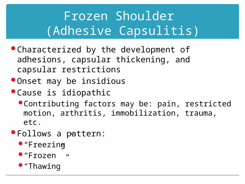
Frozen Shoulder (Adhesive Capsulitis)
Characterized by the development of adhesions, capsular thickening, and capsular restrictions
Onset may be insidiousCause is idiopathic
Contributing factors may be: pain, restricted motion, arthritis, immobilization, trauma, etc.
Follows a pattern:“Freezing” “Frozen”“Thawing”

Frozen Shoulder (Adhesive Capsulitis)
Common Impairments: Night pain and disturbed sleepPain with motionDecreased mobilityMuscle weakness Substitution patternsFunctional limitations
Treatment: Prevention, Stretch/ROM, joint mobilization,
strengthening, and modalities

Shoulder Dislocation
The GH joint is the most commonly dislocated joint in the body
Usually caused by a severe blow to the arm with arm held in a position of external rotation and abduction
Anterior dislocations occur most frequentlyClosed reduction-
Skilled technique to reduce the dislocationProtection Phase, activity restriction for 6-8 weeks
Avoid position of dislocationProtected ROM, isometrics

Shoulder Dislocation
Controlled motion phaseIncrease mobilityIncrease stability and strength of RC and periscapular
musclesReturn to function phase
Restore functional control; balance strength of shoulder and scapular musculature
CoordinationEnduranceEccentric trainingIncrease speed and controlSimulate functional patterns

Bicipital Tendinitis
Lesion is typically located on the long head of the biceps tendon in the bicipital groove
Pain is elicited with resisted shoulder flexion while the arm is supinated
Tenderness to palpation of the bicipital grooveTreatment:
Isometric exercises, Stretching, Cross-Friction massage, and modalities


Shoulder Instability
Multidirectional InstabilityIndividuals have lax connective tissue which allows
for mobilityThe humeral head will translate to a greater degree
than normal in all directionsIndividuals involved in overhead throwing or lifting
activities may be more prone to develop laxity of the shoulder capsule
Hypermobility may also lead to impingement, subluxation, dislocation, or tendinitis

Multi-directional Instability

Unidirectional Instability
May occur in one of the following directions: AnteriorPosteriorInferior
Usually the result of traumaTypically involves rotator cuff tearsDamage to the glenoid labrum is also common

Shoulder Instability
AMBRI: Atraumatic, Multidirectional, often Bilateral, requires Rehabilitation, Inferior capsular shift is the best alternative surgical
therapyUsually initiated without traumaOften multidirectional (anterior, inferior and posterior)Occurring in patients with generalized joint laxity

Shoulder Instability
AMBRI Usually does not have surgeryTreatment consists of a program of shoulder
strengthening and stabilization exercises

Shoulder Instability
TUBS (Traumatic, Unidirectional, Bankart, Surgery) One of most common shoulder injuries in athletes
• Most common in contact athletesMay present as traumatic dislocation/subluxation Mechanism is a posteriorly directed force on an
abducted and externally rotated armHigh recurrence rate that correlates directly
with age at dislocation • Up to 80-90% in teenagers

Traumatic Shoulder Dislocation

Glenoid Labral Tear - CAUSES
Falling on an outstretched arm A direct blow to the shoulder A sudden pull, such as when trying to lift a heavy
object A violent overhead reach
May occur while trying to stop a fall or slide Throwing athletes or weightlifters may experience
glenoid labrum tears as a result of repetitive shoulder motion

Labral Tear

SLAP Lesion
Tear of the superior labrumSLAP (Superior Labrum extending Anterior to
Posterior)Often associated with a tear of the proximal
attachment of the long head of the biceps and recurrent anterior instability of the GH joint
Surgery involves debridement of the superior labrum and reattachment of the labrum and biceps tendon

Bankart Repair
Bankart LesionDetachment of the capsulolabral complex from the
anterior rim of the glenoidCommonly occurs as a result of a traumatic
anterior dislocationThe repair involves an anterior capsulolabral
reconstruction to reattach the labrum to the surface of the glenoid lip

Acromioclavicular Sprain
Most AC sprains are NOT surgically repairedSometimes requires initial immobilizationModalities used to relieve pain, swelling and
muscle spasmsEarly active and AAROM exercises to regain and
maintain mobilityIsometric strengthening exercises

A-C Sprain / Dislocation

Common Surgical Procedures
Glenohumeral Arthroplasty
Arthrodesis of the Shoulder
RCR- Rotator Cuff Repair
SAD- Subacromial Decompression

Glenohumeral Arthroplasty
Total shoulder arthroplasty (TSA)The glenoid and humeral surfaces are replaced
Hemireplacement arthroplastyThe humeral head is replaced
Both are open surgical proceduresIndications for surgery:
Persistent and incapacitating painLoss of shoulder mobility or stabilityInability to perform functional tasks

TSA Postoperative Management
Progression is influenced by the integrity of the rotator cuff musculature
Shoulder is typically immobilizedMaximum Protection Phase:
Day 1 post-op -> 6 weeks post-opControl of pain and inflammationMaintain mobility of adjacent jointsRestore shoulder mobilityMinimize muscle guarding and atrophy

TSA Postoperative Management
Moderate Protection/Controlled Motion Phase6 weeks -> 12-16 weeks post-opContinue to increase PROM of the shoulderDevelop active control and dynamic stabilityImprove muscle performance (strength and
endurance)

TSA Postoperative Management
Minimum Protection/Return to Functional Activity PhaseBegins around 12-16 weeks post-opExtends for several more monthsContinue to improve or maintain shoulder mobilityContinue to improve active control of the shoulderProgress muscle strengthening and stabilization
exercisesReturn to functional activities

Arthrodesis of the Shoulder
The GH joint is fused with pins and bone graftsIndications for surgery
Incapacitating painGross instability of the GH jointComplete paralysis of the deltoid and rotator cuff
musclesSevere joint destruction due to infectionFailed TSA

Arthrodesis of the Shoulder
Postoperative ManagementEmphasis is placed on maintaining mobility of
peripheral joints (wrist and hand) while the shoulder and elbow are immobilized
Following immobilization, begin active scapulothoracic ROM

Rotator Cuff Repair
May be appropriate for either partial-thickness tears or full-thickness tears
Indications for surgical repair are:PainImpaired function
Surgical repair is not indicated for patients who are asymptomatic despite imaging reports confirming presence of a cuff tear
Surgical approach may be arthroscopic or open

Rotator Cuff Repair
Postoperative management depends upon many factors: Size and location of tearOnset of injuryPreoperative functional mobility and strengthAge of patientType of approachType of repair

RCR Postoperative Management
Maximum Protection Phase (up to 8 weeks)Patient will most likely be immobilizedProtection of the repaired tendon(s) is the primary goal
during this phaseControl pain and inflammationAAROM exercises for elbowAROM exercises for wrist and handPrevent shoulder stiffness Restore shoulder mobilityPosture re-educationScapular stabilization exercisesGentle isometrics for GH joint musculature

RCR Postoperative Management
Moderate Protection PhaseRestore nearly full, nonpainful, passive mobility of
the shoulderIncrease muscular strength and endurance of
shoulder musculatureRe-establish dynamic stability of the shoulderAROM is allowed in pain free rangesStrengthening typically begins around 8 weeks post-
op, but may begin as late as 12 weeks for larger repairs

RCR Postoperative Management
Minimum Protection/Return to Function PhaseBegins around 12-16 weeks post-op, and lasts for 6
months to a yearContinue to work towards full ROM
• Passive stretching of GH musculature• Joint mobilization
Advance task-specific exercisesPatients are not allowed to return to high demand
activities for 6 months, up to 1 year

Subacromial Decompression
Designed to increase the volume of subacromial space and provide adequate gliding room for tendons
Indications for surgery:• Pain during overhead activities• Loss of shoulder functional mobility• Intact or minor rotator cuff tear• Impingement
Performed using an arthroscopic or open approach

Subacromial Decompression
Maximum Protection Phase (0-4 weeks)Patient will have shoulder immobilized for 1-2 weeksPain control and inflammation controlROM activities (PROM, AAROM, AROM)Patient educationPostural re-education exercisesIsometric exercises

Subacromial Decompression
Moderate Protection Phase (4-8 weeks)Joint mobilizationStretchingPostural re-education Isotonic strengthening exercisesFunctional activities with light resistance
Minimum Protection Phase (8 weeks – 6 months)Continued strengtheningMaintain full, pain-free AROMFunctional and activity-specific exercises

Exercise Interventions for the Shoulder Girdle

Early Glenohumeral Joint Motion
AAROM Wand ExercisesFlexion, ABDuction, ER, etc.
Ball rolling or Table top washingWall washingPendulums
Ensure that patient is performing this exercise correctly
Wall pulleys
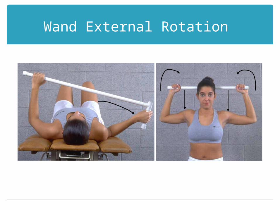
Wand External Rotation

Wand Horizontal Abduction/Adduction

Wand Abduction

Wand Internal Rotation

Pendulum
Bend forward 90 degrees at the waist, using a table for support move body in a circular pattern to move arm

Self-stretching Techniques
Posterior Capsule StretchTable slides-
Flexion and ABDuctionPect doorway stretch“Sleeper Stretch”Latissimus Stretch

Stretches - Latissimus

Latissimus Stretch

Exercises for Muscle Performance
Isometric exercisesDynamic strengthening exercises—scapular
musclesDynamic strengthening exercises—GH musclesFunctional activities

Isometric Strengthening
Isolated sustained submaximal muscle contraction without movementScapular isometricsShoulder flexionShoulder extensionShoulder ABDuctionERIRShoulder Horizontal ABD/ADD

Stabilization/Dynamic Strengthening Exercises
Open and Closed Chain Stabilization Dynamic Strengthening
Prone scapular retractionScapular retraction combined with Horizontal
ABDuctionScapular Retraction and Shoulder Horizontal
Abduction Combined with External RotationScapular Protraction
• “Push-up with a Plus”

GH Dynamic Strengthening
Isotonic StrengtheningPNF PatternsIsokinetic TrainingHand walking on a treadmillProFitterUBE

Advanced Closed-Chain Stabilization and Balance
Quadruped with hands on unstable surfacePhysioball Push-up position walking stairsBOSU Ball push-up, clapsPlyometrics

Functional Activities
Endurance TrainingEccentric TrainingPlyometricsTotal Body Training


Orthopedic Special Tests

Anterior Instability
Apprehension (Crank) TestPositive test is indicated by a
look or feeling of apprehension or alarm on the patient’s face and the patient’s resistance to further motion
This test is used to evaluate for anterior shoulder instability. This test may also be used to assess a labral tear.
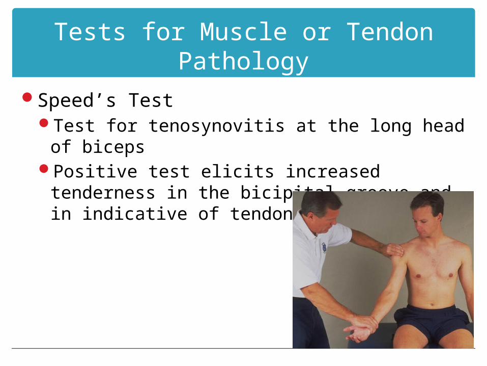
Tests for Muscle or Tendon Pathology
Speed’s TestTest for tenosynovitis at the long head of bicepsPositive test elicits increased tenderness in the
bicipital groove and in indicative of tendonitis

Tests for Muscle or Tendon Pathology
Yergason’s TestA positive result is tenderness in the bicipital groove
(or the tendon may pop out of the groove) and is indicative of bicipital tendonitis

Tests for Muscle or Tendon Pathology
Supraspinatus “Empty Can” TestThe examiner looks for weakness or pain, reflecting
a positive test resultA positive test result indicates a tear in the
supraspinatus tendon or muscle, or neuropathy of the subscapular nerve

Tests for Muscle or Tendon Pathology
Drop Arm (Codman’s) TestA positive test is indicated if the patient is unable to
return the arm to the side slowly or has severe pain when attempting to do so.
A positive result indicates a tear in the rotator complex

Tests for Impingement
Neer Impingement TestThe patient’s face shows pain, reflecting a positive
test result

Tests for Impingement
Hawkins-Kennedy Impingement TestPain indicates a positive test for supraspinatus
tenditintis

Tests for Thoracic Outlet Syndrome
Roos Test+ is unable to keep arms in starting position,
ischemic pain, heaviness, profound weakness, numbness, tingling

Tests for Thoracic Outlet Syndrome
Adson ManeuverTests for subclavian artery compression or TOSA disappearance in the pulse is a positive test.

Questions