Embed Size (px)
Citation preview
Proc. Nati. Acad. Sci. USAVol. 91, pp. 1366-1370, February 1994Biochemistry
Pseudomonas aeruginosa possesses homologues of mammalianphenylalanine hydroxylase and 4a-carbinolamine dehydratase/DCoH as part of a three-component gene clusterGENSHI ZHAO*, TIANHUI XIA, JIAN SONG, AND RoY A. JENSENtDepartment of Microbiology and Cell Science, University of Florida, Gainesville, FL 32611
Communicated by David B. Sprinson, October 22, 1993 (receivedfor review July 12, 1993)
ABSTRACT Pseudomonas aeruginosa possesses a multi-gene operon that includes phenylalanine hydroxylase (PhhA;phenylalaine 4-monooxygenase, EC 1.14.16.1). phhA encodesPhhA (Mr = 30,288), phhB (Mr = 13,333) encodes a homologueof mammalian 4a-carbinolamine dehydratase/homeodomainprotein transregulator, and phhC encodes an aromatic ami-notsferase (Mr = 43,237). The reading frames specifyingphhB and phhC overlap by 2 bases. The P. aeruginosa PhhAappears to contain iron and is pterin dependent. Unlike themultimeric mammalian hydroxylase, the native P. aeruginosaenzyme is a monomer. The P. aeruginosa PhhA is homologouswith mammalian PhhA, tryptophan hydroxylase, and tyrosinehydroxylase. Expression of PhhA from its native promoterrequired phhB. This may suggest a positive regulatory role forphk8, consistent with the dual catalytic and regulatory roles ofthe corresponding mammalian homologue.
Phenylalanine hydroxylase (PhhA; phenylalanine 4-monoox-ygenase, EC 1.14.16.1) catalyzes the irreversible conversionofphenylalanine to tyrosine, using tetrahydrobiopterin (BH4)as a reducing agent. In humans and other animals the enzymecatalyzes the rate-limiting step in the catabolism of dietaryphenylalanine (1). BH4 is oxidized to 4a-hydroxytetrahydro-biopterin, which is then dehydrated via 4a-carbinolaminedehydratase to form dihydrobiopterin (2). BH4 is regeneratedfrom dihydrobiopterin by dihydropteridine reductase.PhhA has generally been considered to be rare in prokary-
otes, where scattered reports of its existence have beenlimited to a single phylogenetic division of Gram-negativebacteria (3-7). Of these microbial pterin- and metal-depen-dent monooxygenases, only the PhhA proteins from Pseu-domonas acidovorans (3, 7) and Chromobacterium viola-ceum (8, 9) have been purified and characterized. Recentlythe C. violaceum gene encoding PhhA has been sequenced(10), and PhhA was found to be a member of the mammalianhydroxylase gene family. C. violaceum PhhA differs from themammalian enzymes in its smaller subunit size, its existenceas a monomer (rather than as a homotetramer), and bindingof copper (instead of iron) at the active site.
In this paper, we report the striking observation that PhhAfrom Pseudomonas aeruginosa is encoded by a gene orga-nized within an apparent operon including genes producinghomologues of mammalian 4a-carbinolamine dehydratase/DCoH and aromatic aminotransferase.*
MATERIALS AND METHODSStrains, Plasmids, and Media. P. aeruginosa PA01 (11) and
Pseudomonas stutzeri JM300 (12) are prototrophic strains.Escherichia coli strains used were JM83 [ara A(proAB-lac)rpsL thi 480 lacZAM15] obtained from GIBCO/BRL,
AT2471 (thi tyrA) obtained from Barbara Bachmann, JP2255(aroF363 pheA361 pheO352 tyrA382 thi strR712 lacY) xylS)(13), AB3257 (aroF, aroG, aroH) (14), and DG44 (thri leu6thil proA2 argE:3 hisG4 lacYl galK2 aral4 xyl5 mtll rpsL31tsx33 supE44) (15). Plasmid pUC18 (lacZ Apr) was obtainedfrom GIBCO/BRL. LB and M9 (16) were used as growthmedia for E. coli and P. aeruginosa. Additions of ampicillin(50 pg/ml), phenylalanine (50 ug/ml), and thiamine (17ttg/ml) were made when appropriate. Agar was added at 20g/liter to solidify media.
Molecular Cloning. Construction of a P. aeruginosa genelibrary was performed as described (17). Recombinant plas-mids were purified from the library (18) and used to transformE. coli AT2471 (tyrA), auxotrophic for tyrosine. Transform-ants able to grow on M9 minimal plates lacking tyrosine wereselected. Plasmids were isolated from the transformants andused to retransform E. coli AT2471.DNA Manipulations. Restriction-site mapping and subclon-
ing were done by standard methods (16). Southern blothybridization was carried out following the instructions ofPromega with biotinylated probes.DNA Sequencing and Data Analysis. Subcloned fragments,
obtained after purification of plasmids by CsCl gradientcentrifugation (18), were sequenced in both directions at theDNA Core Facility of the University of Florida. Data anal-ysis was carried out with the Genetics Computer Groupsoftware package (19).Crude Extract Preparation and Enzyme Assay. P. aerugin-
osa PA01 and E. coli JP2255 carrying various plasmids weregrown in M9 and LB media (450 ml), respectively. The cellsharvested at the late exponential phase of growth weredisrupted by sonication, and the resulting extract was cen-trifuged at 150,000 x g for 1 hr at 4TC. The supernatant (crudeextract) was collected and used for enzyme assay. PhhA wasassayed by following tyrosine formation (8). Cyclohexadie-nyl dehydrogenase was assayed by a continuous spectropho-tometric method (20).
Anion-Exchange Chromatography. E. coliJP2255 (pJZ9-3a)was grown in 2 liters of LB broth supplemented with ampi-cillin (50 pg/ml) at 370C and harvested by centrifugationduring midexponential growth. The cell pellets were resus-pended in 20 ml of 10 mM potassium phosphate buffer (pH7.4) containing 50 mM KCl and 1 mM dithiothreitol (DTT)(buffer A) and were disrupted by sonication. The resultingsuspension was centrifuged at 150,000 x g for 60 min. Thesupernatant was applied to a DEAE-cellulose column (2.5 x19 cm) that had been equilibrated with buffer A. The columnwas washed with 200 ml of buffer A and then eluted with a500-ml linear salt gradient (50-300 mM KCI) in buffer A.
Abbreviation: BH4, tetrahydrobiopterin.*Present address: Department of Microbiology, University of TexasMedical School at Houston, Houston, TX 77054.tTo whom reprint requests should be addressed.'The sequence reported in this paper has been deposited in theGenBank data base (accession no. M88627).
1366
The publication costs of this article were defrayed in part by page chargepayment. This article must therefore be hereby marked "advertisement"in accordance with 18 U.S.C. §1734 solely to indicate this fact.
Proc. Natl. Acad. Sci. USA 91 (1994) 1367
Fractions of 5 ml were collected, and those exhibiting phhAactivity were pooled and concentrated by use of an AmiconPM-10 membrane. This preparation had a specific activity of153 nmol min-1 mg-1 and represented a purification of 4.3-fold with respect to crude extracts.
Gel-Filtration Chromatography. The Mr of the native PhhAwas estimated by gel filtration. The concentrated preparationobtained after DEAE-cellulose fractionation was washedwith buffer B (10 mM potassium phosphate, pH 7.4/0.1 MKCl/1 mM DTT) and was applied to a Sephadex G-100column (2.8 x 56 cm) that had been equilibrated with bufferB and run at 40C. Column fractions (3 ml) were collected.Molecular weight standards were bovine serum albumin (Mr,66,000), ovalbumin (Mr, 45,000), and cytochrome c (Mr,12,300) (Sigma). The fractions were monitored for protein atA280 and for PhhA activity. The peak fraction exhibited aspecific activity of 1.32 gnmol min-'nmg-1. This purificationprocedure yielded a 37.2-fold purification with a 45% yield.Relative to the enzyme level present in wild-type P. aeru-ginosa, a purification exceeding 600-fold was obtained.
RESULTSMolecular Cloning and Identification of the P. aeruginosa
phhA Gene. Since PhhA produces tyrosine, a reasonablestrategy for cloning the phhA gene would utilize functionalcomplementation of E. coli tyrosine auxotrophs. Two E. coliauxotrophs, AT2471 (tyrA) and JP2255 (tyrA pheA), wereused. E. coli AT2471, used in the primary screening studies,would be expected to grow in the presence of plasmid-bornephhA or tyrC, the latter gene encoding cyclohexadienyldehydrogenase in P. aeruginosa (21). We then used E. coliJP2255 to verify the identity of those plasmids carryingphhA.Plasmids carrying the phhA gene would be able to comple-ment E. coli JP2255 in the presence of phenylalanine,whereas those carrying the tyrC gene would be unable to doso because of the absence of chorismate mutase activity inthis strain (13). E. coli AB3257, lacking all three isoenzymesthat catalyze the first step of aromatic biosynthesis andtherefore unable to generate prephenate, was also used toconfirm the presence ofphhA and the absence of tyrC.
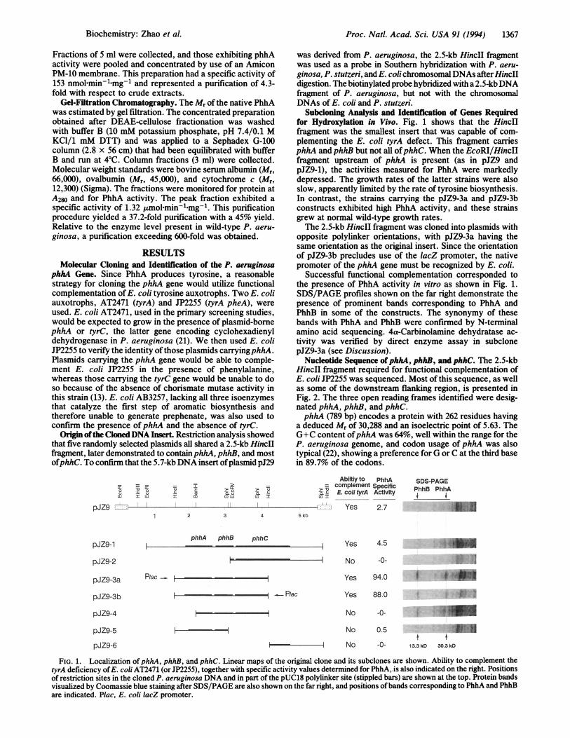
Origin ofthe ClonedDNA Insert. Restriction analysis showedthat five randomly selected plasmids all shared a 2.5-kb HinclIfragment, later demonstrated to contain phhA, phhB, and mostofphhC. To confirm that the 5.7-kbDNA insert ofplasmid pJ29
was derived from P. aeruginosa, the 2.5-kb HincII fragmentwas used as a probe in Southern hybridization with P. aeru-ginosa, P. stutzeri, and E. colichromosomalDNAs afterHincIIdigestion. The biotinylated probe hybridized with a 2.5-kbDNAfragment of P. aeruginosa, but not with the chromosomalDNAs of E. coli and P. stutzeri.
Subcloning Analysis and Identification of Genes Requiredfor Hydroxylation in Vivo. Fig. 1 shows that the HincIIfragment was the smallest insert that was capable of com-plementing the E. coli tyrA defect. This fragment carriesphhA and phhB but not all ofphhC. When the EcoRI/HincIIfragment upstream of phhA is present (as in pJZ9 andpJZ9-1), the activities measured for PhhA were markedlydepressed. The growth rates of the latter strains were alsoslow, apparently limited by the rate of tyrosine biosynthesis.In contrast, the strains carrying the pJZ9-3a and pJZ9-3bconstructs exhibited high PhhA activity, and these strainsgrew at normal wild-type growth rates.The 2.5-kb HincII fragment was cloned into plasmids with
opposite polylinker orientations, with pJZ9-3a having thesame orientation as the original insert. Since the orientationof pJZ9-3b precludes use of the lacZ promoter, the nativepromoter of the phhA gene must be recognized by E. coli.
Successful functional complementation corresponded tothe presence of PhhA activity in vitro as shown in Fig. 1.SDS/PAGE profiles shown on the far right demonstrate thepresence of prominent bands corresponding to PhhA andPhhB in some of the constructs. The synonymy of thesebands with PhhA and PhhB were confirmed by N-terminalamino acid sequencing. 4a-Carbinolamine dehydratase ac-tivity was verified by direct enzyme assay in subclonepJZ9-3a (see Discussion).
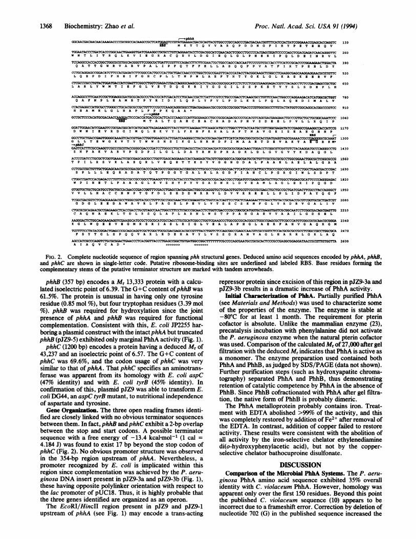
Nucleotide Sequence ofphhA, phhB, and phhC. The 2.5-kbHincII fragment required for functional complementation ofE. coli JP2255 was sequenced. Most of this sequence, as wellas some of the downstream flanking region, is presented inFig. 2. The three open reading frames identified were desig-nated phhA, phhB, and phhC.phhA (789 bp) encodes a protein with 262 residues having
a deduced Mr of 30,288 and an isoelectric point of 5.63. TheG+C content ofphhA was 64%, well within the range for theP. aeruginosa genome, and codon usage of phhA was alsotypical (22), showing a preference for G or C at the third basein 89.7% of the codons.
0 Ia a ELU T1W T
pJZ9 =.
pJZ9-1
r- z=
0 IDUn uj (O I
1 2 3 4 5 kb
phhA phhB phhC
Ablitly to PhhA_- complement SpecificDC.C E. coli tyrA Activity
_- Yes 2.7
Yes 4.5
SDS-PAGEPhhB PhhA
aN
pJZ9-2
pJZ9-3a P/ac -_ [
I No -0-
Yes 94.0
_ P/ac Yes 88.0 ;
No -0- i 4'.No 0.5 4 .
t fNo -0- 13.3 kD 30.3kD
FIG. 1. Localization of phhA, phhB, and phhC. Linear maps of the original clone and its subclones are shown. Ability to complement thetyrA deficiency ofE. coli AT2471 (or JP2255), together with specific activity values determined for PhhA,.is also indicated on the right. Positionsof restriction sites in the cloned P. aeruginosa DNA and in part of the pUC18 polylinker site (stippled bars) are shown at the top. Protein bandsvisualized by Coomassie blue staining after SDS/PAGE are also shown on the far right, and positions ofbands corresponding to PhhA and PhhBare indicated. Plac, E. coli lacZ promoter.
pJZ9-3b
pJZ9-4
pJZ9-5
pJZ9-6
,r..
i
I ,# qr
-
Biochemistry: Zhao et al.
Proc. Natl. Acad. Sci. USA 91 (1994)
GGCAACGACACAACAAAGCCCGGCAAAGACCGCTCAaTr.GAGTCCGTATGAAACGACGCAGTALGG~XCMCGCACls%=CATlaaGACGTM K T T Q Y V A RQ P D D N G P I H Y P B T 8 HQ V
TGAAACCTATACCGGCAAC~rx3AAGTGTCGAAGGCGGCCGCG.clslACGAACCAGCTGAMCC~~C~zGTACG=W N T L I T R Q L K V I E G R A C Q E Y L D G I B Q L G L P H E R I P Q L D 8 I N R V L
TCCGCAG C_--CS1CCCACGETCCCACTACGACCGATQ A T T G W R V A R V P A L I P P Q T PFP B L L A S Q Q P P V A T P I R T P 8 8 L D Y
CCGCCCGCCTATCTGATGCCACGAGATIrCGGCCACTGCGACACCGACCAGGCCAACCCTCTACGCCGGCACCATACGGCAAGCTCGGACAGCCTCC A GCCGAICL QA P D I P H B I P G H C P L L T N P W L AK F T H T Y G K L G L K A S K ES R V F
CTCGC CTCTCCCACT(GA~CCTCA A CCCCG GACGACICGCL A R L Y W M T I B P G L V 8 T D Q G K R I Y G G G I L S S P K B T V Y S L S D 8 P L H
ACCAGGCCTTCAATCGTAGCGAIXTGCGCACGCCCTACCGCATCGACATCCtCACGCCATrMCCAATGCACQ A P N P L B A M R T P Y R I D I L Q P L Y P V L P D L K R L F Q L A Q E D I M A L V
CCAC_ CGCTGTCCCCCCAAGCCWCTC&'ACGAAACGCCGCCGCCGCGGC7GACCCCGTlGCGGCCTTGTACTATGGTIGAGCImCCGCH 8 A M R L G L H A P L P P P X Q A A*
r-ophhB
G L L T E W KYVTYVTWI S H S I K G L H RND F I A A R TD EVA KTAFR KNP
-~~~~phhC~ *h
PTCCCA AA C A C ACGARV AD LGVGVYGACKDAQGL:GCTCCC
D W N A 8 V AR D G I M Q IB R VL L P R NP R H R A F T N A V G B I S B A B A H H P
130
260
390
520
650
780
910
1040
1170
1300
1430
ACCC _ G a ; C _7 _ CGCGCACIGCCMGCOGTAC CG 1560T P I L R S V K L A 8 Q R L V B Q 8 T T K S Y V G G H G D A L P A A R L A 8 L A L G A A
C CGGC~:C~CCCACCCAGACGCCCGCGACGGCCTTGCTGCrGCCGGCATATCA CGCGGCTCGGCTCxCCOGACS P L L L B Q R A D A T Q T P G G T G A L R L A G D F I A H C L P G R G I W L S D P T
CTGGCCGATCCACGAGACCCTGTTCGCCGCCGCCGGCr"GMATCCCCTACGTCAGCGCCGACAACCGC _TGGCCTTCCCAGGCW P I H B T L P A A A G L K V S H Y P Y V S A D N R L D V B A M L A G L E R I P Q G D
HAsCCTGCTrCCACAACCCGACHCHTG RRLAGCDRRACRL AYCAGGGCTV V L L H A C C H N P T G F D L S H D D W R R V L D V V R R R B L L P L I D P A Y Q G F
TCGGCGACGTGAGGAAGAC.CGCGGTACGCCTGTTCGCCGGCGAACTr.CGGTCGGCCCAGTTC CCACCGGCGrCGCTGATCTG D G LB B D A W A V R L P A G B L P B V L V T S S C S K N F G L Y R D R V G A L I V
<:IUC-sC~ ~~CGACCTGCGTAGCCAACTIGGCCTTCCTCGCCCGAACCTGTGCCCGCGCCAG TGCCGGTTCCGAACTGCACTAT7GC A Q N A B R L T D L R S Q L A F L A R N L W S T P P A H G A B V V A A I L G D S 9 L
_CICIGGCATCG~rCGCG CGCTCCGCGCATCGCACr.CATCGGCCTGTGAC TGCGCGCTGGCCGAGGCACTGCGCGGCCGCGCACCGGAK G L W Q B E V 8 G M R S R I A S L R I G L V 8 A L A P H G L A B R F A H V G A Q R G M
7TTTTCCTA'rACCGCGaAGCCwGC~=ICCACICa!GCAGCAGOCGI: A = CTGC3P S Y T G L S P Q Q V A R L R D E H A V Y L V S S G R A N V A G L H A R R L G R L A Q
AG CATa:GGCAACCCTICACGG7TAC CAGCAICCCCCTCCCGCCGAXGCGGAGGATAACCGC-ul-lujGGTAA I A Q V C A D * >>>>>>> <<<
1690
1820
1950
2080
2210
2340
2470
FIG. 2. Complete nucleotide sequence of region spanning phh structural genes. Deduced amino acid sequences encoded by phhA, phhB,and phhC are shown in single-letter code. Putative ribosome-binding sites are underlined and labeled RBS. Base residues forming thecomplementary stems of the putative terminator structure are marked with tandem arrowheads.
phhB (357 bp) encodes a Mr 13,333 protein with a calcu-lated isoelectric point of 6.39. The G+C content ofphhB was61.5%. The protein is unusual in having only one tyrosineresidue (0.85 mol %), but four tryptophan residues (3.39 mol%). phhB was required for hydroxylation since the jointpresence of phhA and phhB was required for functionalcomplementation. Consistent with this, E. coli JP2255 har-boring a plasmid construct with the intactphhA but truncatedphhB (pJZ9-5) exhibited only marginal PhhA activity (Fig. 1).phhC (1200 bp) encodes a protein having a deduced Mr of
43,237 and an isoelectric point of 6.57. The G+C content ofphhC was 69.6%, and the codon usage of phhC was verysimilar to that ofphhA. That phhC specifies an aminotrans-ferase was apparent from its homology with E. coli aspC(47% identity) and with E. coli tyrB (45% identity). Inconfirmation of this, plasmid pJZ9 was able to transform E.coli DG44, an aspC tyrB mutant, to nutritional independenceof aspartate and tyrosine.Gene Orgnition. The three open reading frames identi-
fied are closely linked with no obvious terminator sequencesbetween them. In fact,phhB andphhC exhibit a 2-bp overlapbetween the stop and start codons. A possible terminatorsequence with a free energy of -13.4 kcal-mol-' (1 cal =4.184 J) was found to exist 17 bp beyond the stop codon ofphhC (Fig. 2). No obvious promoter structure was observedin the 354-bp region upstream of phhA. Nevertheless, apromoter recognized by E. coli is implicated within thisregion since complementation was achieved by the P. aeru-ginosa DNA insert present in pJZ9-3a and pJZ9-3b (Fig. 1),these having opposite polylinker orientation with respect tothe lac promoter of pUC18. Thus, it is highly probable thatthe three genes identified are organized as an operon.The EcoRI/HincII region present in pJZ9 and pJZ9-1
upstream of phhA (see Fig. 1) may encode a trans-acting
repressor protein since excision of this region in pJZ9-3a andpJZ9-3b results in a dramatic increase of PhhA activity.
Initial Characterization of PhhA. Partially purified PhhA(see Materials and Methods) was used to characterize someof the properties of the enzyme. The enzyme is stable at-80°C for at least 1 month. The requirement for pterincofactor is absolute. Unlike the mammalian enzyme (23),precatalysis incubation with phenylalanine did not activatethe P. aeruginosa enzyme when the natural pterin cofactorwas used. Comparison ofthe calculated Mr of27,000 after gelfiltration with the deduced Mr indicates that PhhA is active asa monomer. The enzyme preparation used contained bothPhhA and PhhB, as judged by SDS/PAGE (data not shown).Further purification steps (such as hydroxyapatite chroma-tography) separated PhhA and PhhB, thus demonstratingretention of catalytic competence by PhhA in the absence ofPhhB. Since PhhB cofractionated with PhhA after gel filtra-tion, the native form of PhhB is probably dimeric.The PhhA metalloprotein probably contains iron. Treat-
ment with EDTA abolished >99%o of the activity, and thiswas completely restored by addition of Fe2+ after removal ofthe EDTA. In contrast, addition of copper failed to restoreactivity. These results were consistent with the abolition ofall activity by the iron-selective chelator ethylenediaminedi(o-hydroxyphenylacetic acid), but not by the copper-selective chelator bathocuproine disulfonate.
DISCUSSIONComparison of the Microbial PhhA Systems. The P. aeru-
ginosa PhhA amino acid sequence exhibited 35% overallidentity with C. violaceum PhhA. However, homology wasapparent only over the first 150 residues. Beyond this pointthe published C. violaceum sequence (10) appears to beincorrect due to a frameshift error. Correction by deletion ofnucleotide 702 (G) in the published sequence increased the
1368 Biochemistry: Zhao et al.
Biochemistry: Zhao et al. Proc. Natl. Acad. Sci. USA 91 (1994) 1369
v P wv P wV L WF H WV PFWV FMWV FWV PFWV FMWV PFWV PFW
*AIIAF
FADE L AM S Y KYHF AD L.A M N Y KMHF AD I A YMNYKRM
F AD I G Y NY KMH
A ND F TL ...
*- *
EL L E R F CG YK
N A EIX F CQ Y N
FL L SK Y C G Y RF L L SKY CG Y RF L L S AD CG Y R
P L AS R YC KYR
FL L K Y CG F RF L L S K Y CG F H
P A DI A G FRH
SQ L .... .G LEARLL EVD
R V FQ C YIRR V FQ CT GYIRR V FQ CT GYINR V F Q CT GYIRR V F H CT GYVRR V F H CT GYVRR V F H CT GYVRR V F H C T Q Y V R
R V F H C T Q Y I R
R V F Q C T Q Y I R
R V F H C T Q Y I RQ Q F P VC T F I RR R F P V T W W I R
LR V S D E E I E KL G A TD E Y IERL G A S EC TVQ KL G A S EC TVQ KL G A S EV A V QIK
L G A S E E A I Q KL G A P D E Y I E KL G A P D E Y I E KL G A P D D Y I E KL K A S K E ET V FV K A K A L G A A D
E E AP EVE A F D PE E AP EIE A F D PE E PE I R IF DKD E PE V R IF DKLGH A K V K P I D PG H A SKV K P F DFGK HKAK A KG A ADP
G H A K V K A F D PD K P K I L A L E LE K P K I L A L E LD K P Q L K D F E PD E A L H Q A F N PA S A N V G F D L
F S A K F D P Y T LF S A K F D P Y T LF S V K F D P Y T LF S V K Y E P Y T H
F G LK Y AP Y TPF G V K Y N P Y T Q
F G V K Y N P Y T RF G V K Y N P Y T RF S V R Y D P Y T Q
F S V R Y D P Y T Q
F G V R Y N A Y TQL V H E A M R L G LL Y L
F P R K V S E L D KF P R K V S 8 L D KF P R K V S E L D KF P R K I C E L D KF P K K I S D L D FF P K K I S D L D FF P K X I S D L D HF P K K I S D L D HF P R T I QE L D RF P R T I QE L D RF P R R I R D L D R
GE P I P H V E Y TG D P I P R V E Y TG D P I P H V E Y TG D P I P R V E Y T JG D P I P K I E F TG D P I P K I E F TG D P I P K V E F TG D P I P K V E F TG Q P I P R V E Y TG Q P I P R V E Y MG Q P L P H V D Y T.... ........H Y P..P QP L D R Y S J
E D S I P QL E D VE D N I P QL E D VE D R I P QL E D VE N N I P QL E E VE D N I P QL E D VE D N I P Q L E D I
E D N I P QL E D VE D N I P QL E D VE D N I P QL E D VE D N I P QL E D VH E R I P QL D E IA D R V P D F N K L
pterin-binding doain?
HA S S P M H S PTH A S S P M H S P EH A S S P M H S P EH A S S P M H S P EH S S D P L Y T P EH S S D P L Y T P EH S S D P F Y T P EH S S D P F Y T P EH G S K P M Y T P EH G S K P M Y T P EH P S K P M Y T P ET P E E L D Y L QE P H Q L D Y L QE
L S T V Y W F T V EL S T L S W F T V EL S T L Y W F T V EL A T L Y W F T V EL A T C Y F F T V EL A T C Y F F T V EL A T C Y F F T V EL A T C Y F F T V EL A T I Y W F T V EL A T I Y W F T V EL S T I F W F T V EL A R L Y W M T I EL A R L Y W Y T V E
D T A A V QP Y QDE A A A V QP Y QDD A A A V QP Y QDD A A A V QP C QDK I A C K QE C L IK V A C K QE C L IK I T Y K Q E C L IK I T C K Q E C L IE K T A C Q E Y S VE K T A I Q N Y T VE S T G V T K Y P IL E A M R T P Y R IM R I M N T R Y R I
A I D V L D S P H TA I D V L D S P Q AA I D V L D S P H AS I E L L D S P Q TS V Q V L R D T K SS I Q V L R D S K SS I Q I L K D A K SS I Q I L K D T K SR V E V L D N T Q QR I E V L D N T Q QS V E V L D S K P QH A P L F P P K Q AQ L A D A Q P W G A
C H H L V T KC H H L V T KC H H L V T KC H H L V T KC A N R V L LC A N R V L LC A N R V L MC A N R V L MF A N Q I L SF A N Q I L SF A N Q I L S
A E E I A T WA E E I A T WA E E T A T WA E E I
A T WE E E I K T WE E E I K T WE E E I K T W
E E E K Q T WE E E K K T WX E E I E T WE T E H Q V WA E D H A T W
S R F L K E RS R F L K E RS R F L K E RS R F L K E RS N F L K E RS N F L K E RS N F L K E RS N F L K E RS Q F L Q T CS Q F L Q T CS N F L R D CN R V L Q A TN E K L M A A
P D C C H E LP D C C H E LP D C C H E LP D C C H E LP D T C H E LP D T C H E LP D T C H E LP D T C H E LP D I C H E LP D I C H E LP D V C H E LP D I F H E IP D V F H D L
* A
F G L C K Q NF G L C K Q NF G L C K Q NF G L C R Q NF G L C K Q DF G L C K Q DFC L C K Q DF G L C K Q DF G L C K E GF G L C K Q GY G L L A K EF G L V E T DF G L I N T P
Q T Y Q P V YQ T Y Q S V YQ T Y Q P V YQ P Y Q P VYT S F Q D V YT S F Q D V YT T F QD V YT T F QD V YT E F Q P L YT E F Q P L YT Q F Q P L YD I L Q P L YD T F Q K T Y
I Q R S L E GV R R S L E GI R R A L D GI C H S L E SI T S A M N EI T S A M N EI T N A M N EI T S A M N EL K I L A D SL K I L A D SI S N L M D NA -
A T S R R T T
:F D P D L D L D H P G F S:F D P D L D L D H P G F S:F D P D L D L D H P G F SF D P D L D L D H P G Y SY G S E L D A D H P G F KY G S E L D A D H P G F K
IY G S E L D A D H P G F KIY G S E L D A D H P G F KY G A E L D A D H P G F KY G A E L D A D H P G F KY G S E L D A D H P G F T
M KM N D R A D F V V P D I T
K E V Y V T L K G L Y A TK E V Y T T L K G L Y A TK E V Y S T L R G L Y P T
IK E V Y S T L K S L Y P TG T I F R E L N K L Y P T
IG T I F R E L N K L Y P TIG T V F R E L N K L Y P TG T V F Q E L N K L Y P TG T V F R T L K A L Y K TG T V F K T L K S L Y K TG I I F R N L T K L Y K TN T L I T R Q L K V I E GA T L Y Q R Q C K L L P G
T G F Q L R P V A G L L ST G F QL R P V A G L L ST G F QL R P V A G L L ST G F Q L R P V R G L L ST G F S I R P V A G Y L ST G F S I R P V A G Y L ST G F S I R P V A G Y L ST G F S I R P V A G Y L ST G F R L R P V A G L L ST G F R L R P V A G L L ST G F T L R P V A G L L ST G W R V A R V P A L I PT G W K I V A V P G L I P
.L G H V P M L A D R T F A
.L G H V P M L A D R T F AL A H G P M L A D R T F AL G H V P M L A D K T F A.L G H V P L L A E P S F A.L G H V P L L A E P S F A.L G H V P L L A E P S F A.L G H V P L L A E P S F A.L G H V P L F S D R S F A.L G H V P L F S D R S F AM G H V P L F A D P S F A!F G H C P L L T N P W L A.F G H V P L L I N P V F A
G E L K A Y G A G L L S SG E V K A Y G A G L L S SG E V N A Y G A G L L S SG I V K A Y G A G L L S SG Q L R V F G A G L L S S)G Q L R V F G A G L L S SG QL R V F G A G L L S SG QL R V F G A G L L S S;D S I K A Y G A G L L S S;D S I K A Y G A G L L S SG E L K A Y G A G L L S SQ G K R I Y G G G I L S SA G M R I Y G A G I L S S
F V S E S F N D A K D K LF V S E S F S D A K D K LF V S E S F S D A K D K LF V S E S F S D A K N K LF V S E S F E D A K E K MF V S E S F E D A K E K MF V S E S F E D A K E K MF V S E S F E D A K E K MY V A B S F S D A K E K VY V A E S F N D A K E K VY V A D S F E T A K E K TF V L P D L . . .. K R LF V I D S F . . .. K Q L
V Q D E L H T L A H A L SV Q D E L D T L A H A L SGVQ D E M A L A H A L N;V R D E L H T L I N A L NL R Y D L D V I S D A L AL R H D L D V V N D A L AL R H D L D V V S D A L GL Q H D L D V V S D A L AI N S E V G I L C N A L QI N S E I G I L C S A L QGI N S E F Q I L Q N A V A
w c t
D Q V Y R Q R R K LD Q V Y R Q R R K LD Q A Y R Q R R K LD Q V Y R Q R R K SD N V Y R R R R K YD N V Y R R R R K YD N V Y R K R R K YD N V Y R K R R K YD P V Y R A R R K QD P V Y R A R R K QD P E Y A K R R K YT T Q Y V A R Q P DT R K N V G L S H D
H A C R E H L E G FH A C G E H L E A FH A C R E H L E A FH A C K E Y L E A FH A C R E Y L R N LH A C R E Y L R N LH A C R E Y L K N LH A C R E Y L K N LH A C Y E H N H I FH A C Y E Y N H I FH A C R E Y N H V FR A C Q E Y L D G IR A C D E F L E G L
* A - A A -
A R D F L A S L A FA R D F L A S L A FA R D F L A S L A FA R D F L A S L A FP R D F L S G L A FP R D F L S G L A FP R D F L S G L A FP R D F L S G L A FS R D F L G G L A FS R D F L G G L A FS R D F L A G L A FF Q T F F E L L A SD D V F F E H L A N
2052351981981501461461461591591591224
2552852482482001961961962092092095266
30533529829825024624624625925925998
112
G L 355
Q F S Q D I GL A S 385Q F S Q D I GL A S 348Q F S Q D I GL A S 348
Q F S Q EI GL A S 300
Q F S Q E I GL A S 296Q F S Q E I G L A S 296Q F S Q E I GL A S 296
Q F S Q E I GL A S 309Q F S Q E I GL A S 309Q F S Q E I GL A S 309E F T H T Y GK L G 148D Y L E A Y GK G G 162
* * *--- ,
Y G E L L H S L S 404Y G E L L H C L S 434Y G E L L it SL .S 397Y G E L I H S L S 397I S E L K H AL .S 349I S E L R H AL . S 345I S E L K H V L S 345I S E L K H AL .S 345F G E L Q Y C L S 358F G E L Q Y CL .S 358Y G E L E Y C L T 358P K E T V Y SL S 197K S E S I Y C L D S 212
R N Y A S R I QIR P 454R S Y A S RI Q R P 484R S Y A S R I Q R P 447R N Y A A H I K R P 447R E F A K T VK R P 399R E F A K T VK R P 395R E F K K T IK R P 395R E F T K T IK R P 395R T F A A T IP R P 408R N F A A T IP R P 408I K F A N S IP R P 408F Q L A Q E DI M A 243F D A T A P DF A P 258
A I S IA I G IA I S IV I S IR V T R W P S V I
R V S R W P S V I
X V S R Q L S V I
K V S R K P S I I
K I K S I
K I K IK L R V I
497
527
490
490448
444444444452
451
452
264
280
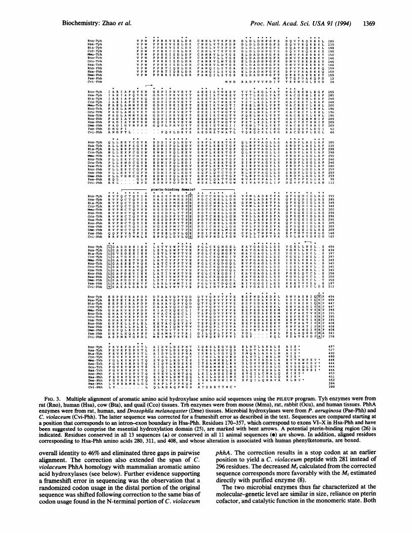
FIG. 3. Multiple alignment of aromatic amino acid hydroxylase amino acid sequences using the PILEUP program. Tyh enzymes were fromrat (Rno), human (Hsa), cow (Bta), and quail (Cco) tissues. Trh enzymes were from mouse (Mmu), rat, rabbit (Ocu), and human tissues. PhhAenzymes were from rat, human, and Drosophila melanogaster (Dme) tissues. Microbial hydroxylases were from P. aeruginosa (Pae-Phh) andC. violaceum (Cvi-Phh). The latter sequence was corrected for a frameshift error as described in the text. Sequences are compared starting ata position that corresponds to an intron-exon boundary in Hsa-Phh. Residues 170-357, which correspond to exons VI-X in Hsa-Phh and havebeen suggested to comprise the essential hydroxylation domain (25), are marked with bent arrows. A potential pterin-binding region (26) isindicated. Residues conserved in all 13 sequences (A) or conserved in all 11 animal sequences (e) are shown. In addition, aligned residuescorresponding to Hsa-Phh amino acids 280, 311, and 408, and whose alteration is associated with human phenylketonuria, are boxed.
overall identity to 46% and eliminated three gaps in pairwise phhA. The correction results in a stop codon at an earlieralignment. The correction also extended the span of C. position to yield a C. violaceum peptide with 281 instead of
violaceum PhhA homology with mammalian aromatic amino 296 residues. The decreased Mr calculated from the correctedacid hydroxylases (see below). Further evidence supporting sequence corresponds more favorably with the Mr estimated
a frameshift error in sequencing was the observation that a directly with purified enzyme (8).randomized codon usage in the distal portion of the original The two microbial enzymes thus far characterized at the
sequence was shifted following correction to the same bias of molecular-genetic level are similar in size, reliance on pterincodon usage found in the N-terminal portion of C. violaceum cofactor, and catalytic function in the monomeric state. Both
Rno-TyhNsa-TyhBta-TyhCcO -TyhMmu-TrhRno-TrhOcu-TrhHsa-TrhRno- PhhHsa-PhhDme - PhhPae-PhhCvi - Phh
Rno-TyhHsa-TyhBea-TyhCco-TyhMmu-TrhRno-TrhOcu-TrhHsa-TrhRno- PhhHsa- PhhDme -PhhPae-PhhCvi - Phh
Rno-TyhHsa-TyhBta-TyhCco-TyhMmu-TrhRno-TrhOcu-TrhHsa-TrhRno- PhhHsa- PhhDme- PhhPae - PhhCvi - Phh
Rno-TyhHsa-TyhBta-TyhCco-TyhMmu-TrhRno-TrhOcu-TrhHsa -TrhRno- PhhHsa- PhhDme- PhhPae-PhhCvi - Phh
Rno-TyhHsa-TyhBta-TyhCco-TyhMmu-TrhRno-TrhOcu-TrhHsa-TrhRno- PhhHsa- PhhDme - PhhPae-PhhCvi - Phh
Rno-TyhHsa-TyhBta-TyhCco-TyhMmu-TrhRno-TrhOcu -TrhHsa -TrhRno -PhhHsa - PhhDme- PhhPae- PhhCvi-Phh
Rno-TyhHsa-TyhBta-TyhCco-TyhMmu-TrhRno-TrhOcu-TrhHsa-TrhRno- PhhHsa- PhhDme - PhhPae- PhhCvi - Phh
Proc. Natl. Acad. Sci. USA 91(1994)
Pae-PhhB WTALTQAHCNACRADAPHVSDBELPVLLRQIP. .DWN. IHVRDGIMQLRKVYLFKNKHaLAPTNAVGHISHABGHHPGLLTNWGKVTVTWWSHSIKGLHRNDFIMAARTDEVAKTABGRK 118-1-1 -II1.11-- - 1-111 1: . 1: 1I I: :.I 1-11 -1- :1 *1 -*
Rno-DCoH ..... MAGFAHRLSARDQLLPNLRAVGLDGRDAIF... KQFHFKDFNRAFGFMTRVALQABKLDHHPHWFNVYNKVHITLSTHBCAGLSERDINLASFIBQVAVSMT ... 104
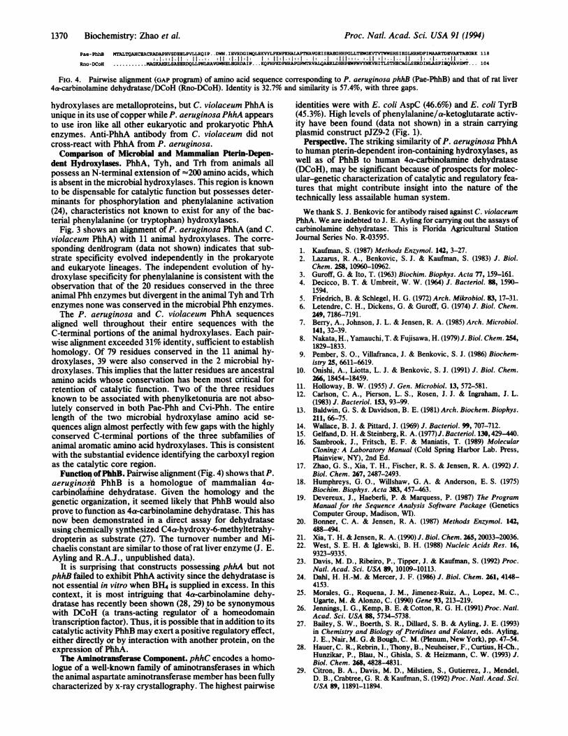
FIG. 4. Pairwise alignment (GAP program) of amino acid sequence corresponding to P. aeruginosa phhB (Pae-PhhB) and that of rat liver4a-carbinolamine dehydratase/DCoH (Rno-DCoH). Identity is 32.7% and similarity is 57.4%, with three gaps.
hydroxylases are metalloproteins, but C. violaceum PhhA isunique in its use of copper while P. aeruginosa PhhA appearsto use iron like all other eukaryotic and prokaryotic PhhAenzymes. Anti-PhhA antibody from C. violaceum did notcross-react with PhhA from P. aeruginosa.Comparison of Microbial and Mammaiian Pterin-Depen-
dent Hydroxylases. PhhA, Tyh, and Trh from animals allpossess an N-terminal extension of -200 amino acids, whichis absent in the microbial hydroxylases. This region is knownto be dispensable for catalytic function but possesses deter-minants for phosphorylation and phenylalanine activation(24), characteristics not known to exist for any of the bac-terial phenylalanine (or tryptophan) hydroxylases.
Fig. 3 shows an alignment of P. aeruginosa PhhA (and C.violaceum PhhA) with 11 animal hydroxylases. The corre-sponding den'drogram (data not shown) indicates that sub-strate specificity evolved independently in the prokaryoteand eukaryote lineages. The independent evolution of hy-droxylase specificity for phenylalanine is consistent with theobservation that of the 20 residues conserved in the threeanimal Phh enzymes but divergent in the animal Tyh and Trhenzymes none was conserved in the microbial Phh enzymes.The P. aeruginosa and C. violaceum PhhA sequences
aligned well throughout their entire sequences with theC-terminal portions of the animal hydroxylases. Each pair-wise alignment exceeded 31% identity, sufficient to establishhomology. Of 79 residues conserved in the 11 animal hy-droxylases, 39 were also conserved in the 2 microbial hy-droxylases. This implies that the latter residues are ancestralamino acids whose conservation has been most critical forretention of catalytic function. Two of the three residuesknown to be associated with phenylketonuria are not abso-lutely conserved in both Pae-Phh and Cvi-Phh. The entirelength of the two microbial hydroxylase amino acid se-quences align almost perfectly with few gaps with the highlyconserved C-terminal portions of the three subfamilies ofanimal aromatic amino acid hydroxylases. This is consistentwith the substantial evidence identifying the carboxyl regionas the catalytic core region.
Function ofPhhB. Pairwise alignment (Fig. 4) shows that P.aeruginoid PhhB is a homologue of mamihalian 4a-carbindlafiine dehydratase. Given the homology and thegenetic organization, it seemed likely that PhhB would alsoprove to function as 4a-carbinolamine dehydratase. This hasnow been demonstrated in a direct assay for dehydrataseusing chemically synthesized C4a-hydroxy-6-methyltetrahy-dropterin as substrate (27). The turnover number and Mi-chaelis constant are similar to those of rat liver enzyme (J. E.Ayling and R.A.J., unpublished data).
It is surprising that constructs possessing phhA but notphhB failed to exhibit PhhA activity since the dehydratase isnot essential in vitro when BH4 is supplied in excess. In thiscontext, it is most intriguing that 4a-carbinolamine dehy-dratase has recently been shown (28, 29) to be synonymouswith DCoH (a trans-acting regulator of a homeodomaintranscription factor). Thus, it is possible that in addition to itscatalytic activity PhhB may exert a positive regulatory effect,either directly or by interaction with another protein, on theexpression of PhhA.The Aminotransferase Component. phhC encodes a homo-
logue of a well-known family of aminotransferases in whichthe animal aspartate aminotransferase member has been fullycharacterized by x-ray crystallography. The highest pairwise
identities were with E. coli AspC (46.6%) and E. coli TyrB(45.3%). High levels of phenylalanine/a-ketoglutarate activ-ity have been found (data not shown) in a strain carryingplasmid construct pJZ9-2 (Fig. 1).
Perspective. The striking similarity of P. aeruginosa PhhAto human pterin-dependent iron-containing hydroxylases, aswell as of PhhB to human 4a-carbinolamine dehydratase(DCoH), may be significant because of prospects for molec-ular-genetic characterization of catalytic and regulatory fea-tures that might contribute insight into the nature of thetechnically less assailable human system.
We thank S. J. Benkovic for antibody raised against C. violaceumPhhA. We are indebted to J. E. Ayling for carrying out the assays ofcarbinolamine dehydratase. This is Florida Agricultural StationJournal Series No. R-03595.
1. Kaufman, S. (1987) Methods Enzymol. 142, 3-27.2. Lazarus, R. A., Benkovic, S. J. & Kaufman, S. (1983) J. Biol.
Chem. 258, 10960-10962.3. Guroff, G. & Ito, T. (1963) Biochim. Biophys. Acta 77, 159-161.4. Decicco, B. T. & Umbreit, W. W. (1964) J. Bacteriol. 88, 1590-
1594.5. Friedrich, B. & Schlegel, H. G. (1972) Arch. Mikrobiol. 83, 17-31.6. Letendre, C. H., Dickens, G. & Guroff, G. (1974) J. Biol. Chem.
249, 7186-7191.7. Berry, A., Johnson, J. L. & Jensen, R. A. (1985) Arch. Microbiol.
141, 32-39.8. Nakata, H., Yamauchi, T. & Fujisawa, H. (1979) J. Biol. Chem. 254,
1829-1833.9. Pember, S. O., Villafranca, J. & Benkovic, S. J. (1986) Biochem-
istry 25, 6611-6619.10. Onishi, A., Liotta, L. J. & Benkovic, S. J. (1991) J. Biol. Chem.
266, 18454-18459.11. Holloway, B. W. (1955) J. Gen. Microbiol. 13, 572-581.12. Carlson, C. A., Pierson, L. S., Rosen, J. J. & Ingraham, J. L.
(1983) J. Bacteriol. 153, 93-99.13. Baldwin, G. S. & Davidson, B. E. (1981) Arch. Biochem. Biophys.
211, 66-75.14. Wallace, B. J. & Pittard, J. (1969) J. Bacteriol. 99, 707-712.15. Gelfand, D. H. & Steinberg, R. A. (1977)J. Bacteriol. 130,429-440.16. Sambrook, J., Fritsch, E. F. & Maniatis, T. (1989) Molecular
Cloning: A Laboratory Manual (Cold Spring Harbor Lab. Press,Plainview, NY), 2nd Ed.
17. Zhao, G. S., Xia, T. H., Fischer, R. S. & Jensen, R. A. (1992) J.Biol. Chem. 267, 2487-2493.
18. Humphreys, G. O., Willshaw, G. A. & Anderson, E. S. (1975)Biochim. Biophys. Acta 383, 457-463.
19. Devereux, J., Haeberli, P. & Marquess, P. (1987) The ProgramManual for the Sequence Analysis Software Package (GeneticsComputer Group, Madison, WI).
20. Bonner, C. A. & Jensen, R. A. (1987) Methods Enzymol. 142,488-494.
21. Xia, T. H. & Jensen, R. A. (1990) J. Biol. Chem. 265, 20033-20036.22. West, S. E. H. & Iglewski, B. H. (1988) Nucleic Acids Res. 16,
9323-9335.23. Davis, M. D., Ribeiro, P., Tipper, J. & Kaufman, S. (1992) Proc.
Natl. Acad. Sci. USA 89, 10109-10113.24. Dahl, H. H.-M. & Mercer, J. F. (1986) J. Biol. Chem. 261, 4148-
4153.25. Morales, G., Requena, J. M., Jimenez-Ruiz, A., Lopez, M. C.,
Ugarte, M. & Alonzo, C. (1990) Gene 93, 213-219.26. Jennings, I. G., Kemp, B. E. & Cotton, R. G. H. (1991) Proc. Natl.
Acad. Sci. USA 88, 5734-5738.27. Bailey, S. W., Boerth, S. R., Dillard, S. B. & Ayling, J. E. (1993)
in Chemistry and Biology of Pteridines and Folates, eds. Ayling,J. E., Nair, M. G. & Bough, C. M. (Plenum, New York), pp. 47-54.
28. Hauer, C. R., Rebrin, I., Thony, B., Neuheiser, F., Curtius, H-Ch.,Hunzikar, P., Blau, N., Ghisla, S. & Heizmann, C. W. (1993) J.Biol. Chem. 268, 4828-4831.
29. Citron, B. A., Davis, M. D., Milstien, S., Gutierrez, J., Mendel,D. B., Crabtree, G. R. & Kaufman, S. (1992) Proc. Natl. Acad. Sci.USA 89, 11891-11894.
1370 Biochemistry: Zhao et al.


![The Dehydratase ADT3 Affects ROS Homeostasis and … · The Dehydratase ADT3 Affects ROS Homeostasis and Cotyledon Development1[OPEN] Alessia Para, DurreShahwar Muhammad, Danielle](https://img.dokumen.tips/doc/110x75/5cca391988c993570d8d8d21/the-dehydratase-adt3-affects-ros-homeostasis-and-the-dehydratase-adt3-affects.jpg)