Embed Size (px)
Citation preview

Fottea, Olomouc, 16(1): 1–11, 2016 1DOI: 10.5507/fot.2016.006
Pseudanabaena foetida sp. nov. and P. subfoetida sp. nov. (Cyanophyta/ Cya-nobacteria) producing 2–methylisoborneol from Japan
Yuko Niiyama1*, Akihiro Tuji1, Kuniko TakemoTo2 & Satoshi ichise3
1 Department of Botany, National Museum of Nature and Science, 4–1–1 Amakubo, Tsukuba, Ibaraki 305–0005, Japan;*Corresponding author e–mail: [email protected]
2 Department of physics, Kansai Medical University, 2–5–1 Shin–machi, Hirakata, Osaka 573–1010, Japan3 Lake Biwa Environmental Research Institute, 5–34 Yanagasaki, Otsu, Shiga 520–0022, Japan
Abstract: Nasty smell of tap water supplied from Lake Biwa caused a great trouble. Then many researches have been conducted from the point of view of water supply management or water quality in Japan. The matter of this bad smell was identified as 2–methylisoborneol (2–MIB) and the source organism of this bad odor was then reported as Phormidium tenue and later two different cultured strains were established. One of these strains shows green color, produce 2–MIB, and is marked PTG. The other shows brown color, does not produce 2–MIB and is marked PTB. However their nomenclatural description has not been done yet and, in fact, they have morphological characters of genus Pseudanabaena rather than Phormidium. Pseudanabaena species are also observed in Lake Kasumigaura. PS1306 produces 2–MIB and other strain PS1303 has no smell. This study focuses on morphological and genetical (16S rRNA) comparison of strains from both lakes. In addition, the ultrastructure of cells of PTB and PTG are demonstrated. On the basis of this comparison we propose description of two new planktic species producing 2–MIB: Pseudanabaena foetida Niiyama, Tuji et ichise sp. nov. and P. subfoetida Niiyama et Tuji sp. nov.
Key words: Lake Biwa, Lake Kasumigaura, 2–methylisoborneol (2–MIB), Phormidium tenue, Pseudanabaena foetida, Pseudanabaena subfoetida, Pseudanabaenaceae, PTB, PTG, 16S rRNA gene
IntroductIon
In 1969, nasty smell of tap water supplied from Lake Biwa (Fig. 1), which is the largest lake in Japan, caused a great trouble. The source organism of this trouble was investigated afterward and became known to be a thin filamentous cyanobacteria. This cyanobacteria was reported then as Phormidium tenue (meNegh.) gomoNT and the cultured strains were established. The substance causing the musty odor was identified as 2–methylisoborneol (2–MIB) (yagi 1983; yamada et al. 1985, 1986). Later, it was apparent that two kinds of cyanobacteria species were contained in the above mentioned cultured strains. One shows green color, produces 2–MIB and has musty odor, and is called PTG, the other shows brown color, does not produce 2–MIB and is called PTB.
PTG and PTB have morphological characteris-tics of genus Pseudanabaena as follows; trichomes are solitary or in small clusters, straight or slightly curved, with conspicuous constrictions at cross–walls, less than 2 µm wide and without sheath; apical cells are not differentiated and without calyptra; cells are cy-
lindrical, longer than wide. Although many researches about these strains had been conducted in Japan from the point of view of water supply management or water quality, nomenclatural description has not been done yet. The species name Phormidium tenue is still used for both strains and similar cyanobacteria producing 2–MIB especially in the field of applied biology in Japan.
In Japan, it is reported that several species and cultured strains of bloom–forming cyaobacterial algae, especially Microcystis and Dolichospermum (Anabae-na) produce the toxin as microcystin, anatoxin or cylin-drospermopsin (WaTaNabe et al. 1994). Although a red colonial Pseudanabaena rutilus–viridis from Canadian large lakes is indicated to have the ability to produce the toxin microcystin (kliNg et al. 2012), no species of the family Pseudanabaenaceae is reported to produce the toxin in Japan. The matter of concern in Japan is water quality or smell of drinking water supplied from reservoirs and bad smell of fish and shellfish caused by thin filamentous algae, so called Phormidium tenue like species. The substances causing the musty odor are identified as 2–methylisoborneol (2–MIB) or geos-min (yagi 1983), and troubles caused by the musty

2 Niiyama et al.: Pseudanabaena foetida & P. subfoetida
odor producing cyanobacteria are reported from sev-eral lakes, reservoirs or ponds in Japan (cf. morii et al. 1982; yamada et al. 1985, 1986; oikaWa et al. 2000; oikaWa & ishibashi 2004), but there is almost no sys-tematic study about the organisms causing the above mentioned troubles. Recently, the species names in Ja-pan are even more confusing because several research-ers observe the light–microscopic morphology of odor producing cyanobacteria and use species names quoted from the new systematics of Oscillatoriales (komárek & aNagNosTidis 2005) and others still use name Phor-midium tenue. NguyeN et al. (2012) describe Annam-ia toxica gen. et sp. nov. based on the ultrastructural and phylogenetic study. This species is first identi-fied and reported as Pseudanabaena cf. moniliformis (NguyeN et al. 2007). dvořák et al. (2015) point out the importance of consideration of cryptic diversity in cyanobacteria and describe a new monospecific genus Pinocchia with Pseudanabaena–like morphology us-ing molecular, ecological and morphological data. Therefore systematic study being composed of detail morphological description, ecological observation and phylogenetic analysis is needed to clarify the Japanese species producing 2–MIB.
Then we began to analyze the morphology and 16S rRNA gene sequences of PTB and PTG strains. Comparison of the ultrastructure in cells, especially the arrangement of thylakoids in cells are so use-ful for taxonomic evaluations of cyanophyta (WhiT-ToN 1972; aNagNosTidis & komárek 1988; komárek & Čáslavská 1991; komárek & KašTovský 2003;
komárek & aNagNosTidis 2005). Three main kinds of thylakoid arrangements correspond with three families in Oscillatoriales; parietal, radial and irregular arrange-ment of thylakoids are typical of Pseudanabaenaceae, Phormidiaceae and Oscillatoriaceae, respectively. Light and electron microscopic observations of PTB and PTG are conducted in this research to confirm their taxonomic position.
In the course of the study of PTB and PTG, we observed Pseudanabaena species almost throughout the year in Lake Kasumigaura (Fig. 1), which is the second largest lake in Japan. We could establish sev-eral Pseudanabaena strains from Lake Kasumigaura. Strains originated from Lake Kasumigaura resemble PTG and PTB in their morphology when they are ob-served under the light microscope of low magnifica-tion. One strain from Lake Kasumigaura produces 2–MIB and has same extreme musty smell as PTG (Tuji et al. unpublished data).
This study focuses on comparison of the mor-phology and genetic characteristics of four cultured strains from both lakes. Moreover, morphological data of other nature and fixed materials and ultrastructure of PTB and PTG strains are demonstrated.
MaterIals and Methods
Cultured strains. The strains PTG and PTB were collected from Lake Biwa during the summer in 1985. The area of Lake Biwa is ca. 670 km2 and its maximum depth is 103.58m and mean depth is 41.2 m. The strain PS1303 and the strain PS1306 were collected in March and June, respectively, from Lake Kasumigaura in 2013. The area of Lake Kasumigaura is ca. 220 km2 and its maximum depth is 7.1 m and mean depth is 4.0 m. Isolation was done by the pipette washing me-thod under a binocular or agar plate method (Tuji & Niiyama 2014) with d medium (modified WC medium: Tuji 2000). 10 ml of culture medium contained in a test tube was used for maintenance of strains. CT medium (ichimura & WaTaNa-be 1977) or M–11 medium (yagi et al. 1979) was used for strains PTG and PTB, and modified C medium (ichimura & WaTaNabe 1977, Niiyama et al. 2011) was used for strains originated from Lake Kasumigaura and PTG and PTB. The cultures were illuminated by cool–white fluorescent lamps, with photon flux density of ca. 20 µmol.m–2.s–1, a photoperi-od of 8 hours light and 16 hours dark, and a temperature of 18 °C or 20 °C. Morphological observation was performed for these cultured strains under the light microscope (BH–2, Olympus Corporation, Tokyo, Japan). Microphotographs were taken with a Canon digital camera EOS Kiss X5 (Ca-non Inc., Tokyo, Japan). Fifty measurements were conduc-ted for calculating the cell width and length of each strains. Cultured strains PTB and PTG are maintained in the Lake Biwa Environmental Research Institute, and strains PS1303 and PS1306 are maintained in the Department of Botany, National Museum of Nature and Science in Tsukuba, Japan. The specimens (TNS–AL in TNS) are kept in the herbari-um of the Department of Botany, National Museum of Na-ture and Science (Table 1). To compare with the European strain, the morphology and 16S rRNA gene of cultured strain Fig. 1. Site map of Lake Biwa and Lake Kasumigaura in Japan.

NIVA–CYA 276/6 originating from Sweden were also ana-lyzed. Modified C medium and above mentioned illumina-tion, temperature and photoperiod conditions were used for NIVA–CYA 276/6.
Fixed material. Photographs of trichomes of Pseudanabae-na were taken, which were in a sample collected from Lake Biwa in 1980 and fixed with formaldehyde. The trichomes in this sample, called Biwa1980 in this paper, had been iden-tified as Phromidium tenue green–type with bad smell. The cell width and length of Biwa1980 were measured for com-paring with those of PTG.
Ultrastructure (Transmission Electron Microscopy). The laboratory–cultured cyanobacterial cells were harvested by centrifugation (PTG: 12,000 rpm, 10 min, 15 °C. PTB: 3,000 rpm, 10 min, 15 °C). After washing with a cacodylate buffer 0.1M (7.4 pH), the pelleted cells were prefixed with 2.5% glutaraldehyde (TAAB Laboratories Equipment Ltd) in the cacodylate buffer at room temperature for 30 min. After wa-shing with the buffer, the material was postfixed with 1 % os-mium tetroxide (TAAB Laboratories Equipment Ltd) in the buffer for 30 min at room temperature. Fixed cells were rin-sed again and dehydrated by ethanol and embedded in Epon 812 resin (TAAB Laboratories Equipment Ltd). The samples were sectioned using an ultramicrotome (Reichert–Nissei), and mounted on EM grids. Sections (80 nm) for the ultra-structural analyses were stained for 15 min with uranyl aceta-te followed by 5 min in lead citrate. Transmission electron microscope (TEM) images were acquired by using JEOL JEM 1400 TEM operating at 80 kV accelerating voltage.
Genomic DNA extraction, PCR amplification and se-quencing. 1.5ml of fresh culture material was centrifuged at 10,000 rpm for 5 min at room temperature. The supernatant was removed and the cell pellets were kept in freezer at 4 ºC until extraction. Total genomic DNA was extracted using the extraction kit (PP–207, Jena bioscience) following the instructions of the manufacturer. 16S rRNA, 16S–23S in-ternal transcribed spacer (ITS) and partial 23S rRNA gene were amplified by using the two primers sets, set A: universal primer 27f (5’–AGA GTT TGA TCM TGG CTC AG–3’ : laNe 1991) and Pseudanabaena specific primer designed in this study, PS16s600r (5’–TCC TGC CCC TAT CTC TCT C–3’: designed in this study) and set B: Pseudanabaena spe-cific primer, PS16s440f (5’–ACC TCT TTT GTT AGG GAA GAT AAT G–3’: designed in this study) and cyano–specifric primer, pitsE–cyanR (5’–CTC TGT GTG CCA AGG TAT C–3’ : erNsT et al. 2003). PCR was performed on a Thermal Cycler (iCycler, BioRad), using 10µl KAPA plant PCR buf-fer, 0.1µl KAPA3G Plant DNA polymerase, 0.04µl KAPA Plant PCR Enhancer (KAPA Biosystems), 7.46µl sterile de-ionized water, 0.7µl each of 10pM concentration of forward and reverse primers, and 1µl DNA template. The temperature cycling program was used the following conditions: 95 °C for 4 min; 40 cycles of 95 °C for 20 sec, 54 °C for 15 sec, 72 °C for 40 sec; the final elongation step was 72 °C for 7 min. The concentration of the amplified products was verified on a 1% agarose gel. Direct sequencing of PCR products, with primers 27f and PS16s600r for set A, and PS16s440f, F3L (5’–GTC CCG CAA CGA GCG CAA C–3’) and pitsE–cy-anR for set B, were undertaken using Big Dye Terminator Chemistry and an ABI 3130xl Genetic Analyzer (Applied Biosystems, Foster City, CA). The Basic Local Alignment Search Tool (BLAST) of the National Center for Biotech-nology Information (NCBI) was utilized for locating strains/
sequences similar to our species. We used 5 new sequences obtained in this study, and 18 sequences from NCBI after performing BLAST. The short sequences from NCBI, were delimited from our analysis. Phylogenetic and molecular evolutionary analyses of the obtained sequences were con-ducted using the MEGA 5 computer program (Tamura et al. 2011). Alignments were computed using MUSCLE in MEGA 5, and checked manually. A maximum likelihood (ML) and Neighbour–joining (NJ) trees were calculated us-ing MEGA software with the best fit model determined by Akaike Information Criterion (AIC) corrected scores, and the substitution nucleotide matrix parameters were calcu-lated by the software. A sequence for Pseudanabaena sp. 1a–03 (FR798944.1) was used for an out–group. One thou-sand bootstrap replicates were performed for NJ and ML analysis. The DNA sequences obtained in this study have been deposited in the DDBJ under the GenBank accession numbers as follows (see Table 1); PTB: LC016774, PTG: LC016773, PS1303: LC016775, PS1306: LC016779, and NIVA–CYA276/6: LC016776.
Secondary structure models of ITS regions. The putative secondary structures of the 16S– 23S rRNA ITS region were predicted with the Mfold Web server using version 2.3 (Zuk-er 2003), with temperature set at 20 °C, but all other settings at default followed by siegesmuNd et al. (2008).
results
The parietal arrangement of thylakoids in cells was observed for both PTB (Fig. 2) and PTG (Fig. 3). Be-cause the parietal pattern of thylakoid arrangement is characteristic in the family Pseudanabaenaceae, PTB and PTG are considered to be a member of Pseudana-baenaceae, thus the name Phormidium tenue is wrong. The morphology of trichomes and cells of Japanese strains PTG, PTB, PS1303 and PS1306 and Swedish one NIVA–CYA 276/6 is seemingly similar under the light microscope of low magnification (Figs. 4, 6, 8, 10, 12), but the cell size measured under the light microscope of high magnification and other characters such as trichome color, odor, etc. of each strain are dif-ferent as shown in Tables 1 and 2.
Then differences of cell width and cell length among PTG, PS1306 and Biwa1980 were evaluated using analysis of variance (ANOVA). As the calcula-ted F values are higher than the F2
111(0.01) = 4.802, there is a significant difference among PTG, PS1306 and Biwa1980. The differences of cell width and cell length between PTB and PS1303 were evaluated by t–test. There are significant differences between the cell width and the cell length between PTB and PS1303 (p<0.001).
The trichome color of PTB is brownish green but those of PTG, PS1303, PS1306 and NIVA–CYA 276/6 are bright blue–green (Table 2). Trichomes of every strains have no sheath at the beginning of cul-tivation. Trichomes of PTG, PS1306 and NIVA–CYA 276/6 are growing separately but trichomes of PTB and
Fottea, Olomouc, 16(1): 1–11, 2016 3DOI: 10.5507/fot.2016.006

PS1303 are sticky and sometimes grow close together and form small colonies (Figs. 4, 8). Only the old tri-chomes of PS1303 have very thin sheath and they stick together to form a thin membranous colony, but other cultured strains have no sheath even in the late growing stage.
The cell color of PTB is pale blue–green to oli-ve green. The cell color of PTG, PS1303, PS1306 and NIVA–CYA 276/6 is bright blue–green. Cells of PTB and PTG are long cylindrical and clearly longer than wide. The mean value of L/W of PTB and PTG are larger than other strains (Table 1). The cells of PS1303 are short to long cylindrical, and those of PS1306 and
NIVA–CYA 276/6 are nearly isodiametric to longer than width and with rounded ends. The ends of cells of PTG, PS1306 and NIVA–CYA 276/6 are rounded and there is a clear aerotope at the both ends of each cell of these three strains. The aerotopes of PTG, PS1306 and NIVA–CYA 276/6 are colorless and transparent, and their trichomes are intensely constricted at the cross–walls, then each cell seems to be connected with bri-ght shining papillae and trichomes seem to have small conical protrusions at the apical cells when observed under the light microscope of lower magnification. PTB and PS1303 have an apical small aerotope at the both ends of each cell and the cells of their trichomes also seem to be connected with small bright papillae under the light microscope of lower magnification. Cell content of PS1306, NIVA–CYA 276/6 and some-times PTG are differentiated between centroplasmic and chromatoplasmic regions. There are several small granules in the cells of PS1306. Cell content of PTB and PS1303 seems to be almost homogeneous.
Phylogenetic analysis of our four strains (PTB, PTG, PS1303, PS1306), NIVA–CYA 276/6 and rela-ted 18 taxa, is shown in Fig. 16. PTB and PS1303 are included in the same cluster with high bootstrap values (96 for NJ and 92 for ML) and cluster with Oscillato-ria limnetica (synonym of Pseudanabaena limnetica). PTG and PS1306 are included in another cluster with high bootstrap values (92 for NJ and 93 for ML). PTG and PS1306 differ 12 base positions (0.8% of 1462bp) in 16S rRNA gene region and 26 base positions (4.9% of 534bp) excluding 45 bp gaps in ITS region. The di-fference of the secondary structures of the 16S–23S rRNA ITS region of PTG and PS1306 was also found (Figures S1 and S2), and it suggests the species level difference for both strains.
As a result of phylogenetic and morphological analysis, PTB and PS1303 are concluded to be Pseu-danabaena limnetica. On the other hand, both PTG and PS1306 have very unique character producing 2–MIB which has strong musty odor but their trichome width, cell morphology and phylogenetic result are different from each other, so we propose two new species, Pseu-danabaena foetida Niiyama, Tuji et ichise for PTG and Pseudanabaena subfoetida Niiyama et Tuji for PS1306 as follows.
Pseudanabaena foetida nIIyaMa, tujI et IchIse sp. nov. (Figs 6, 7)Description: Trichomes solitary, straight or slightly curved, bright blue–green colored, with conspicuous constrictions at cross–walls, 1.0–1.7 (–2.2) µm wide, without mucilage or sheath, not attenuated nor diffe-rentiated at the ends, without calyptra, infrequently move. Cells long cylindrical with rounded ends, bri-ght blue–green, longer than wide, 3.4–11.0 µm long, ratio of width to length ca.1.7–8.5, with aerotopes at both ends of cells, differentiated in centro– and chro-matoplasmic regions. Cell division perpendicular to
Fig. 3. TEM photo of cells of PTG, Pseudanabaena. foetida sp. nov. Scale bar 0.5 µm.
Fig. 2. TEM photo of cells of PTB, Pseudanabaena limnetica. Scale bar 0.2 µm.
4 Niiyama et al.: Pseudanabaena foetida & P. subfoetida

odor producing strains established at the same period of PTG (morii et al. 1982; yamada et al. 1986) are com-pare to PTG, trichome or cell width of those strains are 1.4 to 1.8 µm and somewhat wider than those of PTG. The cell width of Biwa1980 is discontinuously wider than those of PTG (Table 1, Fig. 17). Takemoto et al. (2012) point out that PTB has thin mucilaginous sheath but PTG has not any sheath when observed under the low temperature/low vacuum SEM, and that the cell width of PTG is 1.0–1.7 µm (mean value 1.35 µm) and is 1.3 times larger than that of PTB. It is unclear that the trichome width of PTG have become smaller or not after the 30 years cultivation, but PTG has kept produ-cing 2–MIB for 30 years.
yamada et al. (1985) report that the optimum temperature, pH and light intensity for growth and 2–MIB production of the strain are 20–25 °C, 8–9 and 1000–2000 lux, respectively, and its production rate of 2–MIB is directly proportional to its growth rate. iWase & abe (2010) also report that 2–MIB synthe-sis increases as cells grow in strain NIES–512. iWase and abe (2010) also point out that cells grow between 10–35 °C and the concentration of 2–MIB is highest at 25 °C but 2–MIB is significantly synthesized even at 10 °C and furthermore the concentration of 2–MIB is considerably high at exponential growth phase when cells are incubated under phosphate– and light–limi-ting conditions. Therefore the production of 2–MIB is considered to be one of the taxonomic characters for genus Pseudanabaena.
There are aerotopes at both ends of cells of Pseudanabaena foetida and P. subfoetida. Then the apical part of the terminal cells and the cross–walls of each cell seem to shine under the light microscope. The shape of aerotopes of P. foetida and P. subfoetida are helmet–shape to hemispherical as those of P. galeata böcher (böcher 1949). Cell content of P. subfoetida sometimes has several small granules and the cell con-tent of trichomes seems to be separated with centro– and chromatoplasmic regions as P. biceps (böcher 1946). The morphology of P. foetida (Figs. 6, 7, 14) looks like P. galeata and that of P. subfoetida (Figs. 10, 11, 15) looks like P. biceps, although the apical cells of P. subfoetida are rounded and not rounded–pointed at the ends. P. biceps and P. galeata are ben-thic in small and shallow brackish lakes with muddy bottom (böcher 1946, 1949) or P. galeata is epiphytic or endogloeic and solitary trichomes rarely second-ary in plankton (komárek & aNagNosTidis 2005). P. foetida and P. subfoetida are planktic in relatively large freshwater lakes. P. biceps shows vivacious creeping or gliding movement (Böcher 1946) and P. galeata also shows movement. On the other hand, P. foetida and P. subfoetida rarely show motility, and the trichomes infrequently slowly move forwards. Shorter trichomes consisting of two to several cells (hormogonia) some-times tremble or move forwards faster than the lon-ger ones but they do not glide or creep as species of
the longitudinal axis of a trichome. Trichomes separate between two neighboring cells or by fragmentation wi-thout necridic cells. Hormogonia trembling. Heterocy-tes are not known. Thallus has the extreme musty odor that comes from 2–methylisoborneol.Holotype: A formalin fixed specimen, TNS–AL–57781 in TNS (Department of Botany, National Muse-um of Nature and Science), from cultured strain PTG.Type strain: PTG maintained in the Lake Biwa Envi-ronmental Research Institute, Japan.Iconotype: Fig. 14.Type locality: Lake Biwa, Shiga Pref., Japan.Habitat: Plankton in lakes.Etymology: the epithet name foetida reflects the bad smelling of this species.
Pseudanabaena subfoetida nIIyaMa et tujI sp. nov. (Figs 10, 11)Description: Trichomes solitary, straight, bright blue–green colored, with conspicuous constrictions at cross–walls, 2.1–2.9 µm wide, without mucilage or sheath, not attenuated nor differentiated at the ends, without calyptra, infrequently move. Cells isodiametric to lon-ger than wide with rounded ends, bright blue–green, 2.5–8.5 µm long, ratio of width to length ca.0.9–4.0, with aerotopes at both ends of cells, sometimes with several small granules, clearly differentiated in centro– and chromatoplasmic regions. Cell division perpendi-cular to the longitudinal axis of a trichome. Trichomes separate between two neighboring cells or by fragmen-tation without necridic cells. Hormogonia trembling. Heterocytes are not known. Thallus has the extreme musty odor that comes from 2–methylisoborneol as same as Pseudanabaena foetida.Holotype: A formalin fixed specimen, TNS–AL–58650 in TNS (Department of Botany, National Museum of Nature and Science), from cultured strain PS1306.Type strain: PS1306 maintained in the Department of Botany, National Museum of Nature and Science, Japan.Iconotype: Fig. 15.Type locality: Lake Kasumigaura, Ibaraki Pref., Japan.Habitat: Plankton in lakes.Etymology: the epithet name subfoetida reflects the same bad smelling as and different cell morphology from Pseudanabaena foetida.
dIscussIon
The cultured strains PTB and PTG have been cultiva-ted for about 30 years, so their morphology may have been changed, for example their trichome width may have been thinner than the initial stage. Unfortunately, we have no data of trichome width or cell size of PTB and PTG at the beginning of their cultivation. As far as the photographs and descriptions of so called musty
Fottea, Olomouc, 16(1): 1–11, 2016 5DOI: 10.5507/fot.2016.006

Tabl
e 1.
Cel
l siz
e of
cul
ture
d st
rain
s PTB
, PTG
, PS1
303,
PS1
306
and
NIV
A–C
YA27
6/6,
and
that
of B
iwa1
980
[(L/
W) c
ell l
engt
h/ce
ll w
idth
; (st
d) st
anda
rd d
evia
tion;
n=1
4 in
Biw
a198
0 an
d n
= 50
in o
ther
s].
stra
ince
ll w
idth
cell
leng
thL
/Wsp
ecim
en n
o.A
cces
sion
num
ber
min
–max
(μm
)m
ean
μm (s
td.)
min
–max
(μm
)m
ean
μm (s
td.)
min
–max
m
ean
(std
.)T
NS–
AL
16S
rRN
A
PTB
0.9–
1.3
1.03
(0.1
31)
2.5–
7.1
4.47
(1.2
76)
2.5–
7.0
4.39
(1.2
69)
5778
0LC
0167
74
PS13
031.
1–1.
51.
38 (0
.113
)1.
5–5.
03.
20 (0
.947
)1.
1–4.
22.
34 (0
.735
)58
649
LC01
6775
PTG
1.0–
1.5
1.28
(0.1
53)
3.9–
11.0
6.18
(1.8
03)
2.7–
8.5
4.90
(1.4
77)
5778
1LC
0167
73
PS13
062.
1–2.
92.
42 (0
.204
)2.
5–8.
54.
61 (1
.391
)0.
9–4.
01.
94 (0
.664
)58
650
LC01
6779
Biw
a198
01.
6–2.
21.
89 (0
.175
)3.
4–10
.46.
06 (1
.968
)1.
7–5.
23.
22 (0
.994
)–
-
NIV
A–C
YA27
6/6
2.0–
3.0
2.52
(0.2
82)
3.0–
8.0
5.35
(1.4
89)
1.0–
3.6
2.15
(0.6
66)
5865
1LC
0167
76
6 Niiyama et al.: Pseudanabaena foetida & P. subfoetida
PTB
PTG
PS13
03PS
1306
NIV
A–C
YA27
6/6
trich
ome
colo
rpa
le b
row
nish
gre
enbr
ight
blu
e–gr
een
brig
ht b
lue–
gree
nbr
ight
blu
e–gr
een
brig
ht b
lue–
gree
n
shea
th–
–+
––
mot
lity
of tr
icho
me
som
etim
es tr
embl
ing
rare
ly tr
embl
ing
som
etim
es sl
owly
go
for-
war
dra
rely
trem
blin
g or
slow
ly g
o fo
rwar
dra
rely
slow
ly g
o fo
rwar
d
cell
colo
rpa
le b
lue–
gree
n to
oliv
e bl
ue–g
reen
brig
ht b
lue–
gree
nbr
ight
blu
e–gr
een
brig
ht b
lue–
gree
nbr
ight
blu
e–gr
een
cell
mor
phol
ogy
long
and
thin
cyl
indr
ical
long
and
thin
cyl
indr
ical
long
and
thin
cyl
indr
ical
isod
iam
etric
to lo
nger
than
wid
eis
odia
met
ric to
long
er th
an w
ide
apic
al c
ell
long
cyl
ingr
ical
with
ro
unde
d en
dlo
ng c
ylin
gric
al w
ith
roun
ded
end
long
cyl
ingr
ical
with
ro
unde
d en
dis
odia
mte
tric
to c
ylin
dric
al w
ith
roun
ded
end
isod
iam
tetri
c to
cyl
indr
ical
with
ro
unde
d en
d
pole
r aer
otop
es+
++
++
cons
trict
ion
at c
ross
–wal
l+
++
++
mus
ty o
dor
–+
–+
–
Tabl
e 2.
Mor
phol
ogic
al c
hara
cter
istic
s of P
TB, P
TG, P
S130
3, P
S130
6 an
d N
IVA
–CYA
276/
6.

Figs. 4–13. Photos of five Pseudanabaena strains, PTB, PTG, PS1303, PS1306 and NIVA–CYA 276/6: (4, 5) PTB, Pseudanabaena limnetica; (6, 7) PTG, P. foetida sp. nov.; (8, 9) PS1303, Pseudanabaena limnetica; (10, 11) PS1306, P. subfoetida sp nov.; (12, 13) NIVA–CYA 276/6. Scale bars 10 µm.
Fottea, Olomouc, 16(1): 1–11, 2016 7DOI: 10.5507/fot.2016.006

Fig. 14. Drawings of P. foetida sp. nov. Scale bars 10 µm.
Fig. 15. Drawings of P. subfoetida sp. nov. Scale bars 10 µm.
Geitlerinema or Phormidium. And P. foetida and P. subfoetida live in solitary and do not form fine mats nor cluster in small groups. These characteristics are also different from those of P. galeata or P. biceps and other members of subgen. Illyonema species (komárek & aNagNosTidis 2005). böcher (1946, 1949) points out that P. biceps and P. galeata live on the mud which smells strongly of sulphureted hydrogen but he does not describe the smell of themselves.
From the result of our phylogenetic analysis, P. foetida and P. subfoetida are in a different clade of P. galeata and seem to be closely related to Pseudana-baena sp. dph15 and NIVA–CYA 276/6 (Fig. 16). The Chinese strain collected from Lake Dongqian, Pseu-danabaena sp. dph15, is in the same clade of P. foe-teida and P. subfoetida (Fig. 16) and reported to have 2–MIB synthesis associated operon (HQ830028.1). NIVA–CYA 276/6 seems to be misidentified as Pseu-danabaena limnetica, as it is morphologically and ge-netically different from P. limnetica (Figs. 12, 13 and 16). Although the morphology of NIVA–CYA 276/6 is very similar to that of P. subfoetida, NIVA–CYA 276/6 has no smell and is considered to be a distinct species
from P. foetida and P. subfoetida.
acknowledgeMents
We would like to thank Dr. J. Komárek for answering our questions, Dr. Otakar Strunecký for discussing the phylogenetic position of our strains, and Dr. Athena Economou–Amilli for giving us an advice on the specific name. We thank to Dr. N. Takamura, Dr. S. Matsu-zaki and Ms. M. Nakagawa, Center for Environmental Biology and Ecosystem Studies, National Institute for Environmental Studies, Ja-pan. We also thank to Mr. A. Saito, Institute of Biomedical Science Central Research Center, Kansai Medical University, and colleagues of Lake Biwa Environmental Research Institute. A part of this stu-dy was supported by a Grant–in–aid for Plant Research (The New Technology Development Foundation, Japan), JSPS KAKENHI no. 24510039 (K. Takemoto).
8 Niiyama et al.: Pseudanabaena foetida & P. subfoetida
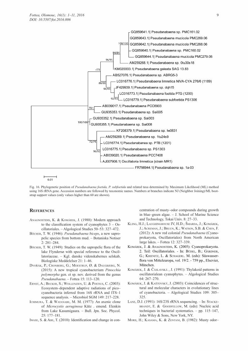
Fig. 16. Phylogenetic position of Pseudanabaena foetida, P. subfoetida and related taxa determined by Maximum Likelihood (ML) method using 16S rRNA gene. Accession numbers are followed by taxonomic names. Numbers at branches indicate NJ (Neighbor Joining)/ML boot-strap support values (only values higher than 60 are shown).
references
aNagNosTidis, K. & komárek, J. (1988): Modern approach to the classification system of cyanophytes 3 – Os-cillatoriales. – Algological Studies 50–53: 327–472.
böcher, T. W. (1946): Pseudanabaena biceps, a new sapro-pelic species from bottom mud. – Botaniska Notiser 2: 281–284.
böcher, T. W. (1949): Studies on the sapropelic flora of the lake Flyndersø with special reference to the Oscil-latoriaceae. – Kgl. danske videnskabernes selskab, Biologiske Meddelelser 21: 1–46.
dvořák, P.; croNberg, g.; moesTruP, Ø. & daugbjerg, N. (2015): A new tropical cyanobacterium Pinocchia polymorpha gen. et sp. nov. derived from the genus Pseudanabaena. – Fottea 15: 113–120.
ErNsT, a.; becker, s.; WolleNZieN, u. & PosTius, c. (2003): Ecosystem–dependent adaptive radiations of pico– cyanobacteria inferred from 16S rRNA and ITS–1 sequence analysis. – Microbiol SGM 149: 217–228.
ichimura, T. & WaTaNabe, M. M. (1977): An axenic clone of Microcystis aeruginosa Kütz . emend. Elenkin from Lake Kasumigaura. – Bull. Jpn. Soc. Phycol. 25: 177–181.
iWase, S. & abe, T. (2010): Identification and change in con-
centration of musty–odor compounds during growth in blue–green algae. – J. School of Marine Science and Technology, Tokai Univ. 8: 27–33.
kliNg, H.J.; laughiNghouse IV, H.D.; šmarda, J.; komárek, J.; acremaN, J.; bruuN, K.; WaTsoN, S.B. & cheN, F. (2012): A new red colonial Pseudanabaena (Cyano-prokaryota, Oscillatoriales) from North American large lakes. – Fottea 12: 327–339.
komárek, j. & aNagNosTidis, k. (2005): Cyanoprokaryota. 2. Teil: Oscillatoriales. - In: büdel, b.; gärdNer, g.; krieNiTZ, l. & schagerl. m. (eds): Süswasser-flora von Mitteleuropa, vol. 19/2. - 759 pp., Elsevier, München.
komárek, J. & Čáslavská , J. (1991): Thylakoid patterns in oscillatorialean cyanophytes. – Algological Studies 64: 267–270.
komárek, j. & KašTovský, J. (2003): Coincidences of struc-tural and molecular characters in evolutionary lines of cyanobacteria. – Algological Studies 109: 305–325.
laNe, D.J. (1991): 16S/23S rRNA sequencing. – In: sTacke-braNdT, e. & goodfelloW, m. (eds): Nucleic acid techniques in bacterial systematics. – pp. 115–147, John Wiley & Sons, New York, NY.
morii, H.; kasama, K. & ZeNTaNi, B. (1982): Musty odor–
Fottea, Olomouc, 16(1): 1–11, 2016 9DOI: 10.5507/fot.2016.006
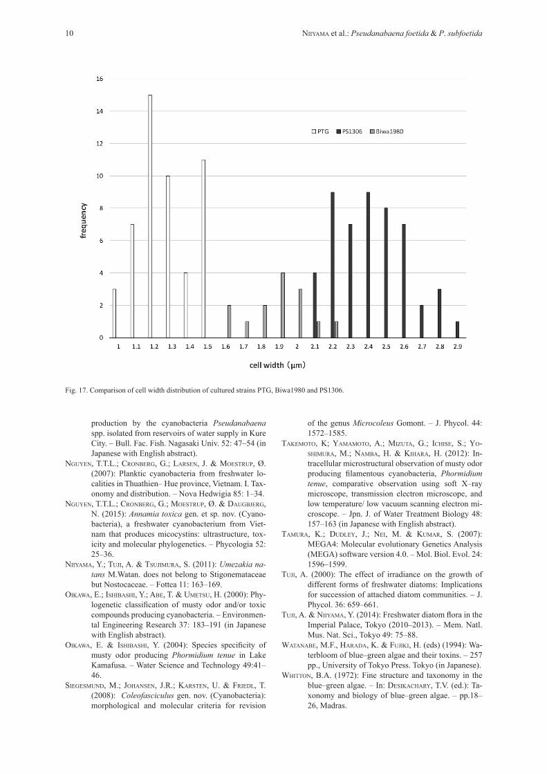
Fig. 17. Comparison of cell width distribution of cultured strains PTG, Biwa1980 and PS1306.
of the genus Microcoleus Gomont. – J. Phycol. 44: 1572–1585.
TakemoTo, K; yamamoTo, A.; miZuTa, G.; ichise, S.; yo-shimura, M.; Namba, H. & kihara, H. (2012): In-tracellular microstructural observation of musty odor producing filamentous cyanobacteria, Phormidium tenue, comparative observation using soft X–ray microscope, transmission electron microscope, and low temperature/ low vacuum scanning electron mi-croscope. – Jpn. J. of Water Treatment Biology 48: 157–163 (in Japanese with English abstract).
Tamura, K.; dudley, J.; Nei, M. & kumar, S. (2007): MEGA4: Molecular evolutionary Genetics Analysis (MEGA) software version 4.0. – Mol. Biol. Evol. 24: 1596–1599.
Tuji, A. (2000): The effect of irradiance on the growth of different forms of freshwater diatoms: Implications for succession of attached diatom communities. – J. Phycol. 36: 659–661.
Tuji, a. & Niiyama, y. (2014): Freshwater diatom flora in the Imperial Palace, Tokyo (2010–2013). – Mem. Natl. Mus. Nat. Sci., Tokyo 49: 75–88.
WaTaNabe, M.F., harada, K. & fujiki, H. (eds) (1994): Wa-terbloom of blue–green algae and their toxins. – 257 pp., University of Tokyo Press. Tokyo (in Japanese).
WhiTToN, B.A. (1972): Fine structure and taxonomy in the blue–green algae. – In: desikachary, T.V. (ed.): Ta-xonomy and biology of blue–green algae. – pp.18–26, Madras.
production by the cyanobacteria Pseudanabaena spp. isolated from reservoirs of water supply in Kure City. – Bull. Fac. Fish. Nagasaki Univ. 52: 47–54 (in Japanese with English abstract).
NguyeN, T.T.L.; croNberg, g.; larseN, j. & moesTruP, Ø. (2007): Planktic cyanobacteria from freshwater lo-calities in Thuathien– Hue province, Vietnam. I. Tax-onomy and distribution. – Nova Hedwigia 85: 1–34.
NguyeN, T.T.L.; croNberg, g.; moesTruP, Ø. & daugbjerg, N. (2015): Annamia toxica gen. et sp. nov. (Cyano-bacteria), a freshwater cyanobacterium from Viet-nam that produces micocystins: ultrastructure, tox-icity and molecular phylogenetics. – Phycologia 52: 25–36.
Niiyama, Y.; Tuji, A. & Tsujimura, S. (2011): Umezakia na-tans M.Watan. does not belong to Stigonemataceae but Nostocaceae. – Fottea 11: 163–169.
oikaWa, E.; ishibashi, Y.; abe, T. & umeTsu, H. (2000): Phy-logenetic classification of musty odor and/or toxic compounds producing cyanobacteria. – Environmen-tal Engineering Research 37: 183–191 (in Japanese with English abstract).
oikaWa, E. & ishibashi, Y. (2004): Species specificity of musty odor producing Phormidium tenue in Lake Kamafusa. – Water Science and Technology 49:41–46.
siegesmuNd, m.; johaNseN, j.r.; karsTeN, u. & friedl, T. (2008): Coleofasciculus gen. nov. (Cyanobacteria): morphological and molecular criteria for revision
10 Niiyama et al.: Pseudanabaena foetida & P. subfoetida

yagi, M. (1983): Odor produced by blue green algae. – Ei-sei Kagaku 29:16–22 (in Japanese with English abstract).
yagi, O.; okada, M. & sudo, R. (1979): Cultivation of Microcystis and red–tide–organisms. – Res. Rep. Natl. Insti. Environ. Stud. 6: 223–223 (in Japanese with English abstract).
yamada, N.; aoyama, K.; yamada, M. & hamamura, N. (1985): Studied on earthy–musty odor in natural wa-ter (1). Growth characteristics and 2–methylisobor-neol production of Phormidium tenue. – Research in Pollution of Water Supplies 8: 515–521 (in Japanese
© Czech Phycological Society (2016)Received May 28, 2014Accepted September 23, 2015
Supplementary material
the following supplementary material is available for this article:
Fig. S1. Secondary structure of the 16S–23S rRNA ITS region of P. foetida (PTG).
Fig. S2. Secondary structure of the 16S–23S rRNA ITS region of P. subfoetida (PS1306).
This material is available as part of the online article (http://fottea.czechphycology.cz/contents)
Fottea, Olomouc, 16(1): 1–11, 2016 11DOI: 10.5507/fot.2016.006
with English abstract).yamada, N.; aoyama, K.; yamada, M. & hamamura, N.
(1986): Studied on earthy–musty odor in natural wa-ter (3). Isolation of bacteria–free Phormidium tenue and the effect of associated bacteria on the growth of axenic P. tenue. – Research in Pollution of Wa-ter Supplies 9: 379–385 (in Japanese with English abstract).
Zuker, M. (2003): Mfold web server for nucleic acid folding and hybridization prediction. Nucleic Acids Res. 31: 3406–15.