Embed Size (px)
Citation preview
Provided by the author(s) and University College Dublin Library in accordance with publisher
policies., Please cite the published version when available.
Title BMP7-induced-Pten inhibits Akt and prevents renal fibrosis
Authors(s) Higgins, Debra F.; Weart, Leah; Masterson, Enda; Tennant, Sadhbh; Grebnev, Gleb;
Martin, Finian; Godson, Catherine; et al.
Publication date 2017-09-18
Publication information Biochimica et Biophysica Acta (BBA) - Molecular Basis of Disease, 1863 (12): 3095-3104
Publisher Elsevier
Item record/more information http://hdl.handle.net/10197/9296
Publisher's statement þÿ�T�h�i�s� �i�s� �t�h�e� �a�u�t�h�o�r ��s� �v�e�r�s�i�o�n� �o�f� �a� �w�o�r�k� �t�h�a�t� �w�a�s� �a�c�c�e�p�t�e�d� �f�o�r� �p�u�b�l�i�c�a�t�i�o�n� �i�n� �B�i�o�c�h�i�m�i�c�a� �e�t�
Biophysica Acta (BBA) - Molecular Basis of Disease. Changes resulting from the publishing
process, such as peer review, editing, corrections, structural formatting, and other quality
control mechanisms may not be reflected in this document. Changes may have been made
to this work since it was submitted for publication. A definitive version was subsequently
published in Biochimica et Biophysica Acta (BBA) - Molecular Basis of Disease (1863, 12,
(2017)) DOI:10.1016/j.bbadis.2017.09.011
Publisher's version (DOI) 10.1016/j.bbadis.2017.09.011
Downloaded 2019-06-26T17:02:48Z
The UCD community has made this article openly available. Please share how this access
benefits you. Your story matters! (@ucd_oa)
Some rights reserved. For more information, please see the item record link above.

Page 1 of 28
BMP7- induced-Pten inhibits Akt and prevents renal fibrosis
Debra F Higgins1, Leah Ewart1, Enda Masterson1, Sadhbh Tennant1,Gleb Grebnev1, Marco Prunotto2,
Sylvia Pomposiello2, Karin Conde-Knape2, Finian M Martin1, Catherine Godson1.
1UCD Diabetes Complications Research Centre, School of Medicine, Conway Institute, University
College Dublin, Belfield, Dublin 4, Ireland.
2F. Hoffmann-La Roche, Dept of Metabolic and Vascular Disease, CH4070 Basel, Switzerland
Corresponding Author:
Name: Dr Debra Higgins
Address: UCD Diabetes Complications Research Centre,
School of Medicine, Conway Institute,
University College Dublin,
Belfield, Dublin 4,
Ireland.
Phone: +353 87 660 1549
Email: [email protected]
Sources of Support: Irish Health Research Board Translational Medicine Fellowship (HRB PDTM
2011/13), Science Foundation Ireland (06/IN.1/B114), Hoffmann La Roche, Basel
Running title: BMP-7 abrogates fibrosis by inhibiting Akt
Word count: Abstract: 240
Text: 3863
Page 2 of 28
Abstract:
Bone morphogenetic protein-7 (BMP-7) counteracts pro-fibrotic effects of TGFβ1 in cultured renal
cells and protects from fibrosis in acute and chronic renal injury models. Using the unilateral ureteral
obstruction (UUO) model of chronic renal fibrosis, we investigated the effect of exogenous-rhBMP-7
on pro-fibrotic signaling pathways mediated by TGFβ1 and hypoxia. Mice undergoing UUO were
treated with vehicle or rhBMP-7 (300μg/kg i.p.) every other day for eight days and kidneys analysed
for markers of fibrosis and SMAD, MAPK, and PI3K signaling. In the kidney, collecting duct and
tubular epithelial cells respond to BMP-7 via activation of SMAD1/5/8. Phosphorylation of
SMAD1/5/8 was reduced in UUO kidneys from vehicle-treated animals yet maintained in UUO
kidneys from BMP-7-treated animals, confirming renal bioactivity of exogenous rhBMP-7. BMP-7
inhibited Collagen Iα1 and Collagen IIIα1 gene expression and Collagen I protein accumulation, while
increasing expression of Collagen IVα1 in UUO kidneys. Activation of SMAD2, SMAD3, ERK, p38 and
PI3K / Akt signaling occurred during fibrogenesis and BMP-7 significantly attenuated SMAD3 and Akt
signaling in vivo. Analysis of renal collecting duct (mIMCD) and tubular epithelial (HK-2) cells
stimulated with TGFβ1 or hypoxia (1% oxygen) to activate Akt provided further evidence that BMP-7
specifically inhibited PI3K / Akt signaling. PTEN is a negative regulator of PI3K and BMP-7 increased
PTEN expression in vivo and in vitro. These data demonstrate an important mechanism by which
BMP-7 orchestrates renal protection through Akt inhibition and highlights Akt inhibitors as anti-
fibrotic therapeutics.
Keywords:
Fibrosis, Anti-fibrotic, PTEN, Akt, TGFβ1, hypoxia

Page 3 of 28
Introduction:
Bone morphogenetic protein -7 (BMP-7 , or osteogenic protein (OP)-1) is a 35kDa member of the
transforming growth factor beta (TGFβ) family of secreted extracellular proteins. In addition to its
role in regulating bone and cartilage formation, BMP-7 is essential for renal morphogenesis,
stimulating condensation of the metanephric mesenchyme around the ureteric bud, leading to the
formation of collecting duct epithelia through promotion of mesenchymal to epithelial transition
(MET). 1-4 BMP-7 null mice die soon after birth due to renal hypoplasia associated with reduced
ureteric bud branching, loss of metanephric mesenchyme, cessation of nephrogenesis and a marked
reduction of glomerular density. 2, 5 BMP-7 expression is retained in adult kidneys and is thought to
be critical for maintaining tubular epithelial integrity. 6-13 During renal disease, interaction of BMP-7
with distinct serine / threonine kinase receptors is prevented by binding to endogenously generated
antagonists such as gremlin, noggin and USAG-1 (Uterine sensitization-associated gene -1, also
referred to as Sclerostin domain containing -1 , Sostdc1), 13 facilitating damage to the tubular
epithelium. Several studies have reported that introduction of exogenous recombinant BMP-7 or
reactivation of endogenous BMP-7 signaling through peptide mediated receptor activation can
prevent progression of renal disease. 7, 8, 10, 14-17
BMP-7 is reno-protective in acute and chronic renal injury models (reviewed in 18) including ischemia
reperfusion injury (IRI), 7, 14 unilateral ureteral obstruction (UUO), 8, 19-21 5/6 nephrectomy, 3, 22 lupus
nephritis, nephrotoxic serum nephritis (NTN), Collagen type 4α3-null 'Alport' mice, 23 and STZ-
induced diabetic nephropathy. 24, 25 BMP-7 is efficacious in other fibrotic diseases including cardiac,
26 liver, 27, 28 corneal, 29 and silica induced pulmonary fibrosis. 30 Preservation of renal function,
normalisation of serum creatinine, reduction of extracellular matrix accumulation, and reduction in
pro-inflammatory mediators have been demonstrated. 7, 8, 14, 19, 23, 31 However, the molecular
mechanisms involved in this protection have not been fully defined. In this report, we describe the
effect of BMP-7 on key signaling pathways underlying fibrogenesis.

Page 4 of 28
TGFβ1 is the prototypic pro-sclerotic cytokine which is aberrantly up-regulated during fibrotic
disease. It is responsible for the induction and accumulation of interstitial matrix, which is
considered the pathological hallmark of all kidney injuries regardless of the aetiology. 32 TGFβ1
promotes pro-fibrotic responses of renal epithelia and pericytes and activates resident fibroblasts. 33
TGFβ1 binds type I and type II serine / threonine receptors on the plasma membrane activating
receptor-associated SMAD proteins (R-SMADs), predominantly SMAD2 and SMAD3.
Phosphorylated R-SMADs associate with co-SMAD-4 and bind specific SMAD binding elements in
gene promoters. SMAD3 mediated signaling plays a central role in renal fibrogenesis 34-37 and SMAD3
knockout mice are protected against STZ-induced diabetic glomerulopathy, 38 and UUO-induced
tubulointerstitial fibrosis. 39 In addition, TGFβ1 signals through non-SMAD proteins to effect a variety
of cellular changes, including the mitogen activated protein kinases (MAPK) p38 and ERK, and
phosphoinositide -3 kinase (PI3K). 10, 40, 41 These pathways may further enhance or oppose SMAD
signaling in a context dependent manner. 42, 43
BMP-7 is predominantly expressed in cortical and corticomedullary proximal tubular cells, distal
convoluted tubules, collecting duct epithelia and glomerular podocytes, 6, 7, 10, 13, 14, 25, 31, 44, 45 and
signals through three type I serine / threonine receptors (termed activin-like kinase (Alk) receptors;
Alk 3 (BMPR-1A), Alk6 (BMPR-1B) and Alk2 (Type 1A activin receptor) and one type II receptor
(BMPRII) to phosphorylate SMAD1, SMAD5, and SMAD8. 10, 17, 46-48 SMADs 1, 5, and 8 also bind
SMAD4 and translocate to the nucleus to activate gene transcription. Alk3 is predominantly
expressed in renal tubular epithelial cells, with Alk6 expressed in osteoblasts 16 and Alk2 in
cartilage.10, 31 The importance of Alk3 in mediating the protective effects of renal BMP-7 signaling
was borne out by the enhanced pro-fibrotic effects of TGFβ1 observed upon deletion of Alk3 in the
tubular epithelium. 16
The principle drivers of injury in UUO include TGFβ1 and hypoxia. 49 In this report, we describe the
effect of BMP-7 on hypoxia- and TGFβ1-induced signaling pathways in the unilateral ureteral

Page 5 of 28
obstruction (UUO) model of renal fibrosis. We report that BMP-7 inhibits activation of PI3K signaling
via Akt in addition to inhibition of SMAD3 in vivo. We demonstrate that BMP-7 specifically inhibits
TGFβ1- and hypoxia-induced PI3K activity in renal collecting duct and tubular epithelial cells via up-
regulation of the PI3K inhibitor phosphatase and tensin homolog (PTEN).
Results:
BMP-7 preserves pSMAD1/5/8 activity in fibrotic kidneys
Phosphorylation of SMAD1/5/8 was analysed in contralateral (CTL) and obstructed (UUO) kidneys to
determine whether exogenous recombinant human BMP-7 (rhBMP-7) administered via i.p. injection
could activate BMP-7 signaling in renal tissue. In vehicle treated animals, pSMAD1/5/8 was detected
in the contralateral kidney (veh-CTL) but lost during development of fibrosis in the corresponding
obstructed kidneys (veh-UUO) (P<0.001, Figure 1A and Supplemental Table 1). In contrast, in
animals treated with rhBMP-7, pSMAD1/5/8 was detected in the obstructed kidneys (BMP-7-UUO)
suggesting maintenance of BMP-7 responsive pathways, confirming functional activity of
exogenously applied rhBMP-7 in the kidney (Figure 1A).
As a further readout for activation of BMP-7 pathways, expression of the BMP-7-target gene Id-1
was analysed. In vehicle-UUO kidneys, Id-1 mRNA was significantly down-regulated (P<0.01, Figure
1A and Supplemental Table 1), mirroring loss of SMAD1/5/8 activity in these kidneys, however there
was no significant decrease of Id-1 in BMP-7-UUO kidneys confirming preservation of BMP-7
regulated pathways (Figure 1B).
Expression of Bmp-7 mRNA was significantly down-regulated in vehicle-UUO kidneys compared with
vehicle-CTL kidneys (78.4% reduction, P<0.001) while BMP-7-UUO kidneys maintained Bmp-7 mRNA
expression (56.8% reduction, P<0.001) (Figure 1B). BMP-7 antagonist Usag1 mRNA was significantly
increased in vehicle-UUO (P<0.05), consistent with loss of BMP-7 signaling in veh-UUO, whereas

Page 6 of 28
Usag1 mRNA was not increased in BMP-7-UUO kidneys consistent with maintenance of BMP-7
signaling. BMP-7 antagonist Grem1 mRNA was not changed in response to UUO but was significantly
increased in BMP-7-CTL kidneys (P<0.05 compared to either veh-CTL or BMP7-UUO, Figure 1B).
Figure 1: Exogenous BMP-7 activates SMAD1/5/8 signaling in fibrotic kidneys in vivo. A Western
blot of 20µg whole cell lysate from vehicle (veh) or BMP-7 treated contralateral (CTL, C) and 8d-post
obstructed (UUO , U) kidneys probed for phosphorylated SMAD1/5/8 and GAPDH, n= 5 vehicle and
n=4 BMP-7 groups, representative blots shown. Graph shows densitometric analysis of pSMAD1/5/8
normalised to ponceau. B Quantitative PCR analysis of Id-1, Bmp-7, Usag1, and Gremlin 1 (Grem1)
gene expression normalised to 18S mRNA levels. Graphs show fold-change in mRNA expression with
Veh-CTL arbitrarily set to 1, n=5 in vehicle and BMP-7 groups, statistical analysis performed using
One-way ANOVA and Bonferroni's multiple comparison test, *P<0.05, **P<0.001.
BMP-7 reduces extracellular collagen, but not α-SMA, accumulation in vivo
The protective effect of BMP-7 on the development of fibrosis in vivo was investigated by
quantifying extracellular matrix (ECM) accumulation in contralateral and obstructed kidneys from

Page 7 of 28
vehicle- or BMP-7-treated mice. A significant increase in sirius red staining of newly deposited
collagen fibrils was observed in vehicle-UUO kidneys compared with vehicle-CTL kidneys (P<0.01) , in
contrast there was no detectable increase in sirius red-stained collagen in BMP-7-UUO kidneys
(P>0.05, Figure 2A & B and Supplemental Table 1) confirming reduced ECM accumulation.
Despite reducing collagen accumulation, BMP-7 had no effect on UUO-induced α-SMA protein
(P<0.05 in veh-UUO and BMP-7 UUO compared to respective contralateral kidneys, Figure 2A&B) or
mRNA expression (P<0.001, in veh-UUO and BMP-7-UUO compared to respective contralateral
kidneys, Figure 2C and Supplemental Table 1). Additionally, BMP-7 had no effect on Fsp-1 mRNA
expression as a marker of interstitial injury in fibrotic kidneys (P<0.001 in veh-UUO and BMP-7-UUO
compared to respective contralateral kidneys, Figure 2C and Supplemental Table 1).
Figure 2: Exogenous BMP-7 reduces renal fibrosis and collagen deposition, but not fibroblast
activation, in vivo. A Extracellular matrix deposition in contralateral (CTL, C) and 8d-post obstructed
(UUO, U) kidneys, from mice treated with vehicle (Veh) or BMP-7 as described in Methods, detected
by Sirius Red or immunohistochemical staining for α-SMA, magnification x20, scale bar 100µm. Neg:
negative control with sections processed in absence of primary antibody. B Morphometric analysis

Page 8 of 28
of positive IHC stained area (mm2). C Quantitative PCR analysis of Fsp-1 and α-Sma mRNA
normalised to 18S mRNA. n=5 in vehicle and BMP-7 groups. Statistical analysis performed using One-
way ANOVA with Bonferroni's multiple comparison post-test,*P<0.05, **P<0.001.
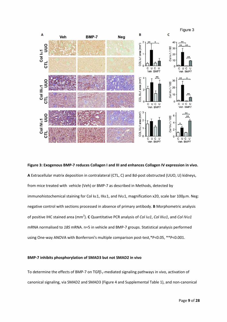
BMP-7 reduces renal fibrosis in vivo via reduced Collagen I and III and enhanced Collagen IV
To determine the specific collagen types that were altered by BMP-7 treatment, Collagen Iα1, IIIα1,
and IVα1 proteins were assessed by immunohistochemistry (Figure 3A and B). Collagen Iα1 was
significantly increased in veh-UUO kidneys (P<0.01) whereas there was no significant increase
observed in BMP-7-UUO kidneys (P>0.05, Figure 3A and B and Supplemental Table 1). Similarly, Col
Iα1 mRNA was significantly increased in veh-UUO compared to veh-CTL kidneys (P<0.001, Figure 3C).
While there was a significant increase in Col Iα1 mRNA between BMP-7-CTL and BMP-7-UUO kidneys
(P<0.001, Figure 2C), Col Iα1 gene expression was significantly reduced in both contralateral and
obstructed kidneys of BMP-7-treated animals compared with vehicle treated animals (P<0.001).
Collagen IIIα1 protein was significantly increased in BMP-7-UUO kidneys compared to BMP-7-CTL
kidneys (P<0.01), and unlike Collagen Iα1, there was no significant difference in the quantity of
Collagen IIIα1 protein between veh-UUO kidneys and BMP-7-UUO (P>0.05, Figure 3A & B and
Supplemental Table 1). However, induction of Collagen IIIα1 mRNA in UUO kidneys was significantly
impaired (P=0.002) in animals treated with BMP-7 (P<0.001in veh-UUO and BMP-7 UUO versus
respective CTL kidneys, Figure 3C).
Collagen IVα1 protein was not significantly increased in UUO kidneys compared with CTL kidneys of
either group (P>0.05, Figure 2A & B and Supplemental Table 1). However, Col IVα1 mRNA was
significantly increased in BMP-7-UUO kidneys compared with vehicle-UUO kidneys (P<0.001, Figure
3C).
Page 9 of 28
Figure 3: Exogenous BMP-7 reduces Collagen I and III and enhances Collagen IV expression in vivo.
A Extracellular matrix deposition in contralateral (CTL, C) and 8d-post obstructed (UUO, U) kidneys,
from mice treated with vehicle (Veh) or BMP-7 as described in Methods, detected by
immunohistochemical staining for Col Iα1, IIIα1, and IVα1, magnification x20, scale bar 100µm. Neg:
negative control with sections processed in absence of primary antibody. B Morphometric analysis
of positive IHC stained area (mm2). C Quantitative PCR analysis of Col Iα1, Col IIIα1, and Col IVα1
mRNA normalised to 18S mRNA. n=5 in vehicle and BMP-7 groups. Statistical analysis performed
using One-way ANOVA with Bonferroni's multiple comparison post-test,*P<0.05, **P<0.001.
BMP-7 inhibits phosphorylation of SMAD3 but not SMAD2 in vivo
To determine the effects of BMP-7 on TGFβ1-mediated signaling pathways in vivo, activation of
canonical signaling, via SMAD2 and SMAD3 (Figure 4 and Supplemental Table 1), and non-canonical
Page 10 of 28
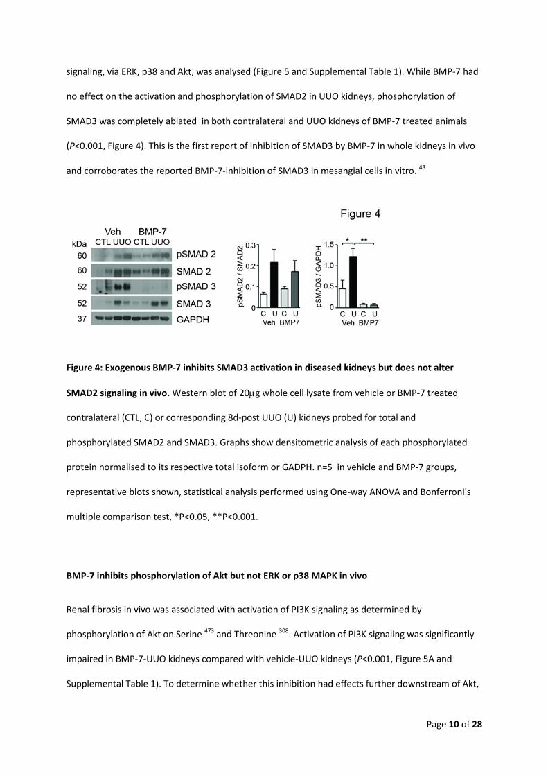
signaling, via ERK, p38 and Akt, was analysed (Figure 5 and Supplemental Table 1). While BMP-7 had
no effect on the activation and phosphorylation of SMAD2 in UUO kidneys, phosphorylation of
SMAD3 was completely ablated in both contralateral and UUO kidneys of BMP-7 treated animals
(P<0.001, Figure 4). This is the first report of inhibition of SMAD3 by BMP-7 in whole kidneys in vivo
and corroborates the reported BMP-7-inhibition of SMAD3 in mesangial cells in vitro. 43
Figure 4: Exogenous BMP-7 inhibits SMAD3 activation in diseased kidneys but does not alter
SMAD2 signaling in vivo. Western blot of 20µg whole cell lysate from vehicle or BMP-7 treated
contralateral (CTL, C) or corresponding 8d-post UUO (U) kidneys probed for total and
phosphorylated SMAD2 and SMAD3. Graphs show densitometric analysis of each phosphorylated
protein normalised to its respective total isoform or GADPH. n=5 in vehicle and BMP-7 groups,
representative blots shown, statistical analysis performed using One-way ANOVA and Bonferroni's
multiple comparison test, *P<0.05, **P<0.001.
BMP-7 inhibits phosphorylation of Akt but not ERK or p38 MAPK in vivo
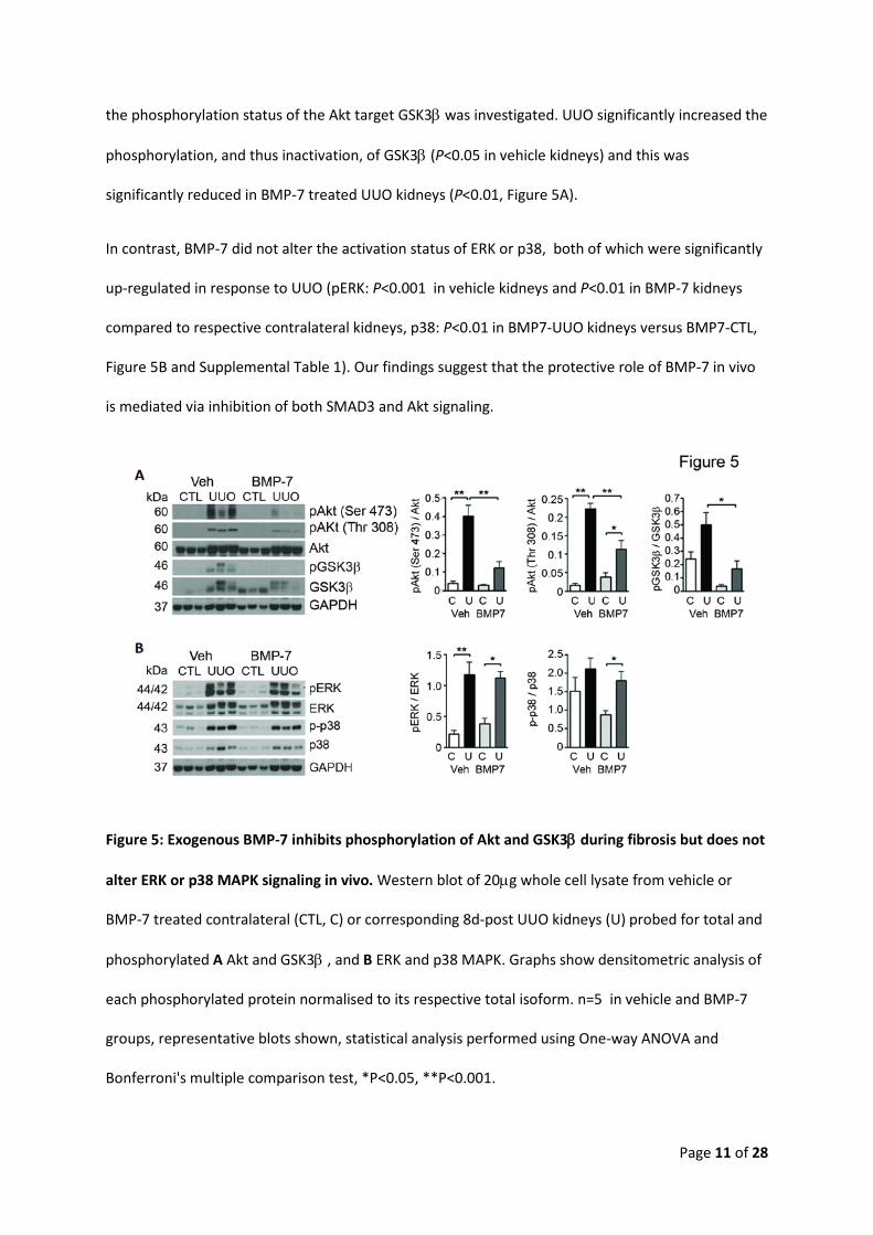
Renal fibrosis in vivo was associated with activation of PI3K signaling as determined by
phosphorylation of Akt on Serine 473 and Threonine 308. Activation of PI3K signaling was significantly
impaired in BMP-7-UUO kidneys compared with vehicle-UUO kidneys (P<0.001, Figure 5A and
Supplemental Table 1). To determine whether this inhibition had effects further downstream of Akt,
Page 11 of 28
the phosphorylation status of the Akt target GSK3β was investigated. UUO significantly increased the
phosphorylation, and thus inactivation, of GSK3β (P<0.05 in vehicle kidneys) and this was
significantly reduced in BMP-7 treated UUO kidneys (P<0.01, Figure 5A).
In contrast, BMP-7 did not alter the activation status of ERK or p38, both of which were significantly
up-regulated in response to UUO (pERK: P<0.001 in vehicle kidneys and P<0.01 in BMP-7 kidneys
compared to respective contralateral kidneys, p38: P<0.01 in BMP7-UUO kidneys versus BMP7-CTL,
Figure 5B and Supplemental Table 1). Our findings suggest that the protective role of BMP-7 in vivo
is mediated via inhibition of both SMAD3 and Akt signaling.
Figure 5: Exogenous BMP-7 inhibits phosphorylation of Akt and GSK3β during fibrosis but does not
alter ERK or p38 MAPK signaling in vivo. Western blot of 20µg whole cell lysate from vehicle or
BMP-7 treated contralateral (CTL, C) or corresponding 8d-post UUO kidneys (U) probed for total and
phosphorylated A Akt and GSK3β , and B ERK and p38 MAPK. Graphs show densitometric analysis of
each phosphorylated protein normalised to its respective total isoform. n=5 in vehicle and BMP-7
groups, representative blots shown, statistical analysis performed using One-way ANOVA and
Bonferroni's multiple comparison test, *P<0.05, **P<0.001.

Page 12 of 28
BMP-7 inhibits TGFβ1- and hypoxia-induced-Akt signaling in renal epithelia
To investigate the effects of BMP-7 on Akt signaling in renal epithelial cells in culture, collecting duct
(mIMCD) and proximal tubular (HK-2) cells were treated with 5-100ng/ml BMP-7. BMP-7 inhibited
phosphorylation of Akt-Ser473 and Akt-Thr308 in a dose dependent manner in mIMCD and induced
expression of E-cadherin (Supplemental Figure 1A). HK-2 tubular epithelial cells were stimulated
with hypoxia (1% O2) to activate Akt signaling, BMP-7 inhibited phosphorylation of Akt-Ser473 in a
dose dependent manner but not Akt-Thr308 (Supplemental Figure 1B). Interestingly, in both cell types
BMP-7 dose-dependently increased phosphorylation of SMAD2 (Supplemental Figure 1A&B).
Supplemental Figure 1: BMP-7 inhibits Akt activation in renal collecting duct (mIMCD) and tubular
epithelia (HK-2) in a dose dependent manner. A Western blot of 20µg whole cell lysate from
mIMCD cells treated with BMP-7 (0-100ng/ml for 150 minutes) and probed for phosphorylated and
total Akt , SMAD2, SMAD 1/5/8 and E-Cadherin, GAPDH was analysed as a loading control. B
Western blot of 20µg whole cell lysate from HK-2 cells pre-treated with rhBMP-7 (0-100ng/ml for 30
minutes) followed by 1% O2 for 60 minutes. Blots were probed for phosphorylated and total forms of
Akt and SMAD2. The experiments were repeated twice, representative blots shown.

Page 13 of 28
To determine the effects of BMP-7 on TGFβ1-induced signaling in vitro, collecting duct epithelia
(mIMCD) were co-stimulated with 100ng/ml BMP-7 and 5ng/ml TGFβ1. BMP-7 inhibited TGFβ1-
induced phosphorylation of Akt at Ser473 but not at Thr308 (P<0. 05, Figure 6A and Supplemental
Table 1). TGFβ1 induced phopshorylation of GSK3β (P<0.05, Figure 6A) and reduced E-cadherin
protein (P<0.01, Figure 6A), although BMP-7 did not alter the phosphorylation status of GSK3β it
restored E-cadherin protein expression (P<0.05 versus TGFβ, Figure 6A). BMP-7 had no effect on
TGFβ1-induced phosphorylation of ERK and p38 (Figure 6B).
Figure 6: BMP-7 inhibits TGFβ1-induced Akt activation and restores E-cadherin expression in
collecting duct epithelial cells. Western blot of 20µg whole cell lysate from mIMCDs pre-treated
with vehicle or rhBMP-7 (100ng/ml for 30 minutes) followed by vehicle or TGFβ1 (5ng/ml for 60
minutes) probed for phosphorylated and total isoforms of A Akt and GSK3β, and E-cadherin, and B
ERK and p-38 MAPK. GAPDH was analysed as a loading control. Graphs show densitometric analysis,
V: Vehicle, B: BMP-7, T: TGFβ1, BT: BMP-7 & TGFβ1. Treatments were performed in triplicate and
experiments repeated three times, statistical significance was determined by one-way ANOVA and
Bonferroni's multiple comparison test, *P<0.05, **P<0.001.
Page 14 of 28
BMP-7 inhibits Akt signaling via up-regulation of PTEN
Activation of PI3K signaling involves conversion of phosphatidylinositol 4,5-bisphosphate (PIP2) to
phosphatidylinositol 3,4,5-triphosphate ( PIP3) enabling subsequent phosphorylation of Akt.
Phosphatase and tensin homologue (PTEN) negatively regulates PI3K signaling by converting PIP3
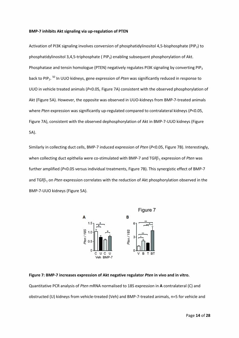
back to PIP2. 50 In UUO kidneys, gene expression of Pten was significantly reduced in response to
UUO in vehicle treated animals (P<0.05, Figure 7A) consistent with the observed phosphorylation of
Akt (Figure 5A). However, the opposite was observed in UUO-kidneys from BMP-7-treated animals
where Pten expression was significantly up-regulated compared to contralateral kidneys (P<0.05,
Figure 7A), consistent with the observed dephosphorylation of Akt in BMP-7-UUO kidneys (Figure
5A).
Similarly in collecting duct cells, BMP-7 induced expression of Pten (P<0.05, Figure 7B). Interestingly,
when collecting duct epithelia were co-stimulated with BMP-7 and TGFβ1 expression of Pten was
further amplified (P<0.05 versus individual treatments, Figure 7B). This synergistic effect of BMP-7
and TGFβ1 on Pten expression correlates with the reduction of Akt phosphorylation observed in the
BMP-7-UUO kidneys (Figure 5A).
Figure 7: BMP-7 increases expression of Akt negative regulator Pten in vivo and in vitro.
Quantitative PCR analysis of Pten mRNA normalised to 18S expression in A contralateral (C) and
obstructed (U) kidneys from vehicle-treated (Veh) and BMP-7-treated animals, n=5 for vehicle and
Page 15 of 28
BMP-7 groups, B collecting duct epithelial cells treated with vehicle (V), BMP-7 (B, 100ng/ml), TGFβ1
(T, 5ng/ml), or combined BMP-7 and TGFβ1 (BT). Treatments were performed in triplicate and the
experiments repeated three times. Statistical analysis was determined by one-way ANOVA and
Bonferroni's multiple comparison test, *P<0.05, **P<0.001.
Discussion:
A protective role for BMP-7 has been reported in a number of renal injury models however, the
molecular mechanisms by which BMP-7 exerts this protection have focussed primarily in vitro on the
effect of BMP-7 on TGFβ1-induced extracellular matrix (ECM) accumulation, 10, 51 and E-cadherin
expression in cultured renal cells 11, and genomic methylation. 52-57 Here we describe specific effects
of BMP-7 on TGFβ1-signaling and show in vivo and in vitro that BMP-7 not only inhibits activation of
SMAD3 but also inhibits Akt resulting in loss of pro-fibrotic signals and protection from fibrogenesis.
Exogenous rhBMP-7 activated SMAD1/5/8 signaling within the obstructed kidney, maintaining the
expression of the BMP-7 target gene Id1 (Figure 1). Gene expression of Bmp-7 was significantly
down-regulated in response to injury in vehicle treated animals, similar to reports in human renal
disease 58-60, however rhBMP-7 induced a significant induction of Bmp-7 mRNA expression
suggesting that BMP-7 can regulate its own expression in a positive feed forward loop (Figure 1B).
BMP-7 prevented the up-regulation of Usag1 expression observed in veh-UUO kidneys (Figure 1B)
suggesting that BMP-7 can regulate it's bioactivity by inhibiting expression of its antagonists.
Additionally, in BMP-7-CTL kidneys Grem1 expression was significantly up-regulated, perhaps in an
attempt to regulate the bioactivity of exogenous BMP-7 under physiologic conditions. This induction
of Grem1 was not observed in BMP-7-UUO kidneys suggesting the involvement of active BMP-7
under pathophysiologic conditions as a protective mechanism against fibrosis.
Page 16 of 28
Remarkably, BMP-7 abrogated fibrosis and ECM deposition despite an inability to alter myofibroblast
activation. Similar induction of α-SMA, the hallmark marker of the myofibroblast, was observed in
fibrotic kidneys from both vehicle- and BMP7-treated animals suggesting that BMP-7 works
downstream of myofibroblast activation to block ECM production (Figure 2). Myofibroblast numbers
are increased during renal fibrosis and they are considered to contribute to collagen and ECM
production in fibrotic diseases. 61 Interestingly, Takeji et al reported that α-SMA null mice had
significantly worse renal disease, in two separate in vivo models, than mice wild-type for α-SMA
expression, concluding that myofibroblasts play a role in suppressing renal fibrosis. Given their
findings, it is perhaps not surprising that the beneficial effects of BMP-7 require that myofibroblast
activation and α-SMA expression remains intact. 62
Induction of Collagen Iα1 and IIIα1 gene expression during fibrosis was abrogated by BMP-7-
treatment and a significant reduction of Collagen Iα1 protein accumulation was observed in BMP-7-
UUO kidneys (Figure 3). Collagen accumulation during fibrosis is dependent not only on increased
synthesis of proteins but also on decreased collagen turn-over. BMP-7 increases collagen
degradation through up-regulation of matrix metalloproteinases (MMP)-2 and MMP-9 23 and down-
regulation of plasminogen activator inhibitor-1 (PAI-1). 51 We propose that the reduced type I
collagen accumulation observed in BMP-7-UUO kidneys is likely due not only to reduced production
but enhanced degradation of ECM molecules. TGFβ1-induced Collagen I and III expression is
mediated via activation of SMAD3, 50, 63 therefore inhibition of SMAD3 activity by BMP-7 in vivo
(Figure 4) may explain the reduced gene expression of Col Iα1 and IIIα1 observed in BMP-7-UUO
kidneys (Figure 3). Our findings support those of Wang and Hirschberg in cultured renal mesangial
cells whereby a BMP-7-SMAD5-dependent up-regulation of SMAD6 inhibited nuclear accumulation
of phospho-SMAD3 leading to loss of TGFβ1-induced Col Iα1 gene expression. 43, 64 Our results
highlight that BMP-7 inhibits SMAD3 mediated TGFβ1 signaling in vivo and expands the repertoire of

Page 17 of 28
cell responses from the previously reported mesangial cells to collecting duct (Figure 6) and tubular
epithelial cells (Supplemental Figure 1).
In contrast to the inhibitory effects of BMP-7 on SMAD3, BMP-7 had no effect on ERK and p38 MAPK
signaling pathways in vivo or in cultured epithelia, consistent with the findings of Wang and
Hirschberg in mesangial cells. 43 However, BMP-7 significantly reduced phosphorylation of PI3K, Akt
and GSK3β. In veh-UUO kidneys, both phosphorylated Akt-Ser 473 and -Thr308 were increased,
however both phosphorylation events were inhibited in the presence of BMP-7 (Figure 5). In vitro,
BMP-7 inhibited TGFβ1-induced phosphorylation of Akt-Ser473 in collecting duct epithelial cells
(Figure 6) and hypoxia-induced phosphorylation of Akt-Ser473 in proximal tubular epithelial cells
(Supplemental Figure 1). However, only the initial phosphorylation event at Ser473 was successfully
inhibited by BMP-7 whereas phopsho-Akt -Thr308 was still detected in cultured cells in vitro. This may
be due to increased secretion of endogenous TGFβ1 or other cytokines that may overcome BMP-7's
ability to inhibit PI3K signaling in vitro.
Under physiologic conditions, active GSK3β phosphorylates β-catenin causing its ubiquitination and
degradation. In response to renal injury in veh-UUO kidneys (Figure 5) or to TGFβ1 (Figure 6),
phosphorylation and inactivation of GSK3β results in stabilisation and nuclear translocation of β-
catenin inducing the expression of its target gene Snai1. 65, 66 Snail is an E-cadherin E-box
transcriptional repressor which decreases E-cadherin protein as part of the epithelial injury process.
67 Inactivation of GSK3β, observed during renal injury, inhibits interaction between GSK3β and Snail,
resulting in nuclear translocation of Snail and repression of E-cadherin transcription 65, 66. In the
presence of BMP-7, GSK3β remains active and phosphorylates Snail promoting ubiquitin-mediated
degradation of Snail 65 and stabilisation of E-cadherin expression. Our results in collecting duct cells
confirmed this regulation, where TGFβ1 increased phosphorylation of GSK-3β and reduced
expression of E-cadherin protein (Figure 6). Furthermore addition of BMP-7 reversed the TGFβ1
down-regulation and restored E-cadherin protein expression. Our findings further delineate the

Page 18 of 28
mechanism by which BMP-7 impacts cellular signaling pathways to orchestrate protection from
fibrogenesis.
PTEN dephosphorylates PIP3 to PIP2 antagonizing PI3K signaling. Loss of PTEN leads to accumulation
of PIP3 and activation of Akt. Conversely, over-expression of PTEN leads to accumulation of PIP2 and
subsequent inhibition of PI3K signaling and Akt phosphorylation. A recent publication by
Samarakoon et al reported that expression of PTEN was lost in several models of renal injury
including UUO, aristocholic acid induced nephropathy (AAN), and streptozotocin-mediated injury.68
We now report that BMP-7 can prevent the loss of PTEN during UUO-induced fibrosis ensuring that
activation of Akt does not occur (Figure 7).
Akt has been implicated in renal fibrogenesis previously, however this is the first report of BMP-7 as
an inhibitor of Akt signaling. Crosstalk between Akt and SMAD signaling pathways has been reported
by a number of independent studies, Kattla et al reported that while TGFβ1-activation of PI3K and
SMAD3 occurred independently, inhibition of Akt, by LY294002 or Akt inhibitor II or PTEN over-
expression, abrogated TGFβ1-induced pro-fibrotic responses in renal epithelial cells. 69 Similarly Bakin
et al reported that Akt was required for TGFβ1-induced EMT of mammary epithelial cells. 70 Finer et
al reported that Akt mediated pro-fibrotic changes in a mouse adriamycin nephropathy model. By
treating mice with a specific inhibitor of PI3K they observed reduced phospho-Akt-Ser473 in glomeruli
and tubular epithelial cells which abrogated fibrosis and proteinuria in response to adriamycin. 71 We
previously reported that the protective effect of Lipoxin A4 in a model of rapidly progressing renal
fibrosis was associated with reduced activation of Akt and reduced Collagen I accumulation in the
obstructed kidney. 72 Runyan et al reported that inhibition of PI3K, by LY294002 or over-expression
of a dominant negative kinase deficient Akt construct, abrogated TGFβ1-induced collagen I
expression via reduced SMAD3 activity. 50 Connective tissue growth factor (CTGF) mediates the pro-
fibrotic effects of TGFβ1 through interaction with β3-integrins, resulting in activation of Akt and
subsequent induction of fibronectin in primary human mesangial cells. 73 BMP-7 binds and inhibits

Page 19 of 28
CTGF 74 highlighting a further potential mechanism through which BMP-7 may inhibit Akt activation
to prevent fibrosis.
Our data shows that exogenous BMP-7 activates SMAD1/5/8 signaling in the kidney as determined
via maintenance of pSMAD1/5/8 and Id-1 gene expression in BMP-7-UUO kidneys (Figure 1), BMP-7
inhibits collagen type I and III gene and protein expression in renal injury (Figure 2&3) via inhibition
of SMAD3 and Akt signaling (Figures 4-6), and that these effects are mediated by up-regulation of
PTEN (Figure 7). In addition to inhibition of SMAD3, BMP-7 promoted activation of SMAD2-mediated
signaling (Supplemental Figure 1), consistent with previous reports describing a protective role for
SMAD2 against fibrogenesis. 75 This report delineates the key role that Akt plays in mediating the
pro-fibrotic changes that occur during kidney injury and describes how BMP-7 can up-regulate PTEN
to prevent activation of Akt signaling. We highlight the potential of Akt inhibition as a therapeutic
approach to preventing progression of renal disease. It will be interesting to investigate whether Akt
inhibitors, developed as anti-cancer and anti-inflammatory therapeutics 76 may also be useful as
anti-fibrotic agents.
Methods:
Unilateral ureteral obstruction (UUO):
UUO was performed on 8-10 week old C57BL/6, FVB mice and kidneys harvested at 8 days. 49
Animals were randomized into two groups (n=5 each) receiving vehicle (20mM histidine, 140mM
NaCl, Roche) or 300µg/kg recombinant human BMP-7 (R&D systems) i.p. on days 0, 2, 4, and 6. One
third of each contralateral and obstructed kidney was formalin fixed, one third methyl Carnoy's
fixed, and one third divided for RNA and protein extraction. The animal study was performed in
accordance with ethical guidelines of UCD AREC and under license from the Department of Health.
Immunohistochemical analysis of kidney tissue:
Page 20 of 28
4µm thick sections were prepared from formalin and Methyl Carnoys fixed paraffin embedded
kidneys. Sirius red stain of collagen was detected on formalin fixed tissue. 49 IHC for type I, type III,
type IV Collagen (Southern Biotech) and α-SMA (Sigma) was performed on Methyl Carnoy's fixed
tissue. 77 Sections were scanned using Aperio Scanscope and analysed mophometrically using Aperio
Imagescope software. 49
Cell culture:
Murine inner medullary collecting duct (mIMCD) and human tubular epithelial (HK-2) cells were
cultured as described. 78, 79 Cells were serum starved for 24 hours prior to stimulation with 0-
100ng/ml rhBMP-7, 5ng/ml TGFβ1 (both R&D systems), or 1% oxygen (Coy hypoxia chamber) for 60
minutes before lysis with either TRIzol or Cellytic. 49
Quantitative real time PCR:
2µg RNA was reverse-transcribed using 200U superscript reverse transcriptase II and the cDNA
analysed for Collagen type Iα1, IIIα1, IVα1, α-Sma, Fsp-1, Id-1, Bmp-7, and Pten using Taqman
primer probes and SYBR green analysis of Usag1 (forward: GCATTTCAGTAGCACTGGAC and reverse:
ATGTATTTGGTGGACCGCAG), and Grem1 (forward: TCATTGTGTTCCATGTGCCT and reverse:
CAGCCCTCAGAGTTACCTCC). 80 qPCR results were analysed using ∆∆Ct normalised against 18S rRNA
and calibrated against vehicle treated contralateral kidneys or vehicle-treated cells.
Western blot protein analysis:
20µg denatured whole cell lysates were probed with primary antibodies against α-SMA, pSMAD1
(Ser465/465), 5 (Ser463/365), 8 (Ser426/458), SMAD5, pSMAD2 (Ser 465/467), SMAD2, pERK1/2 (Thr202/Tyr204),
ERK, p-p38 (Thr180/Tyr182), p38, pAkt (Ser 473 and Thr308), Akt, pGSK3β (Ser9) and GAPDH (Cell
Signaling), pSMAD3 (Ser423/425), SMAD3 (Abcam), followed by incubation with appropriate secondary
HRP-IgG antibody. Bands were detected by chemiluminescence.

Page 21 of 28
Acknowledgements:
This work was supported by grants from the Irish Health Research Board to DFH (HRB PDTM-2011-
13), Science Foundation Ireland to CG (06/IN.1/B114) and financial support from Hoffmann La
Roche, Basel. The Authors would like to thank Mr Andrew Gaffney for technical support, Ms
Catherine Moss for assistance with quantitative PCR, and Ms Janet McCormack for assistance with
slide scanning and morphometry.
Disclosure:
There are no competing financial interests.

Page 22 of 28
mean SEM mean SEM mean SEM mean SEM
1A pSMAD1/5/8 / SMAD5 2.89 0.37 0.49 0.17 <0.001 1.96 0.22 1.74 0.43 n.s.
1B Id-1 mRNA 1.03 0.09 0.59 0.08 <0.01 0.88 0.09 0.64 0.07 n.s.
1B Bmp-7 mRNA 1.02 0.10 0.22 0.05 <0.001 0.95 0.10 0.41 0.05 <0.001
1B Usag1 mRNA 1.00 0.06 1.58 0.18 <0.05 0.80 0.05 1.15 0.03 n.s.
1B Grem1 mRNA 1.04 0.17 0.96 0.08 n.s. 1.87 0.08 0.85 0.13 <0.01
2B Sirius red (mm2) 0.04 0.01 0.11 0.02 <0.01 0.04 0.01 0.06 0.01 n.s.
2B α-SMA (mm2) 0.48 0.46 2.98 0.30 <0.05 0.67 0.07 3.11 0.59 <0.05
2C α -SMA mRNA 1.07 0.12 4.21 0.51 <0.001 0.91 0.08 4.47 0.65 <0.001
2C Fsp-1 mRNA 1.00 0.05 7.21 0.96 <0.001 0.90 0.20 5.03 0.43 <0.001
3B Collagen Iα1 (mm2) 0.35 0.13 1.22 0.49 <0.01 0.32 0.08 0.59 0.24 n.s.
3B Collagen IIIα1 (mm2) 2.19 0.39 3.21 0.63 n.s. 1.35 0.44 2.98 0.62 <0.01
3B Collagen IVα1 (mm2) 4.38 0.77 5.26 1.05 n.s. 3.54 0.35 4.69 0.82 n.s.
3C Collagen Iα 1 mRNA 1.30 0.35 23.16 8.86 <0.001 0.31 0.05 8.78 0.56 <0.001
3C Collagen IIIα 1 mRNA 1.26 0.30 23.83 5.42 <0.001 0.47 0.09 5.62 0.81 <0.001
3C Collagen IVα 1 mRNA 1.08 0.14 2.47 0.41 <0.05 1.09 0.05 4.51 0.50 <0.001
4 pSMAD2 / SMAD2 0.06 0.01 0.22 0.06 n.s. 0.09 0.01 0.17 0.05 n.s.
4 pSMAD3 / GAPDH 0.45 0.20 1.22 0.19 <0.01 0.08 0.02 0.06 0.03 n.s.
5A pAkt Ser 473 / Akt 0.04 0.11 0.40 0.06 <0.001 0.03 0.01 0.12 0.03 <0.05
5A pAkt Thr 308 / Akt 0.01 0.00 0.20 0.02 <0.001 0.03 0.01 0.10 0.02 <0.05
5A pGSK3β / GSK3β 0.24 0.05 0.05 0.09 n.s. 0.04 0.01 0.17 0.06 n.s.
5B pERK / ERK 0.21 0.05 1,17 0.20 <0.001 0.38 0.09 1.12 0.10 <0.01
5B p-p38 / p38 1.51 0.37 2.11 0.29 n.s. 0.87 0.11 1.80 0.24 <0.01
7A Pten mRNA 1.05 0.12 0.74 0.09 <0.05 0.60 0.04 0.79 0.06 <0.05
P vs BMP7-CTL
CTL UUO
P vs Veh-CTL
CTL UUO
Figure Target
Veh BMP-7

Page 23 of 28
mean SEM mean SEM mean SEM mean SEM
6A pAkt Ser 473 / Akt 1.00 0.12 0.62 0.28 n.s. 2.04 0.50 n.s. 0.50 0.18 <0.05 <0.05
6A pAkt Thr 308 / Akt 1.00 0.08 0.50 0.06 <0.01 1.09 0.09 n.s. 1.02 0.08 n.s. n.s.
6A pGSK3β / GSK3β 1.00 0.09 2.04 0.06 n.s. 2.94 0.29 <0.05 2.76 0.30 <0.05 n.s.
6A E-cadherin / GAPDH 0.96 0.09 0.54 0.15 n.s. 0.44 0.01 <0.01 0.83 0.13 n.s. <0.05
6B pERK / ERK 1.00 0.06 0.44 0.08 <0.001 0.74 0.09 <0.05 0.82 0.07 n.s. n.s.
6B p-p38/ p-38 1.00 0.29 1.56 0.69 n.s. 0.99 0.55 n.s. 0.89 0.40 n.s. n.s.
7B Pten mRNA 1.01 0.07 2.15 0.29 <0.05 1.69 0.04 <0.001 4.39 0.85 <0.001 <0.05
Figure TargetVeh BMP-7
P vs VehTGFβ1 P vs
VehBMP-7 & TGFβ1
P vs Veh P vs TGFβ1
Supplemental Table 1: Statistical analysis for quantitative PCR, morphometric analysis of immunohistochemical staining, and densitometric analysis of western
blot protein analysis for Figures 1-7.
Page 24 of 28
References:
1. Luo, G, Hofmann, C, Bronckers, AL, Sohocki, M, Bradley, A, Karsenty, G: BMP-7 is an inducer of nephrogenesis, and is also required for eye development and skeletal patterning. Genes & development, 9: 2808-2820, 1995.
2. Dudley, AT, Lyons, KM, Robertson, EJ: A requirement for bone morphogenetic protein-7 during development of the mammalian kidney and eye. Genes & development, 9: 2795-2807, 1995.
3. Vukicevic, S, Kopp, JB, Luyten, FP, Sampath, TK: Induction of nephrogenic mesenchyme by osteogenic protein 1 (bone morphogenetic protein 7). Proceedings of the National Academy of Sciences of the United States of America, 93: 9021-9026, 1996.
4. Zeisberg, M, Shah, AA, Kalluri, R: Bone morphogenic protein-7 induces mesenchymal to epithelial transition in adult renal fibroblasts and facilitates regeneration of injured kidney. The Journal of biological chemistry, 280: 8094-8100, 2005.
5. Dudley, AT, Robertson, EJ: Overlapping expression domains of bone morphogenetic protein family members potentially account for limited tissue defects in BMP7 deficient embryos. Dev Dynam, 208: 349-362, 1997.
6. Ozkaynak, E, Schnegelsberg, PNJ, Oppermann, H: Murine Osteogenic Protein (Op-1) - High-Levels of Messenger-Rna in Kidney. Biochem Bioph Res Co, 179: 116-123, 1991.
7. Simon, M, Maresh, JG, Harris, SE, Hernandez, JD, Arar, M, Olson, MS, Abboud, HE: Expression of bone morphogenetic protein-7 mRNA in normal and ischemic adult rat kidney. The American journal of physiology, 276: F382-389, 1999.
8. Hruska, KA, Guo, G, Wozniak, M, Martin, D, Miller, S, Liapis, H, Loveday, K, Klahr, S, Sampath, TK, Morrissey, J: Osteogenic protein-1 prevents renal fibrogenesis associated with ureteral obstruction. American journal of physiology Renal physiology, 279: F130-143, 2000.
9. Bosukonda, D, Shih, MS, Sampath, KT, Vukicevic, S: Characterization of receptors for osteogenic protein-1/bone morphogenetic protein-7 (OP-1/BMP-7) in rat kidneys. Kidney international, 58: 1902-1911, 2000.
10. Wang, SN, Lapage, J, Hirschberg, R: Loss of tubular bone morphogenetic protein-7 in diabetic nephropathy. Journal of the American Society of Nephrology : JASN, 12: 2392-2399, 2001.
11. Zeisberg, M, Hanai, J, Sugimoto, H, Mammoto, T, Charytan, D, Strutz, F, Kalluri, R: BMP-7 counteracts TGF-beta1-induced epithelial-to-mesenchymal transition and reverses chronic renal injury. Nature medicine, 9: 964-968, 2003.
12. Klahr, S, Morrissey, J: Obstructive nephropathy and renal fibrosis: The role of bone morphogenic protein-7 and hepatocyte growth factor. Kidney international Supplement: S105-112, 2003.
13. Tanaka, M, Endo, S, Okuda, T, Economides, AN, Valenzuela, DM, Murphy, AJ, Robertson, E, Sakurai, T, Fukatsu, A, Yancopoulos, GD, Kita, T, Yanagita, M: Expression of BMP-7 and USAG-1 (a BMP antagonist) in kidney development and injury. Kidney international, 73: 181-191, 2008.
14. Vukicevic, S, Basic, V, Rogic, D, Basic, N, Shih, MS, Shepard, A, Jin, D, Dattatreyamurty, B, Jones, W, Dorai, H, Ryan, S, Griffiths, D, Maliakal, J, Jelic, M, Pastorcic, M, Stavljenic, A, Sampath, TK: Osteogenic protein-1 (bone morphogenetic protein-7) reduces severity of injury after ischemic acute renal failure in rat. The Journal of clinical investigation, 102: 202-214, 1998.
15. Almanzar, MM, Frazier, KS, Dube, PH, Piqueras, AI, Jones, WK, Charette, MF, Paredes, AL: Osteogenic protein-1 mRNA expression is selectively modulated after acute ischemic renal injury. Journal of the American Society of Nephrology : JASN, 9: 1456-1463, 1998.
16. Sugimoto, H, LeBleu, VS, Bosukonda, D, Keck, P, Taduri, G, Bechtel, W, Okada, H, Carlson, W, Jr., Bey, P, Rusckowski, M, Tampe, B, Tampe, D, Kanasaki, K, Zeisberg, M, Kalluri, R: Activin-like kinase 3 is important for kidney regeneration and reversal of fibrosis. Nature medicine, 18: 396-404, 2012.
17. Vrijens, K, Lin, W, Cui, J, Farmer, D, Low, J, Pronier, E, Zeng, FY, Shelat, AA, Guy, K, Taylor, MR, Chen, T, Roussel, MF: Identification of small molecule activators of BMP signaling. PloS one, 8: e59045, 2013.
18. Kalluri, R, Zeisberg, M: Exploring the connection between chronic renal fibrosis and bone morphogenic protein-7. Histology and histopathology, 18: 217-224, 2003.

Page 25 of 28
19. Morrissey, J, Hruska, K, Guo, G, Wang, S, Chen, Q, Klahr, S: Bone morphogenetic protein-7 improves renal fibrosis and accelerates the return of renal function. Journal of the American Society of Nephrology : JASN, 13 Suppl 1: S14-21, 2002.
20. Manson, SR, Niederhoff, RA, Hruska, KA, Austin, PF: Endogenous BMP-7 is a critical molecular determinant of the reversibility of obstruction-induced renal injuries. American journal of physiology Renal physiology, 301: F1293-1302, 2011.
21. Manson, SR, Niederhoff, RA, Hruska, KA, Austin, PF: The BMP-7-Smad1/5/8 pathway promotes kidney repair after obstruction induced renal injury. The Journal of urology, 185: 2523-2530, 2011.
22. Dube, PH, Almanzar, MM, Frazier, KS, Jones, WK, Charette, MF, Paredes, A: Osteogenic Protein-1: gene expression and treatment in rat remnant kidney model. Toxicologic pathology, 32: 384-392, 2004.
23. Zeisberg, M, Bottiglio, C, Kumar, N, Maeshima, Y, Strutz, F, Muller, GA, Kalluri, R: Bone morphogenic protein-7 inhibits progression of chronic renal fibrosis associated with two genetic mouse models. American journal of physiology Renal physiology, 285: F1060-1067, 2003.
24. Klahr, S, Morrissey, J, Hruska, K, Wang, S, Chen, Q: New approaches to delay the progression of chronic renal failure. Kidney international Supplement: 23-26, 2002.
25. Wang, S, Chen, Q, Simon, TC, Strebeck, F, Chaudhary, L, Morrissey, J, Liapis, H, Klahr, S, Hruska, KA: Bone morphogenic protein-7 (BMP-7), a novel therapy for diabetic nephropathy. Kidney international, 63: 2037-2049, 2003.
26. Zeisberg, EM, Tarnavski, O, Zeisberg, M, Dorfman, AL, McMullen, JR, Gustafsson, E, Chandraker, A, Yuan, X, Pu, WT, Roberts, AB, Neilson, EG, Sayegh, MH, Izumo, S, Kalluri, R: Endothelial-to-mesenchymal transition contributes to cardiac fibrosis. Nature medicine, 13: 952-961, 2007.
27. Wang, SL, Yang, CQ, Qi, XL, Yuan, M, Chang, YZ, Yang, L, Gao, HJ: Inhibitory effect of bone morphogenetic protein-7 on hepatic fibrosis in rats. International journal of clinical and experimental pathology, 6: 897-903, 2013.
28. Hao, ZM, Cai, M, Lv, YF, Huang, YH, Li, HH: Oral administration of recombinant adeno-associated virus-mediated bone morphogenetic protein-7 suppresses CCl(4)-induced hepatic fibrosis in mice. Molecular therapy : the journal of the American Society of Gene Therapy, 20: 2043-2051, 2012.
29. Tandon, A, Sharma, A, Rodier, JT, Klibanov, AM, Rieger, FG, Mohan, RR: BMP7 gene transfer via gold nanoparticles into stroma inhibits corneal fibrosis in vivo. PloS one, 8: e66434, 2013.
30. Yang, G, Zhu, Z, Wang, Y, Gao, A, Niu, P, Tian, L: Bone morphogenetic protein-7 inhibits silica-induced pulmonary fibrosis in rats. Toxicology letters, 220: 103-108, 2013.
31. Gould, SE, Day, M, Jones, SS, Dorai, H: BMP-7 regulates chemokine, cytokine, and hemodynamic gene expression in proximal tubule cells. Kidney international, 61: 51-60, 2002.
32. Ziyadeh, FN: Mediators of diabetic renal disease: the case for tgf-Beta as the major mediator. Journal of the American Society of Nephrology : JASN, 15 Suppl 1: S55-57, 2004.
33. Grgic, I, Duffield, JS, Humphreys, BD: The origin of interstitial myofibroblasts in chronic kidney disease. Pediatr Nephrol, 27: 183-193, 2012.
34. Poncelet, AC, Schnaper, HW: Sp1 and Smad proteins cooperate to mediate transforming growth factor-beta 1-induced alpha 2(I) collagen expression in human glomerular mesangial cells. The Journal of biological chemistry, 276: 6983-6992, 2001.
35. Piek, E, Ju, WJ, Heyer, J, Escalante-Alcalde, D, Stewart, CL, Weinstein, M, Deng, C, Kucherlapati, R, Bottinger, EP, Roberts, AB: Functional characterization of transforming growth factor beta signaling in Smad2- and Smad3-deficient fibroblasts. The Journal of biological chemistry, 276: 19945-19953, 2001.
36. Roberts, AB, Piek, E, Bottinger, EP, Ashcroft, G, Mitchell, JB, Flanders, KC: Is Smad3 a major player in signal transduction pathways leading to fibrogenesis? Chest, 120: 43S-47S, 2001.
37. Patel, SR, Dressler, GR: BMP7 signaling in renal development and disease. Trends in molecular medicine, 11: 512-518, 2005.
38. Fujimoto, M, Maezawa, Y, Yokote, K, Joh, K, Kobayashi, K, Kawamura, H, Nishimura, M, Roberts, AB, Saito, Y, Mori, S: Mice lacking Smad3 are protected against streptozotocin-induced diabetic glomerulopathy. Biochem Biophys Res Commun, 305: 1002-1007, 2003.

Page 26 of 28
39. Sato, M, Muragaki, Y, Saika, S, Roberts, AB, Ooshima, A: Targeted disruption of TGF-beta1/Smad3 signaling protects against renal tubulointerstitial fibrosis induced by unilateral ureteral obstruction. The Journal of clinical investigation, 112: 1486-1494, 2003.
40. Hayashida, T, Poncelet, AC, Hubchak, SC, Schnaper, HW: TGF-beta1 activates MAP kinase in human mesangial cells: a possible role in collagen expression. Kidney international, 56: 1710-1720, 1999.
41. Hayashida, T, Decaestecker, M, Schnaper, HW: Cross-talk between ERK MAP kinase and Smad signaling pathways enhances TGF-beta-dependent responses in human mesangial cells. FASEB journal : official publication of the Federation of American Societies for Experimental Biology, 17: 1576-1578, 2003.
42. Kretzschmar, M, Doody, J, Timokhina, I, Massague, J: A mechanism of repression of TGFbeta/ Smad signaling by oncogenic Ras. Genes & development, 13: 804-816, 1999.
43. Wang, S, Hirschberg, R: Bone morphogenetic protein-7 signals opposing transforming growth factor beta in mesangial cells. The Journal of biological chemistry, 279: 23200-23206, 2004.
44. Kopp, JB: BMP receptors in kidney. Kidney international, 58: 2237-2238, 2000. 45. Leeuwis, JW, Nguyen, TQ, Chuva de Sousa Lopes, SM, van der Giezen, DM, van der Ven, K, Rouw, PJ,
Offerhaus, GJ, Mummery, CL, Goldschmeding, R: Direct visualization of Smad1/5/8-mediated transcriptional activity identifies podocytes and collecting ducts as major targets of BMP signalling in healthy and diseased kidneys. The Journal of pathology, 224: 121-132, 2011.
46. Kawabata, M, Inoue, H, Hanyu, A, Imamura, T, Miyazono, K: Smad proteins exist as monomers in vivo and undergo homo- and hetero-oligomerization upon activation by serine/threonine kinase receptors. The EMBO journal, 17: 4056-4065, 1998.
47. Macias-Silva, M, Hoodless, PA, Tang, SJ, Buchwald, M, Wrana, JL: Specific activation of Smad1 signaling pathways by the BMP7 type I receptor, ALK2. The Journal of biological chemistry, 273: 25628-25636, 1998.
48. Tamaki, K, Souchelnytskyi, S, Itoh, S, Nakao, A, Sampath, K, Heldin, CH, ten Dijke, P: Intracellular signaling of osteogenic protein-1 through Smad5 activation. Journal of cellular physiology, 177: 355-363, 1998.
49. Higgins, DF, Kimura, K, Bernhardt, WM, Shrimanker, N, Akai, Y, Hohenstein, B, Saito, Y, Johnson, RS, Kretzler, M, Cohen, CD, Eckardt, KU, Iwano, M, Haase, VH: Hypoxia promotes fibrogenesis in vivo via HIF-1 stimulation of epithelial-to-mesenchymal transition. The Journal of clinical investigation, 117: 3810-3820, 2007.
50. Runyan, CE, Schnaper, HW, Poncelet, AC: The phosphatidylinositol 3-kinase/Akt pathway enhances Smad3-stimulated mesangial cell collagen I expression in response to transforming growth factor-beta1. The Journal of biological chemistry, 279: 2632-2639, 2004.
51. Wang, S, Hirschberg, R: BMP7 antagonizes TGF-beta -dependent fibrogenesis in mesangial cells. American journal of physiology Renal physiology, 284: F1006-1013, 2003.
52. Tampe, B, Tampe, D, Muller, CA, Sugimoto, H, Lebleu, V, Xu, X, Muller, GA, Zeisberg, EM, Kalluri, R, Zeisberg, M: Tet3-Mediated Hydroxymethylation of Epigenetically Silenced Genes Contributes to Bone Morphogenic Protein 7-Induced Reversal of Kidney Fibrosis. Journal of the American Society of Nephrology : JASN, 2014.
53. Tampe, B, Zeisberg, M: Evidence for the involvement of epigenetics in the progression of renal fibrogenesis. Nephrology, dialysis, transplantation : official publication of the European Dialysis and Transplant Association - European Renal Association, 29 Suppl 1: i1-i8, 2014.
54. Tampe, B, Zeisberg, M: Contribution of genetics and epigenetics to progression of kidney fibrosis. Nephrology, dialysis, transplantation : official publication of the European Dialysis and Transplant Association - European Renal Association, 2013.
55. Zeisberg, EM, Zeisberg, M: The role of promoter hypermethylation in fibroblast activation and fibrogenesis. The Journal of pathology, 229: 264-273, 2013.
56. Bechtel, W, McGoohan, S, Zeisberg, EM, Muller, GA, Kalbacher, H, Salant, DJ, Muller, CA, Kalluri, R, Zeisberg, M: Methylation determines fibroblast activation and fibrogenesis in the kidney. Nature medicine, 16: 544-550, 2010.
57. Higgins, DF, Murphy, M: Epigenetic Unsilencing Reverses Renal Fibrosis. Journal of the American Society of Nephrology : JASN, 2014.

Page 27 of 28
58. Manson, SR, Song, JB, Guo, Q, Liapis, H, Austin, PF: Cell type specific changes in BMP-7 expression contribute to the progression of kidney disease in patients with obstructive uropathy. The Journal of urology, 193: 1860-1869, 2015.
59. Bramlage, CP, Tampe, B, Koziolek, M, Maatouk, I, Bevanda, J, Bramlage, P, Ahrens, K, Lange, K, Schmid, H, Cohen, CD, Kretzler, M, Muller, GA: Bone morphogenetic protein (BMP)-7 expression is decreased in human hypertensive nephrosclerosis. BMC nephrology, 11: 31, 2010.
60. Markic, D, Celic, T, Grskovic, A, Spanjol, J, Fuckar, Z, Grahovac, B, Dordevic, G, Bobinac, D: mRNA expression of bone morphogenetic proteins and their receptors in human renal cell carcinoma. Urologia internationalis, 87: 353-358, 2011.
61. Meran, S, Steadman, R: Fibroblasts and myofibroblasts in renal fibrosis. International journal of experimental pathology, 92: 158-167, 2011.
62. Takeji, M, Moriyama, T, Oseto, S, Kawada, N, Hori, M, Imai, E, Miwa, T: Smooth muscle alpha-actin deficiency in myofibroblasts leads to enhanced renal tissue fibrosis. The Journal of biological chemistry, 281: 40193-40200, 2006.
63. Chen, SJ, Yuan, W, Mori, Y, Levenson, A, Trojanowska, M, Varga, J: Stimulation of type I collagen transcription in human skin fibroblasts by TGF-beta: involvement of Smad 3. The Journal of investigative dermatology, 112: 49-57, 1999.
64. Ishida, W, Hamamoto, T, Kusanagi, K, Yagi, K, Kawabata, M, Takehara, K, Sampath, TK, Kato, M, Miyazono, K: Smad6 is a Smad1/5-induced smad inhibitor. Characterization of bone morphogenetic protein-responsive element in the mouse Smad6 promoter. The Journal of biological chemistry, 275: 6075-6079, 2000.
65. Zhou, BP, Deng, J, Xia, W, Xu, J, Li, YM, Gunduz, M, Hung, MC: Dual regulation of Snail by GSK-3beta-mediated phosphorylation in control of epithelial-mesenchymal transition. Nature cell biology, 6: 931-940, 2004.
66. Wang, H, Fang, R, Wang, XF, Zhang, F, Chen, DY, Zhou, B, Wang, HS, Cai, SH, Du, J: Stabilization of Snail through AKT/GSK-3beta signaling pathway is required for TNF-alpha-induced epithelial-mesenchymal transition in prostate cancer PC3 cells. European journal of pharmacology, 714: 48-55, 2013.
67. Cano, A, Perez-Moreno, MA, Rodrigo, I, Locascio, A, Blanco, MJ, del Barrio, MG, Portillo, F, Nieto, MA: The transcription factor snail controls epithelial-mesenchymal transitions by repressing E-cadherin expression. Nature cell biology, 2: 76-83, 2000.
68. Samarakoon, R, Helo, S, Dobberfuhl, AD, Khakoo, NS, Falke, L, Overstreet, JM, Goldschmeding, R, Higgins, PJ: Loss of tumour suppressor PTEN expression in renal injury initiates SMAD3- and p53-dependent fibrotic responses. The Journal of pathology, 236: 421-432, 2015.
69. Kattla, JJ, Carew, RM, Heljic, M, Godson, C, Brazil, DP: Protein kinase B/Akt activity is involved in renal TGF-beta1-driven epithelial-mesenchymal transition in vitro and in vivo. American journal of physiology Renal physiology, 295: F215-225, 2008.
70. Bakin, AV, Tomlinson, AK, Bhowmick, NA, Moses, HL, Arteaga, CL: Phosphatidylinositol 3-kinase function is required for transforming growth factor beta-mediated epithelial to mesenchymal transition and cell migration. The Journal of biological chemistry, 275: 36803-36810, 2000.
71. Finer, G, Schnaper, HW, Kanwar, YS, Liang, X, Lin, HY, Hayashida, T: Divergent roles of Smad3 and PI3-kinase in murine adriamycin nephropathy indicate distinct mechanisms of proteinuria and fibrogenesis. Kidney international, 82: 525-536, 2012.
72. Borgeson, E, Docherty, NG, Murphy, M, Rodgers, K, Ryan, A, O'Sullivan, TP, Guiry, PJ, Goldschmeding, R, Higgins, DF, Godson, C: Lipoxin A(4) and benzo-lipoxin A(4) attenuate experimental renal fibrosis. FASEB journal : official publication of the Federation of American Societies for Experimental Biology, 25: 2967-2979, 2011.
73. Crean, JK, Finlay, D, Murphy, M, Moss, C, Godson, C, Martin, F, Brady, HR: The role of p42/44 MAPK and protein kinase B in connective tissue growth factor induced extracellular matrix protein production, cell migration, and actin cytoskeletal rearrangement in human mesangial cells. The Journal of biological chemistry, 277: 44187-44194, 2002.
74. Abreu, JG, Ketpura, NI, Reversade, B, De Robertis, EM: Connective-tissue growth factor (CTGF) modulates cell signalling by BMP and TGF-beta. Nature cell biology, 4: 599-604, 2002.

Page 28 of 28
75. Meng, XM, Huang, XR, Chung, AC, Qin, W, Shao, X, Igarashi, P, Ju, W, Bottinger, EP, Lan, HY: Smad2 protects against TGF-beta/Smad3-mediated renal fibrosis. Journal of the American Society of Nephrology : JASN, 21: 1477-1487, 2010.
76. Akinleye, A, Avvaru, P, Furqan, M, Song, Y, Liu, D: Phosphatidylinositol 3-kinase (PI3K) inhibitors as cancer therapeutics. Journal of hematology & oncology, 6: 88, 2013.
77. Eitner, F, Ostendorf, T, Van Roeyen, C, Kitahara, M, Li, X, Aase, K, Grone, HJ, Eriksson, U, Floege, J: Expression of a novel PDGF isoform, PDGF-C, in normal and diseased rat kidney. Journal of the American Society of Nephrology : JASN, 13: 910-917, 2002.
78. Higgins, DF, Lappin, DW, Kieran, NE, Anders, HJ, Watson, RW, Strutz, F, Schlondorff, D, Haase, VH, Fitzpatrick, JM, Godson, C, Brady, HR: DNA oligonucleotide microarray technology identifies fisp-12 among other potential fibrogenic genes following murine unilateral ureteral obstruction (UUO): modulation during epithelial-mesenchymal transition. Kidney international, 64: 2079-2091, 2003.
79. Brennan, EP, Nolan, KA, Borgeson, E, Gough, OS, McEvoy, CM, Docherty, NG, Higgins, DF, Murphy, M, Sadlier, DM, Ali-Shah, ST, Guiry, PJ, Savage, DA, Maxwell, AP, Martin, F, Godson, C: Lipoxins attenuate renal fibrosis by inducing let-7c and suppressing TGFbetaR1. Journal of the American Society of Nephrology : JASN, 24: 627-637, 2013.
80. Marshall, OJ: PerlPrimer: cross-platform, graphical primer design for standard, bisulphite and real-time PCR. Bioinformatics, 20: 2471-2472, 2004.