Embed Size (px)
Citation preview
BioMed CentralBMC Bioinformatics
ss
Open AcceSoftwareProtSA: a web application for calculating sequence specific protein solvent accessibilities in the unfolded ensembleJorge Estrada1,2, Pau Bernadó*3, Martin Blackledge4 and Javier Sancho*1,2Address: 1Departamento de Bioquímica y Biología Molecular y Celular, Facultad de Ciencias, Universidad de Zaragoza, 50009 Zaragoza, Spain, 2Biocomputation and Physics of Complex Systems Institute (BIFI), Universidad de Zaragoza, 50009 Zaragoza, Spain, 3Biomolecular NMR, Institute for Research in Biomedicine, Parc Científic de Barcelona, 08028 Barcelona, Spain and 4Institut de Biologie Structurale Jean-Pierre Ebel, 38027 Grenoble, France
Email: Jorge Estrada - [email protected]; Pau Bernadó* - [email protected]; Martin Blackledge - [email protected]; Javier Sancho* - [email protected]
* Corresponding authors
AbstractBackground: The stability of proteins is governed by the heat capacity, enthalpy and entropychanges of folding, which are strongly correlated to the change in solvent accessible surface areaexperienced by the polypeptide. While the surface exposed in the folded state can be easilydetermined, accessibilities for the unfolded state at the atomic level cannot be obtainedexperimentally and are typically estimated using simplistic models of the unfolded ensemble. A webapplication providing realistic accessibilities of the unfolded ensemble of a given protein at theatomic level will prove useful.
Results: ProtSA, a web application that calculates sequence-specific solvent accessibilities of theunfolded state ensembles of proteins has been developed and made freely available to the scientificcommunity. The input is the amino acid sequence of the protein of interest. ProtSA follows apreviously published calculation protocol which uses the Flexible-Meccano algorithm to generateunfolded conformations representative of the unfolded ensemble of the protein, and uses the exactanalytical software ALPHASURF to calculate atom solvent accessibilities, which are averaged on theensemble.
Conclusion: ProtSA is a novel tool for the researcher investigating protein folding energetics. Thesequence specific atom accessibilities provided by ProtSA will allow obtaining better estimates ofthe contribution of the hydrophobic effect to the free energy of folding, will help to refine existingparameterizations of protein folding energetics, and will be useful to understand the influence ofpoint mutations on protein stability.
BackgroundA detailed understanding of protein folding energetics isfundamental for ab initio prediction of protein 3-D struc-tures from sequences, for the rational engineering of newproteins, and for understanding diseases related to pro-tein misfolding or aggregation [1,2]. The unfolded state of
proteins is central in developing the theoretical frame-work of folding processes because it represents the startingpoint from which proteins evolve to the native state. Thehydrophobic effect operating on apolar side chains is animportant factor driving protein folding [1], and thechange in solvent accessible surface area (SASA) of a pro-
Published: 8 April 2009
BMC Bioinformatics 2009, 10:104 doi:10.1186/1471-2105-10-104
Received: 4 December 2008Accepted: 8 April 2009
This article is available from: http://www.biomedcentral.com/1471-2105/10/104
© 2009 Estrada et al; licensee BioMed Central Ltd. This is an Open Access article distributed under the terms of the Creative Commons Attribution License (http://creativecommons.org/licenses/by/2.0), which permits unrestricted use, distribution, and reproduction in any medium, provided the original work is properly cited.
Page 1 of 8(page number not for citation purposes)
BMC Bioinformatics 2009, 10:104 http://www.biomedcentral.com/1471-2105/10/104
tein upon folding can be used to estimate the contributionof the hydrophobic effect to the free energy of folding[1,3]. Empirical models relate changes in SASA (total,polar or apolar) upon folding to the heat capacity,enthalpy, or entropy of folding, and to equilibrium m-val-ues in chemical unfolding [4,5].
The SASA of a protein was defined [6] as the surfacedescribed around the protein by the centre of a solventsphere in contact with the van der Waals surface of themolecule. Experimental determination of accurate SASAsof folded proteins at the atom level is not yet possible.Fortunately, computation of SASA values in the nativestate is straightforward when 3-D structures are available.Accurate SASA values for the unfolded state are not onlydifficult to determine but also difficult to calculate. Inattempts to calculate the changes in SASA associated to theprotein folding reaction, a variety of models of theunfolded state have been proposed. They include tripep-tides [7-9], peptide-fragment collections in both nativeand extended conformations, extracted from a set ofnative structures [10,11], ensembles of Ac-(Ala)3-X-(Ala)3-Nme peptides [12], and ensembles of polypeptideconformations of a specific selected protein [13]. A com-mon characteristic of all these models, but the last one, isthat they provide mean solvent accessibilities for the 20residue types, but they do not take into account the possi-bility that these accessibilities are modulated by the spe-cific sequence context of the residue of interest.
We have recently developed a way to estimate SASA atatomic resolution in the unfolded ensemble. The methodprovides individual SASAs for each atom of each residuein a given protein sequence [14]. The structural modelchosen to describe the unfolded state consists of hundredsto thousands of unfolded conformations generated byFlexible-Meccano, an algorithm that performs conforma-tional sampling using a coil-library and a simple volumeexclusion term [15]. The ensembles generated in this waysuccessfully describe backbone fluctuations of severalintrinsically unfolded proteins probed by Nuclear Mag-netic Resonance (NMR) and small-angle X-ray scattering(SAXS) [15-19]. Our analysis of solvent exposures inunfolded ensembles of proteins generated with thismethod clearly indicates that the SASA of any residue isstrongly influenced by its sequence neighbours [14] and,therefore, using generic residue-type values is not justi-fied. A detailed benchmarking of the method has beendescribed [14].
Here, we present a web application that calculates SASA ofprotein unfolded-state ensembles, detailed per residueand atom, using the methodology described [14]. As far aswe know, only two related servers exist. BPPred [20] calcu-lates, from the number of residues, an overall proteinchange of SASA upon folding. Unfolded implements the
approach by [11], which is based in generic residue-typevalues. None of these two servers calculates SASA valueson a sequence specific representation of the unfolded-state ensemble of the protein of interest. In this sense,ProtSA is an innovative web application that will provideresearchers with more accurate accessibility data for theparameterization and interpretation of protein foldingthermodynamics.
ImplementationProtSA architecture consists of three parts: the user webbrowser, a middle tier Common Gateway Interface (CGI)application running on a web server, and the server partthat calculates SASA of the protein unfolded-state ensem-ble. The server part uses three external software programs toperform the calculations: Flexible-Meccano for backbone-conformation generation, SCCOMP[21] for side chain build-ing, and ALPHASURF[22] for SASA calculations of each con-formation of the unfolded ensemble of the requestedprotein. The interaction between the ProtSA parts is as fol-lows: the user fills in the input form using the web browser;the browser sends the input data to the CGI application,which checks its completeness and validity, and redirectscomplete and valid requests to the server part; the serverpart is a multithreaded program, with one network threadfor receiving requests, and several worker threads forprocessing requests (one request per thread); the networkthread receives the request from the CGI application,checks for resource availability and replies to the CGI appli-cation with an acceptance or refusal message; the CGIapplication informs the user whether the request isaccepted or not, with the reason for refusal in the latter case;if resources are available, the network thread in the serverpart queues the request and, when a worker threadbecomes available and no earlier requests are queued, thatworker thread processes the request, calculating SASA of theprotein unfolded-state ensemble; finally, the worker threademails the results to the user. Both the CGI application andthe server part were programmed using C++.
ProtSA basically follows the method shown in [14] for calcu-lating SASA of a protein unfolded-state ensemble, thoughProtSA uses ALPHASURF instead of NACCESS[23] for the calcu-lations of each unfolded-state protein conformation. ALPHA-
SURF was chosen because it uses an exact analytical method(based on the alpha shape theory) and is free software; theresults section shows that ALPHASURF and NACCESS give verysimilar results. The steps of the ProtSA method are:
1. Check that all residues in the protein sequencebelong to the set of 20 standard types. If the user pro-vides a 3-D structure, check it for gaps or missing atoms.
2. Generate, from the protein sequence, a set ofunfolded-state backbone-only conformations usingFlexible-Meccano.
Page 2 of 8(page number not for citation purposes)
BMC Bioinformatics 2009, 10:104 http://www.biomedcentral.com/1471-2105/10/104
3. Add side chains to each conformation usingSCCOMP.
4. Calculate SASA of each conformation using ALPHAS-
URF. Obtain mean values per residue and per atom.
5. (Only if the user provides a 3-D structure of the pro-tein) Calculate SASA for the 3-D structure (assumed torepresent the folded state) using ALPHASURF. Calculatedifferences between folded and unfolded SASA, peratom and per residue.
Flexible-Meccano's Monte Carlo algorithm for generatingthe backbone of the unfolded-state conformations uses asubset of the database of amino-acid-specific Φ-and Ψ-torsion angles described in [24]; the subset is obtained byexclusion of all residues in α-helices and β-sheets. Thedatabase includes symmetric values for glycine Φ- and Ψ-torsion angles, and has special cases for residues preced-ing a proline. For each protein unfolded-state conforma-tion the algorithm constructs the backbone starting at theC-terminal, although it has been shown that buildingdirectionality does not influence SASA results [14]. Resi-due i is connected to residue i+1 by selecting a randompair of Φ- and Ψ-angles, for the type of residue i, from thetorsional subset database. If residue i presents clashes withother residues (where residues are represented as spherescentred at the Cβ atom -the Cα atom for glycine residues-using radii derived from Levitt's force-field [25]), the Φ-and Ψ-torsion-angle pair is rejected, and another one israndomly selected. If, after 500 tries, the algorithm doesnot find a non-clashing Φ-and Ψ-torsion-angle pair, thepartially-built conformation is rejected and the algorithmstarts again at the C-terminal residue.
A key factor to the sequence-specificity of SASAs calcu-lated for unfolded ensembles is the decoration of eachpolypeptide backbone with energetically realistic con-formers of the sequence residues. This is performed usingthe iterative method implemented in SCCOMP. Using therotamers of a backbone-dependent library, and a back-bone independent one for special locations in the proteinchain (such as the first and last residues), SCCOMP assignsrotamers, residue by residue, optimizing a scoring func-tion with terms accounting for atom-atom contacts, stericoverlaps, torsion energy, and the hydrophobic effect.SCCOMP repeats the complete assignment of rotamers tothe protein residues until either there is no change instructure in two consecutive iterations or the limit ofallowed iterations is reached.
ResultsThe original method described to calculate solvent accessi-bilities in unfolded ensembles [14] used NACCESS, while theProtSA application relies in ALPHASURF. We have comparedthe performance of these two methods by recalculating
unfolded solvent accessibilities for the set of 19 proteinsused in the original implementation. Another popular pro-gram to calculate exposures, DSSP [26], yields values 5%higher than those of NACCESS and ALPHASURF (not shown).
The results of the new calculations performed with ALPHA-
SURF are shown in Table 1 compared with those obtainedwith NACCESS and previously reported [14]. The two algo-rithms provide very similar exposures for the same proteinwith overall SASA values differing less than 0.36%. Theaverage, minimum and maximum SASA accessibilitiesfound for each residue type within the unfolded ensem-bles of the 19 proteins are shown in Table 2. Differenceswith the original data reported [14] are also minimal. Foraverage residue SASAs, the biggest difference (0.53%) isfor methionine (Table 2 and data in [14]), and the meanof the differences observed for all the residues using thetwo methods is 0.19%. Similarly, for the minimum valueof SASA found for each residue type within the 19 ensem-bles, the biggest difference is at 2.04% for one specificthreonine residue, with a mean of 0.77% for the twentyresidue types. For the maximum SASA values for residuetypes, the biggest difference is 3.15% for one specificglutamic acid residue (mean difference of all maximallyexposed residues being 0.81%). The main utility of ProtSAcalculations is that they can highlight strong divergencesin the exposure of specific residues from the average val-ues exhibited by their corresponding residue types in theunfolded ensemble (Table 2). These divergences aresequence context dependent and can only be revealedwith sequence specific calculations.
ProtSA is available at [27]. The input web form in ProtSAis very simple (Fig. 1). The user can supply a proteinsequence, a PDB-formatted file, or a PDB id. The usermust also specify the number of protein conformations togenerate, and the radius of the solvent probe. Specifica-tion of probe radius may be used for calculating surfaceaccessibility to different ions, not just water molecules.The user also specifies the email address where ProtSA willmail the results. When the user supplies a proteinsequence (which must be a single-chain one), ProtSA cal-culates only the SASA for the unfolded ensemble. Whenthe user supplies a PDB file or a PDB id (which ProtSAuses to fetch the corresponding PDB file from the ProteinData Bank [28]), ProtSA also calculates the SASA for the 3-D structure, which is assumed to represent the foldedstate. ProtSA emails the user the calculated results. Foreach atom and residue, the results include the averagesequence-specific SASA in the unfolded ensemble and, ifit was calculated, the SASA in the folded state, and the dif-ference (SASAfolded – SASAunfolded).
To highlight those residues with unusually high or lowexposures in the unfolded ensemble relative to typical val-ues (calculated as the average exposures in the 19 test pro-
Page 3 of 8(page number not for citation purposes)
BMC Bioinformatics 2009, 10:104 http://www.biomedcentral.com/1471-2105/10/104
Page 4 of 8(page number not for citation purposes)
Table 1: ProtSA solvent accessibilities of unfolded ensembles of test proteins
Protein PDB code Number of residues Accessibilities by ProtSA1 Accessibilities by NACCESS2
[PDB:1LN4] 98 10520 10497[PDB:1T1D] 100 11170 11136[PDB:1BKR] 109 11683 11666[PDB:1BGF] 124 13843 13808[PDB:1JB3] 131 14300 14261[PDB:2LIS] 136 15320 15302
[PDB:1QGV] 142 15648 15632[PDB:1EY4] 149 16437 16430[PDB:1EP0] 185 20568 20529[PDB:1L3K] 196 21442 21436[PDB:1BYI] 224 23476 23393[PDB:1ES9] 232 25251 25250[PDB:1II5] 233 25330 25258
[PDB:1WER] 334 37937 37845[PDB:1FO9] 348 39539 39492[PDB:1FCQ] 350 39941 39877[PDB:1E5M] 416 43551 43429[PDB:1GSO] 431 45765 45628[PDB:2BCE] 579 62744 62631
1Solvent accessibilities (in Å2) calculated with ProtSA, using ALPHASURF. They are averaged over 2000 unfolded structures of each protein. The first and the last five residues of each sequence are not taken into account.2Solvent accessibilities (in Å2) calculated with NACCESS in otherwise identical conditions (taken from Table 3 in reference [14]).
Table 2: Solvent accessibilities (Å2) of amino acid residues in protein unfolded ensembles calculated with ProtSA
Residue Number of Residues1 Average2 Minimum3 Maximum4 % Difference5
Ala 349 73.2 (73.1) 58.2 84.2 31Arg 233 178.9 (178.6) 155.3 192.8 19Asn 198 109.2 (109.1) 91.1 121.5 25Asp 255 102.2 (102.0) 83.8 117.2 29Cys 51 88.7 (88.3) 76.7 98.2 22Glu 287 126.0 (125.9) 108.9 140.9 23Gln 171 125.9 (125.6) 108.6 141.6 23Gly 312 54.3 (54.2) 36.6 65.6 44His 115 129.5 (129.3) 109.0 140.0 22Ile 229 122.5 (122.2) 107.2 135.5 21Leu 407 131.9 (131.5) 110.3 147.7 25Lys 247 149.9 (149.8) 131.2 167.2 22Met 102 134.3 (133.6) 122.0 149.1 18Phe 174 146.1 (146.1) 130.7 163.2 20Pro 217 100.3 (100.0) 81.8 123.4 34Ser 198 76.0 (75.8) 59.3 90.5 34Thr 245 93.3 (93.2) 79.7 107.6 26Trp 70 173.2 (173.0) 161.8 185.1 13Tyr 148 156.9 (156.8) 140.1 173.2 19Val 319 102.2 (102.0) 84.8 115.3 26
Mean 118.7 (118.5) 101.9 133.0 25
1Total number of residues of that kind found in the 19 protein sequences simulated.2Residue-specific solvent exposure averages. The numbers in parenthesis were calculated with NACCESS (see [14])3Minimum solvent exposure found in one of the 19 denatured ensembles.4Maximum solvent exposure found in one of the 19 denatured ensembles.5Percentage difference between maximum and minimum solvent exposures found for one residue type: 100(max-min)/max
BMC Bioinformatics 2009, 10:104 http://www.biomedcentral.com/1471-2105/10/104
Page 5 of 8(page number not for citation purposes)
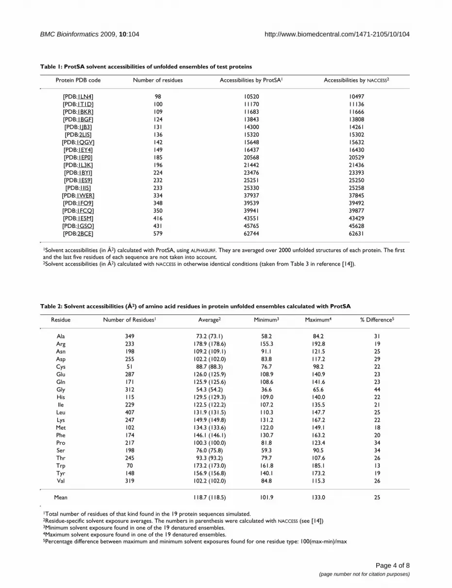
Input form of the ProtSA web applicationFigure 1Input form of the ProtSA web application. Through this simple input form the user can identify the request with a title, and submit the protein information in three different ways (as a PDB file, as a PDB id, or as a chain of residues in text form). The user sets the solvent radius and the number of unfolded conformations to generate for each chain in the protein. ProtSA sends the results of the calculations to the email address of the user.
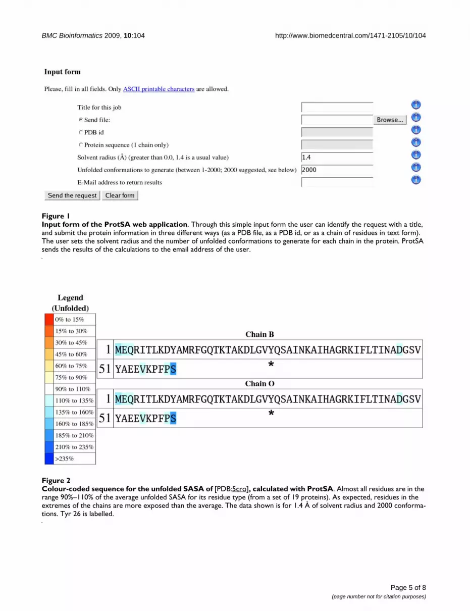
Colour-coded sequence for the unfolded SASA of [PDB:5cro], calculated with ProtSAFigure 2Colour-coded sequence for the unfolded SASA of [PDB:5cro], calculated with ProtSA. Almost all residues are in the range 90%–110% of the average unfolded SASA for its residue type (from a set of 19 proteins). As expected, residues in the extremes of the chains are more exposed than the average. The data shown is for 1.4 Å of solvent radius and 2000 conforma-tions. Tyr 26 is labelled.
BMC Bioinformatics 2009, 10:104 http://www.biomedcentral.com/1471-2105/10/104
Page 6 of 8(page number not for citation purposes)
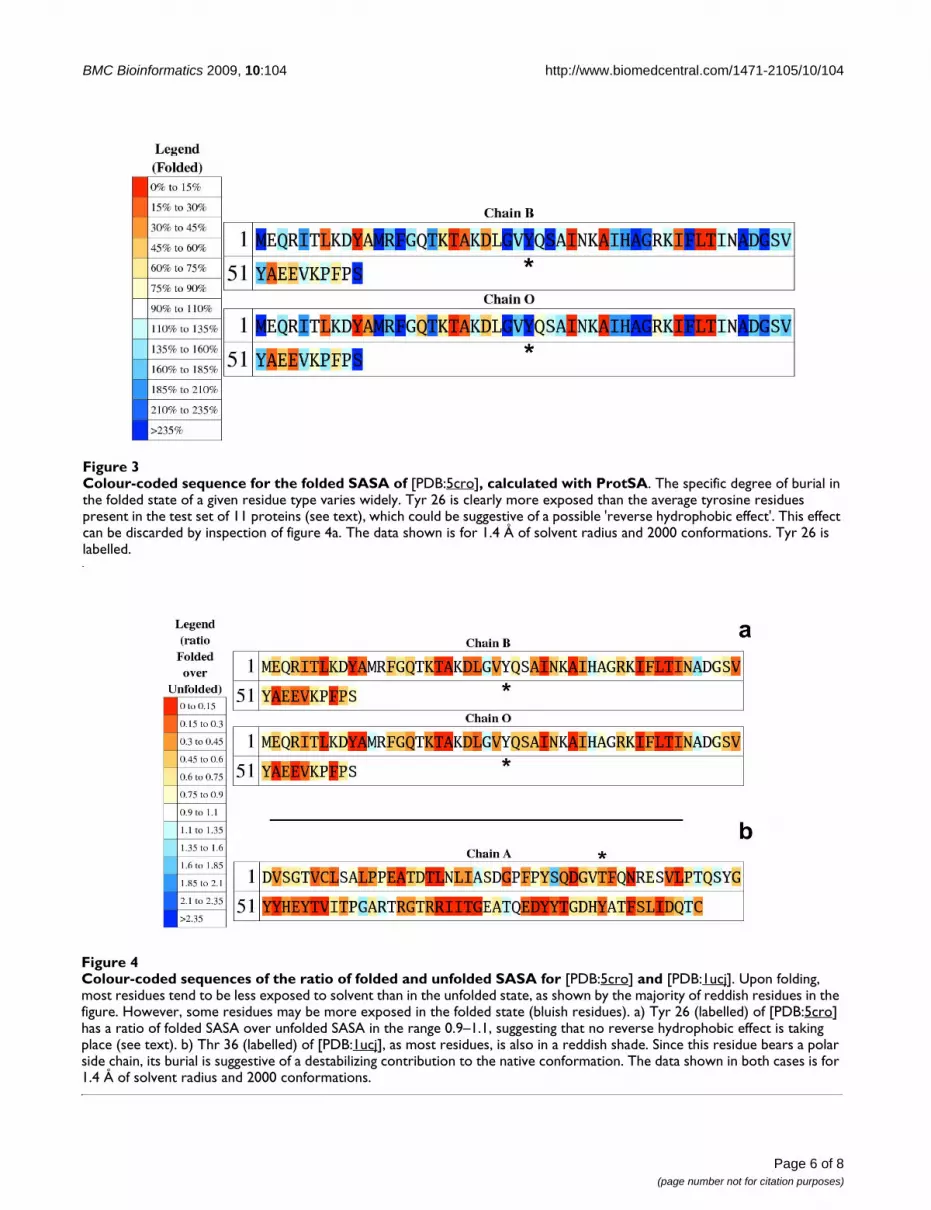
Colour-coded sequence for the folded SASA of [PDB:5cro], calculated with ProtSAFigure 3Colour-coded sequence for the folded SASA of [PDB:5cro], calculated with ProtSA. The specific degree of burial in the folded state of a given residue type varies widely. Tyr 26 is clearly more exposed than the average tyrosine residues present in the test set of 11 proteins (see text), which could be suggestive of a possible 'reverse hydrophobic effect'. This effect can be discarded by inspection of figure 4a. The data shown is for 1.4 Å of solvent radius and 2000 conformations. Tyr 26 is labelled.
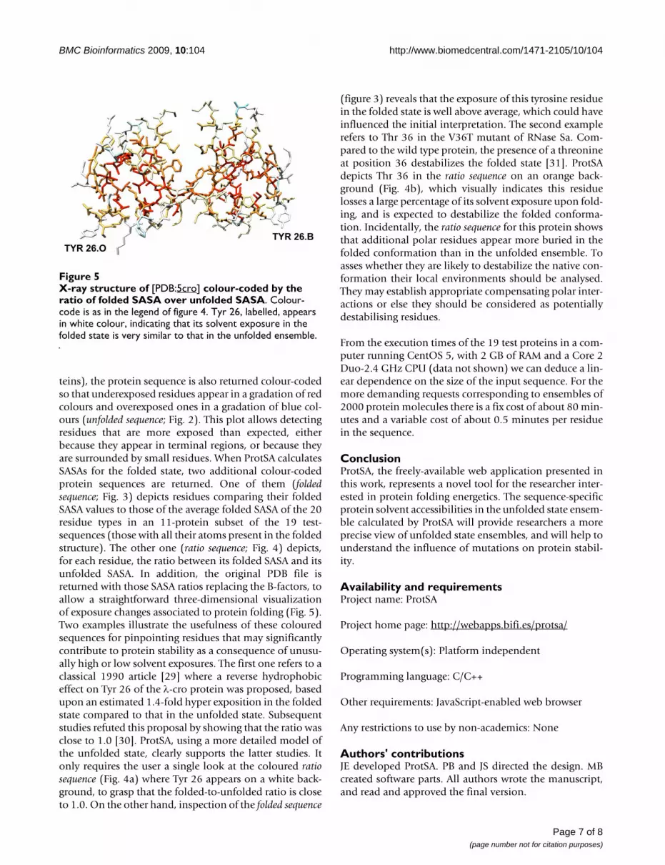
Colour-coded sequences of the ratio of folded and unfolded SASA for [PDB:5cro] and [PDB:1ucj]Figure 4Colour-coded sequences of the ratio of folded and unfolded SASA for [PDB:5cro] and [PDB:1ucj]. Upon folding, most residues tend to be less exposed to solvent than in the unfolded state, as shown by the majority of reddish residues in the figure. However, some residues may be more exposed in the folded state (bluish residues). a) Tyr 26 (labelled) of [PDB:5cro] has a ratio of folded SASA over unfolded SASA in the range 0.9–1.1, suggesting that no reverse hydrophobic effect is taking place (see text). b) Thr 36 (labelled) of [PDB:1ucj], as most residues, is also in a reddish shade. Since this residue bears a polar side chain, its burial is suggestive of a destabilizing contribution to the native conformation. The data shown in both cases is for 1.4 Å of solvent radius and 2000 conformations.
BMC Bioinformatics 2009, 10:104 http://www.biomedcentral.com/1471-2105/10/104
teins), the protein sequence is also returned colour-codedso that underexposed residues appear in a gradation of redcolours and overexposed ones in a gradation of blue col-ours (unfolded sequence; Fig. 2). This plot allows detectingresidues that are more exposed than expected, eitherbecause they appear in terminal regions, or because theyare surrounded by small residues. When ProtSA calculatesSASAs for the folded state, two additional colour-codedprotein sequences are returned. One of them (foldedsequence; Fig. 3) depicts residues comparing their foldedSASA values to those of the average folded SASA of the 20residue types in an 11-protein subset of the 19 test-sequences (those with all their atoms present in the foldedstructure). The other one (ratio sequence; Fig. 4) depicts,for each residue, the ratio between its folded SASA and itsunfolded SASA. In addition, the original PDB file isreturned with those SASA ratios replacing the B-factors, toallow a straightforward three-dimensional visualizationof exposure changes associated to protein folding (Fig. 5).Two examples illustrate the usefulness of these colouredsequences for pinpointing residues that may significantlycontribute to protein stability as a consequence of unusu-ally high or low solvent exposures. The first one refers to aclassical 1990 article [29] where a reverse hydrophobiceffect on Tyr 26 of the λ-cro protein was proposed, basedupon an estimated 1.4-fold hyper exposition in the foldedstate compared to that in the unfolded state. Subsequentstudies refuted this proposal by showing that the ratio wasclose to 1.0 [30]. ProtSA, using a more detailed model ofthe unfolded state, clearly supports the latter studies. Itonly requires the user a single look at the coloured ratiosequence (Fig. 4a) where Tyr 26 appears on a white back-ground, to grasp that the folded-to-unfolded ratio is closeto 1.0. On the other hand, inspection of the folded sequence
(figure 3) reveals that the exposure of this tyrosine residuein the folded state is well above average, which could haveinfluenced the initial interpretation. The second examplerefers to Thr 36 in the V36T mutant of RNase Sa. Com-pared to the wild type protein, the presence of a threonineat position 36 destabilizes the folded state [31]. ProtSAdepicts Thr 36 in the ratio sequence on an orange back-ground (Fig. 4b), which visually indicates this residuelosses a large percentage of its solvent exposure upon fold-ing, and is expected to destabilize the folded conforma-tion. Incidentally, the ratio sequence for this protein showsthat additional polar residues appear more buried in thefolded conformation than in the unfolded ensemble. Toasses whether they are likely to destabilize the native con-formation their local environments should be analysed.They may establish appropriate compensating polar inter-actions or else they should be considered as potentiallydestabilising residues.
From the execution times of the 19 test proteins in a com-puter running CentOS 5, with 2 GB of RAM and a Core 2Duo-2.4 GHz CPU (data not shown) we can deduce a lin-ear dependence on the size of the input sequence. For themore demanding requests corresponding to ensembles of2000 protein molecules there is a fix cost of about 80 min-utes and a variable cost of about 0.5 minutes per residuein the sequence.
ConclusionProtSA, the freely-available web application presented inthis work, represents a novel tool for the researcher inter-ested in protein folding energetics. The sequence-specificprotein solvent accessibilities in the unfolded state ensem-ble calculated by ProtSA will provide researchers a moreprecise view of unfolded state ensembles, and will help tounderstand the influence of mutations on protein stabil-ity.
Availability and requirementsProject name: ProtSA
Project home page: http://webapps.bifi.es/protsa/
Operating system(s): Platform independent
Programming language: C/C++
Other requirements: JavaScript-enabled web browser
Any restrictions to use by non-academics: None
Authors' contributionsJE developed ProtSA. PB and JS directed the design. MBcreated software parts. All authors wrote the manuscript,and read and approved the final version.
X-ray structure of [PDB:5cro] colour-coded by the ratio of folded SASA over unfolded SASAFigure 5X-ray structure of [PDB:5cro] colour-coded by the ratio of folded SASA over unfolded SASA. Colour-code is as in the legend of figure 4. Tyr 26, labelled, appears in white colour, indicating that its solvent exposure in the folded state is very similar to that in the unfolded ensemble.
Page 7 of 8(page number not for citation purposes)
BMC Bioinformatics 2009, 10:104 http://www.biomedcentral.com/1471-2105/10/104
Publish with BioMed Central and every scientist can read your work free of charge
"BioMed Central will be the most significant development for disseminating the results of biomedical research in our lifetime."
Sir Paul Nurse, Cancer Research UK
Your research papers will be:
available free of charge to the entire biomedical community
peer reviewed and published immediately upon acceptance
cited in PubMed and archived on PubMed Central
yours — you keep the copyright
Submit your manuscript here:http://www.biomedcentral.com/info/publishing_adv.asp
BioMedcentral
AcknowledgementsWe thank Sara Ayuso (Univ. Zaragoza, Spain), for help in testing; Patrice Koehl (Univ. California Davis, USA), for help with ALPHASURF; Guillermo Losilla (BIFI, Univ. Zaragoza, Spain), for technical help and José Ramón Per-egrina (Univ. Zaragoza, Spain), for comments and discussions. The molecu-lar graphics image in Fig. 5 was produced using POV-Ray [32] and the UCSF Chimera package [33] from the Resource for Biocomputing, Visualization, and Informatics at the University of California, San Francisco (supported by NIH P41 RR-01081). We acknowledge financial support from MEC (Spain): grant BFU2007-61476/BMC and BIO2007-63458, and from DGA (Spain): grant PI078/08. PB holds a Ramon y Cajal contract financed by MEC (Spain) and the Institute for Research in Biomedicine. JE was a recipient of an FPU doctoral fellowship from MEC (Spain).
References1. Baldwin RL: Energetics of protein folding. J Mol Biol 2007,
371:283-301.2. Chen Y, Ding F, Nie H, Serohijos AW, Sharma S, Wilcox KC, Yin S,
Dokholyan NV: Protein folding: then and now. Arch Biochem Bio-phys 2008, 469:4-19.
3. Wesson L, Eisenberg D: Atomic solvation parameters appliedto molecular dynamics of proteins in solution. Protein Sci 1992,1:227-235.
4. Robertson AD, Murphy KP: Protein structure and the energet-ics of protein stability. Chem Rev 1997, 97:1251-1268.
5. Myers JK, Pace CN, Scholtz JM: Denaturant m values and heatcapacity changes: relation to changes in accessible surfaceareas of protein unfolding. Protein Sci 1995, 4:2138-2148.
6. Lee B, Richards FM: The interpretation of protein structures:estimation of static accessibility. J Mol Biol 1971, 55:379-400.
7. Miller S, Janin J, Lesk AM, Chothia C: Interior and surface of mon-omeric proteins. J Mol Biol 1987, 196:641-656.
8. Shrake A, Rupley JA: Environment and exposure to solvent ofprotein atoms. Lysozyme and insulin. J Mol Biol 1973,79:351-371.
9. Rose GD, Geselowitz AR, Lesser GJ, Lee RH, Zehfus MH: Hydro-phobicity of amino acid residues in globular proteins. Science1985, 229:834-838.
10. Creamer TP, Srinivasan R, Rose GD: Modeling unfolded states ofpeptides and proteins. Biochemistry 1995, 34:16245-16250.
11. Creamer TP, Srinivasan R, Rose GD: Modeling unfolded states ofproteins and peptides. II. Backbone solvent accessibility. Bio-chemistry 1997, 36:2832-2835.
12. Gong H, Rose GD: Assessing the solvent-dependent surfacearea of unfolded proteins using an ensemble model. Proc NatlAcad Sci USA 2008, 105:3321-3326.
13. Goldenberg DP: Computational simulation of the statisticalproperties of unfolded proteins. J Mol Biol 2003, 326:1615-1633.
14. Bernadó P, Blackledge M, Sancho J: Sequence-specific solventaccessibilities of protein residues in unfolded protein ensem-bles. Biophys J 2006, 91:4536-4543.
15. Bernadó P, Blanchard L, Timmins P, Marion D, Ruigrok RW, Black-ledge M: A structural model for unfolded proteins from resid-ual dipolar couplings and small-angle x-ray scattering. ProcNatl Acad Sci USA 2005, 102:17002-17007.
16. Bernadó P, Bertoncini CW, Griesinger C, Zweckstetter M, Black-ledge M: Defining long-range order and local disorder innative α-synuclein using residual dipolar couplings. J Am ChemSoc 2005, 127:17968-17969.
17. Mukrasch MD, Markwick P, Biernat J, Bergen M, Bernadó P, Griesin-ger C, Mandelkow E, Zweckstetter M, Blackledge M: Highly popu-lated turn conformations in natively unfolded tau proteinidentified from residual dipolar couplings and molecular sim-ulation. J Am Chem Soc 2007, 129:5235-5243.
18. Wells M, Tidow H, Rutherford TJ, Markwick P, Jensen MR, MylonasE, Svergun DI, Blackledge M, Fersht AR: Structure of tumor sup-pressor p53 and its intrinsically disordered N-terminal trans-activation domain. Proc Natl Acad Sci USA 2008, 105:5762-5767.
19. Jensen MR, Houben K, Lescop E, Blanchard L, Ruigrok RW, Black-ledge M: Quantitative conformational analysis of partiallyfolded proteins from residual dipolar couplings: applicationto the molecular recognition element of Sendai virus nucle-oprotein. J Am Chem Soc 2008, 130:8055-8061.
20. Geierhaas CD, Nickson AA, Lindorff-Larsen K, Clarke J, VendruscoloM: BPPred: a Web-based computational tool for predictingbiophysical parameters of proteins. Protein Sci 2007,16:125-134.
21. Eyal E, Najmanovich R, McConkey BJ, Edelman M, Sobolev V: Impor-tance of solvent accessibility and contact surfaces in mode-ling side-chain conformations in proteins. J Comput Chem 2004,25:712-724.
22. Edelsbrunner H, Koehl P: The weighted-volume derivative of aspace-filling diagram. Proc Natl Acad Sci USA 2003, 100:2203-2208.
23. Hubbard SJ, Thornton JM: NACCESS Computer Program Department ofBiochemistry and Molecular Biology, University College London, Lon-don, UK; 1993.
24. Lovell SC, Davis IW, Arendall WB 3rd, de Bakker PI, Word JM,Prisant MG, Richardson JS, Richardson DC: Structure validationby Cα geometry: Φ,Ψ and Cβ deviation. Proteins 2003,50(3):437-450.
25. Levitt M: A simplified representation of protein conforma-tions for rapid simulation of protein folding. J Mol Biol 1976,104:59-107.
26. Kabsch W, Sander C: Dictionary of protein secondary struc-ture: pattern recognition of hydrogen-bonded and geometri-cal features. Biopolymers 1983, 22:2577-2637.
27. ProtSA: A web application for calculating sequence specificprotein solvent accessibilities in the unfolded ensemble[http://webapps.bifi.es/protsa/]
28. Berman HM, Westbrook J, Feng Z, Gilliland G, Bhat TN, Weissig H,Shindyalov IN, Bourne PE: The Protein Data Bank. Nucleic AcidsRes 2000, 28:235-242.
29. Pakula AA, Sauer RT: Reverse hydrophobic effects relieved byamino-acid substitutions at a protein surface. Nature 1990,344:363-364.
30. Ohlendorf DH, Tronrud DE, Matthews BW: Refined structure ofCro repressor protein from bacteriophage λ suggests bothflexibility and plasticity. J Mol Biol 1998, 280:129-136.
31. Takano K, Scholtz JM, Sacchettini JC, Pace CN: The contribution ofpolar group burial to protein stability is strongly context-dependent. J Biol Chem 2003, 278:31790-31795.
32. Persistence of Vision Raytracer [http://www.povray.org/]33. Pettersen EF, Goddard TD, Huang CC, Couch GS, Greenblatt DM,
Meng EC, Ferrin TE: UCSF Chimera – A visualization systemfor exploratory research and analysis. J Comput Chem 2004,25:1605-1612.
Page 8 of 8(page number not for citation purposes)