Embed Size (px)
Citation preview

GUIDED THERAPY SYSTEMS, CONFIDENTIAL
Page 1 of 35 Protocol V11 May 4, 2015
PROTOCOL
TITLE: STUDY TO EVALUATE THE SAFETY, EFFICACY AND TOLERANCE OF INTENSE THERAPUETIC ULTRASOUND (ITU) FOR THE TREATMENT OF LATERAL EPICONDYLOSIS
Guided Therapy Systems 33 Sycamore St. Mesa, AZ 85202
Primary Contact;
For The MORE Foundation Name: John Kearney, MD Telephone: 623-537-5600 (24 hours)Email: [email protected]
For Guided Therapy Systems Name: Michael H. Slayton Ph.D
Telephone: 480-649-4399
Email: [email protected]
CONFIDENTIAL Confidential information in the following document is provided to you as an investigator, potential investigator or consultant for review by you and your staff. By accepting this document, you agree that information contained herein will not be disclosed to others without written authorisation from Guided Therapy System except to the extent necessary to obtain informed consent from subjects considering participation in the study. This document may also be disclosed to an Independent Ethics Committee or Institutional Review Board or authorised representatives of national regulatory authorities under the condition that they respect its confidential nature.

GUIDED THERAPY SYSTEMS, CONFIDENTIAL
Page 2 of 35 Protocol V11 May 1, 2015
Approval of the Protocol I have thoroughly read and reviewed the study protocol. This study will be conducted in compliance with the protocol, the Declaration of Helsinki, principles of Good Clinical Practice and any applicable regulatory requirement(s).
PRINCIPAL INVESTIGATOR
Signature: .................................................... Date: .................................................... Name (printed): John A. Kearney, Jr., MDTitle (printed):
SPONSOR - GUIDED THERAPY SYSTEMS
Signature: .................................................... Date: .................................................... Name (printed): Michael H. Slayton, Ph.D. Title (printed): President and CEO

GUIDED THERAPY SYSTEMS, CONFIDENTIAL
Page 3 of 35 Protocol V11 May 1, 2015
CONTACTS
Sponsor Guided Therapy Systems. 33 South Sycamore, Mesa, AZ 85202. Michael Slayton, Ph.D.
Guided Therapy Systems. 33 South Sycamore St., Mesa, AZ 85202.
MONITORS Name: Elizabeth Gleason, M.A., CCRC Title: Manager, Clinical Research Name of Study Site: The MORE Foundation® Address of Study Site: 18444 N. 25th Ave. Suite 110, Phoenix, AZ 85023 Telephone number(s): 623.537.5695 Email: Michael Slayton, Ph.D. Guided Therapy System. 33 South Sycamore Mesa, AZ 85202 USA (phone) 480.649.4399
PRINCIPAL INVESTIGATOR
Name: John Kearney, MD Title: Principal Investigator Name of Study Site: The MORE Foundation® Address of Study Site: 18444 N. 25th Ave. Suite 110, Phoenix, AZ 85023 Telephone number(s): 623-537-5695 Email: [email protected]
REPORTING OF ADVERSE EVENTS
Name: John Kearney, MD Title: Principal Investigator Company name: The MORE Foundation® Company address: 18444 N. 25th Ave. Suite 110, Phoenix, AZ 85023 Telephone number(s): 623-537-5695 Email: [email protected]

GUIDED THERAPY SYSTEMS, CONFIDENTIAL
Page 4 of 35 Protocol V11 May 1, 2015
INTRODUCTION Acute and Chronic pain of the Common Extensor Tendon (CET) region, or lateral epicondylitis/lateral epicondylalgia, or tennis elbow is a common pathology of both athletes and non-athletes affecting 1 to 3 % of the population at large.1,2 The prevalence of chronic problems caused by overuse in tennis players can be as high as 40%.15 Elbow tendinopathy represents an important set of pathologies that account for lost recreation time, decreased quality of life, and work-related disability claims. As an example, medial and lateral epicondylitis was responsible for 11.7% of work-related injury claims in Washington State from 1987 to 1995, resulting in an average direct workers’ compensation cost of $6,593 per case.16 Elbow tendinopathy has widespread social, financial, and clinical implications. Walker-Bone et al2assessed the effect of medial and lateral epicondylitis on workplace absences in a cross-sectional sample of 9,696 working-aged adults. Five percent of patients reported taking a sick day due to their elbow pain within the preceding year.
This condition is most often associated with overuse or a repetitive stress, as opposed to an acute inflammatory reaction. The lack of pathological evidence of inflammation in these types of injuries has lead most authors to now refer to this condition as an Epicondylosis, abandoning the mislabelled “itis”.3–5
Diagnostic Ultrasound has been used to evaluate musculoskeletal anatomy and pathology since the early 1990’s. Ultrasound imaging can be used to evaluate the structure of the Common Extensor tendons, evidence of microtears, partial and full ruptures, and hypoechoic lesions in and about the tendon and peritendinous lesions9.Common Extensor Tendon thickness can change with age10, and should be a useful tool in tracking tendon improvement during a long treatment period.
Conservative treatment of the Epicondylitis or osis is recommended as the initial strategy by most authors. This strategy includes identification and correction of possible etiological factors, and a symptom related approach. Generally, the initial treatment consists of a multifactorial approach that may include a combination of rest (complete or modified activity), medication (NSAIDs for Epicondylitis), stretching and strength training9.
Intense Therapeutic Ultrasound (ITU) is a novel potential treatment for CET Tendinosis. ITU is a highly focused ultrasound wave that delivers enough energy intensity to disrupt soft tissues within a controlled up to 1 mm3 volume, while sparing surrounding tissue structures. Much like a laser is formed by focusing light to concentrate energy over a small area, ultrasound can emit sound waves that can propagate through tissues to deliver selective thermal coagulative changes while leaving the surrounding tissue unaffected.11,12,13
ITU has been used clinically for treating the facial soft tissue for the past decade and it has received FDA approval for non-surgical brow and submental lifting. Over 300,000 treatments have been performed with this technology. Clinical studies in 2005/2006 (IRB#’s 05-06-032, 1253-014) using intense therapeutic ultrasound (ITU) on facial soft tissue whereby the energy is focused underneath the skin tissue to achieve controlled coagulative necrosis (~1 mm3) were performed. Seventy subjects were involved in the studies and 84% of subjects showed an improvement in facial lifting with no significant pain, erythema,

GUIDED THERAPY SYSTEMS, CONFIDENTIAL
Page 5 of 35 Protocol V11 May 1, 2015
inflammation, or scarring11,12. Slight edema and erythema were noted on some subjects, all resolving within 30 minutes after treatment. Histologically, it has been proven that ITU induces greater dermal collagen with thickening of the dermis and straightening of elastic fibers in the reticular dermis11-
15. Due to the proven clinical performance, there is current ongoing research that is specifically designed to determine whether ITU can improve healing of damaged Achilles tendon in a rabbit model15. The main goal of the project was to determine if ITU accelerates the healing process, and/or promotes a response that leads to superior structural and mechanical properties of tendon. Preliminary results from the rabbit research at day 4 post treatment showed an increase in Vascular Endothelial Cell Growth Factor A (VEGFa), Tumor Necrosis Factor Alpha (TNFα), Interleukin 1-Beta (IL-1β), and Transforming Growth Factor Beta 1 (TGFβ1) and a decrease in Collagen Type 1 Alpha 1(COL1α1), and Collagen Type 1 Alpha 2(COL1α2) in the damaged and ITU treated legs. At 14 days post treatment PCR showed an increase in Collagen Type 1 Alpha 1 (COL1a1) and Collagen Type 1 Alpha 2 (COL 1a2), as well as an increase in Vascular Endothelial Cell Growth Factor A (VEGFa), Tumor Necrosis Factor Alpha (TNFα), Interleukin 1-Beta (IL-1β), and Transforming Growth Factor Beta 1 (TGFβ1), in the injured rabbit tendon treated with ITU compared to injured, untreated rabbit tendon15. These results led us to explore the possibility of performing a clinical trial assessing the effectiveness of ITU in actual patients with CET Tendinosis. The purpose of this trial is evaluate the tolerability and efficacy of an ITU device to treat CET tendinosis as measured by Subject Self-assessment survey of pain17, function, and activity (Patient Rated Tennis Elbow Evaluation (PRTEE18), Diagnostic Ultrasound Imaging, to evaluate tendon thickness, homogeneity, and gross anatomy/pathology, and physical examination.
ULTRASOUND THERAPY BACKGROUND
i. Historical Background
Ultrasound is an energy modality that can propagate to deeper layers of tissue while sparing the skin surface. The energy created from intense focused ultrasound (ITU) can be focused into deep tissue layers to achieve controlled coagulative necrosis (≤ 1 mm3).
ii. Previous Ultrasound Research
Ulthera® Studies Clinical studies in 2005/2006 (IRB#’s 05-06-032, 1253-014) using intense therapeutic ultrasound (ITU) on facial soft tissue whereby the energy is focused underneath the skin tissue to achieve controlled coagulative necrosis (~1 mm3) were performed. Seventy subjects were involved in the studies and 84% of subjects showed an improvement in facial lifting with no significant pain, erythema, inflammation, or scarring10,11. In addition, more than 300,000 patients worldwide have been treated with the Ulthera device with no reportable adverse events.

GUIDED THERAPY SYSTEMS, CONFIDENTIAL
Page 6 of 35 Protocol V11 May 1, 2015
For these clinical studies, highly focused sources were used at 4.4 and 7.5 MHz, at source power levels of 20 – 60 W. This sharply focused intense field was capable of achieving selective coagulative necrosis in sub-epidermal skin tissue and resulted in substantial improvement of the skin with no side effects. The Ulthera device has received FDA clearance for non-invasive lifting of the eyebrow, neck and submental regions (K121700). Pre-clinical Therapeutic application of ultrasound energy in skin tissue, has been extensively investigated by our group10-13. The selective thermal coagulation of skin tissue with directive ultrasound energy was demonstrated in vitro as well as in vivo. In order to comprehensively understand the ultrasound energy-skin tissue interaction, a wide range of ultrasound source geometries and source conditions were investigated during the pre-clinical research. The source frequencies investigated ranged from 4 – 10 MHz, whereby the source acoustic power ranged from 5 – 80 W. A summary and scope of the various pre-clinical studies is provided in the Table below.
Table: Summary results of multiple exposures and effect on skin tissue using UltraSite STTM System
Institute Tests Number of Exposures Goals Accomplished MEEI, Harvard Med. School, M White, MD, R. Gliklich, MD
6 Human cadavers 7.5 and 4.4 MHz probes
>1000 exposures placed in facial skin tissue At least 200 sample shots assessed (NBTC + H&E staining)
-- Define clinical procedure steps (skin marking, lesion identification, etc.) -- Verify treatment control, maximum depth, and dose response
Wellman Labs, MGH, H.J. Laubach, MD
Ex vivo human torso/back skin 7.5 and 10 MHz probes
>150 exposures All shots assessed (NBTC and Eosin counter-stain)
-- Understand basic skin tissue effect (depth, dimension, control, etc.) with ultrasound -- Understand biophysical processes – selective thermal ablation and its predictability
UC San Diego, R. Fitzpatrick, MD
6 Human cadavers 7.5, 4.4 and 10 MHz probes
>1000 exposures placed in facial skin tissue At least 300 samples assessed (H&E and Trichrome-Masson staining)
-- Validate clinical treatment logistics (skin marking, lesion identification, etc.) -- Validate visual (clinical) effect of tissue shrinkage -- Verify treatment depth, control, and dose response -- Identify Trichrome-Masson stain as a potential predictor of thermal damage with UltraSite STTM System

GUIDED THERAPY SYSTEMS, CONFIDENTIAL
Page 7 of 35 Protocol V11 May 1, 2015
Ulthera, Inc., I.R.S. Makin, MD. PhD, PG. Barthe, PhD, MH. Slayton, PhD
Ex vivo porcine skin 8 in vivo porcine abdominal skin experiments 7.5, 4.4 and 10 MHz probes
>1000 exposures placed in porcine skin (in vivo and ex vivo) At least 70% of samples assessed histological (NBTC and H&E)
-- Device and application development -- Correlate experiment with numerical simulations -- Understand skin-ultrasound biophysics -- Confirm dose response
In summary, safety and efficacy for the described above studies performed with intense therapeutic ultrasound (ITU) was demonstrated.

GUIDED THERAPY SYSTEMS, CONFIDENTIAL
Page 8 of 35 Protocol V11 May 1, 2015
STUDY OBJECTIVES
To evaluate the tolerance, efficacy and safety of an ultrasound therapy device to treat CET tendinosis as measured by:
Subject survey of pain, function, and activity: Patient Rated Tennis Elbow Evaluation (PRTEE).
Diagnostic Ultrasound Imaging, to evaluate tendon thickness, homogeneity, and gross anatomy/pathology.
Subject self-assessment of improvement, satisfaction and treatment tolerability. Physician physical examination.
STUDY DESIGN
This will be a single center, controlled study involving a total of twenty-five (25) subjects with CET tendinosis (figure 2). Subjects will receive two treatments. Each treatment will be up to ten minutes (10 minutes) in duration.
Figure 1. Study Design.

GUIDED THERAPY SYSTEMS, CONFIDENTIAL
Page 9 of 35 Protocol V11 May 1, 2015
STUDY OUTLINE
This study will be approximately 2 months in duration plus 2 telephone surveys at 12 and 24 weeks. Subjects who meet the criteria and provide written consent to be in the study will be included in the study. Subjects will receive conservative standard of care treatment plus two ITU treatments (week 0 and week 4) on the affected CET (figure 2) using the ultrasound therapy device. Treatments will be up to 10 minutes in duration. A 2, 4, and 8 week follow up period will be included to monitor the effects of the therapy post treatment. A 12 and 24-26 week telephone assessments for the primary objectives will also be included. Telephone assessments will consist of the subjects answering the survey questions over the phone. Subject self-assessments will be conducted before the treatment phase and during the follow-up period. Physical exams will be completed by a physician. Physical therapy will completed by a therapist. ITU treatment and diagnostic ultrasound will be performed by Guided Therapy personnel as shown in the full schedule of events (Appendix 1).
Figure 2: Treatment Area
SELECTION OF SUBJECTS
Planned number of subjects The study population will consist of up to 25 subjects with unilateral CET tendinosis defined as pain lasting more than 3 months with little or no improvement following conservative standard of care alone. The selection of suitable subjects will be made according to the inclusion and exclusion criteria described in the following sections. Inclusion Criteria
1. Subjects who are in general good health and are willing to cooperate and participate by following study requirements for the duration of the study, and to report any adverse symptoms immediately; 2. Subjects who have provided written and verbal informed consent; 3. At time of enrollment subjects are between the ages of 21 and 65 years 4. Subjects literate in English and able to complete all patient self-assessment questionnaires;

GUIDED THERAPY SYSTEMS, CONFIDENTIAL
Page 10 of 35 Protocol V11 May 1, 2015
5. Subjects is willing to refrain from beginning any additional treatment during the duration of the study; 6. Subjects is willing to refrain from changing physical activity during the duration of the study; 7. Subjects diagnosed with unilateral CET tendinosis defined as pain lasting more than 3 months with little or no improvement following conservative standard of care alone. 8. Subjects with BMI ≤ 35 or at the discretion of the PI. 9. Subjects must be able to sit in a chair with their arm on an appropriate table for the treatment and diagnostic ultrasound imaging. 10. Subject must be able and willing to complete the at home therapy regimen. Exclusion Criteria Subjects with any of the following conditions are not eligible for participation:
1. Subjects currently enrolled in any other device, or IND (Investigational New Drug)
clinical trial, or who have participated in a clinical study involving the CET, thirty days prior to study initiation;
2. Subjects who have participated in any other clinical study involving an investigational product 30 days prior to enrollment that, in the opinion of the Principal Investigator, could affect the outcome of this study;
3. Subjects who have received any previous treatment in the symptomatic limb in the past 30 days timeframe (not including standard of care treatment);
4. Subjects diagnosed with bilateral CET tendinopathy.
5. Subjects who have had any previous difficulties or problems with wound healing, bruising, scarring or procedures involving medical devices;
6. Subjects with diagnosed uncontrolled metabolic diseases, such as diabetes,
hypertension, hyperthyroidism, and hypothyroidism as determined by the initial paperwork;
7. Subjects known to be pregnant or nursing as determined by the initial paperwork; 8. Subjects currently taking medications which, in the opinion of the Investigator,
may interfere with the study (e.g., statins, prescription steroids, prescription anti-inflammatory drugs, etc.). Subjects who have been on a stable dose of medication for a minimum of 3 months prior to the study start, and who agree to continue that dose of medication for the duration of the study may be allowed to participate at the Investigator’s discretion
9. Subjects who have taken quinolone antibiotic in the past 3 months; 10. Subjects with a diagnosed auto-immune disorder; 11. Subjects with diagnosed fibromyalgia; 12. Subjects with diagnosed peripheral neuropathy; 13. Subjects that will not be available for on-site follow-ups as noted in the schedule. 14. Subjects with previous CET tendon rupture in the symptomatic limb. 15. Subjects with BMI > 35 or at the discretion of the PI

GUIDED THERAPY SYSTEMS, CONFIDENTIAL
Page 11 of 35 Protocol V11 May 1, 2015
Individuals will be admitted to study at the discretion of the investigator or designate based on medical history (eligibility paperwork) and findings of the pre-study interview and examination. Subject Restrictions Subjects must not change their physical activity during the course of the study. Subjects’ BMI must not increase to > 35 or at the discretion of the PI
WITHDRAWAL OF SUBJECTS.
Subjects may discontinue from the clinical study at any time. In addition, the Principal Investigator (PI) or designee has the right to withdraw a subject for any reason that is in the best interests of the subject. The subject must inform the Investigator immediately if they intend to withdraw. Subjects may be asked to come to the study facility to complete some end of study procedures. The Investigator can remove the subject from this study without their consent for any reason, including, but not limited to:
o His/her judgment that any condition or circumstance may jeopardize their welfare or the integrity of the study.
o Their failure to follow the instructions of the investigator(s) (protocol non-compliance) o Subject BMI exceeds ≥35 during the courses of the study. o If the study is stopped by GTS and/or Principal Investigator of the study prior to
completion.
CLINICAL ASSESSMENTS
Assessment of Efficacy Primary: The primary endpoint(s) for efficacy and tolerability will be the total score of the PRTEE questionnaire (Appendix 2), subject self-assessment of improvement and satisfaction (SROM-Appendix 3-B), and Visual Analog Scale for Pain (VAS) or Universal Pain Assessment Tool (Appendix 3-C). Secondary: The secondary endpoint for efficacy will be a measured reduction in tendon thickness by diagnostic ultrasound, and a physical examination. PRTEE Questionnaire
The PRTEE questionnaire (Appendix 2) evaluates three domains clinically relevant to patients (pain, function, and activity) and was specifically developed to assess the clinical severity of CET tendinopathy. It is composed of 15 questions that cover 3 domains of pain (questions 1 through 5), function for specific activities (questions 6 through 11), and general activities (questions 12 through 15). All questions are scored 0 – 10. Higher scores indicate more severe CET tendinopathy. The PRTEE questionnaire has been validated and shows good test-retest reliability18
Physical Examination
Medical History: Review patient’s medical history questionnaire. Review any diagnostic imaging, tests, or work up listed under longitudinal medical record and centricity. Ask about possible trauma or history of injuries to the symptomatic arm. Review athletic history (tennis, golf, etc) and subject’s pre-study exercise regimen (if any). Height, Weight (BMI) will also be documented.

GUIDED THERAPY SYSTEMS, CONFIDENTIAL
Page 12 of 35 Protocol V11 May 1, 2015
History of Present Illness: Most common complaint is of elbow pain. Social Hx: Frequently found those involved in racquet sports. Medications: non-steroidal anti-inflammatory medications and corticosteroids.
Pain: measured on the VAS scale; note activities that increase symptoms, decrease
symptoms, and the location of symptoms. Examination protocol is simple and will consist of the following:
a. Skin examination (note any rashes, lesions, areas of skin irritation over the lateral elbow region)
b. Palpation over the lateral epicondyle of affected side, note presence of tenderness to palpation vs. no tenderness to palpation
c. Check range of motion of elbow in flexion, extension, pronation, and supination (may use normative template data that is in EMR under elbow exam)
d. Check provocative tests for lateral epicondylitis and note whether or not these elicit pain that refers into the common extensor tendon. The 2 provocative tests will be: 1. Resistance to active wrist extension. 2. Resistance to active middle finger extension
Ultrasound Imaging Ultrasound images will be collected from test sites on both the affected and unaffected CET using the Spark® High Frequency Ultrasound System. CET tendon thickness, homogeneity and gross assessment of the CET tendon and surrounding tissue will be conducted.
Assessment of Safety
Recording of Adverse Events or any other significant event will take place at all visits. Adverse events will be monitored throughout the study. (Appendix 5)
ASSESSMENTS AT EACH VISIT
An overview of the assessments to be conducted at each study visit are presented in a Table in Appendix 1. Subjects will be treated at Visit 1 and Visit 3. Imaging will occur, before the treatments, and at visits 2, and 4. The PRTEE questionnaire will be administered, prior to the first treatment at visits 1 and 3, and at visits 2, and 4. An additional telephone follow-up, where answers to the questionnaires will be recorded, will occur at 12 and 24 weeks post treatment one (1) per the schedule in Appendix 1. The study will last approximately two (2) months with a follow-up phone call at three (3) and six (6) months. Visit 0 (Recruitment): Screening and Enrollment
1. Candidate subjects will read an informed consent (which includes a confidentiality agreement) and will sign (along with a witness) after all questions about the study have been answered. Individuals will be issued a temporary screening number.
2. Eligibility paperwork will include documentation of pregnancy status and pertinent medical history.
If a subject meets all the inclusion/ exclusion criteria, they will be enrolled onto the study, issued a subject number and an X-ray will be scheduled.

GUIDED THERAPY SYSTEMS, CONFIDENTIAL
Page 13 of 35 Protocol V11 May 1, 2015
Visit 1 and 3: (Weeks 0 and 4) Study treatments 1. Record AEs, con meds and other events 2. Administer PRTEE-A questionnaire (Appendix 2) 3. Subject self-assessment questionnaire (Appendix 3) 4. Focused Physical Evaluation 5. Diagnostic Ultrasound images taken 6. ITU treatment, and VAS assessment of treatment tolerability. 7. Physical Therapy will be performed as described (Appendix 4 – Visit 1 only) 8. Home Cold Pack & Stretching Program review and Log (Appendix 6-A and 6-B)
Visit 2, and 4: (Weeks 2 and 8) Diagnostic Ultrasound, Self-Assessments, Physical Examination
1. Record AEs, con meds and other events; 2. Administer PRTEE-A questionnaire (Appendix 2) 3. Subject self-assessment questionnaire (Appendix 3) 4. Diagnostic Ultrasound image taken; 5. Home Cold Pack & Stretching Program review and Log (Appendix 6-A and 6-B)
Phone/Online Interviews at 3 and 6 months post treatment
1. Record AEs, con meds and other events; 2. Follow-up phone interview PRTEE-A verbally administered (Appendix 2). 3. Subject self-assessment questionnaire (Appendix 3)
STUDY TREATMENT
ITU will be performed by a trained technologist, using a clinical prototype (Ulthera FDA cleared in Sept. 2009) GEN II or GEN III designed Guided Therapy Systems, LLC. Mesa, AZ USA (Appendix 7-A). Subjects will be treated seated with their arm resting on a structure that is comfortable for the subject and technologist. Transdermal application of ITU therapy: An average energy up to 3 joules (0.5 to 3 J) will be administered to the affected CET tendon longitudinally until the maximum number of lines (12) are reached. Each line will include individual thermal zones, less than 1 mm3 in volume, centered at a depth between 2 – 6 mm, up to 2 mm apart (figure 3).
Treatment pattern:
1-6 mm
1-2 mm
Ultrasound Probe
TendoTeFigure 3. Zone pitch, and depth.

GUIDED THERAPY SYSTEMS, CONFIDENTIAL
Page 14 of 35 Protocol V11 May 1, 2015
Acoustic Coupling Gel will be applied every 3-5 lines. The study treatment will be a 10 minute session using the ultrasound therapy device with coupling gel. Subjects will be treated seated with their arm resting on a structure that is comfortable for the subject and technologist.
Tendon
Treatment lines
The study treatment will be a
Figure 3-1. Zone pitch, and depth.

GUIDED THERAPY SYSTEMS, CONFIDENTIAL
Page 15 of 35 Protocol V11 May 1, 2015
DIAGNOSTIC ULTRASOUND IMAGING Diagnostic Ultrasound Imaging will be performed by a trained Sonographer using the Spark® Imaging System, manufactured by Ardent Sound, Inc., Mesa, AZ USA (Appendix 7-B) Subjects will be imaged seated with their arm resting on a structure that is comfortable for the subject and sonographer. PolySonic® or AquaSonic® Gel will be applied by the sonographer as required to maintain adequate acoustic coupling. (Appendix 7-C)
I Images will be preserved with the following notations:
Protocol ID Right vs. Left CET Tendon Treated vs. Untreated
Long Axis and Short Axis (if necessary) images will be stored at the Sonographer’s discretion, to demonstrate and track the applicable anatomy/pathology in the peri-CET area. Sonographer will also measure the thickness of the CET tendon in each applicable image.
Sonographer will scan the contralateral CET tendon in the same manner noting the change in the tendon (Right v. Left) and (Treated v. Untreated).
Common Extensor Tendon
Figure 4: representative long axis ultrasound images
Outcome measures Subjects will complete subject reported outcome measures at 0, 2, 4, 8, 12, and 24 weeks and will undergo a focused physical examination and diagnostic ultrasound at 0, 2, 4, and 8 weeks. Adverse events will be recorded at each visit. Treatment tolerability will be assessed immediately after each treatment using the Visual Analog Scale for Pain (VAS) or Universal Pain Assessment Tool (Appendix 3-B). The VAS will be used for 2 different assessments: 1. Pain related to physical activity of the symptomatic arm before treatments and at follow-up visits and phone surveys. 2. Pain tolerance of the therapy treatment. Each of these assessments will be documented and evaluated separately. Aim 1: reported outcomes The principal aim of this study is to assess the efficacy of treatment and time course of response to treatment using validated subject reported outcome measures assessing pain, function and level of activity. Subjects will complete validated subject reported outcome questionnaires assessing pain, function and level of activity prior to initiating treatment and at

GUIDED THERAPY SYSTEMS, CONFIDENTIAL
Page 16 of 35 Protocol V11 May 1, 2015
0, 2, 4, 8, 12, and 24 weeks after starting treatment (Appendix 2 and 3A). Clinical Pain and Treatment Tolerance will be assessed using the Visual Analog Scale for Pain (VAS) or Universal Pain Assessment Tool (Appendix 3-B). Function will be assessed using three validated response forms: PRTEE - An index of the severity of CET Tendinopathy18 (Appendix 2), Self-Reported Outcome Measures (SROM)16. Level of Activity will be assessed using the activity subscale of PRTEE18 (Appendix 2). This subscale scores quantitatively the level of difficulty subjects face when they perform basic activities of daily living. It has shown to be an accurate and reliable predictor of the activity level while performing day-to-day activities. Statistical analysis Data will be assessed for variance homogeneity and normality. PRTEE18, and Visual Analog Scale for Pain (VAS) or Universal Pain Assessment Tool (Appendix 3-B) scores will be assessed before and after intervention. The VAS will be used for 2 different assessments: 1. Pain related to physical activity of the symptomatic arm before and after treatment. 2. Pain tolerance of the therapy treatment. Each of these assessments will be documented and evaluated separately. Aim 2: CET Tendon thickness Diagnostic ultrasound imaging will be performed with an FDA approved ultrasound scanner (Spark® System, Ardent Sound, Inc. – Mesa, AZ, see Appendix 7-B using a coupling gel (Polysonic®, or AquaSonic®, Parker Laboratories, Inc., Fairfield, NJ, see Appendix 7-C) by a trained sonographer or physician. Subjects will be treated seated with their arm resting on a structure that is comfortable for the subject and technologist.
CET Tendon thickness is measured before treatment(s) and at each documented assessment (Appendix 1). Paired t tests are used to compare the CET Tendon thickness means between pre- and post-treatment between the symptomatic and treated arm and the asymptomatic and untreated arm. P<0.05 is considered to be statistically significant. Any peri-CET pathology will also be documented.
Aim 3: Assessment of safety and tolerability Subject self-assessments of treatment tolerability will be conducted immediately after treatments, and at each follow-up visit and phone survey using a 10-point visual analog scale VAS, (Appendix 3-B). The subject will be asked to report their current level of pain and the maximum level of pain experienced since the last treatment. The surveyor will remind the subject of the VAS criteria and record their reply. The VAS is the standard for assessing pain for both clinical and research purposes22. Recording of adverse events will take place at all visits (see adverse event reporting form Appendix 5). Adverse events will be monitored throughout the study. A treatment will not be administered if there is residual pain/discomfort experienced by the subject from the previous treatment.
ACCOUNTABILITY
Treatment administered to the subjects will be documented by the operator and tracked on the Case Report Form (CRF).

GUIDED THERAPY SYSTEMS, CONFIDENTIAL
Page 17 of 35 Protocol V11 May 1, 2015
BREAKING THE CODE IN AN EMERGENCY
Treatment codes and allocation of treatment per subject will be accessible by the Principal Investigator of the study.
SAFETY MONITORING
ADVERSE EVENT
An adverse event (AE) is any untoward medical occurrence in a subject, whether or not related to study product or study procedures. Adverse events include any occurrence that is new in onset, an exacerbation of a pre-existing condition and clinically significant laboratory values. Exceptions The following medical occurrences will not be reported as AEs;
• Pre-treatment Adverse Events; Any medical occurrence that occurs after informed consent, but before first administration of ITU treatment or first study procedure is considered as medical history and only recorded as an AE if it worsens during the study. • Pre-existing medical condition; Events that occur with comparable frequency and severity to the subject’s baseline condition are reported as medical history, not AEs unless they become exacerbated during the study. • Pregnancy; This is not an AE however the PI must report any pregnancies to The Guided Therapy and the Institutional Review Board (IRB) for advice regarding the appropriate course of action.
Study Specific Expected Adverse Event The following AEs are expected due to the use of these study products and/or procedures and for this specific study, they will not be regarded as AEs;
Transient erythema may be expected as a result of the study treatment which is expected to subside after a few hours. This will not be recorded as an AE unless persistent after 24 hours.
Subjects may experience mild-moderate tingling, warming, pain or discomfort during the study treatments. These will not be recorded as AEs unless persistent after 24 hours or if they increase to severe/unbearable sensations.
The following AEs may be expected due to the use of these study products and/or procedures and for this specific study, they will be regarded and recorded as AEs;
While trained personnel will administer the study treatments, there is a possibility for operator error that may result in a burn or high skin irritation response. These will be recorded as AEs.
GUIDED THERAPY SYSTEMS, CONFIDENTIAL
Page 18 of 35 Protocol V11 May 1, 2015
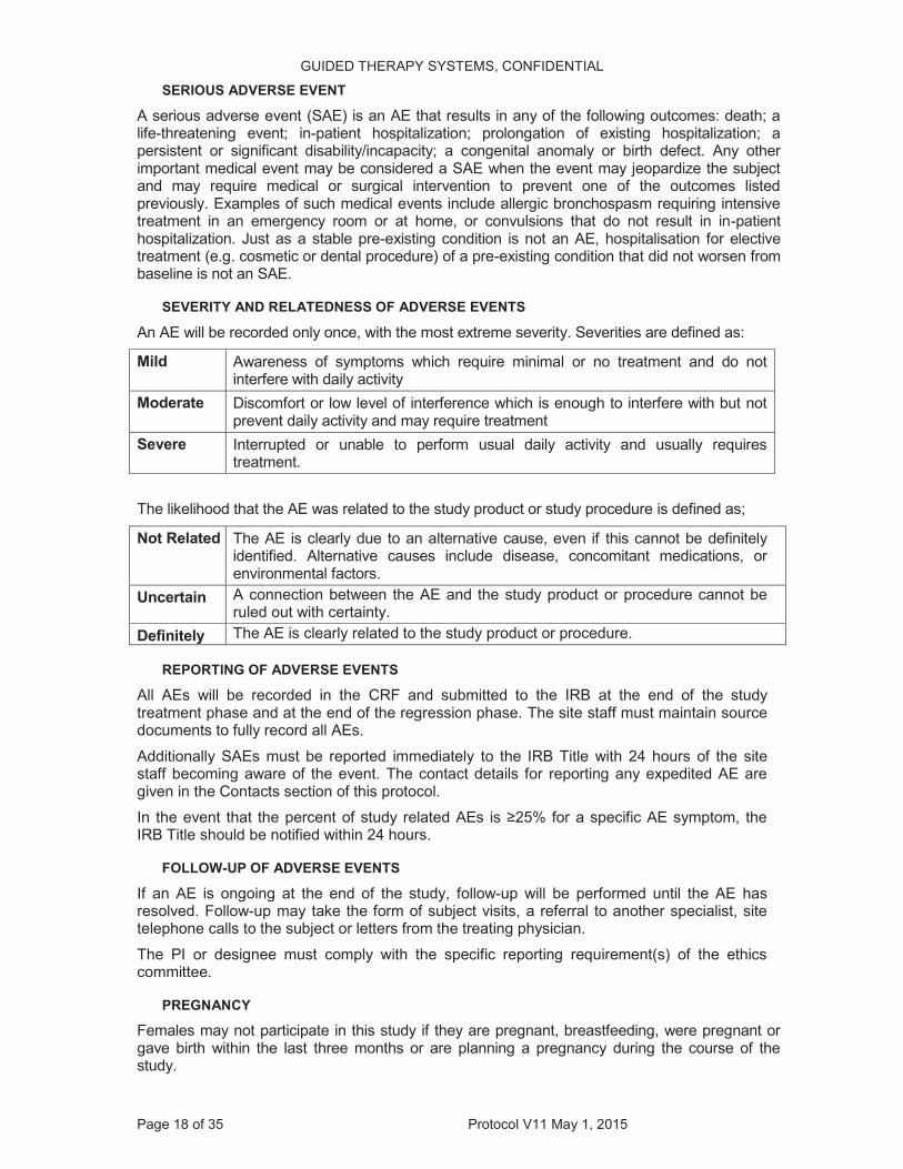
SERIOUS ADVERSE EVENT
A serious adverse event (SAE) is an AE that results in any of the following outcomes: death; a life-threatening event; in-patient hospitalization; prolongation of existing hospitalization; a persistent or significant disability/incapacity; a congenital anomaly or birth defect. Any other important medical event may be considered a SAE when the event may jeopardize the subject and may require medical or surgical intervention to prevent one of the outcomes listed previously. Examples of such medical events include allergic bronchospasm requiring intensive treatment in an emergency room or at home, or convulsions that do not result in in-patient hospitalization. Just as a stable pre-existing condition is not an AE, hospitalisation for elective treatment (e.g. cosmetic or dental procedure) of a pre-existing condition that did not worsen from baseline is not an SAE.
SEVERITY AND RELATEDNESS OF ADVERSE EVENTS
An AE will be recorded only once, with the most extreme severity. Severities are defined as:
Mild Awareness of symptoms which require minimal or no treatment and do not interfere with daily activity
Moderate Discomfort or low level of interference which is enough to interfere with but not prevent daily activity and may require treatment
Severe Interrupted or unable to perform usual daily activity and usually requires treatment.
The likelihood that the AE was related to the study product or study procedure is defined as;
Not Related The AE is clearly due to an alternative cause, even if this cannot be definitely identified. Alternative causes include disease, concomitant medications, or environmental factors.
Uncertain A connection between the AE and the study product or procedure cannot be ruled out with certainty.
Definitely The AE is clearly related to the study product or procedure.
REPORTING OF ADVERSE EVENTS
All AEs will be recorded in the CRF and submitted to the IRB at the end of the study treatment phase and at the end of the regression phase. The site staff must maintain source documents to fully record all AEs. Additionally SAEs must be reported immediately to the IRB Title with 24 hours of the site staff becoming aware of the event. The contact details for reporting any expedited AE are given in the Contacts section of this protocol. In the event that the percent of study related AEs is ≥25% for a specific AE symptom, the IRB Title should be notified within 24 hours.
FOLLOW-UP OF ADVERSE EVENTS
If an AE is ongoing at the end of the study, follow-up will be performed until the AE has resolved. Follow-up may take the form of subject visits, a referral to another specialist, site telephone calls to the subject or letters from the treating physician. The PI or designee must comply with the specific reporting requirement(s) of the ethics committee.
PREGNANCY
Females may not participate in this study if they are pregnant, breastfeeding, were pregnant or gave birth within the last three months or are planning a pregnancy during the course of the study.

GUIDED THERAPY SYSTEMS, CONFIDENTIAL
Page 19 of 35 Protocol V11 May 1, 2015
If the subject thinks they have become pregnant during the study it is important that they inform the study doctor immediately. If the subject becomes pregnant or thinks that they may be pregnant, they will be removed from the study.
STATISTICAL CONSIDERATIONS
SAMPLE SIZE CALCULATION
Up to 25 subjects will be enrolled onto the study to ensure a minimum of 25 subjects complete.
STATISTICAL METHODS
A sample size of 25 per protocol subjects will be required to demonstrate statistical significance (two-sided alpha=0.05; 80% power). Descriptive statistics will be calculated for comparison to the values noted in the existing literature.
MONITORING
The PI agrees that the site will permit, if required, study-related monitoring, audits, IRB/IEC review, and regulatory inspection(s), providing direct access to source data/documents. Sponsor personnel, or their designees, may monitor the study. The monitor has the responsibility to familiarise the PI and the entire centre staff involved in the study with Therapy procedures. The monitor can visit the clinical study centre on a regular basis such as before the first subject has been enrolled, during the course of the study, and at study completion. The monitor must maintain the confidentiality of the study documents. Regular visits may be made to monitor clinical procedures throughout the study duration.
DATA HANDLING AND RECORD KEEPING
Measurement and questionnaire data and images will be acquired by or sent to GTS for analysis. All clinical data will be collected/recorded by clinical site and reported to the GTS within two (2) weeks after the completion of the initial phase of the study (Week 8). Final, QA-approved raw data in SAS or Excel format (where applicable) will be sent to GTS for analysis within 2 weeks following completion of each phone survey. There will be at least one CRF for each subject randomised into the study. It is the responsibility of the PI to ensure the completeness and accuracy of the CRF and to authorise only trained members of staff to complete the CRF. The CRF must be completed legibly, using a black ballpoint pen. Erroneous values and/or text must not be obliterated. Instead, the error must be crossed out with a single line, the correct value/text added, and the correction signed or initialled and dated. At the end of the study, the original CRFs will be sent to The TBD Title and copies held at the study site. All site staff must ensure that the subject's anonymity will be maintained. On all documents that are to be submitted to GTS or external laboratory, subjects must be identified only by an identification code and not by their names. The PI or designee must keep a separate confidential enrollment log that matches identifying codes with the subject's names and addresses. The PI or designee must maintain these documents at the site. It is the responsibility of the PI or designee to maintain adequate clinical study records. Copies of all clinical study material must be archived for a period of at least 15 years after the end of the study (or more as legally required) or until informed by GTS that the documents can be destroyed. All documents must be archived in a secure place and treated as confidential material.
QUALITY STANDARDS
It is the responsibility of the PI to ensure that the study is conducted in accordance with the principles of Good Clinical Practice, the 2008 version of the Declaration of Helsinki and according to applicable local laws and regulations concerning studies conducted on human subjects which are outside of the definition of a medicinal product or medical device.

GUIDED THERAPY SYSTEMS, CONFIDENTIAL
Page 20 of 35 Protocol V11 May 1, 2015
Quality assurance audits may be performed by the clinical site, GTS or any ethics committee or regulatory authority during the course of the study or at study completion. The clinical trial will be conducted in accordance with the approved protocol and protocol amendments (if applicable). However, in the unlikely event of a major protocol deviation (i.e., those deviations which affect the integrity of the study or the safety of subjects) a protocol deviation form will be completed by the Principal Investigator (or designee) and submitted to the GTS and IRB for review and approval. The GTS will review the deviations and determine if the deviation(s) would significantly affect the results, and if deemed necessary, not include such data in the analysis. All other minor deviations should be recorded throughout the study and submitted to the GTS Title on a monthly basis.
ETHICS AND INFORMED CONSENT
The PI or designee must submit a copy of the protocol, informed consent form and all supporting documents to an Independent Ethics Committee or Institutional Review Board who must provide written approval before study specific procedures commence. The IEC/IRB must also approve any other information that is given to subjects such as advertisements and may require other documents such as study product documentation. Any modification to the agreed protocol must be agreed by both GTS and the PI and approved in writing by the IEC/IRB. Written approval must be obtained from the IEC/IRB before any amendment is implemented, unless immediate change is required to eliminate hazards to the subjects or when the change(s) involves only logistical or administrative aspects of the study (e.g., change of monitor(s), telephone number(s)). The PI or designee must obtain informed consent from each subject participating in the study, after explanation of the aims, methods, benefits and potential hazards of the study. The consent must be obtained before any study-specific procedures are performed. It must be made completely and unambiguously clear to each subject that they are free to refuse to participate in the study, or that they can withdraw their consent at any time and for any reason, without incurring any penalty or withholding of treatment. The subject must be given their own copy of the information sheet and signed consent form. The original signed informed consent must be kept on file by the PI or designee.
REPORTING AND PUBLICATION POLICY
Statistical analysis will be performed by GTS. The Investigator or designee will issue a final report of the results of the study following completion of data collection and quality control. The report will include the following: tabulations, a summary of adverse events, a description of the study, the study number, dates the study was conducted, the number of subjects who participated in the study as well as demographics (age, gender, ethnicity) and a summary of deviations. All IRB documentation/approvals must be included in the report. A complete report (electronic is acceptable) will be provided to the Sponsor within four weeks after completion of the 12 week follow-up. A final report will be submitted with four weeks of the 24 week follow-up phone call.

GUIDED THERAPY SYSTEMS, CONFIDENTIAL
Page 21 of 35 Protocol V11 May 1, 2015
REFERENCES
1. Hong QN, Durand MJ, Loisel P. Treatment of lateral epicondylitis: where is the evidence? Joint Bone Spine 2004; 71(5):369–373.
2. Walker-Bone K, Palmer KT, Reading I, Coggon D, Cooper C. Prevalence and impact of musculoskeletal disorders of the upper limb in the general population. Arthritis Rheum 2004; 51(4):642–651.
3. 3 Ollivere CO, Nirschl RP. Tennis elbow:current concepts and rehabilitation. Sports Med 1996; 22(2):133–139.
4. 4 Regan W, Wold LE, Conrad R, Morrey BF. Microscopic histopathology of chronic refractory lateral epicondylitis. Am J Sports Med 1992; 20:746.
5. 5 Kraushaar BS, Nirschl RP. Tendonosis of the elbow (Tennis Elbow). Clinical features and finding of histological, immunohistochemical, and electron microscopy studies. J Bone Joint Surg Am 1999; 81:259–278.
6. Lateral Elbow Tendinopathy: Correlation of Ultrasound Findings With Pain and Functional Disability Am J Sports Med June 2010 38 1209-1214; published online before print March 24, 2010,
7. Maffulli N, Khan K M, Puddu G. Overuse tendon conditions. Time to change a confusing terminology.
Arthroscopy 1998. 14840–843.843. [PubMed] 8. Archambault J M, Wiley J P, Bray R C. Exercise loading of tendons and the development of overuse
injuries. A review of the current literature. Sports Med 1995. 2077–89.89. [PubMed] 9. Sharma P1,Maffulli N. Biology of tendon injury: healing, modeling and remodeling. J Musculoskelet
Neuronal Interact. 2006 10. Ustuner E, Toprak U, Baskan B, Oztuna D. Sonographic examination of the common extensor tendon of
the forearm at three different locations in the normal asymptomatic population; Surg Radiol Anat (2013) 35:547–552 DOI 10.1007/s00276-013-1084-6
11. Apr-Jun;6(2):181-90.Gliklich R, White WM, Barthe PG, Slayton MH, Makin IRS. Clinical pilot study of intense ultrasound (IUS) therapy to deep dermal facial skin and subcutaneous tissues. Arch Facial Plast Surg 2007; 9:88-95.
12. Suh DH, Shin MK, Lee SJ, et al. Intense focused ultrasound tightening in Asian skin: Clinical and pathologic results. Dermatol Surg. 2011;37: 1595-1602
13. Alam, M., L. E. White, et al. (2010). "Ultrasound tightening of facial and neck skin: a rater- blinded prospective cohort study." J Am Acad Dermatol 62(2): 262-269.
14. White, W. M., I. R. Makin, et al. (2007). "Selective creation of thermal injury zones in the superficial musculoaponeurotic system using intense ultrasound therapy: a new target for noninvasive facial rejuvenation." Arch Facial Plast Surg 9(1): 22-29.
15. Slayton M., Barton J, Feasibility of Modulating Healing Tissue Response by ITU (Intense Therapy Ultrasound) in Musculoskeletal Tissue ASLMS, 2014, IEEE, 2014
16. Rose, M., et al., Evaluation of a preliminary physical function item bank supported the expected advantages of the Patient-Reported Outcomes Measurement Information System (PROMIS). J Clin Epidemiol, 2008. 61(1): p. 17-33.
17. Hawker, G.A., et al., Measures of adult pain: Visual Analog Scale for Pain (VAS Pain), Numeric Rating Scale for Pain (NRS Pain), McGill Pain Questionnaire (MPQ), Short-Form McGill Pain Questionnaire (SF-MPQ), Chronic Pain Grade Scale (CPGS), Short Form-36 Bodily Pain Scale (SF-36 BPS), and Measure of Intermittent and Constant Osteoarthritis Pain (ICOAP). Arthritis Care Res (Hoboken), 2011. 63 Suppl 11: p. S240-52
18. Rompe JD1, Overend TJ, MacDermid JC. Validation of the Patient-rated Tennis Elbow Evaluation Questionnaire.J Hand Ther. 2007 Jan-Mar;20(1):3-10; quiz 11.

GUIDED THERAPY SYSTEMS, CONFIDENTIAL
Page 22 of 35 Protocol V11 May 1, 2015
Appendix 1- Calendar of Events

GUIDED THERAPY SYSTEMS, CONFIDENTIAL
Page 23 of 35 Protocol V11 May 1, 2015
Appendix 2 – PRTEE (attachment)

GUIDED THERAPY SYSTEMS, CONFIDENTIAL
Page 24 of 35 Protocol V11 May 1, 2015
Appendix 2 – PRTEE (attachment), continued

GUIDED THERAPY SYSTEMS, CONFIDENTIAL
Page 25 of 35 Protocol V11 May 1, 2015
Appendix 3-A; Self Reported Outcome Measures

GUIDED THERAPY SYSTEMS, CONFIDENTIAL
Page 26 of 35 Protocol V11 May 1, 2015
Appendix 3-B – Subject Self-Assessment Questionnaire, continued:
VAS: Visual Analog Scale for Pain or Universal Pain Assessment Tool.

GUIDED THERAPY SYSTEMS, CONFIDENTIAL
Page 27 of 35 Protocol V11 May 1, 2015
APPENDIX 4 – Clinical Site Physical Therapy
TBD along with clinical site and PI

GUIDED THERAPY SYSTEMS, CONFIDENTIAL
Page 28 of 35 Protocol V11 May 1, 2015
Appendix 5: Adverse Events Report Sample AE Form
AE Term Serious Yes/ No
Severity, Relationship, Action Taken
Treatment of Event (if medication report CM)
Outcome
1 Severity: Relationship: Action taken:
o 01 o 02 o 03 o 04 o 05 (specify)
Onset date: End date:
2 Severity: Relationship: Action taken:
o 01 o 02 o 03 o 04 o 05 (specify)
Onset date: End date:
Severity Relationship Action Taken Treatment of Event: Outcome: Mild Moderate Severe
01= Not Related to treatment 02=Probably related to treatment 03=Definitely related to treatment
01=None 02=Treatment interrupted 03=Treatment stopped
01=None 02=Pharmacological Treatment (report CM) 03=Non-Pharmacological Treatment 04=Hospitalization 05=Other
01=Recovered 02=Recovered w/ Sequelae 03=Recovering 04=Not Recovered 05=Fatal 06=Unknown
Investigator Signature: __________________ Date:______/________/_______

GUIDED THERAPY SYSTEMS, CONFIDENTIAL
Page 29 of 35 Protocol V11 May 1, 2015
Appendix 6-A: Home Treatment example. (PI may suggest other exercise protocols)
Subjects will apply ice or cold pack to the affected area for (times and duration to be confirmed with Clinical Site/PI)

GUIDED THERAPY SYSTEMS, CONFIDENTIAL
Page 30 of 35 Protocol V11 May 1, 2015
Appendix 6-B: Home Treatment Log
Strenthening (Exercise 3 & 4)
Strenthening (Exercise 5 & 6)
Massage (Exercise 7)
Set 1 Set 2 Set 3 Set 4 Set 5 Set 1 Set 1 Set 11/12/20151/13/20151/14/20151/15/20151/16/20151/17/20151/18/20151/19/20151/20/20151/21/20151/22/20151/23/20151/24/20151/25/20151/26/20151/27/20151/28/20151/29/20151/30/20151/31/2015
2/1/20152/2/20152/3/20152/4/20152/5/20152/6/20152/7/20152/8/20152/9/2015
2/10/20152/11/20152/12/20152/13/20152/14/20152/15/20152/16/20152/17/20152/18/20152/19/20152/20/20152/21/20152/22/20152/23/20152/24/20152/25/20152/26/20152/27/20152/28/2015
3/1/20153/2/20153/3/20153/4/20153/5/20153/6/20153/7/20153/8/20153/9/2015
3/10/20153/11/20153/12/20153/13/20153/14/20153/15/20153/16/2015
Sets per Day
CET Stretch (exercise 1 & 2)

GUIDED THERAPY SYSTEMS, CONFIDENTIAL
Page 31 of 35 Protocol V11 May 1, 2015
Appendix 7 – Marketed Materials Information
Appendix 7-A – ITU Systems
Clinical Prototype Systems Gen II and Gen III Power: 3-100 Watts
Frequency: 3 – 10MHz
Energy/Zone: up to 3 Joules (tech. mode)

GUIDED THERAPY SYSTEMS, CONFIDENTIAL
Page 32 of 35 Protocol V11 May 1, 2015
Appendix 7-B – Diagnostic Ultrasound Imaging System

GUIDED THERAPY SYSTEMS, CONFIDENTIAL
Page 33 of 35 Protocol V11 May 1, 2015
Appendix 7-B – Diagnostic Ultrasound Imaging System, continued

GUIDED THERAPY SYSTEMS, CONFIDENTIAL
Page 34 of 35 Protocol V11 May 1, 2015
Appendix 7-C – Acoustic Coupling Gel – Polysonic or similar POLYSONIC® Ultrasound Lotion

GUIDED THERAPY SYSTEMS, CONFIDENTIAL
Page 35 of 35 Protocol V11 May 1, 2015
Appendix 7-C – Acoustic Coupling Gel, continued