Embed Size (px)
Citation preview

Thomas B. ThompsonGunnar F. Schröder, W. Sean Davidson and Xiaodi Deng, Jamie Morris, Catherine Chaton, StabilityImportance of Its Termini for StructuralApolipoprotein A-IV Reveals the Small-angle X-ray Scattering ofProtein Structure and Folding:
doi: 10.1074/jbc.M112.436709 originally published online January 3, 20132013, 288:4854-4866.J. Biol. Chem.
10.1074/jbc.M112.436709Access the most updated version of this article at doi:
.JBC Affinity SitesFind articles, minireviews, Reflections and Classics on similar topics on the
Alerts:
When a correction for this article is posted•
When this article is cited•
to choose from all of JBC's e-mail alertsClick here
http://www.jbc.org/content/288/7/4854.full.html#ref-list-1
This article cites 38 references, 14 of which can be accessed free at
at University of Cincinnati/Medical Center Libraries on November 11, 2013http://www.jbc.org/Downloaded from at University of Cincinnati/Medical Center Libraries on November 11, 2013http://www.jbc.org/Downloaded from

Small-angle X-ray Scattering of Apolipoprotein A-IV Revealsthe Importance of Its Termini for Structural Stability*
Received for publication, November 15, 2012, and in revised form, January 2, 2013 Published, JBC Papers in Press, January 3, 2013, DOI 10.1074/jbc.M112.436709
Xiaodi Deng‡, Jamie Morris§, Catherine Chaton‡, Gunnar F. Schröder¶, W. Sean Davidson§,and Thomas B. Thompson‡1
From the ‡Department of Molecular Genetics, Biochemistry, and Microbiology, College of Medicine, University of Cincinnati,Cincinnati, Ohio 45267, the §Department of Pathology and Laboratory Medicine, College of Medicine, Metabolic Diseases Institute,University of Cincinnati, Cincinnati, Ohio 45215, and the ¶Institute of Complex Systems (ICS-6), Forschungszentrum Jülich,52425 Jülich, Germany
Background: Apolipoproteins are lipid emulsifiers with links to additional protective roles.Results: Small-angle x-ray scattering afforded structural information for full-length apoA-IV.Conclusion: In the head-to-tail dimer, the N/C-terminal globular domainsmodulate the twist and curvature of a central helicalbundle.Significance: The lipid affinity of apoA-IV is regulated by opening and closing a molecular clasp.
ApoA-IV is an amphipathic protein that can emulsify lipidsand has been linked to protective roles against cardiovasculardisease and obesity. We previously reported an x-ray crystalstructure of apoA-IV that was truncated at its N and C termini.Here, we have extended this work by demonstrating that self-associated states of apoA-IV are stable and can be structurallystudied using small-angle x-ray scattering. Both the full-lengthmonomeric and dimeric forms of apoA-IV were examined, withthe dimer showing an elongated rod core with two nodes atopposing ends. The monomer is roughly half the length of thedimer with a single node. Small-angle x-ray scattering visualiza-tion of several deletion mutants revealed that removal of bothtermini can have substantial conformational effects throughoutthemolecule.Additionally, the F334Apointmutation,whichwepreviously showed increases apoA-IV lipid binding, alsoexhibited large conformational effects on the entire dimer.Merging this study’s low-resolution structural informationwith the crystal structure provides insight on the conforma-tion of apoA-IV as a monomer and as a dimer and furtherdefines that a clasp mechanism may control lipid bindingand, ultimately, protein function.
The A class apolipoproteins (apoA-I, apoA-II, apoA-IV, andapoA-V) act as detergents that emulsify lipids to form or asso-ciate with lipoprotein particles, particularly HDL. They areconsidered exchangeable due to their ability to transitionbetween a lipid-free and lipid-bound state, allowing for move-ment between particles in the plasma. These lipoprotein parti-cles are the body’s main mechanism for transporting and deliv-ering lipids. They have distinct functional roles, acting as
structural scaffolds and activators of lipoprotein-remodelingfactors and interacting with cell surface proteins that modulatelipoprotein metabolism (1, 2).Because of their importance in lipid biology, the structural
characterization of apolipoproteins has been a high priority.Unfortunately, many apolipoproteins undergo rapid transi-tions between a constellation of self-associated states (dimers,trimers, tetramers, etc.), thus making them difficult to charac-terize structurally (3). Although some success has beenobtained using site-directed mutagenesis to stabilize particularoligomeric forms of apoA-I and apoE, this approach has thedrawback of potentially altering protein conformation alongwith self-association state (4, 5).We recently demonstrated thatlipid-free human apoA-IV distributes predominantly betweenmonomers and dimers and that their interconversion is slowenough that they can be isolated and studied individually (6).Thus, we have focused on apoA-IV as a model to gain insightinto the structure and self-association of the exchangeableapolipoproteins.ApoA-IV is the largest member of the exchangeable apolipo-
protein family, with a molecular mass of 43 kDa. It is the thirdmost prominent protein found associated with HDL (3, 7). Inaddition to HDL particle formation, apoA-IV has been shownto have a variety of unique biological roles not shared by otherapolipoproteins; for example, in response to lipid absorption,apoA-IV is highly expressed in the intestine and is associatedwith apoB secretion (8, 9). Furthermore, apoA-IV functions as asatiety signal (10, 11), an antioxidant (12), and an anti-inflam-matory agent (13). Recently, apoA-IV has also been shown toregulate insulin secretion, specifically during elevated glucoselevels (14). Intriguingly, there is an 8-fold increase of apoA-IVin patients after Roux-en-Y gastric bypass surgery who showedimprovements in obesity-related comorbidities (15). Despitesuch a diversity of protective functions, little is known about theapoA-IV mechanism of action. ApoA-IV can exist in solution(and serum) without lipid in self-associated forms, predomi-nantly as a monomer or dimer (16), and the lipid-bound formremains poorly characterized.
* This work was supported, in whole or in part, by National Institutes of HealthGrant R01 GM098458 (to T. B. T. and W. S. D.), Grant HL67093 (to W. S. D.),and Training Grant T32 HL007382 (to X. D.).
1 To whom correspondence should be addressed: Dept. of Molecular Genet-ics, Biochemistry, and Microbiology, College of Medicine, University of Cin-cinnati, 231 Albert Sabin Way, MSB 3005B, Cincinnati, OH 45267. Tel.: 513-558-4517; E-mail: [email protected].
THE JOURNAL OF BIOLOGICAL CHEMISTRY VOL. 288, NO. 7, pp. 4854 –4866, February 15, 2013© 2013 by The American Society for Biochemistry and Molecular Biology, Inc. Published in the U.S.A.
4854 JOURNAL OF BIOLOGICAL CHEMISTRY VOLUME 288 • NUMBER 7 • FEBRUARY 15, 2013 at University of Cincinnati/Medical Center Libraries on November 11, 2013http://www.jbc.org/Downloaded from
Structurally, apoA-IV has a large hydrophobic core similar toother strong lipid-binding proteins such as apoA-I and apoE3(4–6).However, it is a relatively poor lipid binder (17). Previouswork from our laboratory strongly suggests that an interactionbetween the N and C termini of apoA-IV holds the protein in aconformation that binds lipids poorly (17–19). Disruption ofthis “clasp” by introducing the point mutation F334A signifi-cantly increased the lipid affinity of apoA-IV (19). Unfortu-nately, our structure of N/C-terminally truncated apoA-IV didnot provide high-resolution structural details pertaining to theclasp mechanism.To understand the conformational states adopted by full-
length apoA-IV, we took advantage of its oligomeric stabilityand performed small-angle x-ray scattering (SAXS).2 Althoughof lower resolution than x-ray crystallography, SAXS allowedusto visualize the molecular envelope of full-length apoA-IV,along with truncation variants and the F334A point mutant.Combining thiswith the partial x-ray structure, we defined howthe individual termini affect the global conformation ofapoA-IV and further supported the clasp mechanism.
EXPERIMENTAL PROCEDURES
Protein Expression and Purification—Recombinant humanapoA-IVWT, apoA-IV64–335, apoA-IV1–335, apoA-IV63–376, andapoA-IVF334A were produced and purified in Escherichia coli asdescribed previously (6). Size-exclusion chromatography (SEC)using a HiLoad 16/60 Superdex 200 column (GE Healthcare)was performed to isolated oligomeric species for the variousconstructs. The isolated oligomeric species were concentratedat 4 °C using Amicon Ultra-15 centrifuge filter units. To main-tain oligomeric homogeneity, concentration was performed in15-min intervals at low speed (1000 � g). At the end of eachinterval, the concentrated sample wasmixedwith the total poolof the less concentrated sample; this minimized the concentra-tion gradient during centrifugation and prevented shiftingbetween oligomeric species. To confirm this, samples werereanalyzed by SEC (Superdex 200 HR 10/300, Amersham Bio-sciences) using 8–25% gradient native gels (PhastGel system,GE Healthcare).Sedimentation Velocity—Analytical ultracentrifugation ex-
periments were performed using a Beckman XL-I ultracentri-fuge with absorbance optics and a four-hole rotor. Sedimenta-tion velocity was performed in a two-channel carbon-filledEpon centerpiece at 36,000 rpm and 10 °C with protein (0.15mg/ml) that had been dialyzed into 20 mMNaPO4 (pH 7.4) and100 mM NaF. Protein was monitored with UV absorbance at230 nm, and data were analyzed using Sedfit (20).SEC—Todetermine the apoA-IV dimer/monomer ratio over
time, a HiLoad 16/60 Superdex 200 column on an ÄKTAExplorer equipped with a UV-900 detector and a P-960 samplepump at 4 °Cwas used. The purified apoA-IVdimerwas dilutedto 0.15 mg/ml in 50 ml of PBS with a single cOmplete EDTA-free protease inhibitor mixture tablet (catalog number05056489001, RocheApplied Science), 1mM sodium azide, and0.05 mM PMSF. The samples were incubated at 4 °C, and at
different time points, a sample was collected and examinedusing a HiLoad 16/60 Superdex 200 gel filtration column. Theelution process was monitored at 215, 235, and 280 nm. Peakintegration was performed with the UV absorbance traces. Thepercent dimerwas determined by dividing the area of the dimerpeak by the total area of both monomer and dimer peaks.1,2-Dimyristoyl-sn-glycero-3-phosphocholine (DMPC)Clear-
ance Assay—Purified protein was tested for its ability to emul-sify suspendedDMPC (Avanti Polar Lipids). The assay was per-formed as described previously (18, 19).SAXS—SAXS data were collected at beamline 12-ID-B at the
Advanced Photon Source at Argonne National Laboratory(Table 1). The beamline was equippedwith a flow cell to reduceradiation damage. A total of 120 �l of sample was loaded intothe flow cell and oscillated during data collection. Data werecollected at room temperature, with an exposure time of 0.5 sand a total of 30 images taken for each sample. Small-anglediffraction images were captured using a PILATUS 2Mdetector.Ab Initio Model Reconstruction—The data collected were
converted from two- to one-dimensional using the Irena pack-age for IGOR software (21), and outliers within each set of 30images were removed and averaged using PRIMUS (22). Theaveraged data were subtracted from the appropriate bufferblank also in PRIMUS. Subsequently, AutoRG and GNOMwere used to determine Rg, Dmax, and I(0) (Table 1). Indirecttransformation was performed with GNOM (23); ab initioreconstructionwas completed usingGASBOR (24). Dependingon the data set, monomer (P1) or dimer (P2) symmetry wasused as a constraint in GASBOR. Ten ab initio reconstructionsfor each construct of apoA-IV were performed and averagedusing the DAMAVER package (25). The apoA-IV64–335 crystalstructure was compared with the scattering data of both apoA-IV64–335 and apoA-IVWT using CRYSOL (26).DireX—Fitting of the crystal structure into the SAXS enve-
lope, which had additional curvature, was achieved by geome-try-based conformational sampling performed by the programDireX (27). An average weighted electron density map was firstcreated from the ab initio reconstruction. Conformationalsampling was dictated by the CONCOORD algorithm in 100total steps (nsteps) withTirion enhancement. A dynamic elasticnetwork was used for further refinement and to prevent over-fitting. The dynamic elastic network used a � value of 0.0(den_gamma) and a � value of 0.2 (den_kappa). A total of 8000restraints were chosen per iteration (den_no) with a dynamic
2 The abbreviations used are: SAXS, small-angle x-ray scattering; SEC, size-exclu-sion chromatography; DMPC, 1,2-dimyristoyl-sn-glycero-3-phosphocholine.
TABLE 1Data collection
Data collection parametersInstrument 12-ID-BMonochromator type Side bounceWavelength (Å) 1.53q range (Å�1) 0.009–0.250Exposure time (s) 0.5Temperature (K) 293
Software employedPrimary data reduction PRIMUSData processing GNOMAb initio analysis GASBORValidation and averaging DAMAVERStructure refinement DireXComputation of model intensities CRYSOL
ApoA-IV Structure Revealed by Small Angle X-ray Scattering
FEBRUARY 15, 2013 • VOLUME 288 • NUMBER 7 JOURNAL OF BIOLOGICAL CHEMISTRY 4855 at University of Cincinnati/Medical Center Libraries on November 11, 2013http://www.jbc.org/Downloaded from

ApoA-IV Structure Revealed by Small Angle X-ray Scattering
4856 JOURNAL OF BIOLOGICAL CHEMISTRY VOLUME 288 • NUMBER 7 • FEBRUARY 15, 2013 at University of Cincinnati/Medical Center Libraries on November 11, 2013http://www.jbc.org/Downloaded from

elastic network strength of 0.4 (den_strength). The structurewas not energy-minimized after DireX fitting.RESULTSStability of ApoA-IV—Lipid-free human apoA-IV was origi-
nally documented to exist as a mixture of a monomer anddimer, with a slow interconversion between the forms (16).Werecently showed that both dimeric and monomeric forms ofapoA-IV can be isolated by SEC (6). This is in sharp contrast toother apolipoproteins, such as apoE (28) and apoA-I (29, 30),indicating that apoA-IV oligomerization is uniquely stable.SAXS analysis requires samples that remain molecularly
homogeneous for the entirety of the analysis.We first looked athow well apoA-IV maintains the dimeric state at 4 °C. At this
temperature, the dimer did not dissociate, even after 600 h (Fig.1A). As SAXS experiments are carried out at room tempera-ture, the apoA-IV dimer was incubated at 20 °C, and sampleswere collected and subjected to sedimentation velocity on days1 and 3. At 20 °C, apoA-IV remained entirely dimeric duringthe course of 3 days (Fig. 1, B andD). This indicated that, in thetime required to collect SAXS data (�5 min), dimeric apoA-IVwould maintain its oligomeric form. As a control to show thatapoA-IV can freely distribute, the same experiment was per-formed at 37 °C. Dissociation of the dimer to themonomer wasreadily observable on day 1 and to amuch greater extent on day3 (Fig. 1,C andD). Thus, at 20 °C, SAXS analyses were justified.Furthermore, the different truncations and mutations of
FIGURE 1. Quantifying the stability of the apoA-IV monomer and dimer. A, the apoA-IV dimer (0.15 mg/ml) was incubated at 4 °C, and samples were takenat various time points and analyzed by SEC. B and C, sedimentation velocity (Sed Vel) was performed to determine the self-associated state with isolated apoA-IVdimers incubated at 20 °C (B) or 37 °C (C) for 1 or 3 days. After fitting for the frictional ratio (f/f0), the c(s) distribution was transformed into a c(M) distribution ofthe molecular masses. D, sedimentation velocity data (dots) and corresponding fits (lines). Shown are 8 of the 32 scans used for fitting and their residuals for thesedimentation velocity experiments. E, native gel of apoA-IV and its variants. F, SEC traces of apoA-IV and variants. A retention volume of 12–14 ml is consistentwith dimeric size, and 14 –16 ml is consistent with a monomer based on molecular size standards.
FIGURE 2. Solution state of apoA-IV64 –335. A, scattering function and Guinier plot (inset) of dimeric apoA-IV64 –335 and simulated scattering function generatedwith CRYSOL using the apoA-IV64 –335 crystal structure (Protein Data Bank code 3S84) and the DireX refined structure. B, pairwise distance distribution functionof apoA-IV64 –335. a.u., arbitrary units. C, three orthogonal views: top, averaged ab initio reconstructions of apoA-IV64 –335 (orange spheres); middle, superpositionof the apoA-IV64 –335 crystal structure envelope (green spheres) with the SAXS envelope (orange spheres); and bottom, ribbon and surface representation of theapoA-IV64 –335 crystal structure. D, the crystal structure was refined using the SAXS envelope with the program DireX. The refined structure of apoA-IV64 –335 isshown as a ribbon, and the ab initio reconstruction of apoA-IV64 –335 is shown as mesh.
ApoA-IV Structure Revealed by Small Angle X-ray Scattering
FEBRUARY 15, 2013 • VOLUME 288 • NUMBER 7 JOURNAL OF BIOLOGICAL CHEMISTRY 4857 at University of Cincinnati/Medical Center Libraries on November 11, 2013http://www.jbc.org/Downloaded from
apoA-IV used in this study did not affect its oligomeric stability.Each variant was isolated and maintained its self-associatedstate as demonstrated by native gel electrophoresis (Fig. 1E).Additionally, after the SAXS data collection, each sample wasanalyzed by SEC, which showed that they still maintained theiroligomeric states (Fig. 1F).DimericApoA-IV64–335—Because apolipoproteins undergo a
variety of molecular transitions, we first determined if theobserved crystal structure of the terminus-free core domain ofapoA-IV (apoA-IV64–335) provides a good model for the pro-tein in solution. We expressed apoA-IV64–335 and isolated itsdimer (Fig. 1, E and F), which remained stably dimeric like thefull-length form. We collected SAXS data on apoA-IV64–335 insolution at four concentrations ranging from 0.5 to 4 mg/ml(Fig. 2A and Table 2). In all cases, the solution remained free ofsignificant aggregation or repulsion as determined by a linearGuinier plot (Fig. 2A), and I(0) remained consistent over theconcentration range examined (Table 2). Although the overallshape of the scattering profile was similar at all protein concen-trations analyzed, the data obtained at 4 mg/ml yielded thestrongest signal-to-noise intensity and was therefore usedfor subsequent analysis. The shape of the scattering and pair-wise distance distribution function profiles indicated a rod-like structure (Fig. 2B). The diameter of the maximal particlesize (Dmax) was determined to be 175 Å using GNOM (Fig. 2,A and B).
From these data, a low-resolution envelope of the proteinwas generated via ab initio reconstructions. Ten independentreconstructions were performed and then averaged withDAMAVER.Overall, the reconstructionswere in agreement, as
the mean normalized spatial discrepancy was only 1.49 � 0.91.The envelope of apoA-IV64–335 reveals a linear elongated rodwith a slight curvature extending 170 Å in length and 20 Å inwidth (Fig. 2C). These dimensions match exceptionally wellwith our recent crystal structure of apoA-IV64–335, in whichresidues 75–312 were resolved. (Residues 64–74 and 313–335were present but were not resolved in the crystal structure.)Additionally, when looking down the long axis, a right-handedtwist of�90° is observed, similar to the crystal structure.More-over, the simulated scattering data from the crystal structure fitthe experimental scattering data well, with a � value of 1.16 asdetermined by CRYSOL, and the experimental radius of gyra-tion (Rg) is consistent with the crystal structure (Fig. 2A andTable 2). These comparisons show that the SAXS and x-raycrystallography data are highly consistent, further validatingthe dimeric apoA-IV64–335 model.
Despite this close agreement, there is a rod curvature differ-ence between the crystal structure and SAXS reconstruction(Fig. 2C). The crystal structure is more linear, whereas theSAXS reconstruction is more curved. When describing thelength of the protein as an arc segment, the central angle ofthe crystal structure measures 44°, whereas the solution struc-ture measures 70° (Fig. 3). This difference in curvature may beattributed to crystallization (31), whereas SAXS is performed insolution. Nevertheless, apoA-IV is still significantly less curvedthan the recent crystal structure of apoA-I1–182 (4).
To better represent the solution state of apoA-IV64–335, werefined the crystal structure against the SAXS envelop with theprogram DireX (Fig. 2D). Success of the refinement was mea-sured by the correlation coefficient, a numerical value compar-
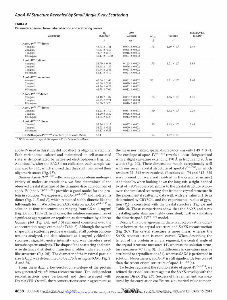
TABLE 2Parameters derived from data collection and scattering curves
ConstructRg
(Guinier)I(0)
(Guinier) Dmax VolumeDAMAVER
(NSD)a
Å cm�1 Å Å3
ApoA-IV64–335 dimer4 mg/ml 48.73 � 1.41 0.074 � 0.002 175 1.19 � 105 1.492 mg/ml 49.37 � 4.53 0.035 � 0.0031 mg/ml 44.74 � 8.16 0.016 � 0.0030.5 mg/ml 43.17 � 17.80 0.007 � 0.002
ApoA-IVWT dimer4 mg/ml 51.70 � 0.89 0.143 � 0.003 175 1.51 � 105 1.932 mg/ml 51.43 � 1.37 0.076 � 0.0021 mg/ml 50.94 � 2.43 0.037 � 0.0020.5 mg/ml 52.11 � 4.10 0.021 � 0.002
ApoA-IVWT monomer4 mg/ml 40.04 � 1.49 0.081 � 0.002 90 8.81 � 104 1.402 mg/ml 40.06 � 2.52 0.040 � 0.0021 mg/ml 41.34 � 4.22 0.022 � 0.0020.5 mg/ml 44.78 � 7.04 0.012 � 0.002
ApoA-IV1–335 dimer2 mg/ml 51.18 � 1.07 0.067 � 0.000 185 1.45 � 105 1.311 mg/ml 49.75 � 2.71 0.032 � 0.0000.5 mg/ml 50.66 � 5.20 0.016 � 0.002
ApoA-IV62–376 dimer2 mg/ml 54.23 � 1.12 0.051 � 0.001 180 1.33 � 105 2.291 mg/ml 51.29 � 3.32 0.026 � 0.0020.5 mg/ml 51.05 � 6.20 0.013 � 0.002
ApoA-IVF334A dimer2 mg/ml 52.26 � 2.17 0.037 � 0.002 195 1.62 � 105 2.491 mg/ml 53.23 � 4.23 0.020 � 0.0020.5 mg/ml 59.17 � 6.58 0.013 � 0.002
CRYSOL apoA-IV64–335 structure (PDB code 3S84) 53.11 174 1.07 � 105a NSD, normalized spatial discrepancy; PDB, Protein Data Bank.
ApoA-IV Structure Revealed by Small Angle X-ray Scattering
4858 JOURNAL OF BIOLOGICAL CHEMISTRY VOLUME 288 • NUMBER 7 • FEBRUARY 15, 2013 at University of Cincinnati/Medical Center Libraries on November 11, 2013http://www.jbc.org/Downloaded from

ing the electron density of the simulationmodel with the exper-imental map (32). During the refinement, the correlation coef-ficient increased from 0.84 to 0.98, indicating that DireXgenerated a model that was a better fit to the envelope than thecrystal structure itself. A low-resolution comparison of the twomodels indicates that the curvature is the result of a distortionto the helical segments caused by a series of repeating prolineresidues. In the crystal structure, these specific proline residuesalign vertically from opposing chains and are thought to pro-vide segmentation and the flexibility needed during lipid bind-ing (6). In our previous description of the crystal structure, wenoted that the amount of distortion caused by the introductionof a proline in a helix or kink angle was much lower than inpreviously reported structures. After modeling with DireX, wemeasured, as described previously (33), the same proline resi-dues and found an increased average kink of 6° (Table 3). Con-
sequently, the kink angle of the solution structure of apoA-IV64–335 dimer is more in line with the average proline kinkangle of 26° seen in other structures (6, 34).Overall, the SAXS data of apoA-IV64–335 match exception-
ally well with the crystal structure and provided constraints tobetter model the solution state. This also gave us confidence inthe implementation of SAXS to study apoA-IVWT and differenttruncation constructs.Dimeric ApoA-IVWT—We next performed a SAXS experi-
ment on the full-length apoA-IV dimer for comparison withapoA-IV64–335 (Fig. 4 and Table 2). We first correlated thesimulated scattering data from the crystal structure of apoA-IV64–335 with the scattering data from apoA-IVWT. Analysiswith the program CRYSOL resulted in a large � value of 3.99(Fig. 4D) compared with the value of 1.16 for apoA-IV64–335.This indicates that there are significant structural differencesbetween the two samples. Interestingly, although the pairwisedistance distribution function of the full-length apoA-IV dimeragain pointed to an elongated structure with a similar Dmax, itdepicted a bimodal distribution, indicating a deviation from apurely rod-like structure (Fig. 4B). Aligning the full-length andtruncated envelopes shows significant differences in the ends ofthe rods. Compared with apoA-IV64–335, the ends of the apoA-IVWT rod are significantly more globular, with a substantialincrease (�27%) in the volume of the envelope (Fig. 4E andTable 2). This is consistent with a 35% increase in themolecularmass of the apoA-IVWT dimer (86.8 kDa) versus the apoA-IV64–335 dimer (62.9 kDa). Moreover, aligning the envelopes of
FIGURE 3. Central arc measurements for the crystal structures of apoA-IV (Protein Data Bank code 3S84) and apoA-I (code 3R2P) and ab initioreconstructions of apoA-IV64 –335, apoA-IV1–335, and apoA-IVWT.
TABLE 3Proline kink angles
Proline Crystal structure DireX �
Chain APro-117 21.5° 32.8° 11.3°Pro-139 14.8° 16.3° 1.5°Pro-161 16.2° 23.5° 7.3°Pro-183 13.1° 16.3° 3.2°Average 16.4° 22.2° 5.8°
Chain BPro-117 23.2° 36.0° 12.8°Pro-139 10.3° 8.5° �1.8°Pro-161 18.7° 30.6° 11.9°Pro-183 10.4° 12.4° 2°Average 15.7° 21.9° 6.2°
ApoA-IV Structure Revealed by Small Angle X-ray Scattering
FEBRUARY 15, 2013 • VOLUME 288 • NUMBER 7 JOURNAL OF BIOLOGICAL CHEMISTRY 4859 at University of Cincinnati/Medical Center Libraries on November 11, 2013http://www.jbc.org/Downloaded from

apoA-IV64–335 and apoA-IVWT at just one end reveals addi-tional density in the apoA-IVWT envelope. This density spa-tially corresponds to the visible N- and C-terminal ends in thecrystal structure (Fig. 2C). This difference in the reconstruc-tions strongly supports the possibility that the N and C terminiinteract with each other at both ends of the rod.In addition to the increase in mass identified at the ends of
the rod, we also observed a difference in the overall twist of the
molecule. More specifically, although both apoA-IVWT andapoA-IV64–335 are slightly curved, with a similar central arc of�70° (Fig. 3), there is a difference in the direction of the twist(Fig. 4, C and D; see Fig. 8). Whereas apoA-IV64–335 has a left-handed twist when looking down the long axis of the rod, apoA-IVWT has a right-handed twist of �120°. This, along with thecurvature, creates a chiral mismatch between the two recon-structions (Fig. 4, D and E; see Fig. 8). Although both apoA-
FIGURE 4. Solution state of apoA-IVWT and comparison with apoA-IV64 –335. A, scattering function and Guinier plot (inset) of dimeric apoA-IVWT andapoA-IV64 –335. B, pairwise distance distribution function of apoA-IVWT and apoA-IV64 –335. a.u., arbitrary units. C, three orthogonal views of the averaged ab initioreconstructions of apoA-IV64 –335 (top) and apoA-IVWT (bottom). D, alignment of apoA-IVWT and apoA-IV64 –335 reconstructions and comparison of apoA-IVWT
SAXS scattering function with the simulated scattering function of the apoA-IV64 –335 crystal structure generated with CRYSOL. E, superposition of one-half ofapoA-IVWT (mesh) and apoA-IV64 –335 (spheres) depicting additional density associated with apoA-IVWT.
ApoA-IV Structure Revealed by Small Angle X-ray Scattering
4860 JOURNAL OF BIOLOGICAL CHEMISTRY VOLUME 288 • NUMBER 7 • FEBRUARY 15, 2013 at University of Cincinnati/Medical Center Libraries on November 11, 2013http://www.jbc.org/Downloaded from
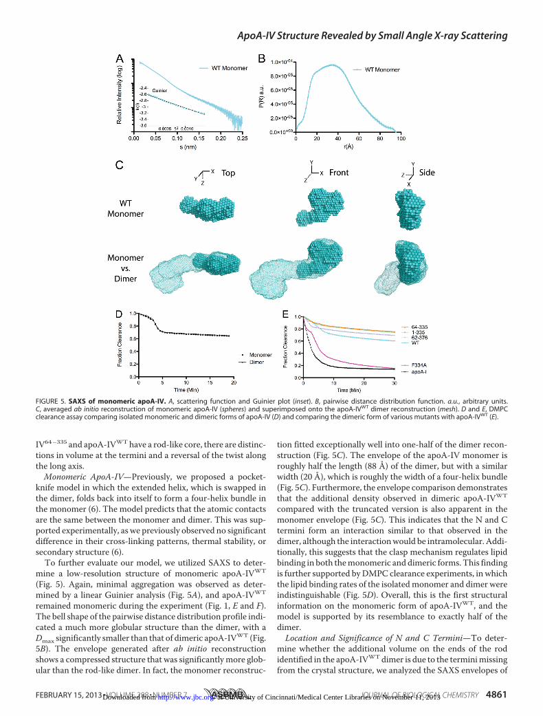
IV64–335 and apoA-IVWThave a rod-like core, there are distinc-tions in volume at the termini and a reversal of the twist alongthe long axis.Monomeric ApoA-IV—Previously, we proposed a pocket-
knife model in which the extended helix, which is swapped inthe dimer, folds back into itself to form a four-helix bundle inthe monomer (6). The model predicts that the atomic contactsare the same between the monomer and dimer. This was sup-ported experimentally, as we previously observed no significantdifference in their cross-linking patterns, thermal stability, orsecondary structure (6).To further evaluate our model, we utilized SAXS to deter-
mine a low-resolution structure of monomeric apoA-IVWT
(Fig. 5). Again, minimal aggregation was observed as deter-mined by a linear Guinier analysis (Fig. 5A), and apoA-IVWT
remained monomeric during the experiment (Fig. 1, E and F).The bell shape of the pairwise distance distribution profile indi-cated a much more globular structure than the dimer, with aDmax significantly smaller than that of dimeric apoA-IVWT (Fig.5B). The envelope generated after ab initio reconstructionshows a compressed structure that was significantlymore glob-ular than the rod-like dimer. In fact, the monomer reconstruc-
tion fitted exceptionally well into one-half of the dimer recon-struction (Fig. 5C). The envelope of the apoA-IV monomer isroughly half the length (88 Å) of the dimer, but with a similarwidth (20 Å), which is roughly the width of a four-helix bundle(Fig. 5C). Furthermore, the envelope comparison demonstratesthat the additional density observed in dimeric apoA-IVWT
compared with the truncated version is also apparent in themonomer envelope (Fig. 5C). This indicates that the N and Ctermini form an interaction similar to that observed in thedimer, although the interactionwould be intramolecular. Addi-tionally, this suggests that the clasp mechanism regulates lipidbinding in both themonomeric and dimeric forms. This findingis further supported byDMPC clearance experiments, in whichthe lipid binding rates of the isolatedmonomer and dimer wereindistinguishable (Fig. 5D). Overall, this is the first structuralinformation on the monomeric form of apoA-IVWT, and themodel is supported by its resemblance to exactly half of thedimer.Location and Significance of N and C Termini—To deter-
mine whether the additional volume on the ends of the rodidentified in the apoA-IVWTdimer is due to the terminimissingfrom the crystal structure, we analyzed the SAXS envelopes of
FIGURE 5. SAXS of monomeric apoA-IV. A, scattering function and Guinier plot (inset). B, pairwise distance distribution function. a.u., arbitrary units.C, averaged ab initio reconstruction of monomeric apoA-IV (spheres) and superimposed onto the apoA-IVWT dimer reconstruction (mesh). D and E, DMPCclearance assay comparing isolated monomeric and dimeric forms of apoA-IV (D) and comparing the dimeric form of various mutants with apoA-IVWT (E).
ApoA-IV Structure Revealed by Small Angle X-ray Scattering
FEBRUARY 15, 2013 • VOLUME 288 • NUMBER 7 JOURNAL OF BIOLOGICAL CHEMISTRY 4861 at University of Cincinnati/Medical Center Libraries on November 11, 2013http://www.jbc.org/Downloaded from

deletion variants with individual N- and C-terminal trunca-tions. ApoA-IV1–335 was used to identify the additional volumeassociated with the N terminus, whereas apoA-IV62–376 wasused for the C terminus. Each variant was expressed and puri-fied as described above, and the dimer was isolated by SEC.Similar to apoA-IVWT and apoA-IV64–335, the isolated dimerswere exceptionally stable (Fig. 1, E and F). ApoA-IV62–376,which lacks residues involved in lipid binding, was shown to bea poor lipid binder (Fig. 5E). Similarly, apoA-IV1–335 also boundlipid poorly. This has been suggested to be a result of an intactclasp, which is highly dependent on interaction of Phe-334withthe N terminus (Fig. 5E) (19). Consistent with previously pub-lished results (19), we observed a similar slow lipid binding pro-file in the DMPC clearance assay when using only the isolateddimer compared with the mixture of monomers and dimers(Fig. 5E).SAXS data were collected on eachmutant, and a summary of
the structural statistics is presented inTable 2. Inspection of thepairwise distance distribution revealed that each construct hasan extended rod structure thatmore resembles the profile fromapoA-IV64–335 than fromapoA-IVWT (Fig. 6C). However, thereare obvious differences in the profile of the two mutants thatresult in distinct molecular envelopes (Fig. 6D). For apoA-IV1–335, the ends of the molecule are less globular compared
with apoA-IVWT and with less volume (Fig. 7), suggesting thatthe missing volume is a result of the C-terminal truncation.Furthermore, comparing apoA-IV1–335 with apoA-IV64–335
shows additional volume that corresponds to the mass of the Nterminus (Table 2). The envelope of apoA-IV1–335 also shows asimilar curvature of the rod compared with apoA-IVWT andapoA-IV64–335, with a central arc of 75° (Fig. 3). Interestingly,apoA-IV1–335 exhibits a right-handed twist comparable withthat of apoA-IVWT (Fig. 8A). This suggests that the addition ofthe N terminus reverses the left-handed twist of apoA-IV64–335
to a right-handed twist, as seen in apoA-IVWT.This is in sharp contrast to the envelope of apoA-IV62–376,
which is significantly less ordered than the previously examinedconstructs. This difference is also apparent upon examinationof the Kratky plot (Fig. 8B), which yields information about theflexibility and folded state of the molecule (35). Folded andordered proteins have a bell-shaped profile, which is signifi-cantly flattened for apoA-IV62–376 compared with otherapoA-IV constructs. Surprisingly, the central rod appears to bekinked at four locations along the length of the core bundle rod(Fig. 6D), thus making it difficult to determine the twist (Fig.8A). In addition, the ends are less compact, again making itdifficult to define the location of the C terminus relative toapoA-IVWT and the other variants (Fig. 7). Taken together,
FIGURE 6. Solution state of apoA-IV1–335 and apoA-IV62–376. A, schematic of the various truncation constructs used for SAXS experiments. B, scatteringfunction and Guinier plot (inset) of dimeric apoA-IV1–335 and apoA-IV62–376. C, pairwise distance distribution function of apoA-IV1–335 and apoA-IV62–376. a.u.,arbitrary units. D, three orthogonal views of the averaged ab initio reconstruction of dimeric apoA-IV1–335 and apoA-IV62–376.
ApoA-IV Structure Revealed by Small Angle X-ray Scattering
4862 JOURNAL OF BIOLOGICAL CHEMISTRY VOLUME 288 • NUMBER 7 • FEBRUARY 15, 2013 at University of Cincinnati/Medical Center Libraries on November 11, 2013http://www.jbc.org/Downloaded from
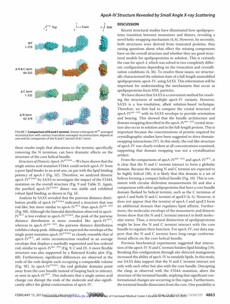
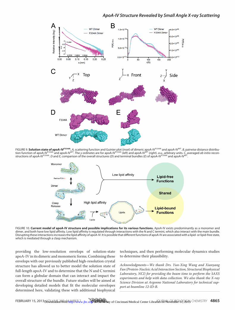
these results imply that alterations to the termini, specificallyremoving the N terminus, can have dramatic effects on thestructure of the core helical bundle.Structure ofDimericApoA-IVF334A—Wehave shown that the
single amino acid mutation F334A could switch apoA-IV froma poor lipid binder to an avid one, on par with the lipid bindingpotency of apoA-I (Fig. 5E). Therefore, we analyzed dimericapoA-IVF334A by SAXS to investigate the impact of the F334Amutation on the overall structure (Fig. 9 and Table 2). Again,the purified apoA-IVF334A dimer was stable and exhibitedrobust lipid binding, as shown in Fig. 5E.Analysis by SAXS revealed that the pairwise distance distri-
bution profile of apoA-IVF334A indicated a structure that wasrod-like, but more similar to apoA-IVWT than apoA-IV64–335
(Fig. 9B). Although the bimodal distribution observed in apoA-IVWT is less evident in apoA-IVF334A, the peak of the pairwisedistance distribution is more rounded like apoA-IVWT,whereas in apoA-IV64–335, the pairwise distance distributionexhibits a sharp peak.Althoughwe expected the envelope of thesingle point mutation apoA-IVF334A to closely resemble that ofapoA-IVWT, ab initio reconstruction resulted in an averageenvelope that displays a markedly segmented and less orderedrod, similar to apoA-IV62–376 (Fig. 9, C and D). A more flexiblestructure was also supported by a flattened Kratky plot (Fig.8B). Furthermore, significant differences are observed at theends of the rods despite each occupying a comparable volume(Fig. 9E). In apoA-IVF334A, the end globular domains pointaway from the core bundle instead of looping back to interact,as seen in apoA-IVWT. This indicates that a single amino acidchange can disrupt the ends of the molecule and also signifi-cantly affect the global conformation of apoA-IV.
DISCUSSION
Recent structural studies have illuminated how apolipopro-teins transition between monomers and dimers, revealing anovel helix-swapping mechanism (4, 6). However, by necessity,both structures were derived from truncated proteins, thusraising questions about what effect the missing componentshave on the overall structure and whether they are good struc-tural models for apolipoproteins in solution. This is certainlythe case for apoA-I, which was solved in two completely differ-ent configurations depending on the truncation and crystalli-zation conditions (4, 36). To resolve these issues, we structur-ally characterized the solution state of a full-length unmodifiedapolipoprotein, apoA-IV, using SAXS. This information will beimportant for understanding the mechanisms that occur asapolipoproteins form HDL particles.Wehave shown that SAXS is a convenientmethod for resolv-
ing the structures of multiple apoA-IV variants. However,SAXS is a low-resolution, albeit solution-based technique.Therefore, we first had to compare the crystal structure ofapoA-IV64–335 with its SAXS envelope to provide orientationand bearing. This showed that the bundle architecture anddomain swapping described in the apoA-IV64–335 crystal struc-ture also occur in solution and in the full-length protein. This isimportant because the concentrations of protein required forcrystallographic studies have been suggested to drive domain-swapping interactions (37). In this study, the rod-like structureof apoA-IV was clearly evident at all concentrations examined,supporting that domain swapping was not a crystallizationartifact.From the comparisons of apoA-IV64–335 and apoA-IVWT, it
is clear that the N and C termini interact to form a globulardomain. Because the missing N and C termini are expected tobe highly helical (38), it is likely that this domain is a set ofhelices forming a compact helical bundle (Fig. 10). This is con-sistent with circular dichroism measurements (6) and also acomparison with other apolipoproteins that have a core bundledomain flanked by helical termini, such as the C terminus ofapoA-I and both N and C termini of apoE3 (4, 5). However, itdoes not appear that the termini of apoA-I and apoE3 forman additional domain that regulates lipid affinity. Further-more, the molecular envelops of the monomeric and dimericforms show that the N and C termini interact in both molec-ular states. Thus, a structural distinction of apolipoproteinsmight be how the N and C termini interact with the corebundle to regulate their function. For apoA-IV, our data sup-port that the N and C termini have long-range conforma-tional affects on the core helical bundle.Previous biochemical experiments suggested that interac-
tion of the apoA-IV N and C termini hinders lipid binding (19).Breaking this configuration through site-directed mutagenesisincreased the ability of apoA-IV to emulsify lipids. In this study,our SAXS data support that the N and C termini interact notonly with each other but also with the core bundle. Disruptingthe clasp, as observed with the F334A mutation, alters thestructure of the terminal bundle, implying that significant con-formational changes are occurring in this region. Furthermore,the terminal bundle dissociates from the core. One possibility is
FIGURE 7. Comparison of N and C termini. Shown is the apoA-IVWT averagedreconstruction with various truncation averaged reconstructions aligned atone end for comparison of the N and C termini (N & C-term).
ApoA-IV Structure Revealed by Small Angle X-ray Scattering
FEBRUARY 15, 2013 • VOLUME 288 • NUMBER 7 JOURNAL OF BIOLOGICAL CHEMISTRY 4863 at University of Cincinnati/Medical Center Libraries on November 11, 2013http://www.jbc.org/Downloaded from

that Phe-334 interacts directly with the bundle, and its muta-tion leads to a dissociation of the two domains. Alternatively,Phe-334 could be necessary for the termini to coalesce into aglobular structure. Our results show that the F334A mutationnot only disrupts the N/C-terminal interaction but also altershow the N/C-terminal bundle interacts with the rod. Withoutthis interaction, the core bundle is not as stable and becomessegmented. This implies that an increase in lipid bindingmightbe associatedwith amore fluid core bundle. A fluid core bundlewas also observed in the N-terminal truncation apoA-IV62–376,but this form remains a poor lipid binder due to deletion ofimportant lipid binding determinants (18). Taken together,these results fundamentally redefine the claspmechanism as aninteraction between the termini and themain bundle instead ofjust the termini and provide a structural explanation for how
the N and C termini can impact lipid binding by altering theoverall bundle architecture.Because apoA-IV exhibits a number of unique biological
functions that are not observed with other apolipoproteins, it isintriguing to speculate that these are related to the clasp mech-anism. One possibility is that certain biological activities ofapoA-IV require a lipid-free state, and therefore, a mechanismis needed to prevent constitutive lipid binding. This could leadto lipid-free functions of apoA-IV (Fig. 10). Disruption of theclasp, possibly through protein or ligand interactions, couldconvert apoA-IV into an avid lipid binder and facilitate lipid-bound functions of apoA-IV (Fig. 10). Future work will need toassociate the functions of apoA-IV with its lipid state.In summary, we have further defined the conformational
starting point of apoA-IV, before it transitions into an HDL, by
FIGURE 8. Conformational affects of N/C termini. A, comparison of the overall averaged envelopes of apoA-IV64 –335, apoA-IV1–335, and apoA-IV62–376 withapoA-IVWT. The envelopes were superimposed by aligning the central helical bundle. N & C-term, N and C termini. B, Kratky plot for each set of SAXS data thatwas used for ab initio reconstructions.
ApoA-IV Structure Revealed by Small Angle X-ray Scattering
4864 JOURNAL OF BIOLOGICAL CHEMISTRY VOLUME 288 • NUMBER 7 • FEBRUARY 15, 2013 at University of Cincinnati/Medical Center Libraries on November 11, 2013http://www.jbc.org/Downloaded from
providing the low-resolution envelops of solution-stateapoA-IV in its dimeric andmonomeric forms.Combining theseenvelops with our previously published high-resolution crystalstructure has allowed us to better model the solution state offull-length apoA-IV and to determine that the N and C terminican form a globular domain that can interact and impact theoverall structure of the bundle. Future studies will be aimed atdeveloping detailed models that fit the molecular envelopesdetermined here, validating these with additional biophysical
techniques, and then performing molecular dynamics studiesto determine their plausibility.
Acknowledgments—We thank Drs. Yun-Xing Wang and XianyangFan (Protein-NucleicAcid Interaction Section, Structural BiophysicalLaboratory, NCI) for providing the beam time to perform the SAXSexperiments and help with data collection. We also thank the X-rayScience Division at Argonne National Laboratory for technical sup-port at beamline 12-ID-B.
FIGURE 9. Solution state of apoA-IVF334A. A, scattering function and Guinier plot (inset) of dimeric apoA-IVF334A and apoA-IVWT. B, pairwise distance distribu-tion function of apoA-IVF334A and apoA-IVWT. The y ordinates are for apoA-IVF334A (left) and apoA-IVWT (right). a.u., arbitrary units. C, averaged ab initio recon-structions of apoA-IVF334A. D and E, comparison of the overall structures (D) and terminal bundles (E) of apoA-IVF334A and apoA-IVWT.
FIGURE 10. Current model of apoA-IV structure and possible implications for its various functions. ApoA-IV exists predominantly as a monomer anddimer, and both have low lipid affinity. Low lipid affinity is regulated through interactions with the N and C termini, which also interact with the main bundle.Disrupting these interactions increases the lipid affinity of apoA-IV. It is possible that different functions of apoA-IV are associated with a lipid- or lipid-free state,which is mediated through a clasp mechanism.
ApoA-IV Structure Revealed by Small Angle X-ray Scattering
FEBRUARY 15, 2013 • VOLUME 288 • NUMBER 7 JOURNAL OF BIOLOGICAL CHEMISTRY 4865 at University of Cincinnati/Medical Center Libraries on November 11, 2013http://www.jbc.org/Downloaded from

REFERENCES1. Lund-Katz, S., and Phillips, M. C. (2010) High density lipoprotein struc-
ture-function and role in reverse cholesterol transport. Subcell. Biochem.51, 183–227
2. Sorci-Thomas, M. G., and Thomas, M. J. (2012) High density lipoproteinbiogenesis, cholesterol efflux, and immune cell function. Arterioscler.Thromb. Vasc. Biol. 32, 2561–2565
3. Davidson, W. S., Hazlett, T., Mantulin, W. W., and Jonas, A. (1996) Therole of apolipoprotein A-I domains in lipid binding. Proc. Natl. Acad. Sci.U.S.A. 93, 13605–13610
4. Mei, X., andAtkinson, D. (2011) Crystal structure of C-terminal truncatedapolipoprotein A-I reveals the assembly of high density lipoprotein (HDL)by dimerization. J. Biol. Chem. 286, 38570–38582
5. Chen, J., Li, Q., andWang, J. (2011) Topology of human apolipoprotein E3uniquely regulates its diverse biological functions. Proc. Natl. Acad. Sci.U.S.A. 108, 14813–14818
6. Deng, X., Morris, J., Dressmen, J., Tubb, M. R., Tso, P., Jerome, W. G.,Davidson, W. S., and Thompson, T. B. (2012) The structure of dimericapolipoprotein A-IV and its mechanism of self-association. Structure 20,767–779
7. Tabet, F., and Rye, K.-A. (2009) High-density lipoproteins, inflammationand oxidative stress. Clinical Science 116, 87–98
8. Tso, P., Sun, W., and Liu, M. (2004) Gastrointestinal satiety signals. IV.Apolipoprotein A-IV. Am. J. Physiol. Gastrointest. Liver Physiol. 286,G885–G890
9. Weinberg, R. B., Gallagher, J. W., Fabritius, M. A., and Shelness, G. S.(2012) ApoA-IV modulates the secretory trafficking of apoB and the sizeof triglyceride-rich lipoproteins. J. Lipid Res. 53, 736–743
10. Tso, P., and Liu, M. (2004) Apolipoprotein A-IV, food intake, and obesity.Physiol. Behav. 83, 631–643
11. Tso, P., Liu, M., and Kalogeris, T. (1999) The role of apolipoprotein A-IVin food intake regulation. J. Nutr. 129, 1503–1506
12. Qin, X., Swertfeger, D. K., Zheng, S., Hui, D. Y., and Tso, P. (1998) Apoli-poprotein A-IV: a potent endogenous inhibitor of lipid oxidation. Am. J.Physiol. 274, H1836–H1840
13. Vowinkel, T., Mori, M., Krieglstein, C. F., Russell, J., Saijo, F., Bharwani, S.,Turnage, R. H., Davidson, W. S., Tso, P., Granger, D. N., and Kalogeris,T. J. (2004) Apolipoprotein A-IV inhibits experimental colitis. J. Clin. In-vest. 114, 260–269
14. Wang, F., Kohan, A. B., Kindel, T. L., Corbin, K. L., Nunemaker, C. S.,Obici, S.,Woods, S. C., Davidson,W. S., andTso, P. (2012)ApolipoproteinA-IV improves glucose homeostasis by enhancing insulin secretion. Proc.Natl. Acad. Sci. U.S.A. 109, 9641–9646
15. Culnan, D. M., Cooney, R. N., Stanley, B., and Lynch, C. J. (2009) Apoli-poprotein A-IV, a putative satiety/antiatherogenic factor, rises after gas-tric bypass. Obesity 17, 46–52
16. Weinberg, R. B., and Spector, M. S. (1985) The self-association of humanapolipoprotein A-IV. Evidence for an in vivo circulating dimeric form.J. Biol. Chem. 260, 14279–14286
17. Pearson, K., Saito, H., Woods, S. C., Lund-Katz, S., Tso, P., Phillips, M. C.,and Davidson, W. S. (2004) Structure of human apolipoprotein A-IV: adistinct domain architecture among exchangeable apolipoproteins withpotential functional implications. Biochemistry 43, 10719–10729
18. Pearson, K., Tubb, M. R., Tanaka, M., Zhang, X. Q., Tso, P., Weinberg,R. B., and Davidson, W. S. (2005) Specific sequences in the N and Ctermini of apolipoprotein A-IVmodulate its conformation and lipid asso-
ciation. J. Biol. Chem. 280, 38576–3858219. Tubb,M. R., Silva, R. A., Pearson, K. J., Tso, P., Liu,M., andDavidson,W. S.
(2007) Modulation of apolipoprotein A-IV lipid binding by an interactionbetween the N and C termini. J. Biol. Chem. 282, 28385–28394
20. Schuck, P. (2000) Size-distribution analysis of macromolecules by sedi-mentation velocity ultracentrifugation and Lamm equation modeling.Biophys. J. 78, 1606–1619
21. Ilavsky, J., and Jemian, P. R. (2009) Irena: tool suite for modeling andanalysis of small-angle scattering. J. Appl. Crystallogr. 42, 347–353
22. Konarev, P. V., Volkov, V. V., Sokolova, A. V., Koch,M. H. J., and Svergun,D. I. (2003) PRIMUS: a Windows PC-based system for small-angle scat-tering data analysis. J. Appl. Crystallogr. 36, 1277–1282
23. Svergun, D. I. (1992) Determination of the regularization parameter inindirect-transformmethods using perceptual criteria. J. Appl. Crystallogr.25, 495–503
24. Svergun, D. I., Petoukhov,M. V., and Koch,M. H. J. (2001) Determinationof domain structure of proteins from x-ray solution scattering. Biophys. J.80, 2946–2953
25. Volkov, V. V., and Svergun, D. I. (2003) Uniqueness of ab initio shapedetermination in small-angle scattering. J. Appl. Crystallogr. 36, 860–864
26. Svergun, D., Barberato, C., and Koch,M. H. J. (1995) CRYSOL–a programto evaluate x-ray solution scattering of biological macromolecules fromatomic coordinates. J. Appl. Crystallogr. 28, 768–773
27. Wang, Z., and Schröder, G. F. (2012) Real-space refinement with DireX:from global fitting to side-chain improvements. Biopolymers 97, 687–697
28. Garai, K., and Frieden, C. (2010) The association-dissociation behavior ofthe ApoE proteins: kinetic and equilibrium studies. Biochemistry 49,9533–9541
29. Vitello, L. B., and Scanu, A. M. (1976) Studies on human serum highdensity lipoproteins. Self-association of apolipoprotein A-I in aqueoussolutions. J. Biol. Chem. 251, 1131–1136
30. Wong, Y. Q., Binger, K. J., Howlett, G. J., and Griffin, M. D. W. (2010)Methionine oxidation induces amyloid fibril formation by full-length apo-lipoprotein A-I. Proc. Natl. Acad. Sci. U.S.A. 107, 1977–1982
31. Deng, X., Davidson, W. S., and Thompson, T. B. (2012) Improving thediffraction of apoA-IV crystals through extreme dehydration. Acta Crys-tallogr. Sect. F Struct. Biol. Cryst. Commun. 68, 105–110
32. Schröder, G. F., Brunger, A. T., and Levitt, M. (2007) Combining efficientconformational sampling with a deformable elastic network model facili-tates structure refinement at low resolution. Structure 15, 1630–1641
33. Visiers, I., Braunheim, B. B., andWeinstein, H. (2000) Prokink: a protocolfor numerical evaluation of helix distortions by proline. Protein Eng. 13,603–606
34. Barlow, D. J., and Thornton, J. M. (1988) Helix geometry in proteins. J.Mol. Biol. 201, 601–619
35. Mertens, H. D. T., and Svergun, D. I. (2010) Structural characterization ofproteins and complexes using small-angle x-ray solution scattering. J.Struct. Biol. 172, 128–141
36. Borhani, D. W., Rogers, D. P., Engler, J. A., and Brouillette, C. G. (1997)Crystal structure of truncated human apolipoprotein A-I suggests a lipid-bound conformation. Proc. Natl. Acad. Sci. U.S.A. 94, 12291–12296
37. Liu, Y., and Eisenberg, D. (2002) 3D domain swapping: as domains con-tinue to swap. Protein Sci. 11, 1285–1299
38. Adamczak, R., Porollo, A., and Meller, J. (2005) Combining prediction ofsecondary structure and solvent accessibility in proteins. Proteins 59,467–475
ApoA-IV Structure Revealed by Small Angle X-ray Scattering
4866 JOURNAL OF BIOLOGICAL CHEMISTRY VOLUME 288 • NUMBER 7 • FEBRUARY 15, 2013 at University of Cincinnati/Medical Center Libraries on November 11, 2013http://www.jbc.org/Downloaded from