Embed Size (px)
Citation preview

rN
m
Neurobiology of Aging 33 (2012) 535–545
Protein oxidation inhibits NO-mediated signaling pathway forsynaptic plasticity
Sho Kakizawaa,b,*, Masahiko Shibazakia, Nozomu Moria,*a Department of Anatomy and Neurobiology, Graduate School of Biomedical Sciences, Nagasaki University, Nagasaki, Japan
b Department of Biological Chemistry, Graduate School of Pharmaceutical Sciences, Kyoto University, Kyoto, Japan
Received 15 December 2009; received in revised form 6 April 2010; accepted 17 April 2010
Abstract
Oxidative stress is a primary factor inducing brain dysfunction in aged animals. However, how oxidation affects brain function is not fullyunderstood. Here we show that oxidation inhibits signaling pathways essential for synaptic plasticities in the cerebellum. We first revealedthat nitric oxide (NO)-dependent plasticities at the parallel fiber-Purkinje cell synapse (PF synapse) were impaired in the cerebellar slicesfrom aged mice, suggesting a possible inhibitory action of protein oxidation by endogenous reactive oxygen species. PF-synaptic plasticitieswere also blocked in the cerebellar slices from young mice preincubated with oxidizing agents or thiol blocker. Because the treatment ofthe slices with the oxidizing agent did not affect basic electrophysiological properties of excitatory postsynaptic current of PF (PF-EPSC)and did not occlude the synaptic plasticities, oxidation was revealed to specifically inhibit signaling pathways essential for PF-synapticplasticities. Finally, biochemical analysis confirmed the idea that inhibitory action of protein oxidation on the PF-synaptic plasticities wasmediated by impairment of nitric oxide-induced protein S-nitrosylation. Therefore, oxidation was revealed to inhibit the S-nitrosylation-dependent signaling pathway essential for synaptic plasticity in a “competitive” manner.© 2012 Elsevier Inc. All rights reserved.
Keywords: Aging; S-nitrosylation; Oxidative agent; Posttetanic potentiation; Long term potentiation; Cerebellum; Purkinje cell; Parallel fiber
www.elsevier.com/locate/neuaging
1. Introduction
Aging is associated with a general decline in physiolog-ical function in biological systems including the nervoussystem (Barnes, 1988, 2003; Finch, 2003; Landfield, 1988;Mattson and Magnus, 2006). With the passage of time, theredox environment of the brain can be altered in favor ofoxidation by an increased production of reactive oxygenspecies (ROS) or by a decreased activity of antioxidantdefenses. This condition, known as oxidative stress or oxi-dative damage, is thought to be a general contributing factorto aging in the nervous systems (Beckman and Ames, 1998;Finkel and Holbrook, 2000; Harman, 1956). Actually, nu-merous studies have demonstrated various correlations be-
* Corresponding authors at: Department of Biological Chemistry, GraduateSchool of Pharmaceutical Sciences, Kyoto University, Kyoto 606-8501 Japan.Tel.: �81 75 753 4552 (S. Kakizawa) or Department of Anatomy and Neu-obiology, Graduate School of Biomedical Sciences, Nagasaki University,agasaki 852-8523, Japan. Tel.: �81 95 819 7019 (N. Mori).
E-mail addresses: [email protected] (S. Kakizawa) or
[email protected] (N. Mori).0197-4580/$ – see front matter © 2012 Elsevier Inc. All rights reserved.doi:10.1016/j.neurobiolaging.2010.04.016
tween age and the accumulation of oxidative damage tocellular macromolecules (Floyd and Hensley, 2002; Stadt-man, 2001). For example, enhanced lipid peroxidation(Calabrese et al., 2004; Devi and Kiran, 2004; Gupta et al.,1991; Murray and Lynch, 1998; O’Donnell and Lynch,1998) and protein oxidation (Cini and Moretti, 1995; For-ster et al., 1996; Sohal et al., 1994; Sultana et al., 2009;Vaishnav et al., 2007) are observed in the brains of agedrodents. Furthermore, several studies have shown that be-havioral deficits of aged animals are associated with in-creases in oxidative stress (Butterfield et al., 2006; Cantuti-Castelvetri et al., 2000; Carney et al., 1991; Forster et al.,1996; Fukui et al., 2001). Although these associations be-tween oxidative damage and brain dysfunction cannot es-tablish a causal link between the 2, they do support the ideathat oxidation is involved in age-related brain dysfunction(Droge and Schipper, 2007; Serrano and Klann, 2004).
One type of cellular process strongly affected by oxida-tion is synaptic plasticity, a cellular process proposed as abiological substrate for learning and memory (Bliss and
Collingridge, 1993; Ito, 2001; Lynch, 2004; Malenka and
witswT
t
sdmsrIa
536 S. Kakizawa et al. / Neurobiology of Aging 33 (2012) 535–545
Nicoll, 1999). However, examination of the effects of oxi-dation on synaptic plasticity in studies where ROS (e.g.,hydrogen peroxide) was applied exogenously to hippocam-pal slices resulted in paradoxical effects (Klann and Thiels,1999; Serrano and Klann, 2004). Some studies suggest thatROS are essential for long term potentiation (LTP) in hip-pocampal slices (Kamsler and Segal, 2003a; Knapp andKlann, 2002), whereas inhibitory effects of ROS are re-ported in other studies (Auerbach and Segal, 1997; Kamslerand Segal, 2003a, 2003b; Pellmar et al., 1991; Watson et al.,2002). Different protocols were used for induction of syn-aptic plasticity and species or age of animals were differentamong these studies, and target signaling pathways of oxi-dation during the induction of synaptic plasticity were un-clear in these experiments. Therefore, the identification ofthe target signaling systems of oxidation in synaptic plas-ticity would provide critical insight concerning how oxida-tive stress results in deficits in synaptic plasticity and brainfunction in aged animals.
Parallel fiber-Purkinje cell synapse (PF synapse) in thecerebellum is a good model for examining molecular mecha-nisms in synaptic plasticity in the central nervous system (Han-sel et al., 2001; Ito, 2006). Purkinje cells (PCs), solely outputfrom the cerebellar cortex, receive 2 types of excitatory inputs:climbing fiber (CF) from inferior olive and PF, the axon ofgranular cells in the cerebellar cortex. Various types of synap-tic plasticity were reported to date at the PF synapse (Evans,2007; Ito, 2006; Jorntell and Hansel, 2006). Among them, thesynaptic potentiation induced by repetitive activity of PF areknown to be dependent on nitric oxide (NO)-mediated signal-ing pathways (Kakegawa and Yuzaki, 2005; Lev-Ram et al.,2002; Namiki et al., 2005). Nitric oxide exerts its effects via 2pathways. One pathway is mediated by soluble guanylyl cy-clase (sGC). Activation of sGC induces increased cytosoliccyclic guanosine monophosphate (GMP) level and activatesprotein kinase G. Another pathway is mediated by S-nitrosy-lation of cysteine residues in various proteins. S-nitrosylationof proteins resulted in modification of function of proteinsincluding ion channels and enzymes (Calabrese et al., 2007;Hess et al., 2005; Jaffrey et al., 2001; Nakamura and Lipton,2007). Because the potentiation of the PF synapse is dependenton NO but not sensitive to 1H-[1,2,4] Oxadiazolo[4,3-a] qui-noxaline-1-1 (ODQ), a specific antagonist for sGC, S-nitrosy-lation-mediated pathway is indicated to be involved in thepotentiation (Lev-Ram et al., 2002; Namiki et al., 2005). Ox-idizing agents including endogenous ROS also modify cys-teine residues and exert its action on proteins (Ansari et al.,2006; Forman et al., 2008; Mikkelsen and Wardman, 2003;Suzuki et al., 2010). Therefore, NO and oxidizing agents com-petitively share cysteine residues to exert their action, and it ishighly possible that oxidizing agents affect S-nitrosylation-mediated signaling pathways essential for the potentiation ofthe PF synapse and impair or occlude the potentiation.
In the present study, we first demonstrated that postte-
tanic potentiation (PTP) and LTP at the PF synapse were hseverely impaired in the cerebellar slices from aged (20- to24-month-old) mice, suggesting involvement of endoge-nous ROS in age-dependent decline in PF-PTP and PF-LTP.This hypothesis was confirmed by subsequent experimentsdemonstrating application of oxidizing agents also inhibitedthe induction of PF-PTP and PF-LTP in the cerebellar slicesfrom young (4–6 weeks old) mice. Because both PF-PTPand PF-LTP is suggested to be dependent on S-nitrosyla-tion, we propose that oxidation by ROS induces its inhibi-tory effects on synaptic plasticity via modification of cys-teine residues whose S-nitrosylation by acute NO signal isessential for the induction of the plasticities, at least at PFsynapses in the cerebellum.
2. Methods
2.1. Slice preparation
All experiments were carried out according to the guide-lines established by the Animal Welfare Committee of Na-gasaki University.
Wild-type C57BL/6 mice at 4 to 6 weeks or 20 to 24months of age were sacrificed by cervical dislocation underdeep anesthesia with diethyl ether. The cerebellum wasexcised, and parasagittal cerebellar slices (250 �m thick)
ere prepared from the vermis (Edwards et al., 1989; Kak-zawa et al., 2000, 2005). Whole-cell recordings were ob-ained from visually identified PCs under an upright micro-cope (BX51WI, Olympus, Tokyo, Japan) using a 40�ater-immersion objective at room temperature (23–25 °C).he resistances of patch pipettes were 2.0–3.5 M� when
filled with an intracellular solution composed of (in mM),130 K-gluconate, 10 KCl, 10 NaCl, 1 ethylene glycol-bis(2-aminoethylether)-N,N,N=,N=-tetraacetic acid (EGTA), 4adenosine triphosphate (ATP)-Mg, 0.4 guanosine triphos-phate (GTP)-Na and 10 4-(2-hydroxyethyl)-1-pipera-zineethanesulfonic acid (HEPES) (pH 7.3; adjusted withpotassium hydroxide [KOH]). The standard bathing solu-tion was composed of (in mM) 125 NaCl, 2.5 KCl, 2 CaCl2,1 MgSO4, 1.25 NaH2PO4, 26 NaHCO3, and 20 glucose,bubbled with 95% CO2 and 5% CO2. An antagonist for theype-A �-aminobutyric acid receptor (GABAA receptor),
bicuculline (10 �M), was always added to block inhibitorypostsynaptic currents.
2.2. Electrophysiology
For the focal stimulation of PF, a stimulation pipette(5–10 �m tip diameter) was filled with the standard bathingolution and used in applying square pulses (0.1 ms inuration, 0–20 V in amplitude) in the molecular layer at theiddle 1-third from the pial surface. The intensity of each
timulus was adjusted to evoke excitatory postsynaptic cur-ents of PF (PF-EPSCs) with amplitudes of 70–150 pA.onic current was recorded from PCs with a patch-clampmplifier (EPC-9, HEKA, Lambrecht/Pfalz, Germany) at a
olding potential of �90 mV or �80 mV, after the com-
mT
lep[f1Ht
a�
nHra
ao
3
3s
ipw(IEtltHpptpihaphLiewB1i
537S. Kakizawa et al. / Neurobiology of Aging 33 (2012) 535–545
pensation of liquid junction potential. The signals werefiltered at 2 kHz and digitized at 20 kHz. Online dataacquisition and offline data analysis were performed usingPULSE (HEKA) software. Synaptic potentiation was in-duced by repetitive burst stimulation (BS) to PF (60 BS at1 Hz; each BS consists of 5 pulses at 50 Hz) after theacquisition of baseline responses (Kakizawa et al., 2007;Namiki et al., 2005). Test stimulus was applied to PFs every10 seconds. The amplitude of PF-EPSC was averaged every60 seconds, and normalized to the mean value observed for10 minutes before the BSs. A 100-ms, �5 mV hyperpolar-izing test pulse preceded each PF stimulus to monitor theseries resistance and input resistance of PCs throughout theexperiment, the data of which were discarded if the resis-tance changed by more than 10% (Kakizawa et al., 2007;Namiki et al., 2005). The data were also discarded when theslope of PF-EPSC amplitude averaged every minute duringthe initial recording for 10 minutes was larger than 2% orwhen the amplitude did not become stable within 20 min-utes after the onset of whole-cell configuration (Kakizawa etal., 2007; Namiki et al., 2005).
2.3. Application of oxidizing agents and thiol blocker
To examine the effects of preincubation of cerebellarslices with oxidizing agents on PF-synaptic plasticities, theslices were transferred to another incubation chamber, andsolution containing hydrogen peroxide (H2O2) or chlora-
ine-T (ChT) was directly added to the incubation media.he final concentration of H2O2 and ChT in the chamber
were 1–100 �M and 0.1–10 �M, respectively. After the10-minute incubation with H2O2 or ChT, the slices weretransferred back to the original incubation chamber and keptfor more than 1 hour until the electrophysiological analysis.In another experiment, cerebellar slices were preincubatedwith 50 �M N-ethyl-maleimide (NEM) for 5 minutes. Toexamine the acute effects of the oxidizing agent on theamplitude of PF-EPSC, extracellular bathing solution wasreplaced with those containing 100 �M H2O2 for 10 min-utes. Flow rate of the perfusion system was 0.67 mL/minute.
2.4. Biotin-switch assay for protein S-nitrosylation
Effects of aging and oxidizing agents on NO-inducedprotein S-nitrosylation in the cerebellar slices were exam-ined by biotin-switch assay, the procedure being describedby Forrester et al. (2007). This assay was performed in thedark. Cerebellar slices (about 100 �g) treated with or with-out NO donor, NOC7, were homogenized with 200 �L ofysis buffer (25 mM HEPES pH 7.7, 50 mM NaCl, 0.1 mMthylenediamine-tetraacetic acid (EDTA), 0.1 mM neocu-roine, 0.1% NP-40, 1 mM phenylmethanesulfonyl fluoridePMSF]). After centrifugation, 100 �L of lysates were usedor the biotin-switch assay. Free cysteines were blocked for
hour at 50 °C in 2 volumes of HEN buffer (250 mMEPES, 1 mM EDTA, 0.1 mM neocuproine, pH 7.7) con-
aining 2.5% sodium dodecyl sulfate (SDS) and 0.1% meth- a
nethiosulfonate. Proteins were acetone precipitated at20 °C and resuspended in 240 �L of HEN buffer con-
taining 1% SDS. After adding fresh ascorbic acid (finalconcentration: 100 mM) and 60 �g of EZ-link N-[6-(bioti-amido)hexyl]-3=-(2=-pyridyldithio)propionamide (biotin-PDP) (Pierce, Rockford, IL), proteins were incubated at
oom temperature for 1 hour. Subsequently, proteins werecetone precipitated again and resuspended in 100 �L of
0.1� HEN buffer containing 1% SDS. Then 100 �l of2�SDS-polyacrylamide gel electrophoresis (PAGE) load-ing buffer (0.1 M Tris-HCl pH 6.8, 4% SDS, 20% glycerol)were added and 20 �l were used for 10% SDS-PAGEseparation. After blotting onto Hybond-P membrane (Amer-sham, Amersham, UK), biotinylated proteins were detectedwith streptavidin-horseradish peroxidase (HRP) (Amersham).Monoclonal antibody TUB2.1 (Sigma-Aldrich, Tokyo, Japan)was used for �-tubulin detection. Protein S-nitrosylation levelswere indicated as values after calibration with �-tubulin levelsnd normalization with the value in vehicle-treated slices with-ut NOC7 application.
. Results
.1. PF-PTP and PF-LTP are impaired in the cerebellarlices from aged mice
We first examined whether PF-synaptic potentiation wasmpaired in cerebellar slices from aged mice. Excitatoryostsynaptic currents (EPSCs) evoked by PF stimulationere recorded from PCs in the cerebellar slices from young
1-month-old) or aged (20- to 24-month-old) mice (Fig 1A).n the cerebellar slices from young mice, the amplitude ofPSCs at the PF synapse (PF-EPSCs) was markedly poten-
iated and maintained stable at about 200% of the prestimu-ation level for at least 30 minutes after repetitive applica-ion of 60 burst stimulations (BSs, each BS � 5 pulses at 50z) at 1 Hz (Fig. 1B, open circles), as is observed in ourrevious study (Namiki et al., 2005). The early phase of theotentiation (1–2 minutes after the 60 BSs; posttetanic po-entiation [PTP]) was accompanied with a decrease inaired-pulse ratio (PPR) of PF-EPSC (Fig. 1C). Because annverse relationship between release probability and PPRad been observed in various synapses including PF syn-pses (Zucker and Regehr, 2002), the PF-PTP was at leastartly dependent on presynaptic enhancement. On the otherand, the late phase of the potentiation (21–30 after 60 BSs;TP) was not accompanied with changes in PPR, and was
ndicated to be postsynaptic changes (Fig. 1C). In the cer-bellar slices from aged mice, the amplitude of PF-EPSCas not markedly changed after repetitive application of 60Ss, except for a slight increase shortly after the BSs (Fig.B, closed circles). The averaged PF-EPSC amplitudes dur-ng the 1- to 2-minute period and 21- to 30-minute period
fter the 60 BSs were 135.6% � 12.5% and 104.5% � 0.6%
1t
ll values
538 S. Kakizawa et al. / Neurobiology of Aging 33 (2012) 535–545
of the prestimulation levels, respectively, and these valueswere significantly lower than those in the young group(231.3% � 22.6% and 199.3% � 7.4%, respectively) (Fig.D and E). Thus, both PF-PTP and PF-LTP were revealed
Fig. 1. Inhibition of parallel fiber (PF)-Purkinje cell synaptic plasticity in age(PC) in the cerebellar slice. Rec, recording pipette; Stim, stimulation pipette. (60 burst stimulations (BSs) repetitively applied at 1 Hz in the cerebellar slicnormalized by the mean value observed for 10 minutes before the BSs. (C) Paired-pin [B]), but not during the 21- to 30-minute period after BSs (dark-gray shadow invalues (right) before BSs (a) and 1 to 2 minutes (b) and 21 to 30 minutes (c) after BBSs. (D) PF-PTP was impaired in aged mice. Typical traces (left) and averagemice before (a) and 1 to 2 minutes after (b) the BSs. **p � 0.01, significantTypical traces (left) and average amplitude (right) of PF-EPSC recorded fromBSs. ***p � 0.001, significantly different from the value in young group. A
o be severely impaired in aged mice.
3.2. PF-PTP and PF-LTP are impaired in cerebellarslices from young mice pretreated with oxidative reagents
Our previous study demonstrated that both PF-PTP and
(A) Schematic image of electrophysiological recording from a Purkinje celltetanic potentiation (PTP) and long term potentiation (LTP) were induced byyoung mice (n � 6), but not from aged mice (n � 5). The amplitude was
o (PPR) was changed during the 1- to 2-minute period after BSs (light gray shadowe cerebellar slices from young mice. Representative traces (left) and average PPR
pulse interval was 100 Ms; *p � 0.05, significantly different from the value beforeude (right) of PF-EPSC recorded from cerebellar slices from young and agedent from the value in young group. (E) PF-LTP was impaired in aged mice.lar slices from young and aged mice before (a) and 21 to 30 minutes after (c)
are expressed as mean � standard error of the mean (SEM).
d mice.B) Postes fromulse rati[B]) in thSs. Inter
amplitly differcerebel
PF-LTP were dependent on NO signals, but not on sGC

at5stght
3p
oafPdeisc
(ea
w
gaavvo
539S. Kakizawa et al. / Neurobiology of Aging 33 (2012) 535–545
activity (Namiki et al., 2005). These facts indicated thatPF-PTP and PF-LTP are dependent on the signals mediatedby S-nitrosylation of cysteine residues. ROS also modifiescysteine residues and exerts its action on cellular proteins. Ithas long been known that protein oxidation is enhanced inthe brains of aged rodents (Cini and Moretti, 1995; Forsteret al., 1996; Sohal et al., 1994; Vaishnav et al., 2007). Thus,it is highly possible that impairment of PF-PTP andPF-LTP in the aged cerebellum could be due to theblockade of protein S-nitrosylation by preceding proteinoxidization by endogenous ROS. To test this hypothesis,effects of protein oxidation in the cerebellar slices fromyoung mice with oxidizing agents on PF-PTP and PF-LTP were examined.
We first incubated acute cerebellar slices from young micewith H2O2, a typical oxidizing agent which is known to inducefunctional changes in ionic channels (Annunziato et al., 2002;Cai and Sesti, 2009). In the cerebellar slices preincubated withH2O2, PF-PTP and PF-LTP were impaired in a dose-dependentmanner (Supplementary Fig. 1). In the cerebellar slices prein-cubated with 100 �M H2O2, the average amplitude PF-EPSCduring the 1- to 2-minute period and the 21- to 30-minuteperiod after the 60 BSs were 132.6% � 11.0% and 104.4% �3.0% of the prestimulation levels, respectively, and these val-ues were significantly lower than those in the vehicle-treatedcontrol group (237.0% � 20.7% and 193.7% � 7.4%, respec-tively) (Fig. 2).
To confirm the involvement of oxidation in impairmentof the plasticities of PF synapses, we incubated the cerebel-lar slices from young mice with another type of oxidizingagent, ChT. Both PF-PTP and PF-LTP were blocked afterthe preincubation of the cerebellar slices with ChT in adose-dependent manner, too (Supplementary Fig. 2). Whenthe slices were pretreated with 10 �M ChT, the averagemplitude PF-EPSC during the 1- to 2-minute period andhe 21- to 30-minute period after the 60 BSs were 101.6% �.1% and 101.8% � 1.0% of the prestimulation levels, re-pectively, and these values were significantly lower thanhose in vehicle-treated control group (Fig. 2). Taken to-ether, oxidation was revealed to be a factor exerting in-ibitory effects on the induction of PF-PTP and PF-LTP inhe cerebellar slices.
.3. Hydrogen peroxide specifically inhibited signalingathways essential for PF-synaptic plasticity
The results shown in Figs. 1–3 indicate that proteinxidation by endogenous ROS (in the cerebellar slices fromged mice) or oxidative reagents (in the cerebellar slicesrom young mice) impaired the induction of PF-PTP andF-LTP possibly through inhibition of S-nitrosylation-me-iated signals essential for the induction of plasticity. How-ver, it is still possible that protein oxidation indirectlynhibited these synaptic plasticities through impairment ofynaptic function itself, because functional losses of ionic
hannels by oxidation had been reported in previous studies WAnnunziato et al., 2002). To exclude these possibilities, wexamined whether oxidation by the oxidizing agent (H2O2)ffects basic electrophysiological properties of PF synapses.
Fig. 2. Impairment of parallel fiber (PF)-Purkinje cell synaptic plasticity byoxidizing agents. (A) Posttetanic potentiation (PTP) and long term poten-tiation (LTP) were induced by 60 burst stimulations (BSs) repetitivelyapplied at 1 Hz in the cerebellar slices preincubated with vehicle (control[cont], open circle; n � 6), but not in the cerebellar slices preincubated
ith 100 �M H2O2 (closed black circle; n � 5) or 10 �M chloramine-T(ChT) (closed gray circle; n � 5). The amplitude was normalized by themean value observed for 10 minutes before the BSs. (B) PF-PTP wasimpaired by oxidizing agents. Typical traces (left) and average amplitude(right) of PF-EPSC before (a) and 1–2 minutes after (b) the BSs. Thecerebellar slices from young mice were treated with vehicle, H2O2 or ChT.**p � 0.01, ***p � 0.001, significantly different from the value in controlroup. (C) PF-LTP was impaired by oxidizing agents. Typical traces (left)nd average amplitude (right) of PF-EPSC before (a) and 21–30 minutesfter (c) the BSs. The cerebellar slices from young mice were treated withehicle or H2O2 or ChT. ***p � 0.001, significantly different from thealue in control group. All values are expressed as mean � standard errorf the mean (SEM).
e first examined effects of H2O2 treatment of the cerebel-

t
i
i
aP
Hv
aPm
540 S. Kakizawa et al. / Neurobiology of Aging 33 (2012) 535–545
lar slices from young mice on PPR. The ratio in the H2O2-treated group was not significantly different than those inthe vehicle-treated control group in all intervals examined(10–300 ms) (Fig. 3A and B). The input-output relation-ships of the amplitudes of PF-EPSCs were not significantlydifferent between H2O2-treated and vehicle-treated groups,oo (Fig. 3C and D).
In some experiments, applications of oxidative reagents
Fig. 3. Basic electrophysiological properties of parallel fiber-Purkinje cellsynaptic-excitatory postsynaptic current (PF-EPSC) are unaffected byH2O2. (A) Paired-pulse ratio of PF-EPSC in H2O2-treated group (n � 9) wasnot significantly different than those in vehicle-treated (cont) group (n �10). (B) Typical current responses of PF synapse to paired-pulse stimula-tion (interpulse interval � 50 ms) from cerebellar slices preincubated withvehicle and H2O2. (C) Input-output relation in H2O2-treated group (n � 5)was not significantly different than those in vehicle-treated group (n � 8).First regression lines of vehicle- and H2O2-treated groups are shown asblack and blue lines, respectively. (D) Typical current responses of PFsynapse evoked by stimulation with increasing intensities from 1 to 10 �Ain vehicle- and H2O2-treated groups. All values are expressed as mean �standard error of the mean (SEM).
nduce synaptic plasticity (Knapp and Klann, 2002). Thus, it v
s still possible that treatment with H2O2 induced synapticpotentiation during the incubation period, and occludedPF-PTP and PF-LTP induced by PF stimulation (60 BSs).Our results excluded this possibility because the amplitudesof PF-EPSCs during and 1–30 minutes after the applicationof H2O2 were not significantly different than those beforepplication of the reagent (Fig. 4). The average amplitudeF-EPSC during the 7- to 9-minute period and 30- to 39-
Fig. 4. Application of H2O2 does not induce potentiation at the parallelfiber (PF)-Purkinje cell synapse. (A) The amplitude of PF-EPSC before,during (shadow), and after bath application of vehicle (cont; n � 5) or
2O2 (n � 4) for 10 minutes. The amplitude was normalized by the meanalue before H2O2 application. (B) Typical traces (left) and average am-
plitude (right) of PF-EPSC recorded from cerebellar slices before (1, blacklines) and 7–9 minutes after (2) the onset of vehicle (blue) or H2O2 (red)pplication. (C) Typical traces (left) and average amplitude (right) ofF-EPSC recorded from cerebellar slices before (1, black lines) and 30–39inutes after (3) the onset of vehicle (blue) or H O (red) application. All
2 2alues are expressed as mean � standard error of the mean (SEM).
1(
iPtocsd
3P
stPopNttabi
ecPm
w(av
541S. Kakizawa et al. / Neurobiology of Aging 33 (2012) 535–545
minute period after the onset of H2O2 application were01.4% � 0.8% and 100.7% � 1.0% of the baseline levelsFig. 4B and C). These results indicate that H2O2 applica-
tion itself induce neither PF-PTP nor PF-LTP, and does notocclude synaptic plasticities induced by PF activity.
Application of H2O2 did not affect basic electrophysiolog-cal properties of the PF synapse (Fig. 3) and did not occludeF-PTP and PF-LTP (Fig. 4). Therefore, these results exclude
he possibility that the impairments of PF-PTP and PF-LTP byxidizing agents were the secondary effects of oxidation in theerebellar slices, and therefore strongly indicate that oxidationpecifically inhibited signaling pathways essential for the in-uction of PF-PTP and PF-LTP.
.4. Thiol residues are essential regulatory sites forF-synaptic plasticities
As is described above, oxidation specifically impairedignaling pathways essential for the induction of the plas-icities of the PF synapse. Previous studies indicated that theF-synaptic potentiation induced by repetitive stimulationf PF were dependent on signaling pathways mediated byrotein S-nitrosylation by NO (Lev-Ram et al., 2002;amiki et al., 2005). Thiol groups in cysteine residue are the
arget of S-nitrosylation of proteins by NO as well as thearget of oxidation by endogenous ROS and oxidizinggents. Thus, it is highly possible that protein oxidationlocks the induction of PF-PTP and PF-LTP through thenhibition of protein S-nitrosylation in the cerebellar slices.
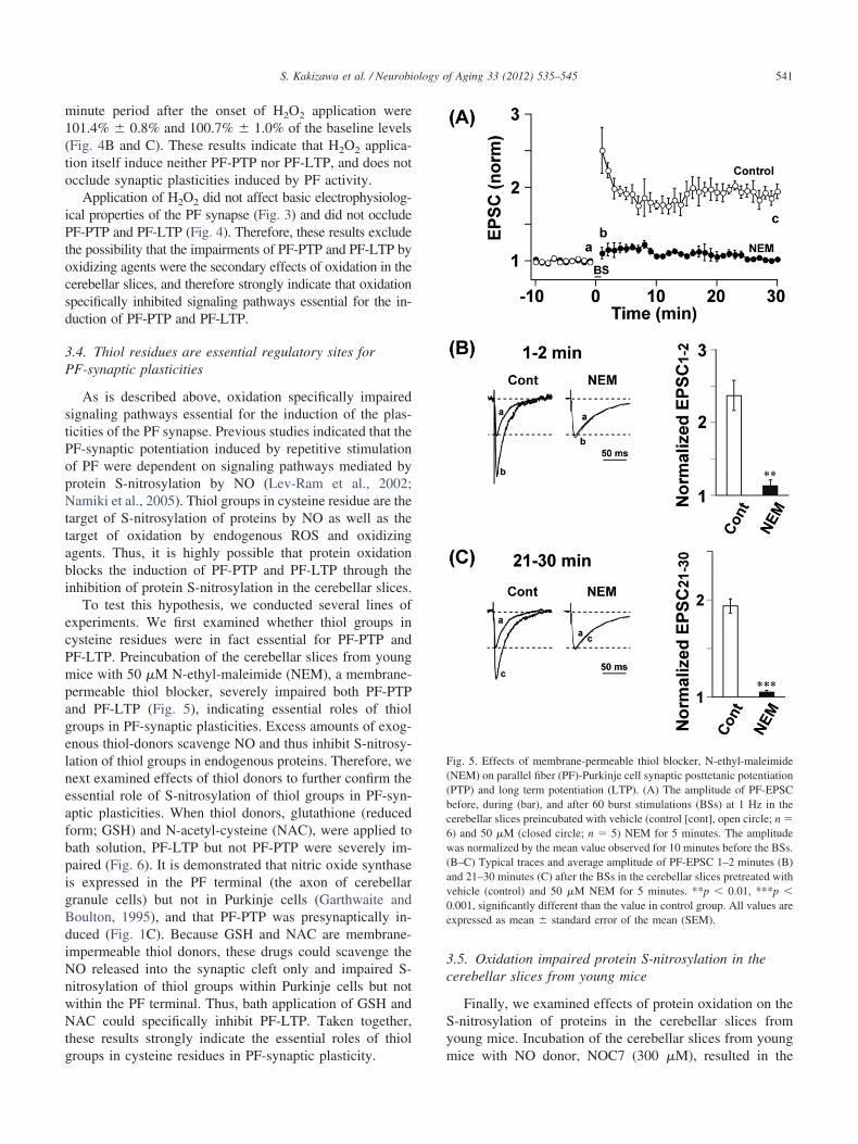
To test this hypothesis, we conducted several lines ofxperiments. We first examined whether thiol groups inysteine residues were in fact essential for PF-PTP andF-LTP. Preincubation of the cerebellar slices from youngice with 50 �M N-ethyl-maleimide (NEM), a membrane-
permeable thiol blocker, severely impaired both PF-PTPand PF-LTP (Fig. 5), indicating essential roles of thiolgroups in PF-synaptic plasticities. Excess amounts of exog-enous thiol-donors scavenge NO and thus inhibit S-nitrosy-lation of thiol groups in endogenous proteins. Therefore, wenext examined effects of thiol donors to further confirm theessential role of S-nitrosylation of thiol groups in PF-syn-aptic plasticities. When thiol donors, glutathione (reducedform; GSH) and N-acetyl-cysteine (NAC), were applied tobath solution, PF-LTP but not PF-PTP were severely im-paired (Fig. 6). It is demonstrated that nitric oxide synthaseis expressed in the PF terminal (the axon of cerebellargranule cells) but not in Purkinje cells (Garthwaite andBoulton, 1995), and that PF-PTP was presynaptically in-duced (Fig. 1C). Because GSH and NAC are membrane-impermeable thiol donors, these drugs could scavenge theNO released into the synaptic cleft only and impaired S-nitrosylation of thiol groups within Purkinje cells but notwithin the PF terminal. Thus, bath application of GSH andNAC could specifically inhibit PF-LTP. Taken together,these results strongly indicate the essential roles of thiol
groups in cysteine residues in PF-synaptic plasticity.3.5. Oxidation impaired protein S-nitrosylation in thecerebellar slices from young mice
Finally, we examined effects of protein oxidation on theS-nitrosylation of proteins in the cerebellar slices fromyoung mice. Incubation of the cerebellar slices from young
Fig. 5. Effects of membrane-permeable thiol blocker, N-ethyl-maleimide(NEM) on parallel fiber (PF)-Purkinje cell synaptic posttetanic potentiation(PTP) and long term potentiation (LTP). (A) The amplitude of PF-EPSCbefore, during (bar), and after 60 burst stimulations (BSs) at 1 Hz in thecerebellar slices preincubated with vehicle (control [cont], open circle; n �6) and 50 �M (closed circle; n � 5) NEM for 5 minutes. The amplitude
as normalized by the mean value observed for 10 minutes before the BSs.B–C) Typical traces and average amplitude of PF-EPSC 1–2 minutes (B)nd 21–30 minutes (C) after the BSs in the cerebellar slices pretreated withehicle (control) and 50 �M NEM for 5 minutes. **p � 0.01, ***p �
0.001, significantly different than the value in control group. All values areexpressed as mean � standard error of the mean (SEM).
mice with NO donor, NOC7 (300 �M), resulted in the

aid
cw(2v
542 S. Kakizawa et al. / Neurobiology of Aging 33 (2012) 535–545
elevation of protein S-nitrosylation levels (p � 0.001; com-pare lanes 1 and 2 in Fig. 7A). However, preincubation ofthe slices with H2O2 severely impaired the NO-inducedincrease in protein S-nitrosylation levels (p � 0.001; com-pare lanes 2 and 4 in Fig. 7A), although pretreatment withH2O2 itself did not affect the levels before the NO treatment(p � 0.05; compare lanes 1 and 3 in Fig. 7A). The normal-ized values of protein S-nitrosylation levels with and with-
Fig. 6. Effects of membrane-impermeable thiol donor, glutathione (reducedform; GSH) and N-acetyl-cysteine (NAC) on parallel fiber (PF)-Purkinjecell synaptic posttetanic potentiation (PTP) and long term potentiation(LTP). (A) The amplitude of PF-EPSC before, during (gray bar), and after60 burst stimulations (BSs) at 1 Hz in the cerebellar slices in the presenceof vehicle (control [cont], open circle; n � 6), 500 �M GSH (closed blackircle; n � 5), and 500 �M NAC (closed gray circle; n � 5). The amplitudeas normalized by the mean value observed for 10 minutes before the BSs.
B–C) Typical traces and average amplitude of PF-EPSC 1–2 minutes (B) and1–30 minutes (C) after the BSs in the cerebellar slices in the presence ofehicle, 500 �M GSH and 500 �M NAC. ***p � 0.001, significantly
different than the value in control group.
out NOC7 application were 2.42 � 0.03 and 1.00 � 0.01 in
the vehicle-pretreated control group (142% increase) and1.10 � 0.02 and 0.94 � 0.02 in the H2O2-pretreated group(16% increase). Nitric oxide-induced increase in proteinS-nitrosylation levels was severely impaired in the cerebel-lar slices from aged mice, when compared with younganimals. The normalized values of protein S-nitrosylationlevels with and without NOC7 application were 2.50 � 0.10and 1.00 � 0.07 in the slices from young mice (150%increase) and 1.52 � 0.06 and 1.05 � 0.01 in the slice fromged mice (47% increase) (Fig. 7B). These biochemical datan Fig. 5 correspond very well with the electrophysiologicalata in Figs. 1 and 2: PF-PTP and PF-LTP as well as
NO-induced increase in protein S-nitrosylation levels wereseverely impaired in the cerebellar slices from aged mice orin the cerebellar slices pretreated with oxidizing agents.Therefore, these results strongly indicate inhibitory actionof protein oxidation on the induction of PF-synaptic plas-ticities through the impairment of protein S-nitrosylation byacute NO signal.
4. Discussion
The strong correlation between increasing age and theaccumulation of oxidative damage has largely supported theoxidative stress hypothesis of aging (Droge and Schipper,2007; Serrano and Klann, 2004). In aged brain of rodents,protein oxidation is enhanced (Cini and Moretti, 1995; For-ster et al., 1996; Sohal et al., 1994; Vaishnav et al., 2007)and behavioral deficits of aged animals are associated withincreases in oxidative stress (Butterfield et al., 2006; Can-tuti-Castelvetri et al., 2000; Carney et al., 1991; Forster etal., 1996; Fukui et al., 2001). These observations stronglyindicate that oxidative stress is a primary factor inducingdeficits in synaptic plasticity and memory in aged animals.However, how oxidative stress results in the impairment ofsynaptic plasticity and brain function are not well under-stood. Previous studies indicate that ROS modulate activityof protein kinase C, extracellular signal-related kinase, pro-tein tyrosine kinases, protein phosphatase 2A, and calcineu-rin (Klann and Thiels, 1999; Serrano and Klann, 2004). Inthese cases, oxidation affects synaptic plasticity through themodulation of enzymatic activities in noncompetitive man-ners. In the present study, preincubation of cerebellar slicesfrom young mice with oxidizing agents resulted in blockadeof PF-synaptic plasticities as well as S-nitrosylation of pro-teins. Because PF-PTP and PF-LTP are indicated to bedependent on S-nitrosylation, the current study provides thefirst evidence that oxidation impaired synaptic plasticitythrough the inhibition of S-nitrosylation in a “competitive”manner.
Various studies demonstrate functional modulation ofionic channels by oxidation (Aizenman et al., 1989; Annun-ziato et al., 2002; Cai and Sesti, 2009). In the present study,treatment of the cerebellar slices with oxidizing agents did
not induce significant changes in basic electrophysiological
H
i�mtpte g.
543S. Kakizawa et al. / Neurobiology of Aging 33 (2012) 535–545
properties of PF-EPSC (Fig. 3), although the treatmentimpaired PF-synaptic plasticity (Fig. 2). Probability of neu-rotransmitter release is dependent on the calcium influxthrough voltage-dependent calcium channels (VDCCs) atpresynaptic terminals, and changes in the functions of a-amino-3-hydroxy-5-methyl-4-isoxazolepropionic acid(AMPA)-type glutamate receptors (AMPARs) affect theamplitude of EPSC. Therefore, functions of ionic chan-nels such as VDCCs and AMPARs should not be affectedby the oxidizing agents, at least in our experimentalcondition. Moreover, these observations indicate that the
Fig. 7. Nitric oxide (NO)-induced protein S-nitrosylation was impaired in toverall protein S-nitrosylation in cerebellar slices treated with 1-hydroxy-2the biotin-switch assay. (A) (upper) Effects of H2O2 on NO-induced propretreated cerebellar slice; lane 2, cerebellar slice treated with 300 �M N
2O2-pretreated cerebellar slice; lane 4, NOC7-treated cerebellar slice afterare given to the left. The values in the bottom are overall protein S-nitrosyn vehicle-pretreated group (lane 1) (lower). Summary of the fold changes
0.01; ***p � 0.001. (B) (upper) NO-induced protein S-nitrosylation in tice treated with vehicle for 10 minutes; lane 2, cerebellar slice from aged
reated with 300 �M NOC7 for 10 minutes; lane 4, cerebellar slice from agroteins are given to the left. The values in the bottom are overall proteinhe value in vehicle-pretreated slices from young mice (lane 1) (lower). Surror of the mean (n � 3). **p � 0.01; ***p � 0.001. A, aged; Y, youn
signaling pathways essential for the induction of PF-
synaptic plasticities are more sensitive to the oxidativestress than those ionic channels involved in the synaptictransmission (e.g., VDCCs and AMPARs).
Although the present study demonstrated that oxidationimpaired PF-synaptic plasticities through the inhibition ofS-nitrosylation essential for the plasticity in a competitivemanner, the target molecule(s) of the oxidation was notidentified. Sixty BSs induced synaptic plasticities accompa-nied with presynaptic change (PF-PTP) as well as postsyn-aptic change (PF-LTP). In the cerebellar slices from theaged mice or pretreated with oxidizing agents (from the
bellar slices from aged mice or slices pretreated with oxidizing agent. The-(N-methyl-3-aminopropyl)-3-methyl-1-triazene (NOC7) was resolved bynitrosylation in the cerebellar slices from young mice. Lane 1, vehicle-r 10 minutes after the pretreatment with vehicle for 10 minutes; lane 3,
treatment with H2O2 for 10 minutes. Molecular weights of marker proteinsvels, calibrated with �-tubulin levels, and then normalized with the valuerosylation levels. Mean � standard error of the mean (SEM) (n � 3). **pbellar slices from young or aged mice. Lane 1, cerebellar slice from youngeated with vehicle for 10 minutes; lane 3, cerebellar slice from young mice
treated with 300 �M NOC7 for 10 minutes. Molecular weights of markersylation levels, calibrated with �-tubulin levels, and then normalized withof the fold changes in S-nitrosylation levels. Values are mean � standard
he cere-oxo-3tein S-OC7 fothe pre
lation lein S-nithe ceremice tred miceS-nitrommary
young mice), both PF-PTP and PF-LTP were abolished.

544 S. Kakizawa et al. / Neurobiology of Aging 33 (2012) 535–545
This evidence suggests that a common target molecule(s)of the oxidation should be localized at both presynapticand postsynaptic sites. Alternatively, it is also possiblethat some distinct molecules involved in PF-synapticplasticities dependent on protein S-nitrosylation are lo-calized at presynaptic and postsynaptic sites and aretargets of oxidation.
It is demonstrated in various studies that behavioral def-icits of aged animals are associated with increases in oxi-dative stress. Because nitric oxide synthase is widely ex-pressed in the nervous system (Bredt and Snyder, 1994;Calabrese et al., 2007; Garthwaite and Boulton, 1995;Huang, 1997; Snyder, 1992), the results in the present studystrongly indicate that many biological events including syn-aptic plasticity are dependent on protein S-nitrosylation byNO and impaired by oxidation in a competitive manner.Identification of cysteine residues in target molecules ofS-nitrosylation as well as oxidation and development ofdrugs specifically reduce the oxidized cysteine residue willopen a new avenue in the study of antiaging for minimizingage-related decline in the brain function.
Disclosure statement
All authors report no actual or potential conflicts.All experiments were carried out according to the guide-
lines established by the Animal Welfare Committee of Na-gasaki University (Approval ID: 0806110667).
Acknowledgements
We thank K. Onga for technical support and C. Tomitafor secretarial assistance.
This work was supported in part by Grant-in-Aid forScientific Research on Priority Areas (Transportsome) fromthe MEXT Japan, Grant-in-Aid for Scientific Research (C)from Japan Society for the Promotion of Sciences, Grantfrom Narishige Neuroscience Research Foundation, and thePresident’s discretionary fund of Nagasaki University toSK. The work was also supported in part by Grant-in-Aidfor Scientific Research (B) from Japan Society for the Pro-motion of Sciences to NM.
Appendix. Supplementary data
Supplementary data associated with this article can befound, in the online version, at doi:10.1016/j.neurobiolaging.2010.04.016.
References
Aizenman, E., Lipton, S.A., Loring, R.H., 1989. Selective modulation ofNMDA responses by reduction and oxidation. Neuron 2, 1257–1263.
Annunziato, L., Pannaccione, A., Cataldi, M., Secondo, A., Castaldo, P., DiRenzo, G., Taglialatela, M., 2002. Modulation of ion channels byreactive oxygen and nitrogen species: a pathophysiological role in brain
aging? Neurobiol. Aging 23, 819–834.Ansari, M.A., Joshi, G., Huang, Q., Opii, W.O., Abdul, H.M., Sultana, R.,Butterfield, D.A., 2006. In vivo administration of D609 leads to pro-tection of subsequently isolated gerbil brain mitochondria subjected toin vitro oxidative stress induced by amyloid beta-peptide and otheroxidative stressors: relevance to Alzheimer’s disease and other oxida-tive stress-related neurodegenerative disorders. Free Radic. Biol. Med.41, 1694–1703.
Auerbach, J.M., Segal, M., 1997. Peroxide modulation of slow onsetpotentiation in rat hippocampus. J. Neurosci. 17, 8695–8701.
Barnes, C.A., 1988. Aging and the physiology of spatial memory. Neuro-biol. Aging 9, 563–568.
Barnes, C.A., 2003. Long-term potentiation and the ageing brain. Philos.Trans. R. Soc. Lond. B Biol. Sci. 358, 765–772.
Beckman, K.B., Ames, B.N., 1998. The free radical theory of agingmatures. Physiol. Rev. 78, 547–581.
Bliss, T.V., Collingridge, G.L., 1993. A synaptic model of memory: long-term potentiation in the hippocampus. Nature 361, 31–39.
Bredt, D.S., Snyder, S.H., 1994. Nitric oxide: a physiologic messengermolecule. Annu. Rev. Biochem. 63, 175–195.
Butterfield, D.A., Perluigi, M., Sultana, R., 2006. Oxidative stress inAlzheimer’s disease brain: new insights from redox proteomics. Eur.J. Pharmacol. 545, 39–50.
Cai, S.Q., Sesti, F., 2009. Oxidation of a potassium channel causes pro-gressive sensory function loss during aging. Nat. Neurosci. 12, 611–617.
Calabrese, V., Scapagnini, G., Ravagna, A., Colombrita, C., Spadaro, F.,Butterfield, D.A., Giuffrida Stella, A.M., 2004. Increased expression ofheat shock proteins in rat brain during aging: relationship with mito-chondrial function and glutathione redox state. Mech. Ageing Dev.125, 325–335.
Calabrese, V., Mancuso, C., Calvani, M., Rizzarelli, E., Butterfield, D.A.,Stella, A.M., 2007. Nitric oxide in the central nervous system: neuro-protection versus neurotoxicity. Nat. Rev. Neurosci. 8, 766–775.
Cantuti-Castelvetri, I., Shukitt-Hale, B., Joseph, J.A., 2000. Neurobehav-ioral aspects of antioxidants in aging. Int. J. Dev. Neurosci. 18, 367–381.
Carney, J.M., Starke-Reed, P.E., Oliver, C.N., Landum, R.W., Cheng,M.S., Wu, J.F., Floyd, R.A., 1991. Reversal of age-related increase inbrain protein oxidation, decrease in enzyme activity, and loss in tem-poral and spatial memory by chronic administration of the spin-trap-ping compound N-tert-butyl-alpha-phenylnitrone. Proc. Natl. Acad.Sci. U. S. A. 88, 3633–3636.
Cini, M., Moretti, A., 1995. Studies on lipid peroxidation and proteinoxidation in the aging brain. Neurobiol. Aging 16, 53–57.
Devi, S.A., Kiran, T.R., 2004. Regional responses in antioxidant system toexercise training and dietary vitamin E in aging rat brain. Neurobiol.Aging 25, 501–508.
Droge, W., Schipper, H.M., 2007. Oxidative stress and aberrant signalingin aging and cognitive decline. Aging Cell 6, 361–370.
Edwards, F.A., Konnerth, A., Sakmann, B., Takahashi, T., 1989. A thinslice preparation for patch clamp recordings from neurones of themammalian central nervous system. Pflügers. Arch. 414, 600–612.
Evans, G.J., 2007. Synaptic signalling in cerebellar plasticity. Biol. Cell 99,363–378.
Finch, C.E., 2003. Neurons, glia, and plasticity in normal brain aging.Neurobiol. Aging 24, S123–S127, Discussion , S131.
Finkel, T., Holbrook, N.J., 2000. Oxidants, oxidative stress and the biologyof ageing. Nature 408, 239–247.
Floyd, R.A., Hensley, K., 2002. Oxidative stress in brain aging. Implica-tions for therapeutics of neurodegenerative diseases. Neurobiol. Aging23, 795–807.
Forman, H.J., Fukuto, J.M., Miller, T., Zhang, H., Rinna, A., Levy, S.,2008. The chemistry of cell signaling by reactive oxygen and nitrogenspecies and 4-hydroxynonenal. Arch. Biochem. Biophys. 477, 183–
195.
545S. Kakizawa et al. / Neurobiology of Aging 33 (2012) 535–545
Forrester, M.T., Foster, M.W., Stamler, J.S., 2007. Assessment and appli-cation of the biotin switch technique for examining protein S-nitrosy-lation under conditions of pharmacologically induced oxidative stress.J. Biol. Chem. 282, 13977–13983.
Forster, M.J., Dubey, A., Dawson, K.M., Stutts, W.A., Lal, H., Sohal, R.S.,1996. Age-related losses of cognitive function and motor skills in miceare associated with oxidative protein damage in the brain. Proc. Natl.Acad. Sci. U. S. A. 93, 4765–4769.
Fukui, K., Onodera, K., Shinkai, T., Suzuki, S., Urano, S., 2001. Impair-ment of learning and memory in rats caused by oxidative stress andaging, and changes in antioxidative defense systems. Ann. N. Y. Acad.Sci. 928, 168–175.
Garthwaite, J., Boulton, C.L., 1995. Nitric oxide signaling in the centralnervous system. Annu. Rev. Physiol. 57, 683–706.
Gupta, A., Hasan, M., Chander, R., Kapoor, N.K., 1991. Age-relatedelevation of lipid peroxidation products: diminution of superoxidedismutase activity in the central nervous system of rats. Gerontology37, 305–309.
Hansel, C., Linden, D.J., D’Angelo, E., 2001. Beyond parallel fiber LTD:the diversity of synaptic and non-synaptic plasticity in the cerebellum.Nat. Neurosci. 4, 467–475.
Harman, D., 1956. Aging: a theory based on free radical and radiationchemistry. J. Gerontol. 11, 298–300.
Hess, D.T., Matsumoto, A., Kim, S.O., Marshall, H.E., Stamler, J.S., 2005.Protein S-nitrosylation: purview and parameters. Nat. Rev. Mol. CellBiol. 6, 150–166.
Huang, E.P., 1997. Synaptic plasticity: a role for nitric oxide in LTP. Curr.Biol. 7, R141–R143.
Ito, M., 2001. Cerebellar long-term depression: characterization, signaltransduction, and functional roles. Physiol. Rev. 81, 1143–1195.
Ito, M., 2006. Cerebellar circuitry as a neuronal machine. Prog. Neurobiol.78, 272–303.
Jaffrey, S.R., Erdjument-Bromage, H., Ferris, C.D., Tempst, P., Snyder,S.H., 2001. Protein S-nitrosylation: a physiological signal for neuronalnitric oxide. Nat. Cell Biol. 3, 193–197.
Jorntell, H., Hansel, C., 2006. Synaptic memories upside down: bidirec-tional plasticity at cerebellar parallel fiber-Purkinje cell synapses. Neu-ron 52, 227–238.
Kakegawa, W., Yuzaki, M., 2005. A mechanism underlying AMPA recep-tor trafficking during cerebellar long-term potentiation. Proc. Natl.Acad. Sci. U. S. A. 102, 17846–17851.
Kakizawa, S., Yamasaki, M., Watanabe, M., Kano, M., 2000. Criticalperiod for activity-dependent synapse elimination in developing cere-bellum. J. Neurosci. 20, 4954–4961.
Kakizawa, S., Miyazaki, T., Yanagihara, D., Iino, M., Watanabe, M.,Kano, M., 2005. Maintenance of presynaptic function by AMPA re-ceptor-mediated excitatory postsynaptic activity in adult brain. Proc.Natl. Acad. Sci. U. S. A. 102, 19180–19185.
Kakizawa, S., Kishimoto, Y., Hashimoto, K., Miyazaki, T., Furutani, K.,Shimizu, H., Fukaya, M., Nishi, M., Sakagami, H., Ikeda, A., Kondo,H., Kano, M., Watanabe, M., Iino, M., Takeshima, H., 2007. Juncto-philin-mediated channel crosstalk essential for cerebellar synaptic plas-ticity. EMBO J. 26, 1924–1933.
Kamsler, A., Segal, M., 2003a. Hydrogen peroxide modulation of synapticplasticity. J. Neurosci. 23, 269–276.
Kamsler, A., Segal, M., 2003b. Paradoxical actions of hydrogen peroxideon long-term potentiation in transgenic superoxide dismutase-1 mice. J.Neurosci. 23, 10359–10367.
Klann, E., Thiels, E., 1999. Modulation of protein kinases and proteinphosphatases by reactive oxygen species: implications for hippocampalsynaptic plasticity. Prog. Neuropsychopharmacol. Biol. Psychiatry 23,
359–376.Knapp, L.T., Klann, E., 2002. Potentiation of hippocampal synaptic trans-mission by superoxide requires the oxidative activation of proteinkinase C. J. Neurosci. 22, 674–683.
Landfield, P.W., 1988. Hippocampal neurobiological mechanisms of age-related memory dysfunction. Neurobiol. Aging 9, 571–579.
Lev-Ram, V., Wong, S.T., Storm, D.R., Tsien, R.Y., 2002. A new formof cerebellar long-term potentiation is postsynaptic and depends onnitric oxide but not cAMP. Proc. Natl. Acad. Sci. U. S. A. 99,8389 – 8393.
Lynch, M.A., 2004. Long-term potentiation and memory. Physiol. Rev. 84,87–136.
Malenka, R.C., Nicoll, R.A., 1999. Long-term potentiation – a decade ofprogress? Science 285, 1870–1874.
Mattson, M.P., Magnus, T., 2006. Ageing and neuronal vulnerability. Nat.Rev. Neurosci. 7, 278–294.
Mikkelsen, R.B., Wardman, P., 2003. Biological chemistry of reactiveoxygen and nitrogen and radiation-induced signal transduction mech-anisms. Oncogene 22, 5734–5754.
Murray, C.A., Lynch, M.A., 1998. Evidence that increased hippocampalexpression of the cytokine interleukin-one beta is a common trigger forage- and stress-induced impairments in long-term potentiation. J. Neu-rosci. 18, 2974–2981.
Nakamura, T., Lipton, S.A., 2007. Molecular mechanisms of nitrosativestress-mediated protein misfolding in neurodegenerative diseases. Cell.Mol. Life Sci. 64, 1609–1620.
Namiki, S., Kakizawa, S., Hirose, K., Iino, M., 2005. NO signallingdecodes frequency of neuronal activity and generates synapse-specificplasticity in mouse cerebellum. J. Physiol. 566, 849–863.
O’Donnell, E., Lynch, M.A., 1998. Dietary antioxidant supplementationreverses age-related neuronal changes. Neurobiol. Aging 19, 461–467.
Pellmar, T.C., Hollinden, G.E., Sarvey, J.M., 1991. Free radicals acceleratethe decay of long-term potentiation in field CA1 of guinea-pig hip-pocampus. Neuroscience 44, 353–359.
Serrano, F., Klann, E., 2004. Reactive oxygen species and synaptic plas-ticity in the aging hippocampus. Ageing Res. Rev. 3, 431–443.
Snyder, S.H., 1992. Nitric oxide: first in a new class of neurotransmitters.Science 257, 494–496.
Sohal, R.S., Ku, H.H., Agarwal, S., Forster, M.J., Lal, H., 1994. Oxidativedamage, mitochondrial oxidant generation and antioxidant defensesduring aging and in response to food restriction in the mouse. Mech.Ageing Dev. 74, 121–133.
Stadtman, E.R., 2001. Protein oxidation in aging and age-related diseases.Ann. N. Y. Acad. Sci. 928, 22–38.
Sultana, R., Newman, S.F., Huang, Q., Butterfield, D.A., 2009. Detectionof carbonylated proteins in two-dimensional sodium dodecyl sulfatepolyacrylamide gel electrophoresis separations. Methods Mol. Biol.476, 149–159.
Suzuki, Y.J., Carini, M., Butterfield, D.A., 2010. Protein carbonylation.Antioxid. Redox. Signal. 12, 323–325.
Vaishnav, R.A., Getchell, M.L., Poon, H.F., Barnett, K.R., Hunter, S.A.,Pierce, W.M., Klein, J.B., Butterfield, D.A., Getchell, T.V., 2007.Oxidative stress in the aging murine olfactory bulb: redox proteomicsand cellular localization. J. Neurosci. Res. 85, 373–385.
Watson, J.B., Khorasani, H., Persson, A., Huang, K.P., Huang, F.L.,O’Dell, T.J., 2002. Age-related deficits in long-term potentiation areinsensitive to hydrogen peroxide: coincidence with enhanced autophos-phorylation of Ca2�/calmodulin-dependent protein kinase. II. J. Neu-rosci. Res. 70, 298–308.
Zucker, R. S., Regehr, W. G., 2002. Short-term synaptic plasticity. Annu.
Rev. Physiol. 64, 355–405.