Embed Size (px)
Citation preview
Presented at the DLSU Research Congress 2016 De La Salle University, Manila, Philippines
March 7-9, 2016
Protective Influence of Virgin Coconut Oil Against the Development of Aspirin- and Hcl/Ethanol- Related Gastric Ulcers in Murine Models
Deanne M. Cuevas1, Pacifico Eric E. Calderon, M.D.1,2*, Rafael C. Cruz1, Krista M. Datuin1, Clifford John G. David1, Nathalie Jane I. De Guzman1, Maria Corazon R. De Jesus1, Peter Johan R. De Leon1, Ace Anthony D. De Mesa1, Bleisha Jessica D. De Peralta1, Norielynn D. De Vega1, Radjan O. Dejarme1, Lizlie Anne B. Dineros1, Mark Lorenzo Y. Dizon1, Ma. Kisha P. Dorado1, Maria Gloriley J. Dumlao1, Jayson G. Espejo1, Fernandino Jose A. Fontanilla, M.D., 1 and Michael B. Ples, M.D.2
1College of Medicine, San Beda College, 638 Mendiola Street, San Miguel, Manila 2Biology Department, College of Science, De La Salle University, 2401 Taft Avenue, Manila
*Corresponding Author: [email protected]
Abstract: This is a pilot investigation on the potential gastroprotective influence of oral supplementation with virgin coconut Cocos nucifera oil (VCO) against aspirin (ASA)- and HCl/ethanol (HE)-induced gastric ulcers in animal models. Forty-eight (48) healthy male mice were randomly assigned to eight treatment groups (n=6 each): two sham controls; two positive controls (ASA and HCl/ethanol); and four groups treated with VCO (5 and 10 mL/kg): VCO 5 + ASA, VCO 10 + ASA, VCO 5 + HE, VCO 10 + HE. Oral pretreatment with VCO was given for seven days via gastric gavage. In the ASA model, a single daily dose of aspirin (300 mg/kg/dose) was given orally from days 8 to 14. In the HE model, a single oral dose of 0.2 mL HCl/Ethanol (0.3 mol/L HCl + ethanol, 40:60 v/v) was given on day 8. The mice were then sacrificed and gastric tissues were harvested for gross and histological evaluation of the mucosa. Overall, in both ASA and HE models, results showed significantly lower ulcer lesion indices (ULI) in VCO-treated groups when compared to positive controls. However, the reduction in ULI was not shown to be dependent on VCO dose. Histology of representative sections of the stomach showed normal epithelia to mild ulcers in the VCO-treated groups. Key Words: virgin coconut oil; gastroprotective; gastric ulcer; upper GI bleeding
1. INTRODUCTION
Under normal conditions, a physiologic balance exists between gastric acid secretion and gastroduodenal defense. This balance may be disrupted by aggressive factors such as nonsteroideal
anti-inflammatory drugs (NSAIDs), Helicobacter pylori infection alcohol, bile salts, acid, pepsin, an impaired defense mechanism and mucosal injury which may lead to ulcers. Individuals with peptic ulcer disease are at risk of complications such as gastroduodenal hemorrhage, perforation and obstruction, end even death (Lau and Cheung, 2009;
Presented at the DLSU Research Congress 2016 De La Salle University, Manila, Philippines
March 7-9, 2016
Yuan, et al., 2006). Interestingly, studies have shown that plant oils may have protective effects against peptic ulcer disease (Romero, et al., 2007; Rodriguez, et al., 2006).
Virgin coconut Cocos nucifera oil (VCO) is
the purest form of coconut oil, water-clear in color, contains natural vitamin E and has not undergone hydrolytic or atmospheric oxidation as shown by its very low, free fatty acid content.. VCO has been demonstrated to possess anti-inflammatory and antioxidant properties which may probably prevent pathophysiologic processes such as gastric erosion or ulceration (Dayrit, et al., 2007; Bawalan and Chapman, 2006). In an effort to explore the possible health benefits of VCO, this study was conducted as a pilot investigation on the gastroprotective potential of VCO in two animal models of gastric ulcer.
The objective of the study is to evaluate the
protective effects of VCO against aspirin (ASA)- and HCl/ethanol-induced gastric ulcers in Swiss albino mice. Specifically, it aims the following: (1) to evaluate the effects of VCO on the development of gastric ulcer in mice, specifically on ulcer lesion index (ULI) and percentage protective ratio (PPR); (2) to determine if the effects of VCO are dose dependent at 5 and 10 mL/kg body weight; and (3) to evaluate histopathologically representative sections of the gastric ulcers.
2. METHODOLOGY 2.1 Experimental Design
The study is pilot experiment that investigated the effectiveness of 5 mL/kg and 10 mL/kg VCO against the development of acute gastric ulcers in mice. Two models (ASA and HCl/ethanol) known to effectively develop experimental ulcers in mice were employed to evaluate the anti-ulcerogenic activity of VCO. 2.2 Subjects and Treatment Groups
Forty-eight healthy male Swiss mice (6–8 weeks old weighing 20–30 g) were procured from the Bureau of Food and Drugs (Muntinlupa City,
Philippines). The animals were acclimatized for 14 days, kept in individual cages at the San Beda College of Medicine Animal Science Laboratory (Manila, Philippines) at 20°C, 50-60% humidity and 12:12 hour light/dark cycle. The mice were assigned randomly into eight groups of six. The mice were given commercial standard chow pellets weighed daily and purified drinking water given ad libitum. The mice were fed by personnel who were blinded to the study. The mice were fasted for 18 h before the experiment was conducted, but were allowed free access to drinking water up until 2 h before the experiment.
The mice were randomly assigned to eight
treatment groups (n=6 each): two sham controls (plain normal saline solution [PNSS]); two positive controls (ASA and HCl/ethanol); and four groups treated with VCO (5 and 10 mL/kg): VCO 5 + ASA, VCO 10 + ASA, VCO 5 + HE, VCO 10 + HE.
2.3 Administration of VCO Pre-treatment and Control
Standard laboratory-grade VCO procured from VMV Skin Research Center (Makati City, Philippines) was used. A VCO sample was sent to the Philippine Institute of Pure and Applied Chemistry (Quezon City, Philippines) for fatty acid analysis by esterification/gas chromatography. VCO used in this study was certified to contain lauric (58%), myristic (18.7%), palmitic (8%), caprylic (7.2%), capric (6.6 %), oleic (5%), stearic (2.4%), linoleic (1%), caproic (0.3%) acids. Oral pretreatment with VCO was given once daily for seven days via gastric gavage. 2.3.1 Protocol A: Aspirin-induced Ulcerogenesis In the ASA model (Clara, et al., 2012), a single daily dose of aspirin (Bayer, Philippines) at 300 mg/kg/dose was given orally to VCO 5 + ASA and VCO 10 + ASA groups from days 8 to 14. On day 15, the mice were sacrificed by cervical dislocation. The stomachs were immediately excised and opened along the greater curvature. The inner surface was rinsed with ice-cold normal saline solution to remove any blood contaminants.
Presented at the DLSU Research Congress 2016 De La Salle University, Manila, Philippines
March 7-9, 2016
2.3.2 Protocol B: HCl/Ethanol-induced Ulcerogenesis In the HCl-ethanol model (Malairajan, et al., 2008), a single oral dose of 0.2 mL HCl/Ethanol (0.3 mol/L HCl + ethanol, 40:60 v/v) was given to VCO 5 + HE and VCO 10 + HE groups on day 8. One hour after administration of HCl/ethanol mixture, the mice were sacrificed by cervical dislocation The stomachs were excised and opened along the greater curvature. After washing with normal saline, the gastric lesions were quantified using a dissecting microscope and the ulcers were scored. 2.4 Evaluation of Gross and Histopathological Changes 2.4.1 Scoring of Ulcer
The gastric mucosal surfaces were examined and the presence of lesions that appeared as hemorrhage bands along the long axes of the stomach were noted (Mahmood, et al., 2012). The lesions in each stomach were visualized under a dissecting microscope (Celestron, USA) under 10 X magnification.
The severity of gastric mucosal lesions was
graded in every subject as follows: (I) petechiae and ulcer area is <1 mm2; (II) ulcer is 1-3 mm2; (III) ulcer area is > 3 mm2.
Ulcer lesion index (ULI) was calculated as
follows: ULI = (1 x number of lesion I) + (2 x number of lesion II) + (3 x number of lesion III).
Percentage protective ratio (PPR) was
calculated as follows: PPR = 100 - [(ULI pretreated / ULI control) x 100] (De Andrade, et al., 2007).
Assessment of the ulcers was done under the
supervision of a veterinary pathologist who was blinded to the study.
2.4.2 Histopathology
Representative sections of the stomach were fixed in buffered formalin (10%) solution, were processed, sectioned every 5µm, were stained with hemotoxylin and eosin using routine procedures, and slides were examined with a light microscope (Eclipse Ni-E, Nikon Instruments Inc., Japan) under 50X magnification by a veterinary pathologist who was blinded to the study.
2.5 Data Analysis
One-way ANOVA was used to analyze the mean ULI between groups with Scheffé's method to account for multiple comparisons. Shapiro-Wilk test and Bartlett’s test are applied to check the assumptions of normality of distribution and homogeneity of variance, respectively. Stata 12 was utilized in the analysis of data. p value <0.05 was considered statistically significant. 3. RESULTS
There was no mortality among the subjects during and after treatment with ASA and HCl/ethanol.
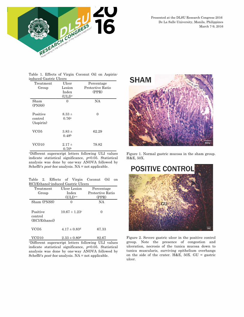
In both ASA and HCl/ethanol models, the
sham group showed normal results while the positive control developed gastric ulcerations, as evidenced by significantly different ulcer lesion indices (Tables 1 and 2). The VCO-treated groups showed significantly lower ulcer lesion index (ULI) for VCO5 and VCO10 when compared with the positive control group. VCO treatment, however, was not dose-dependent.
Histopathology of representative sections of
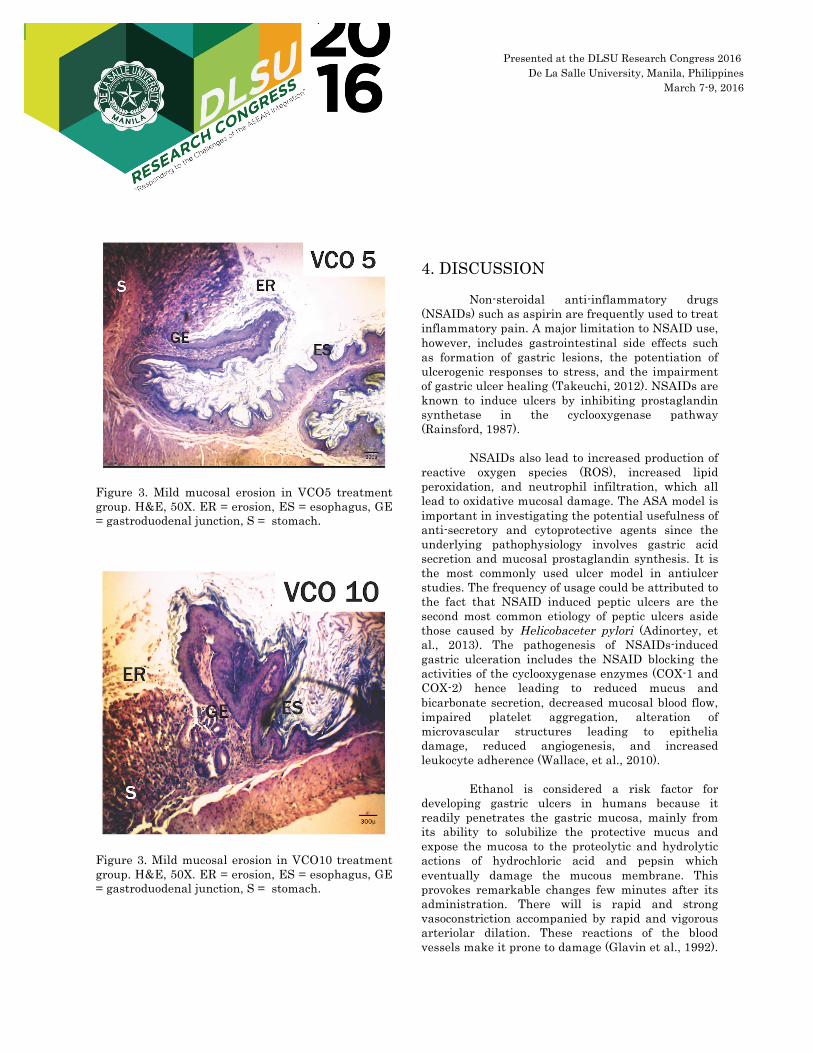
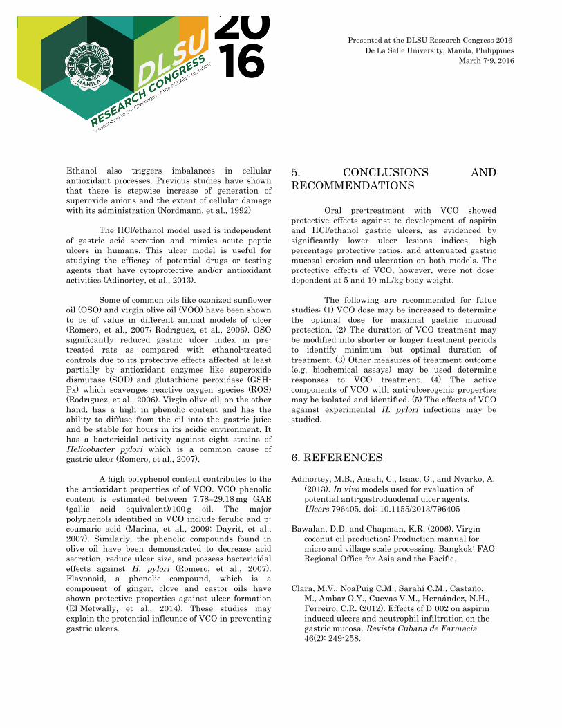
the stomach in both ASA and HCl/ethanol models showed similar results, with normal gastric mucosa in the sham group (Figure 1) and severe gastric ulcers in the positive control group (Figure 2). The VCO-treated groups showed mild ulcerations with partial preservation of normal epithelium (Figures 3 and 4).
Presented at the DLSU Research Congress 2016 De La Salle University, Manila, Philippines
March 7-9, 2016
Table 1. Effects of Virgin Coconut Oil on Aspirin-induced Gastric Ulcers
Treatment Group
Ulcer Lesion Index (ULI)+
Percentage Protective Ratio
(PPR)
Sham (PNSS)
0 NA
Positive control (Aspirin)
8.33 ± 0.76a
0
VCO5
3.83 ± 0.48b
62.29
VCO10
2.17 ± 0.70b
79.82
+Different superscript letters following ULI values indicate statistical significance, p<0.05. Statistical analysis was done by one-way ANOVA followed by Scheffé's post-hoc analysis. NA = not applicable. Table 2. Effects of Virgin Coconut Oil on HCl/Ethanol-induced Gastric Ulcers
Treatment Group
Ulcer Lesion Index
(ULI)++
Percentage Protective Ratio
(PPR) Sham (PNSS) 0 NA Positive control (HCl/Ethanol)
10.67 ± 1.23c
0
VCO5
4.17 ± 0.83d
67.33
VCO10
2.33 ± 0.80d
82.67
+Different superscript letters following ULI values indicate statistical significance, p<0.05. Statistical analysis was done by one-way ANOVA followed by Scheffé's post-hoc analysis. NA = not applicable.
Figure 1. Normal gastric mucosa in the sham group. H&E, 50X.
Figure 2. Severe gastric ulcer in the positive control group. Note the presence of congestion and ulceration, necrosis of the tunica mucosa down to tunica muscularis, surviving epithelium overhangs on the side of the crater. H&E, 50X. GU = gastric ulcer.
300 µ
Presented at the DLSU Research Congress 2016 De La Salle University, Manila, Philippines
March 7-9, 2016
Figure 3. Mild mucosal erosion in VCO5 treatment group. H&E, 50X. ER = erosion, ES = esophagus, GE = gastroduodenal junction, S = stomach.
Figure 3. Mild mucosal erosion in VCO10 treatment group. H&E, 50X. ER = erosion, ES = esophagus, GE = gastroduodenal junction, S = stomach.
4. DISCUSSION
Non-steroidal anti-inflammatory drugs (NSAIDs) such as aspirin are frequently used to treat inflammatory pain. A major limitation to NSAID use, however, includes gastrointestinal side effects such as formation of gastric lesions, the potentiation of ulcerogenic responses to stress, and the impairment of gastric ulcer healing (Takeuchi, 2012). NSAIDs are known to induce ulcers by inhibiting prostaglandin synthetase in the cyclooxygenase pathway (Rainsford, 1987).
NSAIDs also lead to increased production of
reactive oxygen species (ROS), increased lipid peroxidation, and neutrophil infiltration, which all lead to oxidative mucosal damage. The ASA model is important in investigating the potential usefulness of anti-secretory and cytoprotective agents since the underlying pathophysiology involves gastric acid secretion and mucosal prostaglandin synthesis. It is the most commonly used ulcer model in antiulcer studies. The frequency of usage could be attributed to the fact that NSAID induced peptic ulcers are the second most common etiology of peptic ulcers aside those caused by Helicobaceter pylori (Adinortey, et al., 2013). The pathogenesis of NSAIDs-induced gastric ulceration includes the NSAID blocking the activities of the cyclooxygenase enzymes (COX-1 and COX-2) hence leading to reduced mucus and bicarbonate secretion, decreased mucosal blood flow, impaired platelet aggregation, alteration of microvascular structures leading to epithelia damage, reduced angiogenesis, and increased leukocyte adherence (Wallace, et al., 2010).
Ethanol is considered a risk factor for
developing gastric ulcers in humans because it readily penetrates the gastric mucosa, mainly from its ability to solubilize the protective mucus and expose the mucosa to the proteolytic and hydrolytic actions of hydrochloric acid and pepsin which eventually damage the mucous membrane. This provokes remarkable changes few minutes after its administration. There will is rapid and strong vasoconstriction accompanied by rapid and vigorous arteriolar dilation. These reactions of the blood vessels make it prone to damage (Glavin et al., 1992).
Presented at the DLSU Research Congress 2016 De La Salle University, Manila, Philippines
March 7-9, 2016
Ethanol also triggers imbalances in cellular antioxidant processes. Previous studies have shown that there is stepwise increase of generation of superoxide anions and the extent of cellular damage with its administration (Nordmann, et al., 1992)
The HCl/ethanol model used is independent
of gastric acid secretion and mimics acute peptic ulcers in humans. This ulcer model is useful for studying the efficacy of potential drugs or testing agents that have cytoprotective and/or antioxidant activities (Adinortey, et al., 2013).
Some of common oils like ozonized sunflower
oil (OSO) and virgin olive oil (VOO) have been shown to be of value in different animal models of ulcer (Romero, et al., 2007; Rodrıguez, et al., 2006). OSO significantly reduced gastric ulcer index in pre-treated rats as compared with ethanol-treated controls due to its protective effects affected at least partially by antioxidant enzymes like superoxide dismutase (SOD) and glutathione peroxidase (GSH-Px) which scavenges reactive oxygen species (ROS) (Rodrıguez, et al., 2006). Virgin olive oil, on the other hand, has a high in phenolic content and has the ability to diffuse from the oil into the gastric juice and be stable for hours in its acidic environment. It has a bactericidal activity against eight strains of Helicobacter pylori which is a common cause of gastric ulcer (Romero, et al., 2007).
A high polyphenol content contributes to the
the antioxidant properties of of VCO. VCO phenolic content is estimated between 7.78–29.18 mg GAE (gallic acid equivalent)/100 g oil. The major polyphenols identified in VCO include ferulic and p-coumaric acid (Marina, et al., 2009; Dayrit, et al., 2007). Similarly, the phenolic compounds found in olive oil have been demonstrated to decrease acid secretion, reduce ulcer size, and possess bactericidal effects against H. pylori (Romero, et al., 2007). Flavonoid, a phenolic compound, which is a component of ginger, clove and castor oils have shown protective properties against ulcer formation (El-Metwally, et al., 2014). These studies may explain the protential infleunce of VCO in preventing gastric ulcers.
5. CONCLUSIONS AND RECOMMENDATIONS
Oral pre-treatment with VCO showed protective effects against te development of aspirin and HCl/ethanol gastric ulcers, as evidenced by significantly lower ulcer lesions indices, high percentage protective ratios, and attenuated gastric mucosal erosion and ulceration on both models. The protective effects of VCO, however, were not dose-dependent at 5 and 10 mL/kg body weight.
The following are recommended for futue
studies: (1) VCO dose may be increased to determine the optimal dose for maximal gastric mucosal protection. (2) The duration of VCO treatment may be modified into shorter or longer treatment periods to identify minimum but optimal duration of treatment. (3) Other measures of treatment outcome (e.g. biochemical assays) may be used determine responses to VCO treatment. (4) The active components of VCO with anti-ulcerogenic properties may be isolated and identified. (5) The effects of VCO against experimental H. pylori infections may be studied. 6. REFERENCES Adinortey, M.B., Ansah, C., Isaac, G., and Nyarko, A.
(2013). In vivo models used for evaluation of potential anti-gastroduodenal ulcer agents. Ulcers 796405. doi: 10.1155/2013/796405
Bawalan, D.D. and Chapman, K.R. (2006). Virgin
coconut oil production: Production manual for micro and village scale processing. Bangkok: FAO Regional Office for Asia and the Pacific.
Clara, M.V., NoaPuig C.M., Sarahí C.M., Castaño,
M., Ambar O.Y., Cuevas V.M., Hernández, N.H., Ferreiro, C.R. (2012). Effects of D-002 on aspirin-induced ulcers and neutrophil infiltration on the gastric mucosa. Revista Cubana de Farmacia 46(2): 249-258.

Presented at the DLSU Research Congress 2016 De La Salle University, Manila, Philippines
March 7-9, 2016
Dayrit, F., Buenafe, O.E., Chainani, E., de Vera, I.M., Dimzon, I.K., Gonzales, E. and Santos, J.E. (2007) Standards for essential composition and quality factors of commercial virgin coconut oil and its differentiation from RBD coconut oil and copra oil. Philippine Journal of Science 136: 121-131.
De Andrade, S.F., Lemos, M., Comunello, E.,Vania,
Noldin, F., Valdir, C.F., Niero, R. (2007). Evaluation of the antiulcerogenic activity of Maytenus robusta (Celastraceae) in different experimental ulcer models. Journal of Ethnopharmacology 113(2): 252-257.
El-Metwally, Mohamed, E. (2014). Evaluation of
antiulcer activity of ginger, clove and castor oils against aspirin induced gastric ulcers in rats. World Applied Sciences Journal 29(7): 815-824. doi: 10.5829/idosi.wasj.2014.29.07.1396
Glavin, G.B. and Szabo, S. (1992). Experimental
gastric mucosal injury: laboratory models reveal mechanisms of pathogenesis and new therapeutic strategies. FASEB Journal 6(3): 825–831.
Lau J.Y. and Cheung F. K.. (2009). Management of
massive peptic ulcer bleeding. Gastroenterology Clinics of North America 38(2), 231-43. doi: 10.1016/j.gtc.2009.03.003
Mahmood, A.A., Hadi, A.H., Abdelwahab, S.I., Taha,
M.M., Hussiani, J., NurAsykin. (2012). Mechanisms of gastroprotective effects of ethanolic leaf extract of Jasminum sambac against HCl/ethanol-induced gastric mucosal injury in rats. Evidence-Based Complementary and Alternative Med. 2012: 786426.
Malairajan, G.G., Narasimhan, S., Veni, J.K. (2008).
Evalution of anti-ulcer activity of Polyalthia longifolia (Sonn.) Thwaites in experimental animals. Indian Journal of Pharmacology 40(3): 126–128.
Marina, A.M., Man, Y.B., Nazimah S.A., Amin I.
(2009). Antioxidant capacity and phenolic acids of virgin coconut oil. International Journal of Food Sciences and Nutrition 60 Suppl (2): 114-23.
Nordmann, R., Ribiere, C., Rouach, H. (1992). Implication of free radical mechanisms in ethanol-induced cellular injury. Free Radical Biology and Medicine 12(3): 219–240.
Rainsford, K.D. (1987). The effects of 5-lipoxygenase
inhibitors and leukotriene antagonists on the development of gastric lesions induced by nonsteroidal anti-inflammatory drugs in mice. Agents Actions 21(3-4): 316–319.
Rodrıguez, Gonzalez R., Alvarez, Guanche D.,
Merino, N. (2006). Antioxidant mechanism is involved in the gastroprotective effects of ozonized sunflower oil in ethanol-induced ulcers in rats. Mediators of Inflammation 2007: 65873.
Romero, C., Medina, E., Vargas, J., Brenes, M., De
Castro, A. (2007). In vitro activity of olive oil polyphenols against Helicobacter pylori. Journal of Agricultural and Food Chemistry 55(3): 680-686.
Takeuchi, Koji. (2012). Pathogenesis of NSAID-
induced gastric damage: Importance of cyclooxygenase inhibition and gastric hypermotility. World Journal of Gastroenterology 18(18): 2147–2160.
Wallace J. L., McKnight W., Reuter B.K., Vergnolle,
N. (2000) NSAID-Induced gastric damage in rats: requirement for inhibition of both cyclooxygenase 1 and 2. Gastroenterology 119(3): 706–714.
Yuan, Y., Padol, I.T., Hunt, R.H. (2006). Peptic ulcer
disease today. Nature Clinical Practice Gastroenterology and Hepatology 3(2): 80-89.