Embed Size (px)
Citation preview

Indian Journal of Experimental Biology Vol. 44, October 2006, pp. 821-831
Protective effect of ethanolic and water extracts of sea buckthorn (Hippophae rhamnoides L.) against the toxic effects of mustard gas
R Vijayaraghavan1, Anshoo Gautam1, Om Kumar1, S C Pant1, Manoj Sharma1, Seema Singh1, H T Satish Kumar1, Anand Kumar Singh1, Manisha Nivsarkar1, M P Kaushik1, R C Sawhney2,
O P Chaurasia3 & G B K S Prasad4 1Defence Research and Development Establishment, Gwalior 474 002, India 2Defence Institute of Physiology and Allied Sciences, Delhi 110 054, India
3Field Research Laboratory, Leh 56 APO, India 4Department of Biochemistry, Jiwaji University, Gwalior 474 011, India
Received 23 December 2005; revised 7 June 2006
Ethanolic extract of H. rhamnoides L. leaf (HL-EOH), water and ethanolic extract of H. rhamnoides fruit (HF-W and HF-EOH), and H. rhamnoides flavone from fruit (HR-flavone) were evaluated against percutaneously administered sulphur mustard (SM), a chemical warfare agent. The animals administered with SM (9.7, 19.3 and 38.7 mg/kg) died at various days depending upon the dose and there was a significant reduction in the body weight. The H. rhamnoides extracts (1g/kg; 3 doses; po) significantly protected the lethality, with a protective index of 2.4, 1.7, 1.7 and 2.2 for HL-EOH, HF-W, HF-EOH and HR-flavone respectively. Reduced glutathione (GSH) and oxidized glutalthione (GSSG) levels were reduced, and malondialdehyde (MDA) was elevated after percutaneous administration of SM. Oral administration of HL-EOH and HR-flavone significantly protected the body weight loss. Recovery in the levels of GSH, GSSG and MDA were also observed following oral administration of HL-EOH and HR-flavone. All the extracts were non-toxic and the LD50 was more than 5 g/kg. The present study shows that percutaneous administration of SM induces oxidative stress and ethanolic extract of leaf of H. rhamnoides and H. rhamnoides flavone from fruit can significantly protect it.
Keywords: Glutathione, Hippophae rhamnoides, Malondialdehyde, Oxidative stress, Sulphur mustard,
The nerve agents and the blistering agents like sulphur and nitrogen mustards continue to be a threat to the defence forces by the enemy countries and to the civilians by the terrorists1. The chemical weapons convention prohibits the production, storage, transport and use of chemical warfare agents on enemy forces. Though the chemical weapon convention is signed and ratified by several countries and the stockpiled chemical warfare agents are being destroyed, the use of chemical weapons by adversary countries and terrorists is still perceived. Sulphur mustard (SM) commonly known as mustard gas and chemically bis (2-chloro ethyl) sulphide, is an alkylating agent and upon contact with human skin causes serious blisters2. SM was first used in 1917 and from that time onwards it has been used as a warfare agent in several instances3. SM forms a highly reactive sulphonium ion in the body and preferably binds to N7 position of guanine residue of DNA and subsequently leads to
DNA strand breaks and cell death4. Due to the high reactivity of the sulphonium ion, SM binds to a variety of cellular macromolecules5,6. A number of antidotes are being studied as a prophylactic and as therapeutic agent for SM7-11. Decontamination of SM immediately after exposure has been preferred as the best method of minimizing the toxicity12,13.
There is an everlasting interest on a wide variety of plant species for their bioactivity, world over. Several studies have revealed that plants produce potent antioxidants to control the oxidative stress caused by sunlight and oxygen and hence can be considered as a source of complex molecules with antioxidant activity14. Hippophae rhamnoides L. (Eleganaceae) commonly known as Sea Buckthorn, is a temperate shrub and native of Europe and Asia15. It was used as a medicinal plant in Tibet as early as 900 A.D.16. Many medicinal preparations of H. rhamnoides from both wild and cultivated sources have been clinically used to treat radiation damage, burns, oral inflammation and gastric ulcer in China and the former Soviet Republic17. In addition to the medicinal use, the berries of H. rhamnoides are processed to
_____________ Phone: 91-751-2233494 Fax: 91-751-2341148 Email: [email protected]

INDIAN J EXP BIOL, OCTOBER 2006
822
make juice and jam, and flavoring of dairy products because of their unique taste. The berries of H. rhamnoides have a much higher content of ascorbic acid (100-700mg/100g), tocopherol (1-10mg/100g) and carotenoids (3-15mg/100g), compared to citrus fruits18. The fruit juice has low freezing point and the plant can be cultivated in high altitude where temperature is less than 0oC18. The fruit juice is commercially available.
Anti-ulcer19, radioprotectant20 and antioxidant21 properties of H. rhamnoides fruit are reported. One of the toxic mechanisms of SM is due to oxidative stress and flavonoids have been shown to reduce its toxicity22,23. Since H. rhamnoides fruit and leaf contains various flavonoids, extract of fruit and leaf have been studied for their protective efficacy against SM. Materials and Methods
Chemicals⎯SM was synthesized in the Chemistry Division of the Establishment and was found to be above 99% pure by gas chromatographic analysis. O-thalaldehyde (OPT), reduced glutathione (GSH), oxidized glutathione (GSSG) and thiobarbituric acid were purchased from Sigma Chemicals (USA). Other chemicals of high purity were from Qualigens or E-Merck (India). The clinical biochemical analyses were performed using kits purchased from Merck India Ltd.
Utmost care was taken during the synthesis of SM in the declared facility of the establishment (inspected
by the inspectors of the Organization for the Prohibition of Chemical Weapons, The Hague, The Netherlands). The dilution of SM and its administration in animals were carried out in fume hoods. The animals were kept in well-ventilated area after SM administration for 24 hr and then kept in the experimental animal room for further monitoring.
Animals⎯Randomly bred Swiss female mice (25 - 30 g body weight) from the Establishment’s animal facility were used. The animals were kept in polypropylene cages on steam sterilized paddy husk as the bedding material. Free access to food (Amrut Ltd, India) and water was allowed until two hours before the experiment. The care and maintenance of animals were as per the approved guidelines of the Committee for the Purpose of Control and Supervision of Experiments on Animals (CPCSEA), India. A day before percutaneous exposure of SM, hair on the back of the animals was closely clipped using a pair of scissors. The Animal Ethical Committee of the Establishment has approved all the animal procedures.
Preparation of H. rhamnoides extract⎯The leaf and fruit of H. rhamnoides were collected from the natural habitat of Leh region (Laddak) by the Field Research Laboratory (FRL) of DRDO. Various extracts were prepared by Defence Institute of Physiology and Allied Sciences (DIPAS), Delhi and Defence Research and Development Establishment (DRDE), Gwalior. The extraction procedures are given briefly in Table 1.
Table 1⎯Extraction procedure of H. rhamnoides leaf and fruit
S.No. Nomenclature of extract Abbreviation Procedure
1 Alcoholic extract of H. rhamnoides leaf. HL-EOH The fresh leaves were cleaned and dried under shade. The extraction was carried out using powdered leaf by refluxing withethanol at 80oC for 5 hr in Soxhlet. The extract was filtered and the filtrate was dried under reduced pressure in a rotary evaporator.
2 Water extract of H. rhamnoides leaf HL-W The fresh leaves were cleaned and dried under shade. Theextraction was carried out using powdered leaf by refluxing withwater at 80oC for 5 hr in Soxhlet. The extract was filtered and the filtrate was dried under reduced pressure in a rotary evaporator.
3 Alcoholic extract of H. rhamnoides fruit HF-EOH The fresh fruits were cleaned and dried under shade. The extractionwas carried out using the dried fruits by refluxing with ethanol at80oC for 5 hr in Soxhlet. The extract was filtered and the filtrate was dried under reduced pressure in a rotary evaporator.
4 Water extract of H. rhamnoides fruit HF-W The fresh fruits were cleaned and dried under shade. The extractionwas carried out using the dried fruits by refluxing with water at 100oC for 5 hr in Soxhlet. The extract was filtered and the filtrate was dried under reduced pressure in a rotary evaporator.
5 Flavonoids of H. rhamnoides fruit HR-flavone Mixture of flavonoids isolated from the fruit (Beiging Jianghe SeaBuckthorn Company, Beiging, China).

VIJAYARAGHAVAN et al., EFFECT OF HIPPOPHAE RHAMNOIDES ON MUSTARD GAS
823
Safety evaluation of different extracts of H. rhamnoides⎯The extracts were administered to mice orally at 800, 1600 and 3200 mg/kg body weight. For each dose and extract four mice were used. The extracts were either dissolved in water (400 mg/ml) or prepared as an emulsion in 1% hydroxypropyl cellulose (400 mg/ml) and administered to mice using an oral feeding cannula (20 gauge) at a volume of 0.5 to 1.0 ml/100 g body weight. The animals were observed for 14 days. Food and water intake, general behaviour, daily body weight and mortality were recorded. On the 15th day, blood was drawn from ocular plexus under ether anaesthesia. Various biochemical parameters viz., WBC count, RBC count, hemoglobin, urea, cholesterol, total protein, albumin, glutamate pyruvate transaminase (GPT), glutamate oxaloacetate transaminase (GOT), alkaline phosphatase (ALP) and glucose in plasma samples were estimated by automated procedures (Beckman Coulter Cell Counter, and Alfa Wassermann Clinical Analyzer, USA) as per the manufacturers protocol.
Protective efficacy of different extracts of H. rhamnoides against SM toxicity⎯A safe dose of 1000 mg/kg of the extract was taken for the protection studies. Three doses of extracts were administered orally, one simultaneously with sulphur mustard and remaining two on the next two successive days. SM was diluted in PEG-300 and applied on the back of the mice. The dilutions were made in such a manner that the quantity applied was between 80-100 μl. The animals were weighed daily and observed for mortality for 14 days. The LD50 was determined by the moving average method of Gad and Weil24.
Dose dependent protection of H. rhamnoides leaf extracts against SM toxicity⎯Since the leaf extract of H. rhamnoides showed better protection, dose dependent protection was evaluated with three doses of HL-EOH (250, 500 and 1000 mg/kg). The extract and SM were administered as described above.
Protection studies on different biochemical variables and hematological variables⎯The protective effect of various extracts on selected biochemical and hematological variables were estimated by administering three doses, one simultaneously with SM and remaining two on the next two successive days. The extracts were administered orally (1000 mg/kg) and SM in PEG 300
was applied percutaneously (2 LD50). The following groups were kept; each group consisted of 4 mice. I Water, oral = Control group II Water, oral + SM 2 LD50 = SM Group III HL-EOH + SM 2 LD50 = H. rhamnoides leaf,
ethanol extract IV HF-EOH + SM 2 LD50 = H. rhamnoides fruit,
ethanol extract V HF-W + SM 2 LD50 = H. rhamnoides fruit,
water extract VI HR-flavone + SM 2 LD50 = H. rhamnoides
flavones from fruit
SM was diluted in PEG 300 and applied on the back of mice. The dilutions were made in such a manner that the quantity applied was 80-100 μl. The animals were weighed daily and on the 7th day blood was drawn from ocular plexus under ether anesthesia. The animals were sacrificed by cervical dislocation. Pieces of liver were removed, blotted, weighed and used for the estimation of GSH, GSSG and malondialdehyde (MDA). Pieces of liver, spleen and skin on the site of SM application were also removed for light microscopic studies. The blood was used for the measurement of RBC and WBC counts and haemoglobin estimation.
The fluorometric method of Hisin and Hilf25 was used for the determination of hepatic GSH and GSSG concentration. For this 150 mg of liver tissue was homogenized in 4 ml of phosphate EDTA buffer and metaphosphoric acid (25%). The content of the tube was centrifuged and the supernatant was used for the estimation of GSH and GSSG. Hepatic lipid peroxidation was determined by measuring the level of MDA according to the method of Buege and Aust26. Liver (100 mg) was directly homogenized in 5 ml of thiobarbituric acid reagent and boiled for 30 min. The contents of the tube were cooled, centrifuged and absorbance of the clear supernatant was measured at 535 nm. The amount of MDA formed was calculated using a molar extinction coefficient of 1.58×10⁵ /M/cm. The other haematological variables viz., RBC, WBC counts and blood haemoglobin were analysed using Backman Coulter Cell Counter (USA).
Histological studies⎯Samples of liver, spleen and skin tissue were fixed in 10% neutral buffered formalin solution. After proper fixation, small pieces were processed by dehydration and embedded in paraffin wax. Multiple sections of 5-6 µm thickness was prepared and stained with haematoxylin and eosin for light microscopic observation27. From each

INDIAN J EXP BIOL, OCTOBER 2006
824
tissue, 10 slides were prepared. Representative slides from each animal for each tissue was selected and 90 μm square area was identified randomly. Lesions were marked and compared with that of control. The severity of lesions was characterised using LEICA-Qwin-500 Image Analyzer (Leica Orthoplan, Germany) and converted into percentage.
Statistical analysis⎯LD50 and confidence intervals were determined by the moving average method24. The biochemical parameters were analysed by one-way ANOVA with Student-Newman-Keul’s multiple comparisons procedure. A probability of less than 0.05 is taken as statistically significant. SigmaStat (SPSS, Inc., USA) was used for the statistical analysis. Results
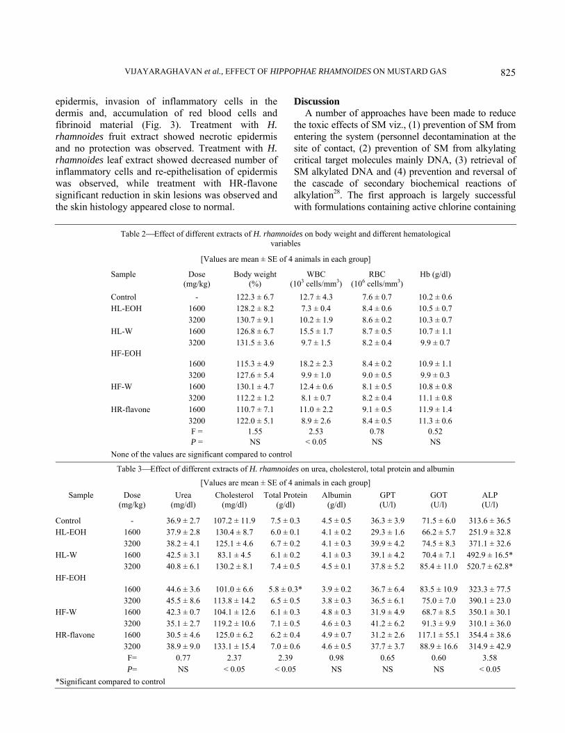
Safety evaluation of H. rhamnoides extracts⎯No mortality was observed up to a dose of 3200 mg/kg following oral administration of water or ethanolic extract of the fruit and leaf of H. rhamnoides. Blood was withdrawn, 14 days after oral administration of the extract, for hematological and biochemical studies, and the data are summarised in Tables 2 and 3. No significant difference was observed in most of the hematological and biochemical parameter in all the extracts of H. rhamnoides.
Protective efficacy of different extracts of H. rhamnoides against SM toxicity⎯LD50 of SM in PEG was found to be 9.7 mg/kg through percutaneous route (Table 4). Different extracts of H. rhamnoides were given orally and protection index was calculated. Maximum protection was observed in HL-EOH and HR-flavone and the LD50 was 22.9 and 21.4 with a protection of 2.4 and 2.2-folds, respectively (Table 4). The protection observed in the case of HF-EOH and HF-W were less (1.7-fold). The body weight decreased significantly after 2 LD50 administration of SM. Seven days after percutaneous administration of SM, the body weight was 58.2% of the pre administration body weight (Table 5). Weight loss was also observed in HF-EOH treated groups. Oral administration of extracts viz., HL-EOH and HR-flavone significantly protected the body weight loss. Since the ethanolic extract of the H. rhamnoides leaf showed maximum protection upon oral administration different doses were evaluated for their protective efficacy. Dose dependent protection was observed, with 1000 mg/kg showing better protection than 500 and 250 mg/kg dose levels (Table 6).
Protection studies on different biochemical variables and hematological variables⎯Significant depletion was observed in reduced glutathione after percutaneous administration of SM. The GSH level was 46.2%, 7 days after SM administration compared to preadministration levels. The HL-EOH and HR- flavone groups did not show significant depletion (Table 7). Though, GSH levels in other groups were better than the SM group, they were still significantly lower than the control group. Oxidized glutathione also showed significant depletion and recovery was observed in HL-EOH and HR-flavone groups. No significant recovery was found in the other groups. Malondialdehyde level showed significant elevation of 130.6% and the effect was protected by oral administration of HL-EOH and HR-flavone. The other groups did not show any significant difference (Table 7). Though WBC count did not show any significant difference, RBC count and Hb concentration were significantly elevated following percutaneous administration of SM (Table 8). Recovery was found in HL-EOH and HR-flavone groups. No recovery was found in the other groups. [
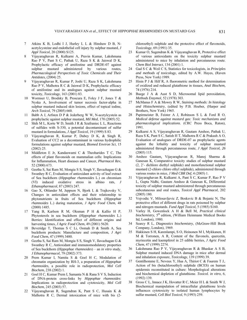
Histological studies in various groups ⎯The liver of SM exposed mice showed severe degenerative changes, obliteration of chromatin material and perinuclear clumping of cytoplasm (Fig.1.) Treatment with H. rhamnoides fruit extract did not offer any protection and the same magnitude of hepatic degeneration was observed, while treatment with H. rhamnoides leaf extract showed fewer disturbances in the lobular pattern and less infiltration of mononuclear cells. However, fatty degeneration persisted. The hepatic lesions were minimal in mice liver treated with HR-flavone. Few inflammatory cells were observed in the vicinity of the central canal. The spleen of mice exposed to SM showed hypocellularity and degeneration of white pulp follicles (Fig. 2). Treatment with H. rhamnoides fruit extract did not protect the mice spleen and degenerative changes were observed. The appearance of megakaryocytes persisted with the treatment of H. rhamnoides fruit extract. Treatment with H. rhamnoides leaf extract showed regression of splenic lesions, and in the case of HR-flavone there were minimal changes and the spleen resembled like that of the control. Sporadic appearance of megakaryocytes was observed in H. rhamnoides leaf extract treated mice spleen, but not in HR-flavone treated mice spleen. The skin of mice at the site of SM application showed complete erosion of
VIJAYARAGHAVAN et al., EFFECT OF HIPPOPHAE RHAMNOIDES ON MUSTARD GAS
825
epidermis, invasion of inflammatory cells in the dermis and, accumulation of red blood cells and fibrinoid material (Fig. 3). Treatment with H. rhamnoides fruit extract showed necrotic epidermis and no protection was observed. Treatment with H. rhamnoides leaf extract showed decreased number of inflammatory cells and re-epithelisation of epidermis was observed, while treatment with HR-flavone significant reduction in skin lesions was observed and the skin histology appeared close to normal.
Discussion A number of approaches have been made to reduce
the toxic effects of SM viz., (1) prevention of SM from entering the system (personnel decontamination at the site of contact, (2) prevention of SM from alkylating critical target molecules mainly DNA, (3) retrieval of SM alkylated DNA and (4) prevention and reversal of the cascade of secondary biochemical reactions of alkylation28. The first approach is largely successful with formulations containing active chlorine containing
Table 2⎯Effect of different extracts of H. rhamnoides on body weight and different hematological variables
[Values are mean ± SE of 4 animals in each group]
Sample Dose (mg/kg)
Body weight (%)
WBC (103 cells/mm3)
RBC (106 cells/mm3)
Hb (g/dl)
Control - 122.3 ± 6.7 12.7 ± 4.3 7.6 ± 0.7 10.2 ± 0.6 HL-EOH 1600 128.2 ± 8.2 7.3 ± 0.4 8.4 ± 0.6 10.5 ± 0.7 3200 130.7 ± 9.1 10.2 ± 1.9 8.6 ± 0.2 10.3 ± 0.7 HL-W 1600 126.8 ± 6.7 15.5 ± 1.7 8.7 ± 0.5 10.7 ± 1.1 3200 131.5 ± 3.6 9.7 ± 1.5 8.2 ± 0.4 9.9 ± 0.7 HF-EOH 1600 115.3 ± 4.9 18.2 ± 2.3 8.4 ± 0.2 10.9 ± 1.1 3200 127.6 ± 5.4 9.9 ± 1.0 9.0 ± 0.5 9.9 ± 0.3 HF-W 1600 130.1 ± 4.7 12.4 ± 0.6 8.1 ± 0.5 10.8 ± 0.8 3200 112.2 ± 1.2 8.1 ± 0.7 8.2 ± 0.4 11.1 ± 0.8 HR-flavone 1600 110.7 ± 7.1 11.0 ± 2.2 9.1 ± 0.5 11.9 ± 1.4 3200 122.0 ± 5.1 8.9 ± 2.6 8.4 ± 0.5 11.3 ± 0.6 F = 1.55 2.53 0.78 0.52 P = NS < 0.05 NS NS None of the values are significant compared to control
Table 3⎯Effect of different extracts of H. rhamnoides on urea, cholesterol, total protein and albumin
[Values are mean ± SE of 4 animals in each group] Sample Dose
(mg/kg) Urea
(mg/dl) Cholesterol
(mg/dl) Total Protein
(g/dl) Albumin
(g/dl) GPT (U/l)
GOT (U/l)
ALP (U/l)
Control - 36.9 ± 2.7 107.2 ± 11.9 7.5 ± 0.3 4.5 ± 0.5 36.3 ± 3.9 71.5 ± 6.0 313.6 ± 36.5 HL-EOH 1600 37.9 ± 2.8 130.4 ± 8.7 6.0 ± 0.1 4.1 ± 0.2 29.3 ± 1.6 66.2 ± 5.7 251.9 ± 32.8 3200 38.2 ± 4.1 125.1 ± 4.6 6.7 ± 0.2 4.1 ± 0.3 39.9 ± 4.2 74.5 ± 8.3 371.1 ± 32.6 HL-W 1600 42.5 ± 3.1 83.1 ± 4.5 6.1 ± 0.2 4.1 ± 0.3 39.1 ± 4.2 70.4 ± 7.1 492.9 ± 16.5* 3200 40.8 ± 6.1 130.2 ± 8.1 7.4 ± 0.5 4.5 ± 0.1 37.8 ± 5.2 85.4 ± 11.0 520.7 ± 62.8*HF-EOH 1600 44.6 ± 3.6 101.0 ± 6.6 5.8 ± 0.3* 3.9 ± 0.2 36.7 ± 6.4 83.5 ± 10.9 323.3 ± 77.5 3200 45.5 ± 8.6 113.8 ± 14.2 6.5 ± 0.5 3.8 ± 0.3 36.5 ± 6.1 75.0 ± 7.0 390.1 ± 23.0 HF-W 1600 42.3 ± 0.7 104.1 ± 12.6 6.1 ± 0.3 4.8 ± 0.3 31.9 ± 4.9 68.7 ± 8.5 350.1 ± 30.1 3200 35.1 ± 2.7 119.2 ± 10.6 7.1 ± 0.5 4.6 ± 0.3 41.2 ± 6.2 91.3 ± 9.9 310.1 ± 36.0 HR-flavone 1600 30.5 ± 4.6 125.0 ± 6.2 6.2 ± 0.4 4.9 ± 0.7 31.2 ± 2.6 117.1 ± 55.1 354.4 ± 38.6 3200 38.9 ± 9.0 133.1 ± 15.4 7.0 ± 0.6 4.6 ± 0.5 37.7 ± 3.7 88.9 ± 16.6 314.9 ± 42.9 F= 0.77 2.37 2.39 0.98 0.65 0.60 3.58 P= NS < 0.05 < 0.05 NS NS NS < 0.05 *Significant compared to control

INDIAN J EXP BIOL, OCTOBER 2006
826
compounds viz., S-330 and CC2, as chemical decontaminants12,13. In spite of several years of active research so far no recommended treatment regimen has evolved for the toxic effects of SM, except for few drugs, that are in the experimental stage9,29. The main reason for the failure is due to the models selected for drug evaluation i.e., either SM is administered by routes other than percutaneous route or simulants of SM is used, or in vitro models have been used30. Many antidotes have been screened for SM toxicity, but those shown to be effective in vitro, are not evaluated using in vivo models. SM is more toxic through the percutaneous route than the subcutaneous or oral routes and the mechanism is route specific31. In the present study SM was administered through the percutaneous route which is the expected route of entry of SM in military or civil threats.
A variety of compounds have been screened as prophylactic agents for SM. Among them amifostine and one of its analogues, DRDE-07 were very promising as prophylactic agents9. These compounds are in experimental stage and are moderately toxic. Other compounds tested offered minimal protection32.
Table 4⎯Protective efficacy of different extracts of H. rhamnoides against percutaneously administered SM.
Group LD50
(mg/kg) 95 % Confidence
limit (mg/kg) PI*
SM only 9.7 6.1-15.2 - HL-EOH + SM 22.9 14.8-34.8 2.4 HF-EOH + SM 16.3 7.4-50.3 1.7 HF-W + SM 16.2 11.0-24.0 1.7 HR- flavone + SM 21.4 10.0-45.8 2.2
* PI = LD50 with treatment/LD50 without treatment
Table 5⎯Protective efficacy of various extracts of H. rhamnoideson body weight change after administration of SM [Values are mean ± SE of 4 animals in each group]
Group 1 day (%)
4 days (%)
7 days (%)
Control 98.6 ± 1.5 99.8 ± 2.1 99.8 ± 2.1 SM only (2 LD50)
99.4 ± 1.5 81.5 ± 8.8 58.2 ± 2.9a
HL-EOH + SM 104.7 ± 2.8 102.8 ± 6.2 103.9 ± 7.3b HF-EOH + SM 98.3 ± 3.3 88.2 ± 6.2 82.2 ± 8.6 HF-W + SM 97.0 ± 1.8 84.2 ± 3.8 64.1 ± 4.4a HR-flavone + SM 98.7 ± 2.1 93.8 ± 9.4 78.3 ± 10.3
F= 1.41 1.68 7.65 P= NS NS < 0.001
Extracts 1g/kg oral, three doses on successive days. P values < 0.05: a - Control vs treatment; b-SM vs treatment
Table 6⎯Protective efficacy of H. rhamnoides leaf (alcoholic extract) at different doses
Group LD50 (mg/kg)
Confidence limit(mg/kg)
PI
SM only 9.7 6.1-15.2 - HL-EOH (250 mg/kg)
+ SM 9.7 6.2-15.3 1.0
HL-EOH (500 mg/kg)
+ SM 13.7 7.2-26.0 1.4
HL-EOH (1000 mg/kg)
+ SM 22.9 14.8-34.8 2.4
Extracts administered orally, three doses on successive days.
Table 7⎯Changes in various biochemical parameters (with or without treatment) after percutaneously administered SM
(Values are mean ± SE of 4 animals in each group)
Group GSH (%)
GSSG (%)
MDA (%)
Control 99.9 ± 7.3 105.5 ± 5.4 100.0 ± 5.0 SM only (2 LD50)
46.2 ± 8.1a 54.8 ± 11.3a 130.6 ± 4.2a
HL-EOH + SM 81.1 ± 8.0b 81.6 ± 7.8 101.9 ± 3.4b HF-EOH + SM 60.0 ± 7.3a 62.4 ± 7.6a 134.3 ± 2.4a HF-W + SM 56.8 ± 8.6a 60.4 ± 8.2a 113.7 ± 8.8 HR- Flavone + SM 85.5 ± 5.0b 88.2 ± 4.8b 116.6 ± 6.1 F = 7.46 6.32 6.93 P = < 0.001 < 0.001 < 0.001
Control values : GSH = 3.88 ± 0.48 μ mole/g tissue GSSG = 1.50 ± 0.34 μ mole/g tissue MDA = 6.36 ± 0.40 n mole/g tissue
Extracts 1g/kg oral, three doses on successive days. P values < 0.05: a - Control vs treatment; b - SM vs treatment
Table 8⎯Changes in various hematological parameters (with or without treatment) after percutaneously administered SM
(Values are mean ± SE of 4 animals in each group)
Group WBC (%)
RBC (%)
Hb (%)
Control 100.0 ± 9.1 100.0 ± 4.0 101.5 ± 4.1SM only (2 LD50)
67.0 ± 15.9 163.3 ± 19.0 146.6 ± 8.8a
HL-EOH + SM 63.0 ± 9.3 114.0 ± 35.4 105.2 ± 8.4b
HF-EOH + SM 77.0 ± 15.4 146.7 ± 22.7 116.3 ± 8.4b
HF-W + SM 75.8 ± 13.8 154.3 ± 36.2 130.3 ± 9.2HR-Flavone + SM ⎯ ⎯ ⎯ F = 1.21 1.08 5.42 P = NS NS < 0.001 Control values : WBC = 10.1 ± 1.7 103 cells/mm3 RBC = 7.3 ± 0.3 106 cells/mm3 Hb= 12.6 ± 0.4 g/dl
Extracts 1g/kg oral, three doses on successive days. P values < 0.05: a - Control vs treatment; b - SM vs treatment

VIJAYARAGHAVAN et al., EFFECT OF HIPPOPHAE RHAMNOIDES ON MUSTARD GAS
827
The reason for this is there are several possible mechanisms of SM toxicity. One of the mechanisms of SM toxicity is related to oxidative stress. SM forms sulphonium ion in the body, which is electrophilic in nature. Antioxidants are nucleophilic in nature and may directly interact with SM intermediates, and also
scavenge reactive oxygen species generated by SM. Since the extracts of H. rhamnoides has been shown to be an effective antioxidant, and its juice usually do not freeze up to –20oC, the oral administration of H. rhamnoides extracts have been studied against SM toxicity18.
Fig. 1⎯Photomicrographs of control and SM administered mice liver (2 LD50), and protection by H. rhamnoides extracts. H & E, ×100. (a) Control mouse liver showing normal hepatic parenchyma, hepatic cord, hepatic lobules and hepatocytes, (b) Liver of SM exposed mouse showing severe degenerative changes (arrow), obliteration of chromatin material and perinuclear clumping of cytoplasm (arrow head), (c), Treatment with water extract of H. rhamnoides fruit showing no protection, and with the same magnitude of hepatic degeneration (arrow), (d) Treatment with ethanolic extract of H. rhamnoides fruit showing no protection, and with the same magnitude of hepatic degeneration, (e) Treatment with H. rhamnoides leaf extract showing fewer disturbances in the lobular pattern and less infiltration of mononuclear cells, (f) Treatment with HR-flavone showing minimal lesions in the liver.

INDIAN J EXP BIOL, OCTOBER 2006
828
No mortality was observed in any of the extracts up to a dose of 3200 mg/kg. Even if all animals die at the next log dose of 6400 mg/kg, the LD50 will be more than 4.5 g/kg, showing that the extracts are nontoxic. In normal conditions the bulk of plasma protein consists of albumin and globulin fractions. The albumin fraction is more than 50 % of total plasma
protein and the albumin to globulin ratio may be altered in liver injury33. No such observation was observed in H. rhamnoides treated groups. Urea concentration increases in the blood in renal and cardiac failure, intestinal and prostate obstruction, and some other infectious diseases33. The urea level was not altered following H. rhamnoides treatment. The
Fig. 2⎯Photomicrographs of control and SM administered mice spleen (2 LD50), and protection by H. rhamnoides extracts. H & E, ×200. (a) Control mouse spleen showing normal architecture of germinal center, red pulp and marginal zone of white pulp, (b) Spleen of mouse exposed to SM showing hypocellularity and degeneration of white pulp follicles (arrow), (c) Treatment with water extract of H. rhamnoides fruit showing degenerative changes in the spleen (arrow), (d) Treatment with ethanolic extract of H. rhamnoides fruit showing degenerative changes in the spleen (arrow), (e) Treatment with H. rhamnoides leaf extract showing regression of splenic lesions, (f) Treatment with HR-flavone showing minimal changes in the spleen and resembling more like that of the control.

VIJAYARAGHAVAN et al., EFFECT OF HIPPOPHAE RHAMNOIDES ON MUSTARD GAS
829
GOT, GPT and ALP values were within the clinical range, which showed that the liver and the heart were not affected. Increased RBC count was observed in partial asphyxia, after administration of certain drugs and also in some disease conditions such as diarrhoea, cholera and dysentery33. Low RBC count was observed
in different forms of anemia34. No such anomalies were observed in any of the extracts of H. rhamnoides. These parameters proved that H. rhamnoides extracts are non-toxic up to 3200 mg/kg dose. No weight loss was observed, and normal food and water intake proved that H. rhamnoides extracts are non-toxic.
Fig. 3⎯Photomicrographs of control and SM administered mice skin (2 LD50), and protection by H. rhamnoides extracts. H & E, ×100. (a) Control mouse skin showing normal epidermis and dermis with flattened keratinocytes, dense granular cells, and basal cells, (b) The skin of mouse at the site of SM application showing erosion of epidermis (arrow), invasion of inflammatory cells in the dermis, accumulation of red blood cells and fibrinoid material, (c) Treatment with water extract of H. rhamnoides fruit showing necrotic epidermis (arrow), (d) Treatment with ethanolic extract of H. rhamnoides fruit showing necrotic epidermis (arrow), (e) Treatment with H. rhamnoides leaf extract showing decreased number of inflammatory cells and re-epithelisation of epidermis (arrow), (f) Treatment with HR-flavone showing significant reduction in skin lesions.

INDIAN J EXP BIOL, OCTOBER 2006
830
The protection offered by H. rhamnoides leaf extract and flavone were good against SM toxicity. It was already proved that toxicity of SM is different through different routes and these extracts gave good protection through the most toxic route of SM. The protection was 2.4 and 2.2-fold for leaf extract and flavone respectively. The protection is probably due to the presence of high content of flavonoids viz., isorhamnetin and quercetin15,35.
SM being a highly reactive molecule may damage all vital organs. SM can act through a variety of mechanisms but two of them are very important. One such mechanism is that SM forms an adduct with N-7 of guanine of the DNA by alkylation. This adduct may induce breakage of strands in the DNA molecule, and prevent DNA from functioning normally during cell division, and may lead to cell death. Damage to the DNA may also lead to mutations and disturbance in the natural repair mechanisms. The second mechanism of action is interaction between sulphur mustard and intracellular glutathione. Glutathione is a small molecule peptide, which takes care of free radicals formed during cell respiration. If SM binds to large amounts of glutathione, then scavenging of free radicals may be reduced. Since free radicals are extremely toxic, this may lead to cell injury and death.
It was earlier reported that a dose and time dependent DNA damage was found after percutaneous and inhalation exposure36. In the present study DNA damage was not observed since the dose used in the earlier studies were very high. The alternative hypothesis for SM toxicity is related to GSH. GSH may act as an intracellular site or scavenger for SM37,38. In the present study there was a significant GSH depletion. GSH has important functions in protecting the cell from oxyradicals and reactive chemicals. It scavenges oxidative radicals and its depletion is related to cytotoxicity. Depletion of GSH due to reaction with electrophiles is expected to expose protein sulphydril to damage via reaction with toxicants or endogenously formed reactive oxygen species. It is also possible that in the absence of GSH the partially reduced oxygen species may be transformed into highly toxic oxidants, which may further react with membrane phospholipid initiating lipid peroxidation. In the present study a significant fall in the hepatic GSH content is indicative of high affinity of SM towards GSH.
H. rhamnoides leaf extract and H. rhamnoides flavone significantly recovered the levels of GSH, GSSG and malondialdehyde, showing their protection
against SM toxicity. The increase in Hb and RBC count after SM administration may be due to increased permeability of the endothelial cells of blood vessels by increased lipid peroxidation. H. rhamnoides leaf extract and H. rhamnoides flavone administration recovered all these changes due to the protection of cell membrane by the antioxidant property of the flavones.
The toxic effects of SM are similar to radiation injury. H. rhamnoides has been shown to be a radioprotectant20,21. The fruit and leaf extracts of H. rhamnoides improved the antioxidant defense system of the cells by increasing the intracellular GSH levels and inhibiting ROS production. The ability of fruit and leaf extracts of H. rhamnoides to relieve the inhibition of lymphocyte proliferation by chromium, indicates that the extracts also have immunomodulating activity15,19. Many studies conducted earlier revealed that the fruit of H. rhamnoides has potent antioxidant, antiulcer and radioprotection properties20. Leaves of H. rhamnoides are claimed to be a rich source of many antioxidant substances like flavonoids and other polyphenolic compounds. The main antioxidant components of leaves are flavonoids, quercetin, isorhamnetin, and flavonols, like epicatechin and leucoanthocyanidins15,35. In the present investigation we have found that percutaneous administration of SM induces oxidative stress and ethanolic extract of leaf of H. rhamnoides and H. rhamnoides flavone from fruit can significantly protect it.
Acknowledgement Thanks are due to Dr. W. Selwamurthy, Chief
Controller of DRDO for his enthusiasm and motivation in herbal drugs, Mr. K. Sekhar, Director, DRDE, for encouragement and support, and Dr. R.C. Malhotra and Dr. K. Ganesan for providing sulphur mustard.
References 1 Marrs T C, Maynard R L & Sidell F R, Opinions of Chemical
Warfare, Chemical Warfare Agents: Toxicology and Treatment, (John Wiley and Sons, England), 1996.
2 Somani S M & Babu S R, Toxicodynamics of Sulphur mustard, Int J Clin Pharmacol Ther Toxicol, 27 (1989) 419.
3 Dacre J C & Goldman M, Toxicology and Pharmacology of the chemical warfare agent Sulphur mustard, Pharmacol Rev, 48 (1996) 290.
4 Wormser U, Toxicology of mustard gas, Trends Pharmacol Sci, 12 (1991) 164.
5 Werrlein R J & Whalley J S M, Effect of Sulphur mustard on the basal cell adhesion complex, J Appl Toxicol, 20 (2000) S115.
6 Rosenthal D S, Rosenthal C M S, Iyer S, Smith W J, Ray R & Smulson M, Calmodulin, Poly (ADP-ribose) polymerase and p53 are the targets for modulating the effects of sulphur mustard, J Appl Toxicol, 20 (2000) S43.
VIJAYARAGHAVAN et al., EFFECT OF HIPPOPHAE RHAMNOIDES ON MUSTARD GAS
831
7 Atkins K B, Lodhi I J, Hurley L L & Hinshaw D B, N-acetylcysteine and endothelial cell injury by sulphur mustard, J Appl Toxicol, 20 (2000) S125.
8 Vijayaraghavan R, Kulkarni A, Pravin Kumar, Lakshmana Rao P V, Pant S C, Pathak U, Raza S K & Jaiswal D K, Prophylactic efficacy of amifostine and DRDE-07 against sulphur mustard administered by various routes, Pharmacological Perspectives of Toxic Chemicals and Their Antidotes, (2004) 25.
9 Vijayaraghavan R, Kumar P, Joshi U, Raza S K, Lakshmana Rao P V, Malhotra R C & Jaiswal D K, Prophylactic efficacy of amifostine and its analogues against sulphur mustard toxicity, Toxicology, 163 (2001) 83.
10 Wormser U, Brodsky B, Proscura E, Foley J F, Jones T & Nyska A, Involvement of tumor necrosis factor-alpha in sulphur mustard induced skin lesions, effect of topical iodine, Arch Toxicol, 79 (2005) 660.
11 Babb A J, Arfsten D P & Jederberg W W, N-acetylcystein as prophylactic against sulphur mustard, Mil Med, 170 (2005) 52.
12 Shih M L, Korte W D, Smith J R & Szafraniec L L, Reactions of sulfides with S-330, a potential decontaminant of sulfur mustard in formulations, J Appl Toxicol, 19 (1999) S 83.
13 Vijayaraghavan R, Kumar P, Dubey D K, & Singh R, Evaluation of CC2 as a decontaminant in various hydrophilic formulations against sulphur mustard, Biomed Environ Sci, 15 (2002) 25.
14 Middleton E Jr, Kandaswami C & Theoharides T C, The effects of plant flavonoids on mammalian cells: Implications for Inflammation, Heart diseases and Cancer, Pharmacol Rev, 52 (2000) 673.
15 Geetha S, Sai Ram M, Mongia S S, Singh V, Ilavazhagan G & Swanhey R C, Evaluation of antioxidant activity of leaf extract of Sea buckthorn (Hippophae rhamnoides L.) on chromium (VI) induced oxidative stress in albino rats, J Ethanopharmacol, 87 (2003) 247.
16 Gao X, Ohlander M, Jeppson N, Bjork L & Trajkovsky V, Changes in antioxidant effects and their relationship to phytonutrients in fruits of Sea buckthorn (Hippophae rhamnoides L.) during maturation, J Agric Food Chem, 48 (2000) 1485.
17 Yang B, Karlson R M, Oksman P H & Kallio H P, Phytosterols in sea buckthorn (Hippophae rhamnoides L.) Berries: Identification and effect of different origins and harvesting times, J Agric Food Chem, 49 (2001) 5620.
18 Beveridge T, Thomas S C Li, Oomah D & Smith A, Sea buckthorn products: Manufacture and composition, J Agri Food Chem, 47 (1999) 3480.
19 Geetha S, Sai Ram M, Mongia S S, Singh V, Ilavazhagan G & Swanhey R C, Antioxidant and immunomodulatory properties of Sea buckthorn (Hippophae rhamnoides) – an in vitro study, J Ethanopharmacol, 79 (2002) 373.
20 Prem Kumar I, Namita S & Goel H C, Modulation of chromatin organization by RH-3, a preparation of Hippophae rhamnoides, a possible role in radioprotection, Mol Cell Biochem, 238 (2002) 1.
21 Goel H C, Kumar Prem I, Samanta N & Rana S V S, Induction of DNA-protein cross-links by Hippophae rhamnoides: Implications in radioprotection and cytotoxicity, Mol Cell Biochem, 245 (2003) 57.
22 Vijayaraghavan R, Sugendran K, Pant S C, Husain K & Malhotra R C, Dermal intoxication of mice with bis (2-
chloroethyl) sulphide and the protective effect of flavonoids, Toxicology, 69 (1991) 35.
23 Kumar O, Sugendran K & Vijayaraghavan R, Protective effect of various antioxidants on the toxicity sulphur mustard administered to mice by inhalation and percutaneous route. Chem Biol Interact, 134 (2001) 1.
24 Gad S C & Weil C S, Statistics for toxicologists, in Principles and methods of toxicology, edited by A.W. Hayes, (Raven Press, New York) 1989.
25 Hisin P J & Hilf R, A fluorometric method for determination of oxidized and reduced glutathione in tissues, Anal Biochem, 74 (1976) 214.
26 Buege J A & Aust S D, Microsomal lipid peroxidation, Methods Enzymol, 52 (1978) 303.
27 McManus F A & Mowry R W, Staining methods: In histology and Histochemistry, (edited by P.B. Hoeber, (Harper and Brothers, New York) 1965.
28 Papirmeister B, Feister A J, Robinson S L & Ford R O, Medical defense against mustard gas: Toxic mechanisms and pharmacological implications. (CRC Press Boca, Raton), 1991.
29 Kulkarni A S, Vijayaraghavan R, Gautam Anshoo, Pathak U, Raza S K, Pant S C, Satish H T, Malhotra R C & Prakash A O, Evaluation of analogues of DRDE-07 as prophylactic agents against the lethality and toxicity of sulphur mustard administered through percutaneous route, J Appl Toxicol, 26 (2005) 115.
30 Anshoo Gautam, Vijayaraghavan R, Manoj Sharma & Ganesan K, Comparative toxicity studies of sulphur mustard (2, 2’- dichloro diethyl sulphide) and monofunctional sulphur mustard (2-chloro chloro ethyl sulphide), administered through various routes in mice, J Med CBR Def, 4 (2005) 1.
31 Vijayaraghavan R, Kulkarni A, Pant S C, Kumar P, Rao P V L, Gupta Nidhi, Gautam Anshoo & Ganesan K, Differential toxicity of sulphur mustard administered through percutaneous subcutaneous and oral routes, Toxicol Appl Pharmacol, 202 (2005) 180.
32 Vojvodic V, Milosavljevic Z, Bosksvic B & Bojanic N, The protective effect of different drugs in rats poisoned by sulphur and nitrogen mustards. Fund Appl Toxicol, 5 (1985) S160.
33 Varley H, Gowenlock A H & Bell M, Practical clinical biochemistry, 5th edition, (William Heinmann Medical Books ltd, London), 1980.
34 Searcy R L, Diagnostics biochemistry, (McGraw-Hill Book Company, London), 1969.
35 Hakkinen S H, Karenlampi, S O, Heinonen M I, Mykkanen, H M & Torronen, A R, Content of the flavonols, quercetin, myriecetin and kaempferol in 25 edible berries, J Agric Food Chem, 47 (1999) 2274.
36 Lakshmana Rao P V, Vijayaraghavan R & Bhaskar A S B, Sulphur mustard induced DNA damage in mice after dermal and inhalation exposure, Toxicology, 139 (1999) 39.
37 Gentilhomme E, Neveux Y, Hua A, Thiriot C & Faurem T J,. Action of bis (betachloroethyl) sulphide (BCES) on human epidermis reconstituted in culture: Morphological alterations and biochemical depletion of glutathione. Toxicol. in vitro, 6, (1992) 139.
38 Gross C L, Innace J K, Hovatter R C, Meier H L & Smith W J, Biochemical manipulation of intracellular glutathione levels influences cytotoxicity to isolated human lymphocytes by sulfur mustard, Cell Biol Toxicol, 9 (1993) 259.