Embed Size (px)
Citation preview

Biomed Environ Sci, 2013; 26(4): 295-302 295
#Correspondence should be addressed to K. VIJAYAVEL: [email protected]; B. ASHOKKUMAR:
[email protected] Biographical note of the first author: K. VIJAYAVEL, male, born in 1978, Ph.D, majoring in environmental toxicology. Received: January 17, 2012; Accepted: February 28, 2012
Original Article
Protective Effect of Coleus aromaticus Benth (Lamiaceae) against Naphthalene-induced Hepatotoxicity
K. VIJAYAVEL1,#, C. ANBUSELVAM2, and B. ASHOKKUMAR3,#
1.Department of Biological Sciences, 5047 Gullen Mall, Wayne State University, Detroit-48202, MI. USA; 2. Department of Pharmacology, Sri Manakula Vinayagar Medical College & Hospital, Madagadipet, Puducherry-605107, India; 3.Department of Genetic Engineering, School of Biotechnology, Madurai Kamaraj University, Madurai-625021, India
Abstract
Objective To investigate protective effect of Coleus aromaticus leaf extract against naphthalene induced hepatotoxicity in rats.
Methods Eighteen male rats were divided into three groups. Group I rats were treated as control. Group II rats were intraperitoneally administered with naphthalene (435 mg/kg b.wt) dissolved in corn oil once a day for a period of 30 days. Group III rats were treated with leaf extract (100 mg/kg b.wt) dissolved in 0.9% saline and naphthalene (435 mg/kg b.wt) dissolved in corn oil once a day for a period of 30 days.
Results Significant protective effect was observed against naphthalene induced liver damage, which appeared evident from the response levels of marker enzymes (aspartate transaminase, alanine transaminase, acid phosphatase, alkaline phosphatase and lactate dehydrogenase). The biochemical components viz. triglycerides, free fatty acids, cholesterol acyl transferase, high-density lipoprotein, low-density lipoprotein, cholesterol and bilirubin were found to be increased in liver and serum of naphthalene stressed rats when compared to control.
Conclusion Treatment of naphthalene intoxicated rats with plant extract reversed these distorted parameters to near normal levels. Liver histology showed supportive evidence regarding the protective nature of plant extract against fatty changes induced by naphthalene. The present study provides a scientific rationale for using C. aromaticus in the management of liver disorders.
Key words: Coleus aromaticu; Hepatoprotection; Naphthalene; Hepatotoxicity; Histopathology
Biomed Environ Sci, 2013; 26(4):295-302 doi: 10.3967/0895-3988.2013.04.008 ISSN:0895-3988
www.besjournal.com(full text) CN: 11-2816/Q Copyright ©2013 by China CDC
INTRODUCTION
he pervasive environmental contaminant, naphthalene, is a volatile polycyclic aromatic hydrocarbon with wide
commercial application. As a raw material, naphthalene is principally used as an intermediate for many synthetic chemicals which include insect
repellents, moth balls, toilet bowel deodorants, dyes, resins and plastics, starters in phthalic anhydride production, and pharmaceuticals including veterinary medicines. Furthermore, it is an identified by-product of coal tar, crude oil and fossil fuel combustion[1-2]. Naphthalene is of significant toxicological interest because of its widespread human exposure and the potential to generate
T

296 Biomed Environ Sci, 2013; 26(4): 295-302
reactive oxygen species (ROS) as it undergoes extensive microsomal metabolism leading to the generation of quinone, hydroquinone and semiquinone free radical intermediates[3].
Occupational and accidental exposure of naphthalene to humans is said to induce toxic hepatic effects like jaundice, hepatomegaly, and elevated serum enzyme levels[4]. Researchers have evidenced elevated levels of hepatic enzymes (such as aspartate aminotransferase and lactic acid dehydrogenase) in plasma[5-6] and liver inflammation[7-8]. The liver plays a key role in the metabolic processes, especially neutralizing toxic chemicals during which the liver cells are subject to damage. The pathogenesis of chemically-induced hepatic injury usually involves the generation of toxic metabolites that have been implicated in hepatocellular injury with an attendant decrease in the hepatic activity and/or increased plasma activity of enzymatic systems and other liver function parameters[9].
Protection of normal tissue and organs during acute and chronic naphthalene exposure is still a matter of special concern. Several agents of natural or synthetic origin have been evaluated for their chemoprotective potentials. However, doubts about the efficacy and safety of some of the synthetic agents at their effective chemoprotective concentrations have potentiated the search for novel, safer, more effective and affordable agents. Thus, over recent years, ethno medical practices and especially the consumption of botanicals have gained worldwide attention and acceptance mostly because of the supposedly less frequent side effects. In this context, attention has been drawn on the chemoprotective potentials of Coleus aromaticus. The Indian borage C. aromaticus Benth (Laminaceae) is an aromatic medicinal herb with several medicinal properties and is routinely used in the Indian system of medicine. The Coleus species contains various biologically active compounds like polyphenols, tannins, flavones and flavonols[10], chlorogenic acid, rosmarinic acid and caffeic acid[11]. Extracts of this Coleus herb species are considered to be an antispasmodic, stimulant and stomachic agent[12-13], used in women’s childbirth and infertility[14] as an antimicrobial agent to treat female reproductive tract infections[15], and serve as a potential antioxidant[16].
Though various researchers have reported the medicinal benefits of C. aromaticus, there is a paucity of information regarding the protective
activity of this plant against hepatotoxicity. In view of the above, we have investigated the hepatoprotective effect of C. aromaticus against naphthalene induced liver damage in Wistar strain albino rats.
MATERIALS AND METHODS
Plant Material and Extract Preparation
Fresh leaves of C. aromaticus (CA) were washed in distilled water, shadow dried and coarsely powdered. The dried leaf powder (5 g) was packed in a permeable cellulose thimble (medium porosity (10-15 µm) and subjected to continuous Soxhlet extraction with 100 mL of 95% ethanol for 12 h at 70 °C. The 100% yield of the extraction was ensured when the recirculation of ethanol became clear by 12 h. The resulting green extract was concentrated to dry residue with a rotary evaporator and refrigerated until use.
Free Radical Scavenging Activity of C. aromaticus Extract
The free radical scavenging activity of ethanolic extract of CA was assessed by the principle involving the bleaching of stable DPPH according to the method of Ursini et al.[17]. A reaction mixture containing methanol, DPPH (10 mmol/L, 30 L) and various concentrations of ethanolic extract of CA in dimethyl sulfoxide (DMSO) was allowed to stand at room temperature for 30 min before mixing with millipore water (1 mL) and toluene (3 mL). The solution was then centrifuged and the absorbance (A) of upper phase was read at 517 nm in a spectrophotometer against a blank without ethanolic extract of CA prepared and processed as described above. Vitamin E was used as standard and was compared with the plant extract. The free radical scavenging activity was calculated as given below.
{ CONTROL - SAMPLE}Radical scavenging (%) = 100
CONTROL×
A A
A
Assessment of Total Phenolic Content of C. aromaticus Leaves
Total phenolic content of CA was estimated by following the method of Malick and Singh[18] using gallic acid as standard. Leaves of C. aromaticus (500 mg) was homogenized in 80% ethanol by using all glass homogenizer and the homogenate was cold

Biomed Environ Sci, 2013; 26(4): 295-302 297
centrifuged at 10 000 x g for 20 min. The supernatant was used for the estimation of total polyphenols. Folin-ciocalteau reagent (0.5 mL) was added to 3 mL of ethanolic leaf extract and then 2 mL of 20% sodium carbonate was added. The mixture was incubated for 5 min at room temperature and the absorbance of blue color was read at 650 nm in a spectrophotometer. A calibration curve was prepared by using gallic acid standards at concentrations of 100 μg to 1 mg/L. Total phenolic content of the leaf extract was calculated as gallic acid equivalents (GAE) with the formula:
c VC =
M
×
Where, C-Total content of phenolic compounds (mg/g) in leaf extract expressed in GAE; c-The concentration of gallic acid (mg/mL); V-The volume of plant extract (mL); M-The weight of leaf sample.
Experimental Design
Male Wistar strain albino rats, weighing about 120±5 g, were maintained in a 12-h light and dark, at 22±3 °C cycle and fed with a commercial pelleted diet and free access to water. The experiments were conducted according to the ethical norms approved by the Ministry of Social Justices and Empowerment, Government of India and Institutional Animal Ethics Committee Guidelines.
In the initial experiment lethal (LD100), median lethal (LD50) and sublethal (LD0) dose of naphthalene were evaluated for the rats. Naphthalene concentration of 435 mg/kg bw was found to be the sublethal level which was the highest concentration where 100% rats survived. Therefore, 435 mg/kg bw of naphthalene was chosen as the experimental dose to rats. In another experiment, three different doses (50, 100, and 150 mg/kg bw) of CA leaf extract were administered to the naphthalene stressed rats for a period of 30 days along with the regular pelleted diet and were tested for the response of lactate dehydrogenase (LDH) enzyme level in liver and serum. No significant difference in the LDH level was observed in rats exposed to 50 mg/kg bw (low dose) of CA leaf extract, while the LDH level exhibited significant recovery levels to 100 and 150 mg/kg bw (medium dose and high dose) of CA leaf extract (Figure 1). The recovery level of LDH was similar to 100 and 150 mg/kg bw of CA leaf extract and therefore 100 mg/kg bw of CA leaf extract was chosen as the treatment concentration for the naphthalene stressed rats.
Figure 1. Dose repose of Coleus aromaticus extract to LDH enzyme levels in naphthalene stressed rats. Values are expressed as mean and standard deviation of three observations. *Symbol indicates significant difference with P<0.05.
The rats were randomized into three groups, with six animals in each group. Group I rats served as normal control which received corn oil alone, Group II rats were administered with naphthalene (Sigma Chemical Co., Ltd, USA) dissolved in corn oil (435 mg/kg body weight) i.p. once a day for 30 days, and Group III rats were treated with CA leaf extract in 0.9% saline (100 mg/kg b.wt orally) and naphthalene dissolved in corn oil (435 mg/kg body weight), i.p. once a day for 30 days. At the end of the experiment, the rats were anesthetized with sodium pentobarbitone (35 mg/kg bwt. i.p.) and sacrificed by cervical dislocation, and their blood was collected to obtain serum. Liver was quickly excised and placed in ice-cold 0.9% NaCl solution.
Biochemical Analysis
Portions (100 mg) of liver tissue and 1 mL of serum were homogenized separately in 5 mL ice-cold 0.125 mol/L Tris-HCl (pH 6.8) and then centrifuged at 5000 g for 15 min. The cleared supernatants were used for biochemical assays. Phosphomonoesterases such as acid phosphatase (ACP) and alkaline phosphatase (ALP) activities were assessed according to the method of Balasubramanian et al.[19] by using p-nitrophenol as standard. Transaminases such as aspartate transaminase (AST) and alanine transaminase (ALT) were quantified with 2, 4-dinitrophenyl hydrazine (DNPH) as standard by the method of Reitman and Frankel[20]. Lactate dehydrogenase (LDH) was estimated with sodium pyruvate as standard by using the method of King[21].
Chloroform and methanol mixture in the ratio of
298 Biomed Environ Sci, 2013; 26(4): 295-302
2:1 was used to the lipid extraction from liver and serum according to the method of Floch et al.[22]. Freshly prepared aliquots of chloroform-methanol mixture were used for the extraction. The extracts of lipid were transferred to separating funnels which contained 2 mL of physiological saline and kept overnight. Then lipid extracts were then transferred into pre-weighed beakers and allowed to dry at room temperature to a constant weight. The total lipid content was estimated by obtaining the difference in the initial and final weight and then re-dissolved in 1.0 mL of chloroform–methanol to estimate lipid components. Cholesterol (CHL) level was assessed by the method of Parekh and Jung[23]. Triglycerides (TG) were estimated according to the method of Rice[24]. Free fatty acid content (FFA) was determined by method of Hron and Menahan[25]. Method of Hitz et al.[26] was used for the estimation of lecithin cholesterol acyl transferase (LCAT). High-density lipoprotein cholesterol (HDL) and low-density lipoprotein cholesterol (LDL) fractions were measured according to the dual precipitation method of Burstein and Scholnick[27]. Bilirubin (BL) level was assayed according to the method of Malloy and Evelyn[28]. The results for all parameters were expressed as g/mg wet tissue for liver and g/mL for serum.
Liver Histopathology
Histological evaluation was performed on a lobe of the liver and a portion of specimen was fixed in 10% formalin and embedded in paraffin wax. Sections were cut at 4 µm in thickness, stained with hematoxylin and eosin and viewed under light microscope for histological architecture of the control and experimental rats.
Statistical Analysis
Values are expressed as mean±S.D. for six rats in each group, and significance of the differences between mean values was determined by one-way analysis of variance (ANOVA). The significance is given as P<0.05, P<0.01, and P<0.001.
RESULTS
Free Radical Scavenging Activity of Coleus aromaticus Extract
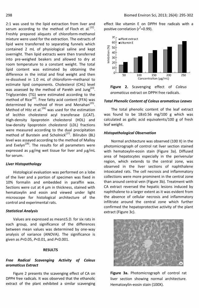
Figure 2 presents the scavenging effect of CA on DPPH free radicals. It was observed that the ethanolic extract of the plant exhibited a similar scavenging
effect like vitamin E on DPPH free radicals with a positive correlation (r2=0.99).
Figure 2. Scavenging effect of Coleus aromaticus extract on DPPH free radicals.
Total Phenolic Content of Coleus aromaticus Leaves
The total phenolic content of the leaf extract was found to be 18±0.56 mg/100 g which was calculated as gallic acid equivalents/100 g of fresh leaf weight.
Histopathological Observation
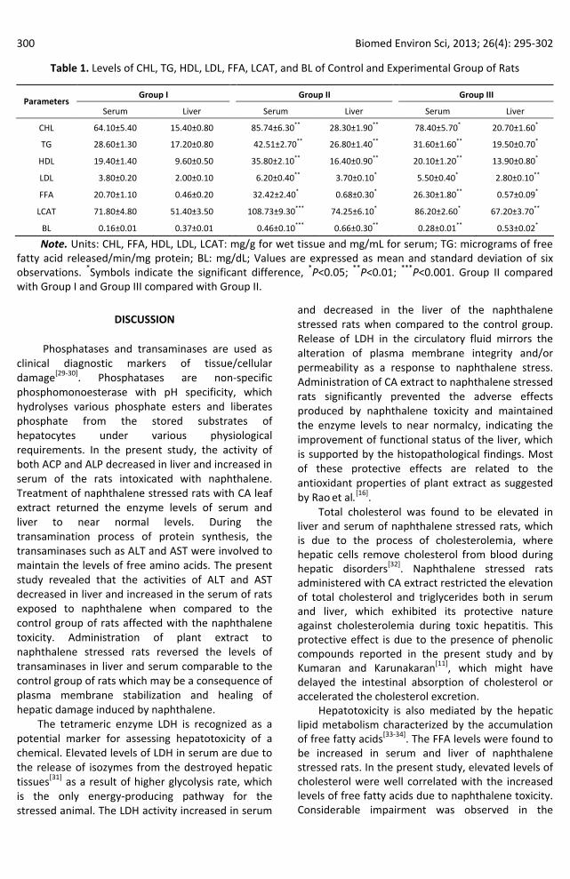
Normal architecture was observed (100 X) in the photomicrograph of control rat liver section stained with hematoxylin-eosin stain (Figure 3a). Diffused area of hepatocytes especially in the perivenular region, which extends to the central zone, was observed in the liver sections of naphthalene intoxicated rats. The cell necrosis and inflammatory collections were more prominent in the central zone than around central vein (Figure 3b). Treatment with CA extract reversed the hepatic lesions induced by naphthalene to a larger extent as it was evident from the absence of cellular necrosis and inflammatory infiltrate around the central zone which further confirmed the hepatoprotective activity of the plant extract (Figure 3c).
Figure 3a. Photomicrograph of control rat liver section showing normal architecture. Hematoxylin-eosin stain (100X).

Biomed Environ Sci, 2013; 26(4): 295-302 299
Figure 3 b. Photomicrograph of naphthalene intoxicated rat liver section shows the loss of architecture (inflammatory collections) and cell necrosis (perivenular) extending to the central zone indicating the appearance of fatty inflammation and infiltration (Hematoxylin-eosin stain 100X).
Figure 3c. Photomicrograph of rat liver section shows no hepatic necrosis in and around central vein zone of naphthalene intoxicated rat treated with Coleus aromaticus extract (Hematoxylin-eosin stain 100X).
Enzyme Systems and Biochemical Components
The activity of AST, ALT, ACP, ALP, and LDH in liver of naphthalene intoxicated rats (Group II) reduced significantly (P<0.01, P<0.01, P<0.01, P<0.05, and P<0.01) and in contrast their activity in serum increased significantly (P<0.001, P<0.01, P<0.05, P<0.01, and P<0.001) when compared to control (Group I). The activity of AST, ALT, ACP, ALP, and LDH in liver of Group III rats increased significantly (P<0.05, P<0.01, P<0.001, P<0.01, and P<0.01) and their activity in serum decreased significantly (P<0.01, P<0.05, P<0.01, P<0.05, and P<0.001) when compared to Group II rats, indicating the hepatocellular damage induced by naphthalene intoxication and the protective effect of plant extract (Figure 4).
A significant increase in the levels of CHL, FFA, LCAT, HDL, LDL, and BL (P<0.01, P<0.01, P<0.01, P<0.01, P<0.05, P<0.001, P<0.001) and (P<0.01, P<0.01, P<0.01, P<0.05, P<0.05, P<0.05, P<0.01) was observed in serum and liver of rats stressed with naphthalene (Group II) when compared to control group rats. On the other hand, the activities of CHL, FFA, LCAT, HDL, LDL, and BL reduced significantly (P<0.05, P<0.01, P<0.01, P<0.05, P<0.01, P<0.05, P<0.01) and (P<0.05, P<0.05, P<0.05, P<0.01, P<0.05, P<0.01, P<0.05) in serum and liver of rats treated with plant extract when compared to naphthalene stressed rats (Table 1). Treatment of CA extract favored a significant protection against naphthalene induced liver injury by maintaining the levels of enzyme systems and biochemical components at a near normal level.
Figure 4. Levels of marker enzymes in liver and serum of control and experimental group of rats. Units: AST: mg pyruvate/mg protein/h; ALT: mg pyruvate/mg protein/h; ACP: mg p-nitrophenol/mg protein/h; ALP: mg p-nitrophenol/mg protein/h; LDH: µ mol of pyruvate liberated/min/mg protein; Values are expressed as mean and standard deviation of six observations. *Symbols indicates the significant difference, *P<0.05, **P<0.01; ***P<0.001. Group II compared with Group I, and Group III compared with Group II.
300 Biomed Environ Sci, 2013; 26(4): 295-302
Table 1. Levels of CHL, TG, HDL, LDL, FFA, LCAT, and BL of Control and Experimental Group of Rats
Group I Group II Group III Parameters
Serum Liver Serum Liver Serum Liver
CHL 64.10±5.40 15.40±0.80 85.74±6.30** 28.30±1.90** 78.40±5.70* 20.70±1.60*
TG 28.60±1.30 17.20±0.80 42.51±2.70** 26.80±1.40** 31.60±1.60** 19.50±0.70*
HDL 19.40±1.40 9.60±0.50 35.80±2.10** 16.40±0.90** 20.10±1.20** 13.90±0.80*
LDL 3.80±0.20 2.00±0.10 6.20±0.40** 3.70±0.10* 5.50±0.40* 2.80±0.10**
FFA 20.70±1.10 0.46±0.20 32.42±2.40* 0.68±0.30* 26.30±1.80** 0.57±0.09*
LCAT 71.80±4.80 51.40±3.50 108.73±9.30*** 74.25±6.10* 86.20±2.60* 67.20±3.70**
BL 0.16±0.01 0.37±0.01 0.46±0.10*** 0.66±0.30** 0.28±0.01** 0.53±0.02*
Note. Units: CHL, FFA, HDL, LDL, LCAT: mg/g for wet tissue and mg/mL for serum; TG: micrograms of free fatty acid released/min/mg protein; BL: mg/dL; Values are expressed as mean and standard deviation of six observations. *Symbols indicate the significant difference, *P<0.05; **P<0.01; ***P<0.001. Group II compared with Group I and Group III compared with Group II.
DISCUSSION
Phosphatases and transaminases are used as clinical diagnostic markers of tissue/cellular damage[29-30]. Phosphatases are non-specific phosphomonoesterase with pH specificity, which hydrolyses various phosphate esters and liberates phosphate from the stored substrates of hepatocytes under various physiological requirements. In the present study, the activity of both ACP and ALP decreased in liver and increased in serum of the rats intoxicated with naphthalene. Treatment of naphthalene stressed rats with CA leaf extract returned the enzyme levels of serum and liver to near normal levels. During the transamination process of protein synthesis, the transaminases such as ALT and AST were involved to maintain the levels of free amino acids. The present study revealed that the activities of ALT and AST decreased in liver and increased in the serum of rats exposed to naphthalene when compared to the control group of rats affected with the naphthalene toxicity. Administration of plant extract to naphthalene stressed rats reversed the levels of transaminases in liver and serum comparable to the control group of rats which may be a consequence of plasma membrane stabilization and healing of hepatic damage induced by naphthalene.
The tetrameric enzyme LDH is recognized as a potential marker for assessing hepatotoxicity of a chemical. Elevated levels of LDH in serum are due to the release of isozymes from the destroyed hepatic tissues[31] as a result of higher glycolysis rate, which is the only energy-producing pathway for the stressed animal. The LDH activity increased in serum
and decreased in the liver of the naphthalene stressed rats when compared to the control group. Release of LDH in the circulatory fluid mirrors the alteration of plasma membrane integrity and/or permeability as a response to naphthalene stress. Administration of CA extract to naphthalene stressed rats significantly prevented the adverse effects produced by naphthalene toxicity and maintained the enzyme levels to near normalcy, indicating the improvement of functional status of the liver, which is supported by the histopathological findings. Most of these protective effects are related to the antioxidant properties of plant extract as suggested by Rao et al.[16].
Total cholesterol was found to be elevated in liver and serum of naphthalene stressed rats, which is due to the process of cholesterolemia, where hepatic cells remove cholesterol from blood during hepatic disorders[32]. Naphthalene stressed rats administered with CA extract restricted the elevation of total cholesterol and triglycerides both in serum and liver, which exhibited its protective nature against cholesterolemia during toxic hepatitis. This protective effect is due to the presence of phenolic compounds reported in the present study and by Kumaran and Karunakaran[11], which might have delayed the intestinal absorption of cholesterol or accelerated the cholesterol excretion.
Hepatotoxicity is also mediated by the hepatic lipid metabolism characterized by the accumulation of free fatty acids[33-34]. The FFA levels were found to be increased in serum and liver of naphthalene stressed rats. In the present study, elevated levels of cholesterol were well correlated with the increased levels of free fatty acids due to naphthalene toxicity. Considerable impairment was observed in the

Biomed Environ Sci, 2013; 26(4): 295-302 301
production of LCAT in naphthalene stressed rats. Rats treated with CA extract exhibited significant reduction in the LCAT levels both in liver and serum, thereby confirming its efficacy.
Triglycerides synthesized by hepatic cells bind to the luminal surface of endothelial cells of liver sinusoid and play a vital role in removing the triglycerides from the partially catabolized very low-density lipoprotein (VLDL), thereby converting VLDL to low-density lipoprotein (LDL). Naphthalene intoxicated animals showed considerable elevation of the TG levels in serum and liver, which was moderately prevented in the animals, treated with plant extract, representing its vital role in preventing the injury triggered by naphthalene in endothelial cells. The serum level of LDL was significantly elevated in naphthalene stressed rats. The phenolic compounds have been shown to form phenoxyl radicals in the presence of peroxidases. Administration of CA extract significantly reduced the LDL levels in naphthalene intoxicated rats, which can be attributed to the antioxidant property of phenolic compounds which are present in the plant extract capable of inhibiting the LDL peroxidation.
It has been suggested that the hypolipidemic compounds with antioxidant properties prevent LDL peroxidation and retard their synthesis[35]. The elevated levels of liver and serum HDL in naphthalene stressed rats might be due to the hypertriglyceridemia prompted by reactive metabolites generated as a result of microsomal metabolism. Oxidative stress is one of the major pathways involving reactive oxygen species to initiate lipid peroxidation[36]. HDL is a free radical scavenger and prevents the peroxidation of beta lipoproteins[37]. Rats administrated with CA extract exhibited reduction in the levels of HDL, which was due to the ability of plant extract to accelerate the breakdown of free radical species generated during naphthalene intoxication. Serum bilirubin is a potential marker for the hepatic dysfunction and any abnormal increase in the levels of bilirubin in the serum denotes hepatobiliary disease and hepatocellular dysfunction[38]. The present investigation reveals that the increased levels of bilirubin in serum and liver are the result of naphthalene induced hepatitis. The extract mediated suppression of bilirubin induction suggests the ability of CA extract to stabilize biliary dysfunction.
The assessment of the enzyme systems and biochemical components is basically supported by the histological observations. Massive fatty changes,
gross necrosis, infiltration of lymphocytes around the central vein and loss of cellular boundaries were observed in the liver tissues of naphthalene intoxicated rats. The histopathological observations of the liver of rats treated with CA extract exhibited a near to normal morphology and recovery from the hepatic lesions induced by the naphthalene. Therefore, it is suggested that C. aromaticus extract protects liver by stabilizing the plasma membrane and the stimulation of hepatic regeneration makes the liver more resistant to damage by the toxin.
CONCLUSION
The therapeutic properties and effects of phenolic compounds have received adequate importance for their potential beneficial effects on human health and have aroused increasing interest in recent years. Plant polyphenols are known to exhibit a wide range of pharmacological activities such as antioxidant, anti-inflammatory and cancer-preventive properties, and the elucidation and quantification of phenolic compounds is of major interest. The phenolic compounds as potential antioxidants play an important role in adsorbing and scavenging free radicals, quenching oxygen (singlet and triplet) and peroxides decomposition. In the present study, the possible hepatoprotective effect of C. aromaticus extract on the naphthalene-induced liver injuries may be associated with the presence of phenolic compounds which might have enhanced the free radical scavenging properties, aiding the stabilization of the hepatocellular membrane, and enhanced protein and glycoprotein biosynthesis etc., showing some promising protection against the toxic hepatitis. The hepatoprotective effect of CA extract might not be confined just to the presence of phenolic compounds. Other antioxidant compounds like flavonoids and bioactive reactions such as oxidative inhibition, hydroxyl radical, DPPH, ABTS, nitric oxide, and superoxide scavenging properties of other unidentified bioactive compounds might also be responsible, as being evidenced by other researchers[11,16,39-40]. The bioactive compounds of the plant extract not only enhanced the plasma membrane integrity, but also boosted the regenerative and reparative capacity of the liver. The results of our investigation clearly suggest that the bioactive compounds present in the plant extract efficiently help the liver to function normally by neutralizing the toxic effects of naphthalene. Yet further studies are needed for better understanding

302 Biomed Environ Sci, 2013; 26(4): 295-302
of the protective mechanisms of action and to evaluate the efficacy of bioactive compounds in C. aromaticus.
REFERENCES
1. ATSDR. Toxicological Profile of Naphthalene, 1-Methylnaphthalene, 2-Methylnaphthalene. U. S. Department of Health and Human Services, Agency for Toxic Substances and Disease Registry, 2005.
2. Ahmed AAE, Fatani AJ. Protective effect of grape seed sproanthocyanidins against naphthalene-induced hepatotoxicity in rats. Saudi Pharmaceutical Journal, 2007; 15 (1), 38-47.
3. Vijayavel K, Abuselvam C, Balasubramanian MP. Antioxidant effect of the marine algae Chlorella vulgaris against naphthalene induced oxidative stress in the albino rats. Mol Cell Biochem, 2007; 303(1-2), 39-44.
4. Bates N. Mothball poisoning. Emerg Nurse, 2002; 10, 24-8. 5. Kurz JM. Naphthalene poisoning: Critical care nursing
techniques. Dimens Crit Care Nurs, 1987; 6, 264-70. 6. Ojwang PJ, Ahmed-Jushuf IH, Abdullah MS. Naphthalene
poisoning following ingestion of moth balls: Case report. East Afr Med J, 1985; 62(1), 71-3.
7. Gupta R, Singhal PC, Muthusethupathy MA, et al. Cerebral oedema and renal failure following naphthalene poisoning. J Assoc Physicians India, 1979; 27, 347-8.
8. MacGregor RR. Naphthalene poisoning from the ingestion of moth balls. Can Med Assoc J, 1954; 70, 313-4.
9. Mitra SK, Venkataranganna MV, Sundaram R, Gopumadhavan S. Protective effect of HD-03, a herbal formulation, against various hepatotoxic agents in rats. J Ethnopharm, 1998; 63, 181-6.
10. Rasineni GK, Siddavattam D, Reddy AR. Free radical quenching activity and polyphenols in three species of Coleus. J Med Plant Res, 2008; 2(10), 285-91.
11. Kumaran A, Karunakaran RJ. Activity-guided isolation and identification of free radical-scavenging components from an aqueous extract of Coleus aromaticus. Food Chem, 2007; 100(1), 356-61.
12. Khory NR, Katrak NN. Materia medica of India and their therapeutics. New Delhi: BDH Printers, 1999; pp.380.
13. Morton JF. Country borage (Coleus ambonicus Lour): a potent flavoring and medicinal plant. J Herb Spice Med Plant, 1992; 1(1-2), 77-90.
14. Lans C. Ethnomedicines used in Trinidad and Tobago for reproductive problems. J Ethnobiol Ethnomed, 2007; 15, 3-13.
15. Pritima RA, Pandian RS. Antimicrobial Activity of Coleus aromaticus (Benth) against microbes of reproductive tract infections among women. African J Infec Disease, 2007; 1(1), 18-2.
16. Rao BS, Shanbhoge R, Upadhya D, et al. Antioxidant, anticlastogenic and radioprotective effect of Coleus aromaticus on Chinese hamster fibroblast cells (V79) exposed to gamma radiation. Mutagen, 2006; 21(4), 237-42.
17. Ursini F, Maiorino M, Morazzoni P, et al. A novel antioxidant flavonoid (IdB 1031) reacting molecular mechanisms of cellular activation. Free Rad Biol Med, 1994; 16, 547-53.
18. Malick C, Singh MB. In: Plant Enzymology and Histoenzymology, (eds) Kalyani Publishers, New Delhi, 1980; pp. 286.
19. Balasubramanian MP, Dhandayuthapani S, Neelaiappan K, et al. Comparative studies on phosphomonoesterase in helminths. Helminthol, 1983; 20, 111-20.
20. Reitman S, Frankel S. Determination of serum glutamate oxaloacetate and glutamic pyruvic acid transaminase. American J Clinic Pathol, 1957; 28, 56-66.
21. King J. Practical Clinical Enzymology. Dvan. Nostrand Co, London, 1965; pp, 83-93.
22. Floch J, Lees M, Stanley GHS. A simple method for the isolation and purification of total lipids from animal tissues. J Biol Chem, 1957; 226, 497-509.
23. Parekh AC, Jung DH. Cholesterol determination with ferric acetateuranyl acetate and sulphuric acid-ferrous sulphate reagents. Anal Chem, 1970; 42, 1423-7.
24. Rice EW. Triglycerides in serum. In: Roedrick P, McDonald RP (eds), Standard Methods in Clinical Chemistry, Academic Press, New York, 1970; pp. 215.
25. Hron WT, Menahan LA. A sensitive method for the determination of free fatty acids in plasma. J Lipid Res, 1981; 122, 377-81.
26. Hitz J, Steinmetz J, Siest G. Plasma lecithin: cholesterol acyl transferase reference values and effects of xenobiotics. Clin Chem Acta, 1983; 133, 85-6.
27. Burstein M, Scholnick HR. Precipitation of chylomicron and very low density lipoprotein from human serum with sodium lauryl sulphate. Life Sci, 1972; 1, 177-84.
28. Malloy E, Evelyn K. The determination of bilirubin with the photoelectric colorimeter. J Bio Chem, 1987; 199, 481-5.
29. Manabe A, Cheng CC, Egashira Y, et al. Dietary wheat gluten alleviates the elevation of serum transaminase activities in d-galactosamine-injected rats. J Nut Scien Vitaminol, 1996; 42, 121-32.
30. Hemieda FA, Abdel-Hady SK, Elnga MA. Biochemical and histological studies on H2-receptor antagonist ranitidine-induced hepatotoxicity in rats. Indian J Exp Biol, 2005; 43(9), 782-5.
31. Saad B, Dakwar S, Said O, et al. Evaluation of medicinal plant hepatotoxicity in co-cultures of hepatocytes and monocytes. Evid Based Comp Alternat Med, 2006; 3(1), 93-8.
32. Glickman RM, Sebesin SM. Lipid metabolism. In: Arias IM, Schachter D, Popper H, Shafritz A (eds), The Liver Biology and Pathobiology. Raven Press, New York, 1982; pp 123-42.
33. Owen JS. Extra hepatic cell membrane lipid abnormalities and cellular dysfunction in liver disease. Drugs, 1990; 40, 73-83.
34. Senthilkumar R., Viswanathan P, Nalini N. Glycine modulates hepatic lipid accumulation in alcohol-induced liver injury. Pol J Pharmacol, 2003; 55(4), 603-11.
35. Daugherty A, Zweifel BS, Schonfeld G. The effects of probucol on the progression of atherosclerosis in mature watanabe heritable hyperlipidemic rabbits. Br J Pharm, 1991; 103, 1013-8.
36. Vijayavel K, Anbuselvam C, Balasubramanian MP. Free radical scavenging activity of the marine mangrove Rhizophora apiculata bark extract with reference to naphthalene induced mitochondrial dysfunction. Chem Biol Int, 2006; 163, 170-5.
37. Chander R, Kapoor NK. High-density lipoprotein is a scavenger of superoxide anions. Biochem Pharmacol, 1990; 40, 1663-5.
38. Martin P, Friedman LS. Assessment of liver function and diagnostic studies. In: Friedman LS, Keeffe EB (Eds.), Hand Book of Liver Disease. Churchill Livingstone, Philadelphia, 1992; pp. 1-14.
39. Kumaran A, Karunakaran RJ. Antioxidant and free radical scavenging activity of an aqueous extract of Coleus aromaticus. Food Chem, 2006; 97, 109-14.
40.Rout OP, Acharya R, Mishra SK. In-Vitro Antioxidant potentials in leaves of Coleus aromaticus Benth and rhizomes of Zingiber zerumbet (L) SM. Journal of Applied Pharmaceutical Science, 2011; 1(8), 194-8.


![ACTIVITY TEST OF EXTRACT OF ILER LEAF (Coleus ...ACTIVITY TEST OF EXTRACT OF ILER LEAF (Coleus atropurpureus [L] Benth ) TO STAPHYLOCOCCUS AUREUS BACTERIA UJI AKTIVITAS EKSTRAK DAUN](https://img.dokumen.tips/doc/110x75/5e6641b624047465221dd574/activity-test-of-extract-of-iler-leaf-coleus-activity-test-of-extract-of-iler.jpg)


![EFFECT OF PLANTING COLEUS BLUME] TARO (COLOCASIA … … · EFFECT OF PLANTING COLEUS BLUME] ON INSECT POPULATIONS IN TARO (COLOCASIA ESCULENTA) FIELDS IN AMERICAN SAMOA A. M. VARGO,](https://img.dokumen.tips/doc/110x75/5f06fa377e708231d41aaead/effect-of-planting-coleus-blume-taro-colocasia-effect-of-planting-coleus-blume.jpg)


