Embed Size (px)
Citation preview
Abstracts
a guidewire using a prototype monorail type system. Digitalized images of theentire OCT examination with a corresponding fluoroscopic image identifying thelocation of the OCT probe and the distance of the probe from the major papillawere recorded during probe withdrawal. Histologic analysis of brush cytology,biopsy, EUS-FNA, and/or surgical resection specimens from the corresponding areaof interest in the BD and PD was reviewed in a blinded fashion. OCT featurescharacterized on the basis of our previous experience in dogs and reported inhumans (reflectance pattern, intensity, and structural organization) were comparedto the fluoroscopic and histologic features of normal epithelium, inflammation,dysplasia, and cancer. Results: 22 patients (14 men; mean age 68, range 35-89) wereenrolled with BD OCT imaging in 17 pts [6 normal and 11 strictures-1 extrinsicHodgkin’s lymphoma, 3 pancreatic cancer, 2 benign PSC-dominant strictures, 1autoimmune pancreatitis (AIP), 3 cholangiocarcinoma, and 1 post-transplantbenign anastomotic stricture] and PD OCT imaging in 3 pts [1 chronic pancreatitisstricture, 1 pancreatic cancer, and 1 intraductal papillary mucinous neoplasia(IPMN)]. Low light scattering and loss of organizational structure on OCTcorrelated with dysplasia and cancer in all patients with cancer. In patients withinflammation, such as with recent passage of a stone and benign post-transplantstricture, a heterogenous reflectance pattern and large gland-like structures werenoted. Large, finger-like projections were noted in the IPMN patient on OCT thatwere not identified by pancreatoscopy. No complications occurred in any patientundergoing OCT. Conclusions: In this pilot study, in vivo intraductal OCT of the BDand PD was both feasible and safe. Intraductal OCT accurately identifies dysplasticcellular changes or cancer in the PD and BD epithelium during ERCP.
1065
Peroral Pancreatoscopy (PP) for Pancreatic Stone Therapy and
Investigation of Susptected Pancreatic Lesions - First Human
Experience Using the Spyglass Direct Visualization System
(SDVS)Yang K. Chen, Paul R. Tarnasky, Isaac Raijman, Douglas Pleskow, RajJ. Shah, Evelina L. Fortajada, Richard a. KozarekIntroduction: PP offers direct access and visualization of the pancreatic ducts withthe potential to enhance visual diagnosis, direct tissue sampling, and guide therapy.Recently, a single operator modular system consisting of a 10Fr disposable catheterwith 4-way tip deflection, reusable optical probe, 1.2 mm working channel anddedicated irrigation channels was released for clinical use in the biliary system(Spyglass Direct Visualization System, Boston Scientific Corporation, Marlboro,MA). Methods: The goal of this study is to document the performance, safety, andutility of the SDVS in patients requiring PP for stone therapy or investigation ofsuspicious lesions with or without biopsy. Procedural success was defined as abilityto access and visualize the target lesion, obtain biopsies, or achieve stone clearanceas needed. Results: 48 consecutive pts (median age 67 yo, IQR 17-88; 26M/22F)underwent PP using the SDVS. Indications for PP were: pancreatic stones (17),indeterminate strictures (11), dilated ducts (5), indeterminated filling defects (11),mass (4). 31 pts had sphincterotomy: 14 had prior sphincterotomy, 14 requiredpancreatic sphincterotomy during the procedure, 2 required extension. The meanpancreatic duct diameter was 7.6 mm. Stones were located in the head (16) and tail(1); mean diameter of largest stone in each case was 9.4 mm (SD 5.7). EHL wasattempted in 10 pts and stone clearance was achieved in 8 pts (80%) after a mean of1.2 SDVS-guided EHL sessions (SD 0.44). EHL was planned but not performed in 2other pts because of technical difficulties (unable to visually target stones for EHL(1); unable to pass the EHL probe through Spyscope (1). PP-guided biopsies wereattempted in 10 pts using a disposable Spybite forceps. A mean of 4.4 (SD 1.7)biopsy specimens per case provided adequate samples for histologic evaluation inall patients. No serious adverse events, and only one mild adverse event, related toPP or SDVS devices were reported. Conclusions: PP using the Spyglass DVS Systemis technically feasible, allowing access to target sites in the main pancreatic duct forvisual inspection, stone therapy and tissue sampling in most patients. Based on thispreliminary experience, PP can be safely performed in patients with pancreaticstones, IPMN and indeterminate strictures, however, its safety and clinical utility inpatients with small ducts and/or non-obstructed ducts remains to be proven. Asmaller catheter diameter is preferred for pancreatic application.
1066
Endoscopic Drainage of Pancreatic Fluid Collections with Fully
Covered Metallic Stents (CSEMS). How Does It Compare to
Conventional Drainage with Plastic Stents?Jayant P. Talreja, Vanessa M. Shami, Jennifer Ku, Kristi Ellen,Michel KahalehBackground: Transenteric drainage of pancreatic fluid collections (PFC) usingcovered metallic stents (CSEMS) has been investigated by our group and otherssince it offers the option of providing a larger diameter access fistula for drainagewhen compared to plastic stents. Aim: To evaluate the efficacy and safety oftransenteric CSEMS placement in the drainage of PFC and compare this techniqueto conventional drainage using plastic stents. Material and methods: BetweenJanuary 2007 and September 2007, 18 patients (51 � 18 y/o, 12 male) underwentplacement of CSEMS (VIABIL�, Conmed) for the drainage of PFC. All but 2 patients
AB108 GASTROINTESTINAL ENDOSCOPY Volume 67, No. 5 : 2008
were drained using EUS-guided drainage. A double pigtail was placed along side (4cases) or into the CSEMS (14 cases) to prevent migration. Follow-up and finalresults were prospectively recorded and compared to a group of 18 patientsmatched by age, gender, complication rate and size of PFC (see table). Results:Etiologies of PFC were gallstone (9), alcohol (5) and other (4). Mean size was 10 �4 cm (range: 4-16). A median of 1 session was required to achieve drainage (range:1-4). Mean time of follow-up until final evaluation was 66 � 56 days (range: 15-240).Complications included superinfection (5), bleeding (2) and inner migration (1).One patient experienced migration of the CSEMS with resolution of the collectionand spontaneous expulsion. A total of 17/18 (95%) patients responded successfullywith 14 (78%) patients achieving complete resolution of their PFC. The patient whofailed required multiple endoscopic sessions and surgical drainage of infectednecrosis. When compared to conventional drainage with plastic stents, patientswho underwent CSEMS drainage had a statistically significant shorter time tosuccessful response (see table). Conclusion: Placement of a CSEMS seems to offera faster alternative for the drainage of PFC when compared to conventionaldrainage with a plastic stent. A prospective and randomized study should beperformed comparing the two techniques to confirm this data.
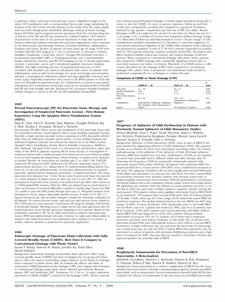
Comparison of CSEMS vs. Plastic Drainage of PFC
CSEMS (N Z 18) Plastic (N Z 18)Pvalue
www.giejournal
AGE (years) 5
1 � 18 50 � 17 0 .429 Gender (male/female) 1 2/6 11/7 0 .331 Size (cm) 1 0 � 4 8 � 3 0 .118 Time till final outcome(days) 6 6 � 56 138 � 118 0 .03Complications S
uperinfection: 5 Bleeding: 2 Migration: 1 Superinfection: 3 Perforation: 2Migration: 1 0 .494ResultsPF
FC resolution: 14 PFC improvement: 3ailure: 1
PFC resolution: 17 Failure: 1 0 .0831067
Frequency of Sphincter of Oddi Dysfunction in Patients with
Previously Normal Sphincter of Oddi Manometry StudiesMouen Khashab, Evan L. Fogel, Stuart Sherman, James L. Watkins,Lee Mchenry, Pradermchai Kongkam, Furqaan Ahmed, Laura Lazzell-Pannell, Suzette E. Schmidt, Glen a. LehmanBackground: Sphincter of Oddi manometry (SOM), done at time of ERCP, is thegold standard for diagnosing sphincter of Oddi dysfunction (SOD). The questionremains as to whether the short term manometric recordings reflect the 24-hourpathophysiology of the sphincter. If the sphincter of Oddi (SO) basal sphincterpressure (BSP) does indeed vary over time, performance of SOM on two separateoccasions may potentially lead to different results and affect therapy. Aim: Todetermine the frequency of SOD in persistently symptomatic patients withpreviously normal SOM studies. Methods: All patients who underwent ERCP forsuspected SOD over a 13-year period (1994-2007) were considered for inclusion inthe study. The subgroup of patients with an intact papilla and a normal SOM study(both biliary and pancreatic) was selected and only those who had a repeat ERCPfor persistent symptoms were included. Patients with previous endoscopic orsurgical papillary interventions were excluded. SOM was performed in conventionalretrograde fashion with a low-compliance infusion pump system and a triple-lumen5Fr aspirating type catheter. SOD was defined as a basal sphincter pressure R 40mm Hg in either the pancreatic or biliary sphincter segments. Results: During thestudy period, 5352 patients without prior papillary intervention had an ERCP/SOMat our institution. 1073 patients had a normal SOM study. Only 30 of these patients(27 Female, 3 Male, mean age 40.1 years, range 23-69) underwent repeat ERCP forpersistent symptoms. The median duration between the two ERCPs was 493.5 days(range, 52-3538). In these 30 patients, SOD classification prior to the initial ERCPwas as follows: type I in 1 patient (not treated in 1994), type II in 12 patients, typeIII in 5 patients. 12/30 (40%) patients had normal pancreatic and biliary SOM atrepeat ERCP. SOD was diagnosed in 18/30 (60%) patients. Post-proceduralpancreatitis occurred in 5/30 (16.7%) patients, all of whom had a temporary,protective pancreatic stent placed. Summary: In this study, 60% of patients withpreviously normal SOM were subsequently found to have SOD at a second ERCP.Conclusion: A single SOM study may not represent the day-to-day SO physiology;one normal exam may not rule out SOD. A repeat ERCP with manometry may bewarranted in a subset of patients with persistent debilitating symptoms and a highindex of suspicion for SOD. Outcome data are needed to determine whether thisapproach justifies the potential risks of ERCP.
1068
Prophylactic Somatostatin for Prevention of Post-ERCP
Pancreatitis: A Meta-AnalysisAbhishek Choudhary, Matthew L. Bechtold, Srinivas R. Puli, MohamedO. Othman, Wilson P. Pais, Mainor R. Antillon, Praveen K. RoyBackground: Acute pancreatitis is a common complication of ERCP. Over the years,attempts have been made to identify a pharmacological agent to prevent post-ERCPpancreatitis, such as somatostatin. Several randomized controlled trials (RCTs) haveevaluated the efficacy of somatostatin in the prevention of post-ERCP pancreatitis
.org
Abstracts
with mixed results. We conducted a meta-analysis to evaluate the efficacy ofprophylactic somatostatin for prevention of post-ERCP pancreatitis. Methods:MEDLINE, Cochrane Central Register of Controlled Trials & Database of SystematicReviews, PubMed, and recent abstracts from major conference proceedings weresearched (through 10/07). RCTs comparing prophylactic somatostatin to placebo orno somatostatin in the setting of post-ERCP pancreatitis were included. Standardforms were used to extract data by two independent reviewers. The effects ofprophylactic somatostatin were analyzed by calculating pooled estimates of post-ERCP pancreatitis, hyperamylasemia, mortality, grade of pancreatitis, and abdominalpain. Separate analyses were performed for each outcome by using odds ratio (OR).Publication bias was assessed by funnel plots. Heterogeneity among studies wasassessed by calculating I2 measure of inconsistency. Results: Fourteen RCTs (N Z2,827) met the inclusion criteria. Five trials administered somatostatin as a bolusinfusion and nine trials as a continuous infusion. There was no significantheterogeneity among the studies. Somatostatin infusion with ERCP decreased post-ERCP pancreatitis (OR 0.44, 95% CI: 0.23-0.83, p Z 0.01; NNT 34), level ofhyperamylasemia (OR 0.66, 95% CI: 0.52-0.85, p ! 0.01), and abdominal pain (OR0.46, 95% CI: 0.23-0.94, p Z 0.03). No significant difference was noted for mortality(OR 0.12, 95% CI: 0.01-2.31, p Z 0.16). Sub-group analysis based on mode ofadministration revealed that bolus infusion decreased the odds of post-ERCPpancreatitis (OR 0.27, 95% CI: 0.14-0.53, p ! 0.01; NNT 13); however, continuousinfusion did not (OR 0.56, 95% CI: 0.26-1.20, p Z 0.14). Analysis of high qualitystudies (Jadad score R 3) revealed similar results (OR 0.36, 95% CI: 0.15-0.86, p Z0.02; NNT 25). No significant publication bias was observed. Conclusions:Prophylactic somatostatin given as a bolus injection is efficacious in decreasing theodds of post-ERCP pancreatitis, hyperamylasemia, and abdominal pain; however, itdid not decrease mortality.
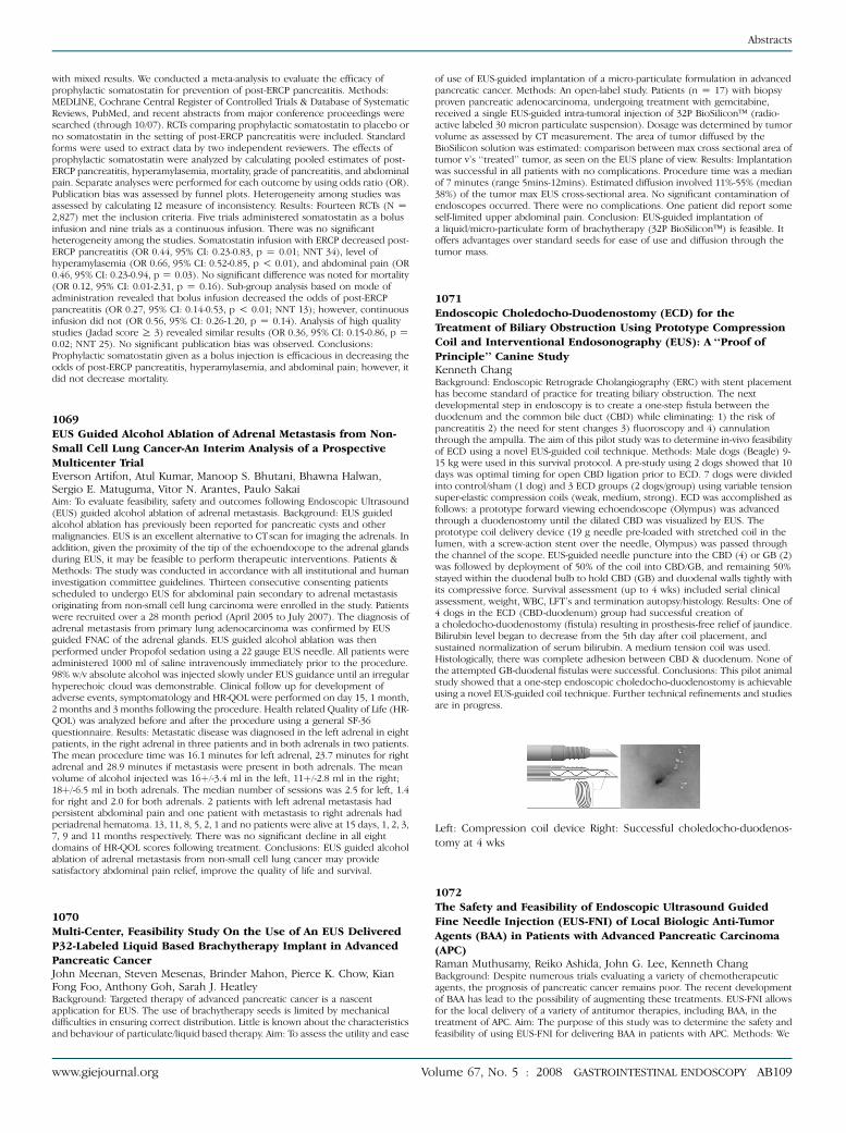
Left: Compression coil device Right: Successful choledocho-duodenos-
tomy at 4 wks
1069
EUS Guided Alcohol Ablation of Adrenal Metastasis from Non-
Small Cell Lung Cancer-An Interim Analysis of a Prospective
Multicenter TrialEverson Artifon, Atul Kumar, Manoop S. Bhutani, Bhawna Halwan,Sergio E. Matuguma, Vitor N. Arantes, Paulo SakaiAim: To evaluate feasibility, safety and outcomes following Endoscopic Ultrasound(EUS) guided alcohol ablation of adrenal metastasis. Background: EUS guidedalcohol ablation has previously been reported for pancreatic cysts and othermalignancies. EUS is an excellent alternative to CT scan for imaging the adrenals. Inaddition, given the proximity of the tip of the echoendocope to the adrenal glandsduring EUS, it may be feasible to perform therapeutic interventions. Patients &Methods: The study was conducted in accordance with all institutional and humaninvestigation committee guidelines. Thirteen consecutive consenting patientsscheduled to undergo EUS for abdominal pain secondary to adrenal metastasisoriginating from non-small cell lung carcinoma were enrolled in the study. Patientswere recruited over a 28 month period (April 2005 to July 2007). The diagnosis ofadrenal metastasis from primary lung adenocarcinoma was confirmed by EUSguided FNAC of the adrenal glands. EUS guided alcohol ablation was thenperformed under Propofol sedation using a 22 gauge EUS needle. All patients wereadministered 1000 ml of saline intravenously immediately prior to the procedure.98% w/v absolute alcohol was injected slowly under EUS guidance until an irregularhyperechoic cloud was demonstrable. Clinical follow up for development ofadverse events, symptomatology and HR-QOL were performed on day 15, 1 month,2 months and 3 months following the procedure. Health related Quality of Life (HR-QOL) was analyzed before and after the procedure using a general SF-36questionnaire. Results: Metastatic disease was diagnosed in the left adrenal in eightpatients, in the right adrenal in three patients and in both adrenals in two patients.The mean procedure time was 16.1 minutes for left adrenal, 23.7 minutes for rightadrenal and 28.9 minutes if metastasis were present in both adrenals. The meanvolume of alcohol injected was 16þ/-3.4 ml in the left, 11þ/-2.8 ml in the right;18þ/-6.5 ml in both adrenals. The median number of sessions was 2.5 for left, 1.4for right and 2.0 for both adrenals. 2 patients with left adrenal metastasis hadpersistent abdominal pain and one patient with metastasis to right adrenals hadperiadrenal hematoma. 13, 11, 8, 5, 2, 1 and no patients were alive at 15 days, 1, 2, 3,7, 9 and 11 months respectively. There was no significant decline in all eightdomains of HR-QOL scores following treatment. Conclusions: EUS guided alcoholablation of adrenal metastasis from non-small cell lung cancer may providesatisfactory abdominal pain relief, improve the quality of life and survival.
1070
Multi-Center, Feasibility Study On the Use of An EUS Delivered
P32-Labeled Liquid Based Brachytherapy Implant in Advanced
Pancreatic CancerJohn Meenan, Steven Mesenas, Brinder Mahon, Pierce K. Chow, KianFong Foo, Anthony Goh, Sarah J. HeatleyBackground: Targeted therapy of advanced pancreatic cancer is a nascentapplication for EUS. The use of brachytherapy seeds is limited by mechanicaldifficulties in ensuring correct distribution. Little is known about the characteristicsand behaviour of particulate/liquid based therapy. Aim: To assess the utility and ease
www.giejournal.org Vo
of use of EUS-guided implantation of a micro-particulate formulation in advancedpancreatic cancer. Methods: An open-label study. Patients (n Z 17) with biopsyproven pancreatic adenocarcinoma, undergoing treatment with gemcitabine,received a single EUS-guided intra-tumoral injection of 32P BioSilicon� (radio-active labeled 30 micron particulate suspension). Dosage was determined by tumorvolume as assessed by CT measurement. The area of tumor diffused by theBioSilicon solution was estimated: comparison between max cross sectional area oftumor v’s ‘‘treated’’ tumor, as seen on the EUS plane of view. Results: Implantationwas successful in all patients with no complications. Procedure time was a medianof 7 minutes (range 5mins-12mins). Estimated diffusion involved 11%-55% (median38%) of the tumor max EUS cross-sectional area. No significant contamination ofendoscopes occurred. There were no complications. One patient did report someself-limited upper abdominal pain. Conclusion: EUS-guided implantation ofa liquid/micro-particulate form of brachytherapy (32P BioSilicon�) is feasible. Itoffers advantages over standard seeds for ease of use and diffusion through thetumor mass.
1071
Endoscopic Choledocho-Duodenostomy (ECD) for the
Treatment of Biliary Obstruction Using Prototype Compression
Coil and Interventional Endosonography (EUS): A ‘‘Proof of
Principle’’ Canine StudyKenneth ChangBackground: Endoscopic Retrograde Cholangiography (ERC) with stent placementhas become standard of practice for treating biliary obstruction. The nextdevelopmental step in endoscopy is to create a one-step fistula between theduodenum and the common bile duct (CBD) while eliminating: 1) the risk ofpancreatitis 2) the need for stent changes 3) fluoroscopy and 4) cannulationthrough the ampulla. The aim of this pilot study was to determine in-vivo feasibilityof ECD using a novel EUS-guided coil technique. Methods: Male dogs (Beagle) 9-15 kg were used in this survival protocol. A pre-study using 2 dogs showed that 10days was optimal timing for open CBD ligation prior to ECD. 7 dogs were dividedinto control/sham (1 dog) and 3 ECD groups (2 dogs/group) using variable tensionsuper-elastic compression coils (weak, medium, strong). ECD was accomplished asfollows: a prototype forward viewing echoendoscope (Olympus) was advancedthrough a duodenostomy until the dilated CBD was visualized by EUS. Theprototype coil delivery device (19 g needle pre-loaded with stretched coil in thelumen, with a screw-action stent over the needle, Olympus) was passed throughthe channel of the scope. EUS-guided needle puncture into the CBD (4) or GB (2)was followed by deployment of 50% of the coil into CBD/GB, and remaining 50%stayed within the duodenal bulb to hold CBD (GB) and duodenal walls tightly withits compressive force. Survival assessment (up to 4 wks) included serial clinicalassessment, weight, WBC, LFT’s and termination autopsy/histology. Results: One of4 dogs in the ECD (CBD-duodenum) group had successful creation ofa choledocho-duodenostomy (fistula) resulting in prosthesis-free relief of jaundice.Bilirubin level began to decrease from the 5th day after coil placement, andsustained normalization of serum bilirubin. A medium tension coil was used.Histologically, there was complete adhesion between CBD & duodenum. None ofthe attempted GB-duodenal fistulas were successful. Conclusions: This pilot animalstudy showed that a one-step endoscopic choledocho-duodenostomy is achievableusing a novel EUS-guided coil technique. Further technical refinements and studiesare in progress.
1072
The Safety and Feasibility of Endoscopic Ultrasound Guided
Fine Needle Injection (EUS-FNI) of Local Biologic Anti-Tumor
Agents (BAA) in Patients with Advanced Pancreatic Carcinoma
(APC)Raman Muthusamy, Reiko Ashida, John G. Lee, Kenneth ChangBackground: Despite numerous trials evaluating a variety of chemotherapeuticagents, the prognosis of pancreatic cancer remains poor. The recent developmentof BAA has lead to the possibility of augmenting these treatments. EUS-FNI allowsfor the local delivery of a variety of antitumor therapies, including BAA, in thetreatment of APC. Aim: The purpose of this study was to determine the safety andfeasibility of using EUS-FNI for delivering BAA in patients with APC. Methods: We
lume 67, No. 5 : 2008 GASTROINTESTINAL ENDOSCOPY AB109