Embed Size (px)
Citation preview
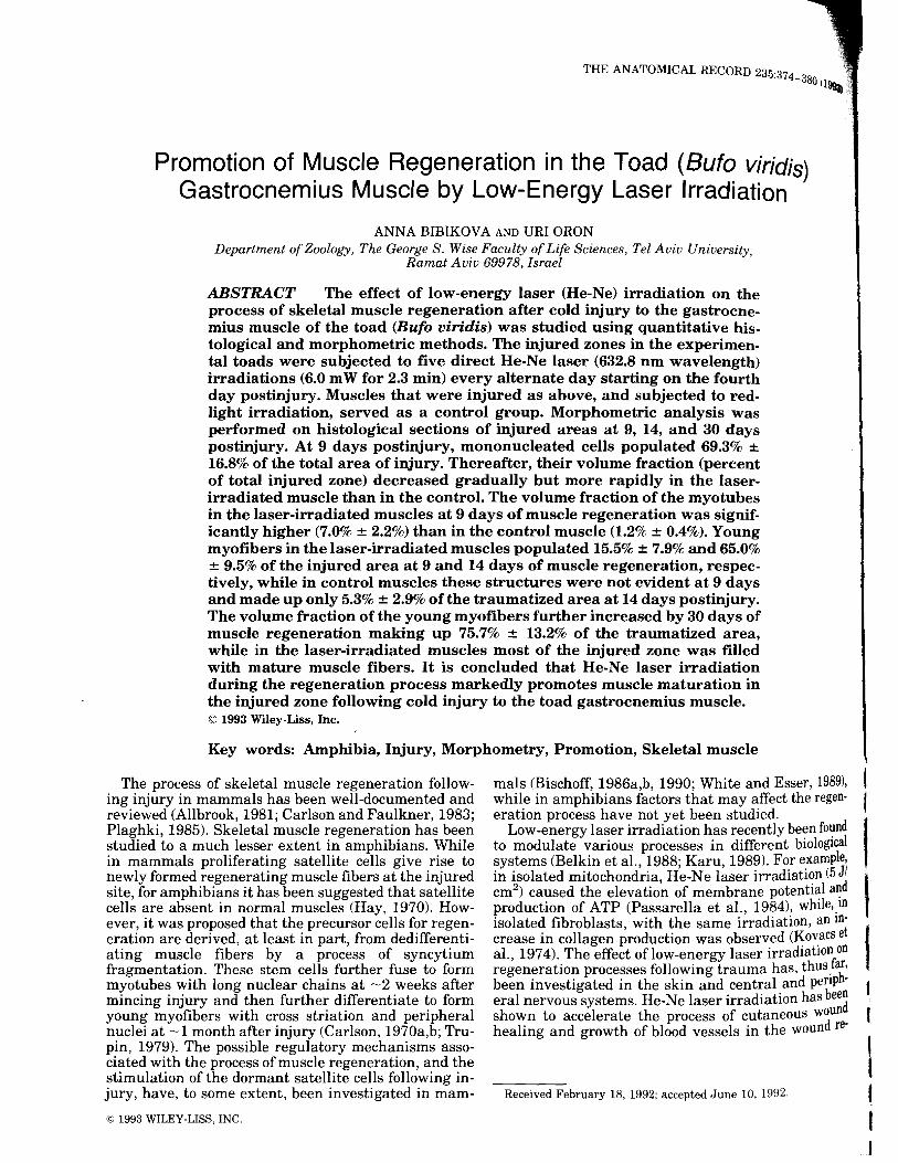
THE ANATOMICAL RECORD 235:374 -380 1199a)
Promotion of Muscle Regeneration in the Toad (Bufo viridis) Gastrocnemius Muscle by Low-Energy Laser Irradiation
ANNA BIBIKOV A AND URI ORON Department of Zoology, The George S. Wise Faculty of Life Sciences, Tel Aviv University,
Ramat Aviv 69978, Israel
ABSTRACT The effect of low-energy laser (He-Ne) irradiation on the process of skeletal muscle regeneration after cold injury to the gastrocnemius muscle of the toad (Bufo viridis) was studied using quantitative histological and morphometric methods. The injured zones in the experimental toads were subjected to five direct He-Ne laser (632.8 nm wavelength) irradiations (6.0 mW for 2.3 min) every alternate day starting on the fourth day postinjury. Muscles that were injured as above, and subjected to redlight irradiation, served as a control group. Morphometric analysis was performed on histological sections of injured areas at 9, 14, and 30 days postinjury. At 9 days postinjury, mononucleated cells populated 69.3% ± 16.8% of the total area of injury. Thereafter, their volume fraction (percent of total injured zone) decreased gradually but more rapidly in the laserirradiated muscle than in the control. The volume fraction of the myotubes in the laser-irradiated muscles at 9 days of muscle regeneration was significantly higher (7.0% ± 2.2%) than in the control muscle (1.2% ± 0.4%). Young myofibers in the laser-irradiated muscles populated 15.5% ± 7.9% and 65.0% ± 9.5% of the injured area at 9 and 14 days of muscle regeneration, respectively, while in control muscles these structures were not evident at 9 days and made up only 5.3% ± 2.9% of the traumatized area at 14 days postinjury. The volume fraction of the young myofibers further increased by 30 days of muscle regeneration making up 75. 7% ± 13.2% of the traumatized area, while in the laser-irradiated muscles most of the injured zone was filled with mature muscle fibers. It is concluded that He-Ne laser irradiation during the regeneration process markedly promotes muscle maturation in the injured zone following cold injury to· the toad gastrocnemius muscle. © 1993 Wiley-Liss, Inc.
Key words: Amphibia, Injury, Morphometry, Promotion, Skeletal muscle
mals (Bischoff, 1986a,b, 1990; White and Esser, 1989), I while in amphibians factors that may affect the regen- I eration process have not yet been studied.
The process of skeletal muscle regeneration following injury in mammals has been well-documented and reviewed (Allbrook, 1981; Carlson and Faulkner, 1983; Plaghki, 1985). Skeletal muscle regeneration has been studied to a much lesser extent in amphibians. While in mammals proliferating satellite cells give rise to newly formed regenerating muscle fibers at the injured site, for amphibians it has been suggested that satellite cells are absent in normal muscles (Hay, 1970). However, it was proposed that the precursor cells for regeneration are derived, at least in part, from dedifferentiating muscle fibers by a process of syncytium fragmentation. These stem cells further fuse to form myotubes with long nuclear chains at -2 weeks after mincing injury and then further differentiate to form young myofibers with cross striation and peripheral nuclei at -1 month after injury (Carlson, 1970a,b; Trupin, 1979). The possible regulatory mechanisms associated with the process of muscle regeneration, and the stimulation of the dormant satellite cells following injury, have, to some extent, been investigated in mam-
Low-energy laser irradiation has recently been found I to modulate various processes in different biological systems (Belkin et al., 1988; Karu, 1989). For example, in isolated mitochondria, He-Ne laser irradiati~n (5 Jd/ ,. cm2
) caused the elevation of membrane potential a~ production of ATP (Passarella et al., 1984), while,_ID isolated fibroblasts, with the same irradiation, an m- I crease in collagen production was observed (~o".acs et al., 1974). The effect oflow-energy laser irradiation on regeneration processes following trauma has, thus _fa~, been investigated in the skin and central and pe~P · era! nervous systems. He-Ne laser irradiation has eed shown to accelerate the process of cutaneous wodun healing and growth of blood vessels in the woun re-
t I
\ Received February 18, 1992; accepted June 10, 1992. (
© 1993 WILEY-LISS, INC. I

PROMOTION OF IU~GENERATION IN THE TOAD
ones
laser used was an MA) He-Ne laser (632.8 nm, 6.0 beam diameter.
At 9, and 30 anesthetized
nemius muscle was "'"'"'"'"r1 tions at ~ 3 mm from toads were then form. The muscles were
in a.u.vutu1,
serial nr.e>n<>r<•rl and stained with
trichrome stain.
entire that the in this
and Statistics
rather random-area samples within area. The borderline of the identified
375
BIBIKOVA
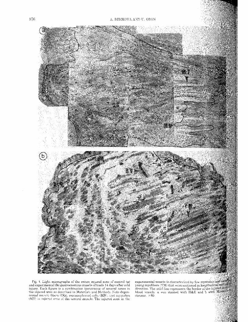
experimental muscle is characterized by few myotu that were sectioned in
line the border with H&E and b

PROMOTION MUSCLE REGENERATION IN THE TOAD 377
The structures were fraction of the various structures in the traumatized ew.1;.u·u between the pnot<)gI'a area at all time intervals
on the The fraction v..,,,..,,JH:OU
cells was not u.,. ...... ~~""' ser-irradiated muscles
made 2L8% ± cles. This in control ""'""''°" in control muscles the cells occu-
15.6% ± 3.1% of the traumatized area, while in muscle
DISCUSSION
data on the sequenskeletal muscle

878 BIBIKOVA AND U. ORON
2. Light m1•croer1rntts days and 30 experimental (b,d,f) In a, the area populated erate, mononucleated cells (MNJ and degenerated fibers (DG), while at have a same time in the laser-irradiated muscle (b) myotubes (MT) are al-
conspicuous in the zone. At 14 days µv'""'Jm (MT) the zone, while
some with central nuclear chains,
The results of this indicate that a.,,,.,.,..,,t1m<> processes follow cold erative and
the toad's muscle and are ~~,'~"'·vu. The fact that the -~~.~-.. ~·
muscle fibers made up the laser-irradiated control at 14 motion of the oe.geirim·a

PROMOTION REGENERATION lN THE TOAD 379
MYOTUBES
15
9 14 30
Daya after injury
YOUNG MYOFIBERS
9 14 30
Days after injury
MATURED MYOFIBERS
9 14 30
Days alter injury
of the volume fraction of the various zones of control columns) and
muscles as a of time after cold are mean ± SE from five or six toads at 9 and 30
postinjury, they are mean± SE ofl0-14 toads in two
DEGENERATED MYOFIBERS
20
~ 16 c:
.!2 Q 12 ., .:: 4l 8 E
" 0 4 >
0 9 14 30
Days after injury
MONONUCLEATED CELLS
90
~ 12 c: .!2 ti 54
; «> 36 E
" 0 18 >
0 9 14 30
Days after injury
30 DAYS AFTER INJURY
100
~ 80 c 0
~ 60
; GI
40 E " 0 20 >
0 MN YM MF
Hislologicsl structures
np1·tm·mf•rl in the winter and the summer. The histogram right corner summarizes the 30-day muscle ref:en,er:1-
tion in control and experimental muscles. MF, mature fibers. levels of ""°'"'b"'~''" group were *P < 0.05 or **P < 0.01.

880 A. BlBIKOVA AND
fibers made To the
the first to irradiation in vertebrates. Since mode of action of the and tissues is not to its dramatic
ACKNOWLEDGMENTS
We thank Mrs. L. Maltz for technical Prof. D. Wool for with the statistical
in culture. Dev. Bischoff, R. 1986b A satellite
Dev. Biol., 115:140-147. Bischoff, R. 1990 Cell commitment of
J. Cell Biol., Carlson, B.M. 1970a The regeneration of entire
fragments. In: Regeneration of Striatial A. Mauro, S.A. Shafiq, and A.T. Milhorat, Amsterdam, pp. 25-37.
Carlson, B.M. 1970b Histological observations mammalian and amphibian muscles. In: Muscle and A. Mauro, S.
eds. Medica, Amsterda and Faulkner 1983 The
following injury. A review.