Embed Size (px)
Citation preview

Proline-Rich Synapse-Associated Protein-1/Cortactin BindingProtein 1 (ProSAP1/CortBP1) Is a PDZ-Domain Protein HighlyEnriched in the Postsynaptic Density
Tobias M. Boeckers,1,2 Michael R. Kreutz,1 Carsten Winter,2 Werner Zuschratter,1 Karl-Heinz Smalla,3Lydia Sanmarti-Vila,1 Heike Wex,1 Kristina Langnaese,1 Juergen Bockmann,2 Craig C. Garner,4 andEckart D. Gundelfinger1
1Leibniz Institute for Neurobiology, 39118 Magdeburg, Germany, 2AG Molecular Neurobiology, Institute for Anatomy,Westfaelische Wilhelms-University, 48149 Muenster, Germany, 3Institute of Medical Neurobiology, University Magdeburg,39120 Magdeburg, Germany, and 4Department of Neurobiology, University of Alabama, Birmingham, Alabama 35294-0027
The postsynaptic density (PSD) is crucially involved in thestructural and functional organization of the postsynaptic neu-rotransmitter reception apparatus. Using antisera against ratbrain synaptic junctional protein preparations, we isolatedcDNAs coding for proline-rich synapse-associated protein-1(ProSAP1), a PDZ-domain protein. This protein was found to beidentical to the recently described cortactin-binding protein-1(CortBP1). Homology screening identified a related protein,ProSAP2. Specific antisera raised against a C-terminal fusionconstruct and a central part of ProSAP1 detect a cluster ofimmunoreactive bands of 180 kDa in the particulate fraction ofrat brain homogenates that copurify with the PSD fraction.Transcripts and immunoreactivity are widely distributed in thebrain and are upregulated during the period of synapse forma-tion in the brain. In addition, two short N-terminal insertions aredetected; they are differentially regulated during brain develop-ment. Confocal microscopy of hippocampal neurons showed
that ProSAP1 is predominantly localized in synapses, and im-munoelectron microscopy in situ revealed a strong associationwith PSDs of hippocampal excitatory synapses. The accumu-lation of ProSAP1 at synaptic structures was analyzed in thedeveloping cerebral cortex. During early postnatal develop-ment, strong immunoreactivity is detectable in neurites andsomata, whereas from postnatal day 10 (P10) onward a punc-tate staining is observed. At the ultrastructural level, the immu-noreactivity accumulates at developing PSDs starting from P8.Both interaction with the actin-binding protein cortactin andearly appearance at postsynaptic sites suggest that ProSAP1/CortBP1 may be involved in the assembly of the PSD duringneuronal differentiation.
Key words: rat brain; synapse; postsynaptic density (PSD);PDZ domain; synaptogenesis; actin-based cytoskeleton; devel-opment; synamon
The postsynaptic density (PSD) is a specialized structure beneaththe postsynaptic membrane that is crucially involved in the orga-nization of the neurotransmitter receptive apparatus and theadhesion of the postsynapse to presynaptic terminals (Ziff, 1997).It constitutes a structural matrix that anchors and clusters neuro-transmitter receptors, synaptic cell adhesion molecules, and com-ponents of intracellular signaling pathways exactly opposite to theneurotransmitter release site and thus must be considered as a
general organizer of the postsynaptic signal transduction machin-ery (Ziff, 1997; Kennedy, 1998; O’Brien et al., 1998). It has beensuggested that important mechanisms of synaptic plasticity, in-cluding activity-dependent changes in postsynaptic signal trans-duction, may have a structural basis in the PSD (Siekevitz, 1985;Lismann and Goldring, 1988). Consequently, major efforts havebeen undertaken to identify the protein components of this struc-ture (Walsh and Kuruc, 1992; Langnaese et al., 1996; Kennedy,1998).
Cytoskeletal elements including actin (Fifkova and Morales,1992), fodrin (Carlin et al., 1983), or a-actinin-2 (Wyszynski etal., 1997) are abundant components of PSDs. This suggests thatthe actin-based cytoskeleton constitutes the basic filamentousmeshwork of the postsynaptic cytomatrix and plays an importantrole in the organization of PSDs (Adam and Matus, 1996).
A number of PDZ-domain proteins have been identified ascentral elements for clustering synaptic membrane proteins at thepostsynapse and anchoring them to the cytoskeleton. For exam-ple, membrane-associated guanylate kinases SAP90/PSD-95,Chapsyn/PSD-93, SAP97, SAP102, and Drosophila DlgA bindand cluster ion channels, including NMDA, kainate receptors,and K1 channels, and cell adhesion molecules, including neuroli-gin and fasciclin II, as well as ephrins and their receptors atsynaptic sites (Kim et al., 1995, 1996; Kornau et al., 1995; Lau et
Received March 15, 1999; revised May 14, 1999; accepted May 18, 1999.This work was supported by grants from the Innovative Medizinische Forschung
(IMF, WWW Muenster), the Land Sachsen-Anhalt, Deutsche Forschungsgemein-schaft, and Fonds der Chemischen Industrie to T.M.B., M.R.K., and E.D.G., andfrom National Institutes of Health to C.C.G (AG 12978-02). We thank A. Ahle, C.Borutzki, G. Gaede, U. Kaempf, A. Lewedag, S. Loheide, and B. Kracht for experttechnical assistance.
The nucleotide sequences reported in this paper have been submitted to theGenBank/EMBL Data Bank with accession numbers AJ131899 (ProSAP1) andAJ133120 (ProSAP2).
Correspondence should be addressed to Dr. T. M. Boeckers, Department ofNeurochemistry and Molecular Biology, Leibniz Institute for Neurobiology, P.O.Box D, 39118 Magdeburg, Germany.
Dr. Sanmarti-Vila’s present address: Memorial Sloan Kettering Cancer Center,Department of Neurology, New York, NY 10021.
Dr. Wex’s present address: Mount Sinai School of Medicine, Department ofHuman Genetics, New York, NY 10029-6514.
Dr. Langnaese’s present address: Institute of Human Genetics, University Mag-deburg, 39120 Magdeburg, Germany.Copyright © 1999 Society for Neuroscience 0270-6474/99/196506-13$05.00/0
The Journal of Neuroscience, August 1, 1999, 19(15):6506–6518
al., 1996; Muller et al., 1996; Irie et al., 1997; Tejedor et al., 1997;Thomas et al., 1997; Zito et al., 1997; Garcia et al., 1998; Torreset al., 1998). In addition, PDZ domains of these proteins recruitintracellular signaling proteins, like neuronal nitric oxide-synthase or SynGAP, to the postsynaptic membrane (Brenman etal., 1996a,b; Chen et al., 1998; Kim et al., 1998). Several otherPDZ-domain proteins, e.g., GRIP (Dong et al., 1997), ABP(Srivastava et al., 1998), CCIP (Kurschner et al., 1998), orS-SCAM (Hirao et al., 1998), were also reported to be involved inthe assembly of postsynaptic structures.
Using antisera against synaptic protein preparations, we cloneda collection of cDNAs by expression screening encoding compo-nents of central synapses (Kistner et al., 1993; Langnaese et al.,1996). Here we report on one of these components, the proline-rich synapse-associated protein-1 (ProSAP1). ProSAP1 is a 180kDa protein that carries a PDZ domain at its N terminus. Bio-chemical analysis and ultrastructural localization studies showedthat ProSAP1 is a component of the PSD. Multiple developmen-tally and spatially regulated processing variants code for ProSAP1isoforms, one of which is identical to the cortactin-bindingprotein-1 (CortBP1) recently identified as a Cortactin SH3 do-main interacting protein (Du et al., 1998). Electron microscopiclocalization studies on the developing cortex suggest a role forProSAP1 in early steps of synaptic assembly.
MATERIALS AND METHODSCloning of rat ProSAP1 cDNA. The initial cDNA clone sap24e wasisolated from a llgt11 expression library with polyclonal antibodiesgenerated against a rat brain synaptic junction preparation as describedpreviously (Kistner et al., 1993; Langnaese et al., 1996). OverlappingcDNA clones were obtained by several rounds of screening of lZAPII(Stratagene, La Jolla, CA) rat hippocampal and total brain cDNA librar-ies with the 32P-labeled cDNA fragments.
Antibody production. Partial cDNAs of the ProSAP1 cDNA [encodingamino acids (aa) 355–509 and aa 826–1259] were subcloned into thebacterial expression vector pGEX-1T (Pharmacia Biotech, Uppsala, Swe-den). A 45 and a 95 kDa glutathione S-transferase (GST)-ProSAP1fusion protein was expressed in Escherichia coli XL 1 Blue and purifiedon glutathione-Sepharose 4B as recommended by the manufacturer(Pharmacia Biotech). The fusion proteins were used to generate Pro-SAP1 antibodies in guinea pigs and rabbits.
Immunohistochemistry. Immunocytochemical staining was performedusing 7 mm microtome sections from rat brains, which were fixed byimmersion in Bouin’s fluid for 48 hr, dehydrated, and embedded inparaplast. ProSAP1 was detected with the C-terminal rabbit anti-ProSAP1 polyclonal antibody diluted 1:600 using the peroxidase–anti-peroxidase method (Sternberger et al., 1970). Antibody binding wasvisualized by incubating sections first with porcine anti-rabbit IgG(Dako, Hamburg, Germany) for 30 min and with rabbit peroxidase–anti-peroxidase complex (Dako) for 30 min. Subsequently, the detectionreagent 3,3-diaminobenzidine/H2O2 (DAB) (Sigma, Munich, Germany)was applied. Some sections were counterstained with hematoxylin formorphological orientation. Controls were performed as follows: (1) pre-absorption of the antibody with the antigen and (2) omitting the primaryor secondary antibody. No staining was observed under either of theseconditions.
Electron microscopy was performed using vibratome sections (50 mm)from rat brains fixed by perfusion with Karnowsky’s solution, i.e., 2.2%glutaraldehyde, 2% paraformaldehyde in 0.1 M phosphate buffer, pH7.35. Cortical tissue from rat brains at postnatal day 5 (P5), P8, and P10was fixed by immersion with the same fixative. Floating sections werestained with the Vectastain ABC Staining Kit (Vector, Burlingame, CA)according to the manufacturer’s instructions. After color reaction withDAB, sections were extensively washed in 0.05 M Tris/HCl buffer, pH7.4, (twice) and 0.1 M cacodylate buffer, pH 7.4, (twice) before beingfixed in 2.5% glutaraldehyde in 0.1 M cacodylate buffer, pH 7.4, for 2 hr(4°C). Subsequently sections were further washed in 0.1 M cacodylatebuffer, pH 7.4, (twice) and doubly distilled water (ddH2O) (four times).
Silver enhancement of the DAB product was performed as follows:solution A, 3% hexamethylenetetramine in ddH2O; solution B, 5% silver
nitrate (AgNO3) in ddH2O; solution C, 2.5% disodium tetraborate inddH2O; solution D, 0.05% tetrachloroauric(III) acid in ddH2O; solutionE, 2.5% sodium thiosulfate in ddH2O. First, sections were incubated for10 min at 60°C in premade mixed solution, 5 ml (A) 1 250 ml (B) 1 500ml (C), and then washed in distilled water (dH2O) (three times for 3 min).Thereafter sections were incubated in solution D at room temperaturefor 3 min, washed with dH2O (three times for 3 min), incubated insolution E for 3 min, and washed again for three times for 3 min in dH2O.Subsequently, the sections were post-fixed in 1% OsO4 , dehydrated inethanol, and embedded in epon. Parallel semithin sections were stainedwith toluidine blue for morphological orientation; ultrathin sections werecontrasted with uranyl acetate/ lead citrate before analysis with a Philipselectron microscope.
For double- or triple-immunofluorescence staining the animals wereperfusion-fixed transcardially with 4% paraformaldehyde in 0.1 M PBS,pH 7.4 (Richter et al., 1996). After post-fixation overnight in the samefixative, brains were cut into 50-mm-thick frontal sections on a vibratomeand pretreated with a mixture of methanol /PBS (1:1) and subsequentlyincubated with 5% goat serum in PBS. After further washing steps inPBS, free-floating sections were incubated in a mixture of two or threeprimary antisera containing 0.4% Triton X-100 for 36 hr. The selectionof the primary antibodies and the detection systems from differentspecies ensured cross talk-free stainings in the colocalization experi-ments: ProSAP1/Bassoon: guinea pig anti-ProSAP1 1:500, anti-guineapig-CY3 1:100; rabbit anti-Bassoon 1:500 (tom Dieck et al., 1998),anti-rabbit-CY5 1:100; ProSAP1/Synaptophysin: rabbit anti-ProSAP11:1000, anti-rabbit-CY3 1:100, mouse monoclonal anti-synaptophysin(Dako) 1:500, anti-mouse CY5 1:100; ProSAP1/Bassoon/MAP2: guineapig anti-ProSAP1 1:500, anti-guinea pig-CY3 1:100; rabbit anti-bassoon1:500, anti-rabbit FITC 1:100 (Sigma Aldrich, Deisenhofen, Germany),mouse monoclonal anti-MAP2 (Sigma Aldrich) 1: 500, anti-mouseCY5 1:100; ProSAP1/Synapsin/MAP2: guinea pig anti-ProSAP1 1:500,anti-guinea pig-CY3 1:100, rabbit anti-synapsin (Sigma Aldrich), anti-rabbit FITC 1:100 (Sigma Aldrich), mouse monoclonal anti-MAP2 (Sig-ma Aldrich) 1:500, anti-mouse CY5 1:100. CY3- and CY5-labeled sec-ondary antibodies were purchased from Jackson Laboratories (BarHarbor, ME).
Image acquisition by confocal laser scanning microscopy. The hippocam-pal regions CA1 and CA3 of immunostained sections were scanned witha confocal laser scanning microscope (Leica TCS 4D, Leica Bensheim)equipped with an Argon-Krypton-Ion laser (488/568/647 nm) and anacousto-optical tunable filter (AOTF) for selection and intensity adap-tation of laser lines. The configuration of the system [excitation beamsplitter, TD (488/568/647); detector beam splitter, RSP 580; barrier filter,BP 535 (channel 1); detector beam splitter, RSP 660; barrier filter, BP600 (channel 2); and barrier filter, 0G 665 (channel 3)] allowed a rapidsimultaneous detection of the CY3 fluorescence in channel 2 (indicativefor ProSAP1) and CY5 in channel 3 (indicative for bassoon or synapto-physin) without an appreciable cross talk. Along the z-axis, usually 9–16thin optical sections with a z-resolution of 0.5–1 mm (focus depth) werescanned in a 512 3 512 or 1024 3 1024 pixel format. Images were takenat various magnifications, usually with a Fluotar 403 oil, NA 1.0–0.5,Plan Apo 633 oil, NA 1.4, or Plan Apo 1003 oil, NA 1.4 objective lensat various zoom factors (1–4) as indicated in the legends. Subsequently,maximum intensity projections (extended focus images) were calculatedfrom each fluorescence channel of the image stack and stored as RGBimages together with original image stacks. For further image analysisand restoration files were transferred to an Apple Macintosh computer,where image processing (contrast enhancement) was performed withAdobe Photoshop (Version 5.0; Adobe Systems, Mountain View, CA).Color prints of the CLSM images from individual focal planes wereprinted on a Pictrography 3000 printer (Fuji, Tokyo, Japan).
Hybridization. In situ hybridization was performed essentially as de-scribed previously (Kreutz et al., 1997). Transcripts encoding ProSAP1were detected with a cDNA antisense oligonucleotide purchased fromMWG-Biotech (Ebersberg, Germany) directed against the 39-end of themRNA: 59-TTC TTA CTG TCT GTA GAG TTG GCT GGT TGGCTG GAG TTC-39 (bp 3155–3113).
The expression of the N-terminal insertions was detected with thefollowing antisense oligonucleotides: presence of insert A (at bp 1050),59-CGG TTT ATC CTT CTT CTT CCG GAC TGA GGC TTT ATCC-39 (bp 1079–1044); absence of insert A (at bp 1050), 59-CGG TTTATC TTT ATC CAC GAG TTC CTC CAA TTC CGC TGT-39 (bp1079–1020 without insert A); presence of insert B (at bp 1287), 59-GCTGTC TAT CGA TTT CTG CCT TCG CAT CGT ACC TCG AGG-39
Boeckers et al. • PSD Protein ProSAP1/CortBP1 J. Neurosci., August 1, 1999, 19(15):6506–6518 6507

Figure 1. Primary structure of ProSAP1. A, Physical map of the rat ProSAP1 cDNA. The protein coding region is boxed. The PDZ and SAM domainare indicated in light and dark gray, respectively. The SH3 interaction module ( ppI ) is hatched; proline-rich elements (5) are indicated by thin lines. Thepositions of two alternatively processed inserts (A and B) are marked. In the 39-untranslated region a polyA-tail (beginning at nucleotide 7688) is found(compare accession no. AJ131899). B, Alignment of the amino acid sequences of rat ProSAP1 (as deduced from the cDNA, top sequence) and synamon(EMBL/GenBank accession no. AF102855, bottom sequence). Regions of high homology are shaded in gray; PDZ domain is (Figure legend continues)
6508 J. Neurosci., August 1, 1999, 19(15):6506–6518 Boeckers et al. • PSD Protein ProSAP1/CortBP1

(bp 1329–1281); absence of insert B (at bp 1287), 59-TCT GCT GTCTCG AGG GAG GCC CAG AAA TGG GCC TTT CGG-39 (bp 1331–1257 without insert B).
Controls were performed as follows: (1) omission of the antisenseoligonucleotide, (2) posthybridizational washing steps above the calcu-lated melting point of the hybrid, (3) hybridization with the correspond-ing sense oligonucleotide, and (4) hybridization in the presence of100-fold excess of unlabeled oligonucleotide. In no case was any specificlabeling observed.
Isolation of subcellular protein f ractions and Western blot analysis. Tis-sue from adult rats (total brain, heart, liver, kidney, thymus, testis,spleen) was homogenized in 20 mM Tris buffer, pH 7.4, containing either2 mM CaCl2 or 1 mM EDTA and protease inhibitor mixture (BoehringerMannheim, Mannheim, Germany). Soluble proteins were obtained as thesupernatant after 100,000 3 g centrifugation. After detergent extractionof the remaining pellet with 1% Triton X-100, the detergent-insolublepellet was extracted with 1% SDS to obtain a fraction of cytoskeletalproteins.
Tissue fractionation was performed essentially as described by Carlinet al. (1980) with some modifications (tom Dieck et al., 1998). Brains of30-d-old rats were homogenized in homogenization buffer (5 mMHEPES, pH 7.4; 320 mM sucrose) containing protease inhibitor mixture(Boehringer Mannheim); cell debris and nuclei were removed by 1000 3g centrifugation. The supernatant was spun for 20 min at 13,000 3 g,resulting in supernatant S2 and pellet P2 (crude membrane fraction). S2was centrifuged at 100,000 3 g for 1 hr, and the resulting supernatant wastaken as cytoplasmic fraction (S100). The P2 pellet was further fraction-ated by centrifugation in a sucrose step gradient as described by Carlinet al. (1980). For isolation of synaptic junctional proteins (PSD fraction),the synaptosomal fraction of the first gradient was diluted with 320 mMsucrose (60 ml/10 gm wet tissue) and an equal volume of 1% TritonX-100, 320 mM sucrose, and 12 mM Tris-HCl, pH 8.1. The suspension waskept on ice for 15 min and was centrifuged for 30 min at 32,800 3 g. Thepellet was resuspended in 320 mM sucrose, 1 mM NaHCO3 (6 ml/10 gmwet tissue), and an equal volume of 1% Triton X-100; 320 mM sucrosewas added, and synaptic junctional proteins were pelleted by a 2 hrcentrifugation at 201,800 3 g. All steps were performed at 4°C.
To study the association of ProSAP1 with the cytoskeleton duringpostnatal development, P2 fractions from brains of 1-, 4-, 7-, 10-, 14-, 21-,30-, and 90-d-old rats were rehomogenized in lysis buffer (0.5 mMNaHCO3 , 2.5 mM Tris/HCl, 0.5% Triton X-100, pH 8.1), incubated for1 hr at 4°C, and subsequently spun for 1 hr at 100,000 3 g. Pellets werewashed once with lysis buffer (1 hr, 100,000 3 g) and finally resuspendedin 500 ml Laemmli buffer. Proteins (20 mg/ lane) were separated bySDS-PAGE on 5–20% gels under fully reducing conditions and trans-ferred onto nitrocellulose. For immunodetection, Western blots wereincubated overnight either with the polyclonal ProSAP1 antiserum (di-lution 1:3000) or monoclonal antisera generated against SAP90/PSD95(clone P43520, dilution 1:250; Transduction Laboratories, Mamhead,UK) or the NR1 subunit of the NMDA receptor (clone 54.1, dilution1:250, PharMingen, San Diego, CA). Immunoreactivity was visualizedusing the ECL detection system (Amersham Buchler, Braunschweig,Germany).
Extraction experiments of P2 pellets were performed with the follow-ing agents: (1) 5 mM HEPES, pH 7.4; 320 mM sucrose; (2) 25 mMTris-HCl, pH 8.0; (3) 0.5 M NaCl; (4) 1 M NaCl; (5) 1 M Tris-HCl; (6) 1%Triton X-100 in 25 mM Tris-HCl; (7) 2.5% CHAPs in 25 mM Tris-HCl;(8) 1% SDS; (9) 0.1% SDS; (10) 8 M urea; (11) 3 M potassium rhodanite(tom Dieck et al., 1998). P2 pellets were resuspended in homogenizationbuffer, aliquoted into six samples (200 mg protein each), and centrifugedat 15,000 3 g for 20 min. Each pellet was then resuspended in 0.5 ml ofone of the extraction agents, incubated for 15 min at 4°C with gentleshaking, and centrifuged again for 15 min at 100,000 3 g. The resultingpellets were washed in homogenization buffer and dissolved in 80 mlgel-loading buffer (Laemmli, 1970). The supernatants were precipitated
with trichloroacetic acid, and the resulting pellets were dissolved in 80 mlloading buffer. Proteins were separated by SDS-PAGE on 5–20% gelsunder fully reducing conditions and transferred onto nitrocellulose. Forimmunodetection, Western blots were incubated overnight with primaryantiserum (dilution 1:2000), and immunoreactivity was visualized usingthe ECL detection system (Amersham Buchler). Subsequently Westernblots were analyzed for the ability of the different extraction agents tosolubilize ProSAP1.
RESULTSFrom a collection of cDNA clones isolated by expression screen-ing with antisera against a rat brain synaptic protein preparation(Langnaese et al., 1996), one cDNA clone, sap24e, encoded aprotein fragment of 80 amino acids (Boeckers et al., 1998).Northern blot analysis revealed that corresponding transcripts areprimarily expressed in the brain (data not shown). The sap24eclone was used to isolate a set of 13 overlapping cDNAs rangingin size from 1.3 to 6.9 kb. Eventually, cDNAs covering the Nterminus of encoded protein were isolated using a 59-terminalfragment of the 6.9 kb clone. Full-length cDNA (7.8 kb) wasconstructed taking advantage of a HindIII restriction site at nu-cleotide 1391 of the assembled sequence (accession no.AJ131899). The cDNA has an open reading frame coding for aproline-rich (12% prolines) protein of 1259 amino acid residues,which we named ProSAP1. Analysis of several cDNA clonesrevealed the existence of at least three processing variants ofProSAP1 (Fig. 1A,B), one of which is identical with the recentlypublished cortactin-binding protein CortBP1 (Du et al., 1998).
Sequence analysis predicts several structural domains of Pro-SAP1/CortBP1 (Fig. 1A,B). These include an N-terminal PDZdomain (aa 38–144), a proline-rich SH3 binding motif (ppI motif,aa 954–960) that has been shown previously to interact with theSH3 domain of the actin-binding protein cortactin (Du et al.,1998), and a C-terminal SAM domain (aa 1193–1257) consistingof four short helices linked by loops (Ponting, 1995) [for a moredetailed analysis of the SAM domain of ProSAP1/CortBP1, seeDu et al. (1998)]. Moreover, prolines frequently occur as clustersof three or more residues (Fig. 1A,B).
The N-terminal PDZ domain found in ProSAP1 shows onlymoderate similarity with previously described PDZ domains (Fig.1C). The highest degree of identity was found to synapse-associated PDZ-domain proteins, e.g., PDZ2 of SAP102 (31%),PDZ2 of Chapsyn-110/PSD-93 (29%), PDZ1 of SAP90/PSD-95(27%), and PDZ2 of SAP97 (27%). The first PDZ domain of theDrosophila disks large tumor suppressor protein (DlgA) is 28%identical. The GLGF motif, a hallmark of most PDZ domainsthat plays an important functional role in binding the C termini ofinteraction partners (Kornau et al., 1997) is substituted by GFGFin ProSAP1. A search for ProSAP1-related proteins by homologyscreening with PDZ-domain probes identified cDNAs for a newprotein, ProSAP2 (accession no. AJ133120), with a PDZ domainthat is 80% identical with that of ProSAP1 (Fig. 1C). Genomicsequences of the human homolog of ProSAP2 were found inpublic databases (Cosmid Clones; accession nos. AC000050 and
4
lined in black; insertions A and B in ProSAP1 are indicated by double-headed arrows; the SH3-binding motif and proline-rich elements are underlinedwith bold and broken lines, respectively; the N-terminal SAM domain (displayed as inverted characters) is not conserved between the two proteins. Theankyrin repeats of synamon are underlined, and the SH3 domain of synamon is boxed in broken lines. C, ProSAP1, ProSAP2, and synamon PDZ domainsdefine a new subfamily. Shown is alignment of the PDZ domains of rat ProSAP1, rat synamon, rat and human ProSAP2 (accession nos. AJ131899,AF102855, AJ133120, AC000050, and AC000036) with examples of a more distantly related PDZ domain of synaptic membrane-associated guanylatekinases of SAP90/PSD-95 (Kistner et al., 1993), SAP97 (Muller et al., 1995), SAP102 (Muller et al., 1996), Chapsyn-110/PSD-93 (Kim et al., 1996;Brenman et al., 1996a,b), and DLG1 (Woods and Bryant, 1989).
Boeckers et al. • PSD Protein ProSAP1/CortBP1 J. Neurosci., August 1, 1999, 19(15):6506–6518 6509
AC000036; mapped at chromosome 22). There is a very highdegree of sequence identity (96%) between rat and human Pro-SAP2 PDZ domains. Like ProSAP1, ProSAP2 is a proline-richprotein. However, regions of high similarity between the twoproteins are restricted to the ppI motif and the SAM domain(Winter C, Kreutz MR, Smalla KH, Bockmann J, Garner CC,Gundelfinger ED, and Boeckers TM, unpublished data). AnotherPDZ domain highly related to that of ProSAP1 and ProSAP2 isfound in synamon, a synaptic protein recently included in publicdatabases (Fig. 1B,C).
Sequencing of several different ProSAP1 cDNA clones sug-gested the expression of alternatively spliced transcripts affectingthe N-terminal part of the protein. Sequence insertions/deletionswere found at nucleotide 1050 (insert A; 21 bp) and nucleotide1287 (insert B; 27 bp). Several independent clones showed thepresence of either one of these insertions or of both inserts. In
none of the sequenced clones the absence of both exons could beobserved. The originally described CortBP1 is missing insert Abut contains insert B (Du et al., 1998). ProSAP1 has severalputative phosphorylation sites for cAMP and cGMP-dependentprotein kinases (1), casein kinase II (20), tyrosine kinase (1), andprotein kinase C (20). Interestingly, both insertions add severalpositive charges to the protein (Fig. 1B) and carry putativeprotein kinase C phosphorylation sites (insertion A: aa 175–178SVR; insertion B: aa 254–257 TMR).
Characterization and expression pattern of ProSAP1Recombinant GST-fusion proteins including either part of thecentral region or the C-terminal region of ProSAP1 were used toraise polyclonal antisera against ProSAP1 in rabbits and guineapigs. In accordance with the staining results obtained by Du et al.(1998), all antisera specifically detect a cluster of protein bands at
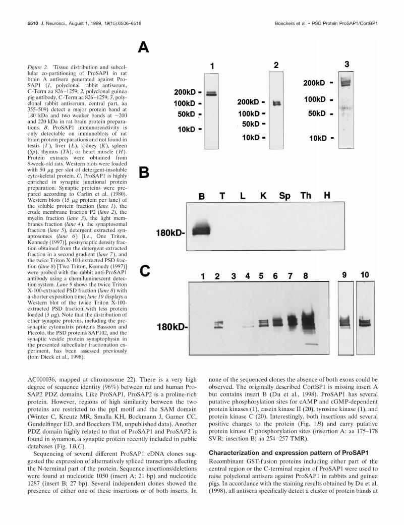
Figure 2. Tissue distribution and subcel-lular co-partitioning of ProSAP1 in ratbrain A antisera generated against Pro-SAP1 (1, polyclonal rabbit antiserum,C-Term aa 826–1259; 2, polyclonal guineapig antibody, C-Term aa 826–1259; 3, poly-clonal rabbit antiserum, central part, aa355–509) detect a major protein band at180 kDa and two weaker bands at ;200and 220 kDa in rat brain protein prepara-tions. B, ProSAP1 immunoreactivity isonly detectable on immunoblots of ratbrain protein preparations and not found intestis (T ), liver (L), kidney (K ), spleen(Sp), thymus (Th), or heart muscle (H ).Protein extracts were obtained from8-week-old rats. Western blots were loadedwith 50 mg per slot of detergent-insolublecytoskeletal protein. C, ProSAP1 is highlyenriched in synaptic junctional proteinpreparation. Synaptic proteins were pre-pared according to Carlin et al. (1980).Western blots (15 mg protein per lane) ofthe soluble protein fraction (lane 1), thecrude membrane fraction P2 (lane 2), themyelin fraction (lane 3), the light mem-branes fraction (lane 4 ), the synaptosomalfraction (lane 5), detergent extracted syn-aptosomes (lane 6 ) [i.e., One Triton,Kennedy (1997)], postsynaptic density frac-tion obtained from the detergent extractedfraction in a second gradient (lane 7 ), andthe twice Triton X-100-extracted PSD frac-tion (lane 8) [Two Triton, Kennedy (1997)]were probed with the rabbit anti-ProSAP1antibody using a chemiluminescent detec-tion system. Lane 9 shows the twice TritonX-100-extracted PSD fraction (lane 8) witha shorter exposition time; lane 10 displays aWestern blot of the twice Triton X-100-extracted PSD fraction with less proteinloaded (3 mg). Note that the distribution ofother synaptic proteins, including the pre-synaptic cytomatrix proteins Bassoon andPiccolo, the PSD proteins SAP102, and thesynaptic vesicle protein synaptophysin inthe presented subcellular fractionation ex-periment, has been assessed previously(tom Dieck et al., 1998).
6510 J. Neurosci., August 1, 1999, 19(15):6506–6518 Boeckers et al. • PSD Protein ProSAP1/CortBP1

a molecular weight in the range of 180–220 kDa on immunoblotsof brain protein preparations (Fig. 2A), but not of other tissuehomogenates, including testes, liver, kidney, spleen, thymus, andheart (Fig. 2B). Thus ProSAP1 migrates in SDS-PAGE slowerthan expected from the calculated molecular weight, a behaviorthat is frequently observed for cytoskeletal proteins. None of thebands were recognized in total brain homogenates by either of thepreimmune sera or after preabsorption with the antigen (data notshown). Because ProSAP1 has been cloned using antisera di-rected against the PSD fraction, the subcellular distribution of theprotein was investigated. ProSAP1 immunoreactivity is present inthe crude membrane fraction (P2) of rat brain (Fig. 2C, lane 2)but not in the soluble protein fraction (Fig. 2C, lane 1). Duringthe further steps of subcellular fractionation by differential cen-trifugation, ProSAP1 immunoreactivity essentially cofractionateswith PSDs (Fig. 2C). To investigate the association of ProSAP1with the cytoskeleton, attempts were made to solubilize theprotein from the crude membrane fractions (tom Dieck et al.,
1998). A partial extraction of immunopositive bands was achievedwith high salt (1 M NaCl, 1 M Tris-HCl) or 0.1% SDS. Virtuallycomplete solubilization was observed when using strongly dena-turing conditions (1% SDS or 8 M urea). Treatment with 0.5 M
NaCl, the detergents CHAPS (2.5%), or Triton X-100 (1%) orwith the chaotropic agent potassium rhodanite (3 M) did not resultin a substantial release of ProSAP1 from the pellet. All ProSAP1isoforms represented by the different immunoreactive bands dis-played a similar extraction behavior. These solubilization charac-teristics of the protein indicate a close cytoskeletal association ofProSAP1.
To examine the spatial distribution of the ProSAP1 transcriptswe performed in situ hybridization studies to horizontal brainsections (Fig. 3A). An antisense oligonucleotide directed againstthe C-terminal part of the mRNA and thus recognizing all knownvariants of ProSAP1 transcripts can be localized to many parts ofthe brain with high levels of expression in the olfactory bulb,cerebral cortex, cerebellum, central gray, and hippocampus. The
Figure 3. Distribution of ProSAP1 in the adult rat brain. A, Distribution of mRNA transcripts. In situ hybridization to a horizontal section from brainwith the 35S-labeled ProSAP1 antisense oligonucleotide shows the overall expression of the transcript in the adult brain. Intense labeling is observed incerebral cortex, cerebellum, hippocampus, and olfactory bulb, whereas putamen, thalamic nuclei, and brainstem show a moderate staining. Magnification:2.53. B, C, Overview of spatial distribution of ProSAP1 protein in rat brain by immunohistochemistry with ProSAP1 antisera. Sagittal (B) and frontal(C) sections are shown. Strong ProSAP1 immunoreactivity is detected in cerebral cortex, hippocampus, and the molecular layer of the cerebellum.Furthermore, the thalamic nuclei, the putamen, and to a much lesser extent the hypothalamus are labeled. Further enlargement of the hippocampalCA2/CA3 region (D) illustrates that cell nuclei and cell bodies are free of staining, whereas in the neuropil a punctate staining (arrows) can be observed.Magnifications: B, C, 2.53; D, 4503. Electron micrographs (E, F ) were taken from hippocampal CA3 sections. Silver enhancement of the DAB reactionproduct results in the punctate appearance of the immunoreactivity. Note that labeling is relatively weak in dendrites (d), enhanced toward dendriticspines (sp), and very strong at PSDs (arrowheads, arrows). Axon terminals (at) are essentially unlabeled. Magnification: F, 45,0003; G, 85,0003.
Boeckers et al. • PSD Protein ProSAP1/CortBP1 J. Neurosci., August 1, 1999, 19(15):6506–6518 6511

Figure 4. Distribution of ProSAP1 in hippocampal neurons as revealed by double- and triple-immunofluorescence labeling. A–D, Double immunoflu-orescence of hippocampal neurons in the CA1 and CA3 region with the rabbit antibody directed against ProSAP1 (CY3, green) and a monoclonalantibody directed against the presynaptic protein Bassoon (A, B, D, CY4, red) or synaptophysin (C, CY4, red). Note that the antigens are largelyco-distributed at hippocampal synapses. At higher magnifications of mossy fiber terminals in the stratum lucidum of the (Figure legend continues)
6512 J. Neurosci., August 1, 1999, 19(15):6506–6518 Boeckers et al. • PSD Protein ProSAP1/CortBP1

caudate putamen and thalamic nuclei as well as the brain stem aremoderately labeled. In control experiments, the use of a senseoligonucleotide, competition with 100-fold excess of unlabeledoligonucleotide, as well as washing steps above the calculatedmelting temperature of the hybrid yielded no labeling abovebackground (data not shown). This broad expression pattern isreflected by the light-microscopic localization of ProSAP1 immu-noreactivity in rat brain (Fig. 3B,C) showing an intense labelingof cerebral cortex, molecular layer of the cerebellum, hippocam-pal formation, thalamic nuclei, and basal ganglia. At higher mag-nification of the hippocampus, a representative light micrograph(Fig. 3D) shows a punctate staining pattern of hippocampal neu-ropil in the stratum oriens and stratum radiatum (CA2/CA3region). Ultrastructural investigation of the hippocampal CA3region identifies the close association of the antigen with the PSD(Fig. 3E,F). Although axon terminals do not show any labelingabove background, the antigen can be found in dendrites with ahighly significant increase of staining toward spines and PSDs.Double- and triple-immunofluorescent stainings of hippocampalsections for ProSAP1 and various marker proteins for differentneuronal compartments further document the primarily synapticlocalization of ProSAP1. Figure 4A,B illustrates the punctatedistribution and close apposition of ProSAP1-immunoreactivestructures and the presynaptic cytomatrix protein Bassoon (tomDieck et al., 1998) at many synapses of the CA1 region. Thevirtually complementary localization of ProSAP1 and the synap-tic vesicle protein synaptophysin or Bassoon at mossy fiber bou-tons of the CA3 region can be seen in Figure 4C,D. Triple stainingwith the dendritic marker protein MAP2, the presynaptic mark-ers Bassoon or synapsin, and ProSAP1 illustrates the dense clus-tering of ProSAP1 on dendrites of hippocampal neurons. Largeclusters are seen at the mossy fiber terminals in the stratumlucidum, whereas smaller PSDs decorate dendrites in the stratumradiatum (Fig. 4E,F).
Expression of ProSAP1 during early postnatal periodTo assess whether ProSAP1 may be involved in synaptic assemblyduring development or whether its function is restricted to pre-formed synapses, we investigated the expression of ProSAP1during the period of synaptogenesis. To this end we performed insitu hybridization, immunocytochemistry studies, and Westernblot analyses. Hybridization to brain sections of days P5, P9, andP18 indicate an increase of ProSAP1 transcripts during earlypostnatal brain development, especially in the caudate putamenand thalamic nuclei (Fig. 5A). ProSAP1 expression in the cerebralcortex, the hippocampus, and the cerebellum is moderate to highalready at P5 and shows a stable expression throughout develop-ment. Immunohistochemical staining of cortical neurons duringthe early postnatal period (P5, P8, P10) demonstrates a strikingchange in the localization of the antigen from being localizedmainly in the cytoplasm of cell bodies and neurites to a closeassociation of the protein with postsynaptic structures (Figs. 5, 6).On P5, ProSAP1 immunoreactivity is seen in the cytoplasms ofdensely packed cortical neurons (Figs. 5B,a). At the ultrastruc-tural level the antigen appears localized in small processes and
lamellopodia (Fig. 6A,B). Interestingly, already at that stage ofdevelopment the antibody detects ProSAP1 only in a subset ofneurites that are in close contact with unlabeled neuritic struc-tures. On P8 the antigen can be localized in the cytoplasm of cellbodies and in larger outgrowing neurites (Fig. 5B,b). Ultrastruc-tural investigations revealed that ProSAP1 is already localized inthe now apparent PSD of early synapses (Fig. 6C–E). On P10 apunctate staining can be recognized at the light microscopic level(Fig. 5B,c) that reflects the specific labeling of PSDs of corticalsynapses (Fig. 6F,G). At this stage of development the stainingpattern does not differ from that in adult animals.
These localization data are consistent with the assumption thatProSAP1 is one of the first protein components assembling intodeveloping PSDs. If so, one prediction would be that ProSAP1becomes anchored to the subsynaptic cytoskeleton earlier thanother known components of the PSD protein fraction, e.g.,SAP90/PSD-95 or NMDA receptors. To test this hypothesis, weanalyzed on immunoblots the appearance during development ofProSAP1, SAP90/PSD-95, and the NR1 subunit of NMDA re-ceptors in cytoskeletal protein fractions that should be enrichedfor PSD components. As shown in Figure 7, the association ofProSAP1 with the PSD-enriched fraction is strongly enhancedbetween P7 and P10, consistent with the period of its appearanceat postsynaptic sites of cortical synapses (Fig. 6). In contrast,enhanced appearance of NR1 and SAP90/PSD-95 immunoreac-tivity in PSD-enriched protein fractions is observed only laterduring development, i.e., from P10 and P14 onward, respectively.
Expression of ProSAP1 isoforms duringpostnatal developmentTo examine whether the expression of ProSAP1 isoforms en-coded by the identified processing variants of ProSAP1 tran-scripts are subject to temporal or spatial regulation, we performedin situ hybridizations at different developmental stages with fourdifferent oligonucleotides. The probes were designed to detect thepresence or absence of inserts A and B (Fig. 1, compare A, B).Insert A-containing transcripts are predominantly expressed dur-ing early stages of brain development (Fig. 8a–d, A1). On days P1and P10 it is found in cortical brain areas as well as in thecerebellum, hippocampus, and thalamic nuclei. After 3 weeks andin the adult brain the hybridization signal intensity is reduced,and the mRNA is found almost exclusively in the cerebellum.Transcripts without insert A (Fig. 8e–h, A2) display stable ex-pression at all postnatal stages in all brain regions expressingProSAP1 (compare Figs. 3A, 5A).
On the contrary, insert B is expressed throughout the rat brainfrom day 1 onward with a pattern similar to the A2 hybridizationsignals (Fig. 8i–l, B1). ProSAP1 transcripts without insert (Fig.8m–p, B2) show only a very weak expression, mainly in cortexand cerebellum on days P1 through P10. At 3 weeks there is asomewhat stronger hybridization signal in the cerebellum. In theadult brain no B2transcript levels above background could bedetected.
4
CA3 region (C, D), the close apposition of ProSAP1 with both presynaptic marker proteins can be seen. The staining with the Bassoon antibody especiallyillustrates the close spatial relationship of the two proteins because Bassoon is mainly restricted to the active zone of the presynapse (tom Dieck et al.,1998; Richter et al., 1999). Confocal images of triple immunofluorescence ProSAP1 ( green), Bassoon (red), and MAP2 (blue) (E) as well as ProSAP1( green), synapsin (red), and MAP2 (blue) (F) show the localization of synaptic structures on dendrites of hippocampal CA3 neurons. Please note thatlabeled shaft and spine synapses are discernible that decorate the MAP2-positive dendritic trees. Scale bars: A–C, E, F, 10 mm; D (all insets), 1 mm.
Boeckers et al. • PSD Protein ProSAP1/CortBP1 J. Neurosci., August 1, 1999, 19(15):6506–6518 6513

DISCUSSIONProSAP1 originally has been isolated as a protein contained insynaptic junctional protein preparations from rat brain (Kistneret al., 1993; Langnaese et al., 1996, Boeckers et al., 1998). Bio-chemical analysis and ultrastructural localization studies revealedthat ProSAP1 is indeed a component of the PSD of excitatorybrain synapses. Analysis of the primary structure of ProSAP1identified several sequence motifs typical for proteins of themembrane-associated cytoskeleton. While this study was inprogress, CortBP1, which is identical with ProSAP1, has beenidentified as an interaction partner of the actin-binding proteincortactin (Du et al., 1998). This indicates that ProSAP1/CortBP1may be one of the elements that links the postsynaptic signalingapparatus to the actin-based cytoskeleton within the PSD.
PDZ domains of ProSAP1 and ProSAP2 define a newsubfamily of PDZ domainsProSAP1 has various structural motifs that are known to beinvolved in protein–protein interactions. As shown by Du et al.(1998), ProSAP1/CortBP1 is able to interact with the cortactinSH3 domain via a ppI motif in the central part of the protein.Additional proline-rich domains identified in ProSAP1 may beinvolved in similar interactions with other proteins. A hallmark ofthe N-terminal part of ProSAP1 is a new type of PDZ domain.PDZ domains are protein interaction modules that mediate thebinding of distinct cell surface and intracellular proteins to thecortical cytoskeleton (Kornau et al., 1997). PDZ domains relatedto that of ProSAP1 include those of ProSAP2, a protein that isalso primarily expressed in the brain (Winter, Kreutz, Smalla,
Bockmann, Garner, Gundelfinger, and Boeckers, unpublisheddata), and of the synaptic SAPAP-interacting protein synamon(accession no. AF102855). PDZ domains of ProSAP1, ProSAP2,and synamon share .80% sequence identity, whereas PDZ do-mains of previously known proteins, such as SAP90/PSD-95,SAP97, Chapsyn-110/PSD-93, and SAP102, are only ;30% iden-tical with this new subfamily of PDZ domains (Fig. 1C).
The sequence similarity between ProSAP1 and ProSAP2 isstriking in the PDZ and SAM domains; the degree of sequencesimilarity in other regions of the two proteins is relatively low.The ppI motif is conserved between the two proteins, suggestingthat ProSAP2 is also a SH3 domain-binding protein. A databasesearch for ProSAP1-related proteins revealed several brain cD-NAs and genomic DNA fragments encoding human ProSAP1(accession nos. M86079, H41098, and HSU73633/chromosome11) and human ProSAP2 (accession nos. AC000050, AC000036/chromosome 22). Synamon is a 2091 aa synaptic protein with fouramino-terminal ankyrin repeats, a central SH3 domain next to aPDZ domain (GenBank accession no. AF102855). The similaritybetween ProSAP1 and synamon is not restricted to the PDZdomain, but clusters of high sequence identity are distributedthroughout the C-terminal part of synamon and the entire lengthof ProSAP1. However, no SAM domain is found in synamon(Fig. 1B).
ProSAP1 is highly enriched in PSDs in the adultrat brainThe actin-based cytoskeleton is thought to play a crucial role inthe regulation of dendritic spine morphology and the assembly of
Figure 5. A, Distribution of the ProSAP1transcripts and protein in the developingrat brain. In situ hybridizations of horizon-tal brain sections from 5 ( a)-, 9 ( b)-, and18-d-old (c) rats. X-ray film images of insitu hybridizations with the ProSAP1 anti-sense oligonucleotide show the dense ex-pression of the transcript in cortex, cere-bellum, and hippocampus at thesedevelopmental stages. The transcript is es-pecially upregulated during developmentin the thalamic nuclei and the caudate pu-tamen. Magnification: 3.53. B, Immuno-histochemical staining of cortical neuronsat P5 (a), P8 (b), and P10 (c). Note thestrong labeling of cytoplasm and small out-growing neurites at P5 (a), whereas theneuropil appears largely unstained. On P8the cytoplasmic staining is reduced, andlarger neurites (arrows) are clearly labeled(b). Two days later (P10) the staining pat-tern changes to a punctate labeling (ar-rows) in the neuropil of the developingcortex (c). Magnification: 5003.
6514 J. Neurosci., August 1, 1999, 19(15):6506–6518 Boeckers et al. • PSD Protein ProSAP1/CortBP1

postsynaptic structures, including the PSD. Thus, dendritic spinesand in particular the PSD are extremely rich in distinct isoformsof actin (Matus et al., 1982; Cohen et al., 1985; Fifkova andMorales, 1992; Kaech et al., 1997). Spine mobility and expressionof synaptic plasticity appear to be intimately associated with themodulation of the actin cytoskeleton (Fifkova and Morales, 1992;Fischer et al., 1998). Various elements of the actin cytoskeleton,including brain spectrin/fodrin (Carlin et al., 1983), dystrophin
(Kim et al., 1992), a-adducin (Seidel et al., 1995), drebrin (Ha-yashi et al., 1996), and a-actinin-2 (Wyszynski et al., 1997) havebeen shown to be components of the PSD. The functional signif-icance of the cortical cytoskeleton is underscored by the fact thatpostsynaptic NMDA receptor activity depends critically on theintegrity of actin filaments (Rosenmund and Westbrook, 1993).Moreover, NMDA receptor linkage to the postsynaptic actincytoskeleton appears to be mediated by a-actinin-2 and is regu-
Figure 6. Ultrastructural localization of ProSAP1 in the developing rat cortex. Electronmicroscopy of immunostained cortical sections at P5 (A, B)shows the primarily cytoplasmic localization of ProSAP1 in a subset of outgrowing neurites. Note the clearcut differentiation between ProSAP1-positiveand -negative neurites. In A, a ProSAP1-positive neurite with putatively pathfinding lamellopodia is displayed. B shows the close contact between aProSAP1-positive and -negative neurite. At P8, strong labeling can be found in the cytoplasm of growing neurites (C); synaptic contacts show strongProSAP1 immunoreactivity in the now appearing PSDs (D, E). At P10 (F, G), differentiation of brain tissue has advanced, and ProSAP1 immunore-activity is primarily found in spines and in particular at PSDs. Magnification: A, B, 46,0003; C, D, 70,0003; E, 90,0003. at, Axon terminal.
Boeckers et al. • PSD Protein ProSAP1/CortBP1 J. Neurosci., August 1, 1999, 19(15):6506–6518 6515

lated by a Ca21/calmodulin-dependent mechanism (Wyszinski etal., 1997).
ProSAP1 is likely to be a component of the actin-based cyto-matrix of the PSD. First, ProSAP1/CortBP1 is linked to the actincytoskeleton via cortactin (Du et al., 1998). Second, it is specifi-cally expressed in brain tissue, and solubilization experiments aswell as Western blot analysis after subcellular fractionation ofbrain tissue revealed that ProSAP1 is a cytoskeletal protein thatis highly enriched in the PSD fraction. Moreover, in situ hybrid-ization and immunohistochemical studies at the light and electronmicroscopic level revealed that ProSAP1 is widely expressed inneurons and mainly located in the submembraneous matrix of thePSD. It occurs primarily at asymmetric type 1 synapses, which arethought to be excitatory (Peters et al., 1991; Ziff, 1997). Thesedata indicate that ProSAP1 is part of the highly specializedcytoskeleton at the PSD, which anchors neurotransmitter recep-tors, cell adhesion molecules, and intracellular signal transductionpathways to the postsynaptic site (Ziff, 1997; Craven and Bredt,1998; O’Brien et al., 1998). Cortactin that links ProSAP1/CortBP1 to the actin cytoskeleton is substrate for the nonrecep-tor protein tyrosine kinase Src (Wu and Parsons, 1993). It is anF-actin binding protein that is thought to mediate aspects of cellsignaling associated with the cortical cytoskeleton (Du et al.,1998). Thus both proteins may contribute to the enormous dy-namic potential of the postsynaptic cytoskeleton supposed toprovide the basic mechanisms for synaptic plasticity (Buchs andMuller, 1996).
ProSAP1 isoforms may be functionally involved in theassembly of the postsynaptic cytomatrixduring developmentIt is still unknown which mechanisms govern the formation of thePSD beneath the postsynaptic membrane. On theoretical groundsthe initial formation requires the docking of several proteins,
including neurotransmitter receptors, protein kinases and phos-phatases, adaptor proteins, and filamentous cytoskeletal proteins,to fulfill the morphological and functional criteria of building thePSD. At least two processes have to occur in parallel: (1) clus-tering of receptor molecules in apposition to the active zone ofthe presynaptic membrane providing the structural basis for anexcitable membrane and (2) anchoring of proteins involved inintracellular signal transduction to this membrane. Currently wedo not know anything about the initiation of these two processes.
ProSAP1/CortBP1 and cortactin are highly enriched in growthcones of hippocampal primary neurons before synaptogenesis(Du et al., 1998). This is consistent with our finding in situ thatProSAP1 immunoreactivity is found in lamellopodia of corticalneurons before synaptogenesis. A striking change in ultrastruc-tural localization of ProSAP1 immunoreactivity occurs whensynaptic contacts are formed. Then ProSAP1 is concentrated atsites where PSDs are thought to form. This early appearance atthe differentiating postsynaptic membrane suggests that ProSAP1could be involved in initial steps of PSD assembly. Interestingly,the association of ProSAP1 with the PSD appears to precede theanchoring of SAP90/PSD-95 and the NR1 subunit of the NMDAreceptor. An important question to be answered in this context is
Figure 7. Developmental association of ProSAP1, SAP90/PSD-95, andthe NR1 subunit of the NMDA receptor with cytoskeletal protein frac-tions. Western blot analysis of a fraction enriched for PSD elements (20mg/ lane) during postnatal development shows that moderate amounts ofProSAP1 are already detectable at P1 with a significant increase of thesignal intensity between P7 and P10. Association of SAP90/PSD-95 withthe protein fraction strongly increases from P14 onward. The detection ofthe NR1 subunit of the NMDA receptor is detectable from P1, but astrong increase in immunoreactivity is seen only between P10 and P21.The star indicates the band resulting from the SAP90/PSD-95 staining.The blot has been reprobed for the detection of the NR1 subunit of theNMDA receptor.
Figure 8. Expression of processing variants of ProSAP1 transcripts inthe brain during postnatal development. During early postnatal develop-ment, insert A (a, b, A1) shows a wide distribution throughout the brain.At later stages the hybridization signal intensity decreases and becomesmainly restricted to the cerebellum (c, d, A1). ProSAP1 transcriptswithout insert A show a wide expression in rat brain at all developmentalstages investigated (e–h, A2). Nearly identical results were obtained withan antisense oligonucleotide designed to recognize insert B (i–l, B1). Incontrast, ProSAP1 mRNA without insert B shows a very weak expressionon P1 and P10; it is largely restricted to the cerebellum after 3 weeks andcannot be detected in the adult rat brain. Therefore in most brain regionsthe A2/B1 transcript seems to be regularly expressed, whereas duringdevelopment, especially in the cerebellum, transcripts with insert A butwithout insert B can also be detected.
6516 J. Neurosci., August 1, 1999, 19(15):6506–6518 Boeckers et al. • PSD Protein ProSAP1/CortBP1

whether ProSAP1 is actively involved in the formation of apostsynaptic cytoskeletal specialization at the synaptic junction orwhether the protein is incorporated in the PSD after the structurehas been formed. Of interest is the observation that duringdevelopment the expression of splice variants containing insertionA as well as those lacking insertion B is restricted to the earlypostnatal period. These transcripts are predominantly expressedin the cerebellum of animals up to the age of ;3 weeks. Inter-estingly, these inserts change the charge to the molecule andintroduce additional potential phosphorylation sites for proteinkinase C, an enzyme that is known to be involved in the regula-tion of synaptic assembly and plasticity (Shearman et al., 1991;Ben-Ari et al., 1992; Wang and Feng, 1992; Abeliovich et al.,1993; Klann et al., 1993; Moriya and Tanaka, 1994; Reymann andStaak, 1994; Pasinelli et al., 1995). Further studies must clarifywhether these developmentally regulated processing variants areinvolved in early synaptogenesis and/or growth cone regulation.
REFERENCESAbeliovich A, Chen C, Goda Y, Silva AJ, Stevens CF, Tonegawa S (1993)
Modified hippocampal long-term potentiation in PKC gamma-mutantmice. Cell 31:75:1253–1262.
Adam G, Matus A (1996) Role of actin in the organisation of brainpostsynaptic densities. Brain Res Mol Brain Res 31:246–250.
Ben-Ari Y, Aniksztejn L, Bregestovski P (1992) Protein kinase C mod-ulation on NMDA currents: an important link for LTP induction.Trends Neurosci 15:333–339.
Boeckers TM, Kreutz MR, Bockmann J, Langnaese K, Sanmarti-Vila L,Garner CC, Gundelfinger ED (1998) SAP24e, a novel postsynapticdensity (PSD) protein. Soc Neurosci Abstr 24:2.1999.
Brenman JE, Christopherson KS, Craven SE, McGee AW, Bredt DS(1996a) Cloning and characterization of postsynaptic density 93, anitric oxide synthase interacting protein. J Neurosci 16:7407–7415.
Brenman JE, Chao DS, Gee SH, McGee AW, Craven SE, Santillano DR,Wu Z, Huang F, Xia H, Peters MF (1996b) Interaction of nitric oxidesynthase with the postsynaptic density protein PSD-95 and alpha1-syntrophin mediated by PDZ domain. Cell 84:757–767.
Buchs PA, Muller D (1996) Induction of long-term potentiation is asso-ciated with major ultrastructural changes of activated synapses. ProcNatl Acad Sci USA 23:8040–8045.
Carlin RK, Grab DJ, Cohen RS, Siekievitz P (1980) Isolation and char-acterization of postsynaptic densities from various brain regions: en-richment of different types of postsynaptic densities. J Cell Biol86:831–843.
Carlin RK, Bartelt DC, Siekevitz P (1983) Identification of fodrin as amajor calmodulin-binding protein in postsynaptic density preparation.J Cell Biol 96:443–448.
Chen H-J, Rojas-Soto M, Oguni A, Kennedy MB (1998) A synapticRas-GTPase activating protein (p135 SynGAP) inhibited by CaMkinase II. Neuron 20:895–904.
Cohen RS, Chung SK, Pfaff DW (1985) Immunochemical localization ofactin in dendritic spines of the cerebral cortex using colloidal gold as aprobe. Cell Mol Neurobiol 5:271–284.
Craven SE, Bredt DS (1998) PDZ proteins organize synaptic signalingpathways. Cell 39:495–498.
Dong H, O’Brien RJ, Fung ET, Lanahan AA, Worley PF, Huganir RL(1997) GRIP. A synaptic PDZ domain containing protein that inter-acts with AMPA receptors. Nature 386:279–284.
Du Y, Weed SA, Wen-Cheng X, Marshall TD, Parsons TJ (1998) Iden-tification of a novel cortactin SH3 domain-binding protein and itslocalization to growth cones of cultured neurons. Mol Cell Biol18:5838–5851.
Fifkova E, Morales M (1992) Actin matrix of dendritic spines, synapticplasticity, and long term potentiation. Int Rev Cytol 139:267–307.
Fischer M, Kaech S, Knutti D, Matus A (1998) Rapid actin-based plas-ticity in dendritic spines. Neuron 20:847–854.
Garcia EP, Mehta S, Blair LA, Wells DG, Shang J, Fukushima T, FallonJR, Garner CC, Marshall J (1998) SAP90 binds and clusters kainatereceptors causing incomplete desensitization. Neuron 21:727–739.
Hayashi K, Ishikawa R, Ye LH, He XL, Takata K, Kohama K, Shirao T
(1996) Modulatory role of drebrin on the cytoskeleton within dendriticspines in the rat cerebral cortex. J Neurosci 16:7161–7170.
Hirao K, Hata Y, Ide N, Takeuchi M, Irie M, Yao I, Deguchi M, ToyodaA, Sudhof TC, Takai Y (1998) A novel multiple PDZ domain-containing molecule interacting with N-methyl-D-aspartate receptorsand neuronal cell adhesion proteins. J Biol Chem 14:21105–21110.
Irie M, Hata Y, Takeuchi M, Ichtchenko K, Toyoda A, Hirao K, Takai Y,Rosahl TW, Sudhof TC (1997) Binding of neuroligins to PSD-95.Science 277:1511–1515.
Kaech S, Fischer M, Doll T, Matus A (1997) Isoform specificity in therelationship of actin to dendritic spines. J Neurosci 17:9565–9572.
Kennedy MB (1997) The postsynaptic density at glutamatergic synapses.Trends Neurosci 20:264–268.
Kennedy MB (1998) Signal transduction molecules at the glutamatergicpostsynaptic membrane. Brain Res Brain Res Rev 26:243–257.
Kim E, Niethammer M, Rothschild A, Jan YN, Sheng M (1995) Clus-tering of Shaker-type K 1 channels by interaction with a family ofmembrane-associated guanylate kinases. Nature 378:85–88.
Kim E, Cho K-O, Rothschild A, Jan YN, Sheng M (1996) Heteromul-timerization and NMDA-receptor-clustering activity of chapsyn-110, amember of the PSD-95 family of proteins. Neuron 17:103–113.
Kim J-H, Liao D, Lau L-F, Huganir RL (1998) SynGAP: a synapticRasGAP that associates with the PSD95/SAP90 protein family. Neuron20:683–691.
Kim TW, Wu K, Xu JL, Black IB (1992) Detection of dystrophin in thepostsynaptic density of rat brain and deficiency in a mouse model forDuchenne muscular dystrophy. Proc Natl Acad Sci USA89:11642–11644.
Kistner U, Wenzel BM, Veh RW, Cases LC, Garner AM, Appeltauer U,Voss B, Gundelfinger ED, Garner CC (1993) SAP90, a rat presynapticprotein related to the product of the Drosophila tumor suppressor genedlg-a. J Biol Chem 268:4580–4583.
Klann E, Chen SJ, Sweatt JD (1993) Mechanism of protein kinase Cactivation during the induction and maintenance of long-term potenti-ation probed using a selective peptide substrate. Proc Natl Acad SciUSA 15:8337–8341.
Kornau HC, Schenker LT, Kennedy MB, Seeburg PH (1995) Domaininteraction between NMDA receptor subunits and the postsynapticdensity protein PSD-95. Science 269:1737–1740.
Kornau HC, Seeburg PH, Kennedy MB (1997) Interaction of ion chan-nels and receptors with PDZ domain proteins. Curr Opin Neurobiol7:368–373.
Kreutz MR, Bockers TM, Sabel BA, Hulser E, Stricker R, Reiser G(1997) Expression and subcellular localization of a p42 IP4/Centaurin-a, a-brain specific, high-affinity receptor for inositol 1,3,4,5-tetrakisphosphate and phosphatidylinositol 3,4,5-trisphosphate in ratbrain. Eur J Neurosci 9:2110–2124.
Kurschner C, Mermelstein PG, Holden WT, Surmeier DJ (1998) CIPP,a novel multivalent PDZ domain protein, selectively interacts withKir4.0 family members, NMDA receptor subunits, neurexins, andneuroligins. Mol Cell Neurosci 11:161–172.
Laemmli UK (1970) Cleavage of structural proteins during the assemblyof the head of bacteriophage T4. Nature 227:680–685.
Langnaese K, Seidenbecher C, Wex H, Seidel B, Hartung K, AppeltauerU, Garner A, Voss B, Mueller B, Garner CC, Gundelfinger ED (1996)Protein components of a rat brain synaptic junctional protein prepara-tion. Mol Brain Res 42:118–122.
Lau LF, Mammen A, Ehlers MD, Kindler S, Chung WJ, Garner CC,Huganir RL (1996) Interaction of the N-methyl-D-aspartate receptorcomplex with a novel synapse associated protein, SAP102. J Biol Chem271:21622–21628.
Lisman JE, Goldring MA (1988) Feasibility of long term storage ofgraded information by the Ca 21/calmodulin-dependent protein kinasemolecules of the postsynaptic density. Proc Natl Acad Sci USA85:5320–5324.
Matus A, Ackermann M, Pehling G, Byers HR, Fujiwara K (1982) Highactin concentration in brain dendritic spines and postsynaptic densities.Proc Natl Acad Sci USA 79:775–782.
Moriya M, Tanaka S (1994) Prominent expression of protein kinase C(g) mRNA in the dendrite-rich neuropil of mice cerebellum at thecritical period for synaptogenesis. NeuroReport 5:929–932.
Muller BM, Kistner U, Veh RW, Cases LC, Becker B, Gundelfinger ED,Garner CC (1995) Molecular characterization and spatial distributionof SAP97, a novel presynaptic protein homologous to SAP90 and the
Boeckers et al. • PSD Protein ProSAP1/CortBP1 J. Neurosci., August 1, 1999, 19(15):6506–6518 6517

Drosophila discs large tumor suppressor protein. J Neurosci15:2355–2366.
Muller BM, Kistner U, Kindler S, Chung WJ, Kuhlendahl S, Fenster SD,Lau LF, Veh RW, Huganir R, Gundelfinger E, Garner CC (1996)SAP102, a novel postsynaptic protein that interacts with NMDA re-ceptor complexes in vivo. Neuron 17:255–265.
O‘Brien RJ, Lau LF, Huganir RL (1998) Molecular mechanisms ofglutamate receptor clustering at excitatory synapses. Curr Opin Neu-robiol 8:364–369.
Pasinelli P, Ramakers GM, Urban IJ, Hens JJ, Oestreicher AB, de GraanPN, Gispen WH (1995) Long-term potentiation and synaptic proteinphosphorylation. Behav Brain Res 23:53–59.
Peters A, Palay SL, Webster H (1991) Synapses. In: The fine structure ofthe nervous system: neurons and their supporting cells, pp 138–211.New York: Oxford UP.
Ponting CP (1995) SAM: a novel motif in yeast sterile and Drosophilapolyhomeotic proteins. Protein Sci 4:1928–1930.
Reymann KG, Staak S (1994) Molecular mechanisms underlying long-term potentiation: postsynaptic glutamate receptors and protein kinaseC. In: Protein kinase C in the CNS focus on neuronal plasticity,proceedings (Canonico P, Scapagnini U, Pamparana F Routtenberg A,eds), pp 31–56. Milano: Masson.
Richter K, Hamprecht B, Scheich H (1996) Ultrastructural localizationof glycogen phosphorylase predominantly in astrocytes of the gerbilbrain. Glia 17:263–273.
Richter K, Langnaese K, Kreutz MR, Olias G, Zhai W, Scheich H,Garner CC, Gundelfinger ED (1999) The presynaptic cytomatrix pro-tein Bassoon is located at both excitatory and inhibitory synapses of ratbrain. J Comp Neurol 408:437–448.
Rosenmund C, Westbrook GL (1993) Calcium-induced actin depoly-merization reduces NMDA channel activity. Neuron 10:805–814.
Seidel B, Zuschratter W, Wex H, Garner CC, Gundelfinger ED (1995)Spatial and sub-cellular localization of the membrane cytoskeleton-associated protein alpha adducin in the rat brain. Brain Res 700:13–24.
Shearman MS, Shinomura T, Oda T, Nishizuka Y (1991) Synaptosomalprotein kinase C subspecies: A. Dynamic changes in the hippocampusand cerebellar cortex concomitant with synaptogenesis. J Neurochem56:1255–1262.
Siekevitz P (1985) The postsynaptic density: a possible role in long-lasting effects in the central nervous system. Proc Natl Acad Sci USA82:3494–3498.
Srivastava S, Osten P, Vilim FS, Khatri L, Inman G, States B, Daly C,DeSouza S, Abagyan R, Valtschanoff JG, Weinberg RJ, Ziff EB (1998)
Novel anchorage of GluR2/3 to the postsynaptic density by the AMPAreceptor-binding protein ABP. Neuron 21:581–591.
Sternberger LA, Hardy PH, Cuculis JJ, Meyer HG (1970) The unla-belled antibody-enzyme method of immunohistochemistry: preparationand properties of a soluble antigen-antibody complex (horseradish-peroxidase-antiperoxidase) and its use in identification of spirochetes.J Histochem Cytochem 18:315–333.
Tejedor FJ, Bokhari A, Rogero O, Gorczyca M, Zhang J, Kim E, ShengM, Budnik V (1997) Essential role for dlg in synaptic clustering ofShaker K 1 channels in vivo. J Neurosci 17:152–159.
Thomas U, Kim E, Kuhlendahl S, Koh YH, Gundelfinger ED, Sheng M,Garner CC, Budnik V (1997) Synaptic clustering of the cell adhesionmolecule fasciclin II by discs-large and its role in the regulation of thepresynaptic structure. Neuron 19:787–799.
tom Dieck S, Sanmarti-Vila L, Langnaese K, Richter K, Kindler S, SoykeA, Wex H, Smalla K-H, Kaempf U, Fraenzer J-T, Stumm M, GarnerCC, Gundelfinger ED (1998) Bassoon, a novel zinc-finger CAG/glutamine-repeat protein selectively localized at the active zone ofpresynaptic nerve terminals. J Cell Biol 142:499–509.
Torres R, Firestein BL, Dong H, Staudinger J, Olson EN, Huganir RL,Bredt DS, Gale NW, Yancopoulos GD (1998) PDZ proteins bind,cluster, and synaptically colocalize with Eph receptors and their ephrinligands. Neuron 21:1453–1463.
Walsh MJ, Kuruc N (1992) The postsynaptic density: constituent andassociated proteins characterized by electrophoresis, immunoblotting,and peptide sequencing. J Neurochem 59:667–678.
Wang JH, Feng DP (1992) Postsynaptic protein kinase C essential toinduction and maintenance of long-term potentiation in the hippocam-pal CA1 region. Proc Natl Acad Sci USA 1:2576–2580.
Woods DF, Bryant PJ (1989) Molecular cloning of the lethal(1)Discslarge-1 oncogene of Drosophila. Dev Biol 134:222–235.
Wu H, Parsons JT (1993) Cortactin, an 80/85-kilodalton pp60src sub-strate, is a filamentous actin-binding protein enriched in the cell cortex.J Cell Biol 120:1417–1426.
Wyszynski M, Lin J, Rao A, Nigh E, Beggs AH, Craig AM, Sheng M(1997) Competitive binding of alpha-actinin and calmodulin to theNMDA receptor. Nature 385:439–442.
Ziff EB (1997) Enlightening the postsynaptic density. Neuron19:1163–1174.
Zito K, Fetter RD, Goodman CS, Isacoff EY (1997) Synaptic clusteringof fascilin II and shaker: essential targeting sequences and role of Dlg.Neuron 19:1007–1016.
6518 J. Neurosci., August 1, 1999, 19(15):6506–6518 Boeckers et al. • PSD Protein ProSAP1/CortBP1