Embed Size (px)
Citation preview

Early Detection of Lung Cancer – A Pan Canadian Study Version 9, Dec 14, 2010
PROJECT TITLE: Early Detection of Lung Cancer – A Pan-Canadian Study PROJECT LEADERS: Stephen Lam MD, FRCPC & Ming-Sound Tsao MD, FRCPC PARTNERSHIP INSTITUTIONS AND INVESTIGATORS:
• British Columbia (BC Cancer Agency & the University of British Columbia): Stephen Lam, Annette McWilliams, John Mayo, Stuart Peacock, Richard Finley John Yee, Ken Evans.
• British Columbia (James Hogg iCapture Center & the Heart and Lung Institute, St. Paul’s hospital, University of British Columbia): Don Sin, Wan Tan
• Alberta (Foothills Hospital & the University of Calgary): Alain Tremblay, Paul Burrowes, Paul MacEachern
• Ontario (Brock University): Martin Tammemagi • Ontario (Princess Margaret Hospital, University of Toronto): Heidi Roberts,
Ming-Sound Tsao, Frances Shepherd, Geoffrey Liu, Kam Sophrati; John Thenganatt; Natasha Leighl
• Ontario (Juravinski Cancer Center, McMaster University): John Goffin, Serge Puksa, , Lori Stewart, Bill Evans
• Ontario (Ottawa Hospital Regional Cancer Centre, University of Ottawa): Garth Nicholas, Glen Goss, Jean M Seely, Kayvan Amjadi
• Quebec (Hôpital Laval & U de Laval): Simon Martel, Francis Laberge, Michel Gingras
• Nova Scotia (QEII HSC, Dalhousie University): Michael Johnston, Daria Manos • Newfoundland (Memorial University of Newfoundland): Rick Bhatia, Scott
Harris, George Fox

Early Detection of Lung Cancer – A Pan Canadian Study Version 9, Dec 14, 2010
2
1.0 EXECUTIVE SUMMARY: Overall Goal: Our overall goal is to develop a new multi-modal screening strategy that integrates risk modeling, autofluorescence bronchoscopy (AFB) and serum biomarkers with low dose spiral computed tomography (CT) for early detection of lung cancer. We hypothesize that the inclusion of low cost risk modeling and biomarkers to select population cohorts with the highest risk of lung cancer development can provide a cost effective application of relatively expensive yet effective detection methods. This could render lung cancer screening an affordable program within the Canadian health care system. Using short-term outcomes from this study including detection rates and costs, we shall be able to compare our results with other existing programs, including those of the major randomized trials in the United States and Europe, and provide an informed decision for the implementation of lung cancer screening in Canada. Study Objectives: 1. To develop a new multi-modal screening strategy and integrated methods to detect
early lung cancer such that screening can become an affordable program within the Canadian health care system.
2. To evaluate the impact of the screening modalities on the quality of life of subjects in the screening study.
3. Leveraging the results of on-going randomized clinical trials in the US and Europe, develop a decision analytic framework for determining the cost and effectiveness of a lung cancer screening in Canada.
Summary of Study: • Recruitment of 2,700 smokers age 50 to 75 from 8 centers across Canada over 20
months with an estimated 6-year lung cancer risk ≥ 2% using the a novel lung cancer risk assessment model.
• Administer a baseline epidemiologic questionnaire to collect socio-demographic, smoking and other exposure and risk factor data.
• Perform spirometry and collect blood specimens for biomarker measurement in all participants.
• Perform low dose non contrast enhanced thoracic spiral CT on all participants at baseline, month 12 and month 48 (additional Month 48 LDCT planned 8 months after study had started).
• Perform AFB in the first 50% of participants • Determine the prevalence of CT & AFB abnormalities across the country • Determine the resource utilization, actual cost of investigations and treatment (e.g.
procedure costs, length of hospital stay, type of hospital ward, chemotherapy, radiotherapy, physician visits related to screening procedure)
• Administer questionnaires to determine quality of life and psychological impact of lung cancer screening
• Administer follow-up questionnaire annually until completion of study to capture lung cancer incidence rate and health care utilization.
Early Detection of Lung Cancer – A Pan Canadian Study Version 9, Dec 14, 2010
3
• Determine incremental value of spirometry and blood biomarkers for lung cancer risk assessment.
Study Deliverables • Prospective evaluation of the ability of (a) blood biomarkers, (b) pulmonary function
test, (c) AFB and (d) low dose spiral CT to independently and jointly detect early (asymptomatic) lung cancer in a high-risk population.
• Development of a unique multi-modal lung cancer screening model that incorporates risk factors, autofluorescence bronchoscopy and biomarkers with low dose spiral CT.
• Evaluation of key screening parameters that may impact the success of widespread population lung cancer screening in Canada, (e.g. detection rate by CT screening, health care costs and management strategies) compared to randomized trials in the United States (NLST) and the EU (Dutch-Belgian NELSON trial) that will be released around 2010-2011.
• Evaluation of the quality of life and psychological well-being in those participating in an early lung cancer detection program
• Estimation of the costs and resource utilization implications to public health payers of implementing a lung cancer screening program provincially and across Canada
• Identification of potential barriers to program implementation and make recommendations on how to overcome these barriers
• Provision of a cadre of highly skilled chest radiologists, respirologists, surgeons, thoracic oncologists and lung pathologists knowledgeable in the detection and management of early lung cancer who will in turn be mentors to other clinicians.
• Based on the evidence obtained in this study and those from randomized clinical trials outside of Canada, provide a GO/NO GO recommendation to Provincial and Federal Health Ministries regarding implementation of lung cancer screening in Canada.
• If the evidence supports lung cancer screening in Canada, the partnership will initiate discussion with stakeholders regarding funding for a screening program across Canada.
• If the randomized clinical trials do not show a mortality reduction benefit of screening, the partnership will apply to funding agencies to design and conduct an alternative randomized clinical trial using our multi-modal screening strategy in Canada alone or in conjunction with other countries.
• Provision of a unique resource to validate other early detection biomarkers and genetic markers of lung cancer susceptibility that may further enhance the performance of our multi-modal lung cancer risk assessment model.
Impact on Patient Health in Canada and Worldwide • Currently, lung cancer is the most common cause of cancer death in Canada and
worldwide. In 2007 in Canada, lung cancer is expected to account for 11,000 and 8,900 cancer deaths in men and women, respectively, which represents 29% and 26% of all cancer deaths. Former smokers remain at an elevated risk for developing lung cancer even decades after they stop smoking. Fifty percent of newly diagnosed lung cancer patients are former smokers. Worldwide, over 1.2 million people die of lung cancer each year. By 2020, it is projected that lung cancer will be the 5th highest killer among all diseases. Even with the best of health care, overall, only 16% of lung
Early Detection of Lung Cancer – A Pan Canadian Study Version 9, Dec 14, 2010
4
cancer patients survive 5 years or more. However, if lung cancer is diagnosed and treated early (Stage 0/IA), the 5 year survival is over 77%. Thus, early detection and treatment of lung cancer is the most promising strategy to reduce lung cancer mortality.
• While this study will not provide information on the mortality benefit of lung cancer screening, it will complement two large randomized studies from outside of Canada that will emerge around 2010 to 2011. With the information on mortality benefits from these trials, this study will give the information on the key elements to allow an informed decision on lung cancer screening in Canada.
• If validated in this study, our multi-modal risk assessment model estimates that approximately 15 highest risk subjects will need to be screened to find one lung cancer, instead of the current ratio of over 40:1 using age and smoking as enrolment criteria.
• If in the future a Canada-wide lung cancer screening program is desired, this study will provide the specialized professional expertise to ensure its implementation. For example, population penetration strategies for large scale screening will have been developed. Radiologists skilled in interpretation of lung cancer screening CT scans, biopsy and localization of small lung nodules, chest physicians skilled in performing AFB and surgeons familiar with minimally invasive resection and treatment methods will be available.
• If the randomized trials outside of Canada are positive, expansion of provincially based early detection programs across Canada will benefit the general population similar to that achieved in cervical and breast cancer screening. A very recent report by the Milken Institute (October 2007, www.chronicdiseaseimpact.com) on the impact of major chronic disease in the age group in which lung cancer is prevalent projected huge social and economic benefits from preventing loss of human capital.
Key Milestones • Month 4: completion of REB approval, hiring of study personnel, quality control
check for site chest radiologists and endoscopists, data server and network interface between BCCA and partner sites.
• Month 12: completion of recruitment, questionnaires administration, spirometry, blood specimen collection, LDCT in 1,000 subjects and autofluorescence bronchoscopy in 500 subjects.
• Month 24: completion of recruitment, questionnaires administration, spirometry, blood specimen collection, LDCT in 2,500 subjects and autofluorescence bronchoscopy in 1,250 subjects.
• Month 36: Completion of annual repeat LDCT and blood biomarker measurements. Completion of recruitment, questionnaire administration, spirometry, blood specimen collection, spiral CT in additional 200 subjects with indeterminate lung nodules).
• Month 37: If the randomized clinical trials (NLST and NELSON) showed a mortality reduction benefit of screening, initiate discussion with Provincial and Federal Health Ministries to implement screening program in Canada.
• Month 48: Completion minimum of 2 years of follow-up and tracking of health care utilization and costs. Completion of annual repeat LDCT and blood biomarker measurements in additional 200 subjects with indeterminate lung nodules.

Early Detection of Lung Cancer – A Pan Canadian Study Version 9, Dec 14, 2010
5
• Completion of Year 4 LDCT. • Month 54: Completion of Terry Fox lung cancer risk assessment model development. • Month 54: If randomized trials outside of Canada are negative, we will apply to
funding agencies to design and conduct an alternative randomized clinical trial using our multi-modal screening strategy in Canada alone or in conjunction with other countries.
• Month 60: Completion of Year 4 LDCT • Month 84: Delivery of final report and a position paper on the costs and quality of life
implications of a publicly delivered early lung cancer detection program in Canada. 2.0 LAY SUMMARY: Lung cancer is the most common cause of cancer death in Canada and worldwide. By 2020, it is projected that lung cancer will be the 5th highest killer among all cancer and non-cancer diseases. Only 16% of lung cancer patients survive 5 years or more because the majority of the patients are diagnosed too late when they present with symptoms. If lung cancer is diagnosed and treated early before it spreads outside the air passages, the 5 year survival is over 77%. Early detection and treatment of lung cancer is the most promising strategy to reduce lung cancer mortality. Previous efforts at screening have not been successful in reducing the death rate from lung cancer because the screening tests that were available were either not sensitive enough to pick out small cancers (chest x-ray) or detect very few cancers (sputum cytology). Surgery was the only treatment even for small superficial cancers in the air passages. Age and smoking were the only means to identify individuals at risk of lung cancer. As a result, over 40 so called high risk smokers needed to be screened to find one lung cancer. New technologies such as spiral CT scan and autofluorescence bronchoscopy (blue light bronchoscopy) can pick out tiny cancers that are not visible by previous tests. However, these newer tests are more expensive. They also pick up abnormal areas that are not related to lung cancer leading to unnecessary additional tests or treatment that are of no benefit and may even be harmful to those who take part in the screening. Our proposed project is unique in several aspects. First, to cut down on the number of persons that need to be screened to find one cancer, we will make use of a population based lung cancer risk assessment model to estimate the level of risk. In addition to age, smoking and occupational exposure, additional information that can be readily and inexpensively obtained such as family history of lung cancer, body height and weight and educational level (an estimator of the socioeconomic status) will be used to predict lung cancer risk more accurately. To determine if we can improve the accuracy of the risk assessment model further, the value of a simple breathing test or a blood test using a marker that was recently discovered at the BC Cancer Agency will be tested independent of the model and also as part of the risk assessment model. Secondly, we will use a combination of spiral CT scan and autofluorescence bronchoscopy to comb the large bronchial tubes, small air passages and lung tissue for signs of early lung cancer. Thirdly, we will track the type of diagnostic tests and treatment as well as their costs to determine the health care resources required and how much it would cost the public if a lung cancer screening program were to be implemented in Canada.

Early Detection of Lung Cancer – A Pan Canadian Study Version 9, Dec 14, 2010
6
The project will screen 2,500 current and former smokers at high risk of lung cancer. Seven major academic centres from coast to coast will take part in the study. The project brings together a group of top Canadian experts in radiology, respiratory medicine, thoracic surgery, pathology, oncology, epidemiology, health economics and health care policy to accomplish this task. Lung Cancer Canada, a public advocacy group for lung cancer patients, will be a partner in the project to inform the public and health care policy decision makers of the results of the study as well as to lobby the government to adopt positive results to improve the outcome of lung cancer patients. This study is timely because the results of two large randomized trials comparing spiral CT with chest x-ray or spiral CT with no screening will emerge around 2010 to 2011. By comparing our results with these studies and take advantage of the information as to whether spiral CT screening saves lives, this study will provide the key elements to allow an informed decision on lung cancer screening in Canada. This study will also provide the specialized professional expertise to ensure its implementation if a Canada-wide lung cancer screening program is desired. 3.0 PROJECT: Overall Goal: Our overall goal is to develop a new multi-modal screening strategy that integrates risk modeling, autofluorescence bronchoscopy (AFB) and serum biomarkers with low dose spiral computed tomography (CT) for the early detection of lung cancer. The inclusion of low cost risk modeling and biomarkers to select population cohorts with the highest risk of lung cancer development can provide a cost effective application of relatively expensive yet effective detection methods. This could render lung cancer screening an affordable program within the Canadian health care system. Using short-term outcomes from this study including detection rates and costs, we will compare our results to those of existing programs, including those within the major randomized trials in the United States and Europe, and provide an informed decision on lung cancer screening in Canada. Specific Aims: 1. To develop a new multi-modal screening strategy and integrated methods to detect
lung cancer early. 2. To evaluate the impact of screening modalities on the quality of life of subjects in the
screening study. 3. To develop a decision analytic framework for determining the effectiveness and cost-
effectiveness of our lung cancer screening strategy leveraging the results from on-going clinical trials (e.g. NLST and NELSON).
3.1 Background
Lung cancer is the most common cause of cancer death worldwide with more than 1.2 million people dying of the disease each year. In Canada, more people die of lung cancer than breast, colon and prostate cancers combined 1. In 2007, there will be an estimated 23,300 Canadians diagnosed with lung cancer and 19, 900 deaths 2. The
Early Detection of Lung Cancer – A Pan Canadian Study Version 9, Dec 14, 2010
7
overall 5-year survival rate is approximately 14 % for non-small cell carcinoma and 5 % for small cell carcinoma 3.The only patients that achieve long-term survival are those with resectable early stage disease, with a 5-year survival rate of 70 – 80 % 4-6. Unfortunately, the majority of patients have advanced inoperable disease at the time of diagnosis. Although smoking rates in Canada are declining, lung cancer will remain a major health issue for decades. Former heavy smokers remain at an elevated risk for developing lung cancer even years after smoking cessation and 50 % of newly diagnosed lung cancer patients are former smokers 7-9. Canada’s current population includes nearly 5 million current smokers and over 7 million former smokers 10. Although lung cancer rates are falling in men, they are still increasing in women 2. While anti-smoking initiatives targeting youth are important in preventing lung cancer in the future, additional measures such as early detection are needed for individuals already at risk due to tobacco smoke exposure.
3.1.1 Issues in Lung Cancer Screening Despite the enormity of the public health problem, there is no screening program for lung cancer. Squamous cell carcinoma and small cell carcinoma usually arise in the central bronchial airways, while adenocarcinoma and large cell carcinoma usually arise peripherally. Thus different approaches may be required for the detection of tumors in different compartments of the lung.
Previous screening studies using sputum cytology and chest x-ray in the late 1970’s and early 1980’s showed no improvement in lung cancer mortality and therefore the concept of screening for this disease was abandoned until the development of thoracic computed tomography (CT) 11-15. Studies comparing the chest radiograph and thoracic CT have shown that chest x-ray fails to detect up to 77% of CT detectable cancers, with CT detecting smaller lung cancers at an earlier stage16-31. The investigation of thoracic CT scanning for lung cancer screening, including randomized trials, is continuing in several centers worldwide32, 33. In Canada, two centers are actively involved in non- randomized lung cancer screening trials. At Princess Margaret Hospital, Dr. Heidi Roberts and colleagues have been performing spiral CT in smokers as part of the I-ELCAP consortium. Over 3,000 individuals have been screened34 (and personal communication). At the British Columbia Cancer Agency and the Vancouver General Hospital, Drs. Stephen Lam, Annette McWilliams and John Mayo have been performing an early lung cancer detection program as part of several NCI sponsored chemoprevention trials using thoracic CT scanning in combination with AFB for the last 7 years 29, 30. Over 1,600 volunteers above 50 years of age with a 30 pack year or greater smoking history have been screened. There are several potential limitations to the use of thoracic CT scanning for lung cancer screening in the general population. Firstly, its high sensitivity is associated with a low specificity due to the detection of small pulmonary nodules
Early Detection of Lung Cancer – A Pan Canadian Study Version 9, Dec 14, 2010
8
of non-malignant etiology. The frequency of these nodules varies from 30-85% of screened subjects depending on the CT technique utilized 16-31. The majority are less than 5mm in diameter and multiple, thereby creating a substantial workload for the reporting radiologist and considerable stress for the patient 29, 30. To confirm the benign nature of these false positive nodules, multiple follow up scans are required to document their benign status. Although the risk of malignancy for each nodule is small (<1%), nodule behavior over time is the only indicator of a benign or malignant process 29, 30. If a nodule shows persistent growth on two consecutive CT scans, the likelihood of malignancy increases from <1% to >70% 29, 30. The current recommendation is that detected nodules are observed with serial CT scans for 24 months to ensure stability and to exclude malignancy 35. This creates ongoing costs, potential anxiety and repetitive ionizing radiation exposure for the screened subjects especially when the impact of screening with thoracic CT scan on lung cancer mortality is currently unknown although it is a subject of on-going randomized clinical trials in the United States and Europe 32, 33. Secondly, although low dose thoracic CT scanning is sensitive for the detection of peripheral lung cancers that are surrounded by low-density air containing lung, it is not sensitive for detecting early central lung cancers that are surrounded by soft tissue. Therefore, the majority of lung cancers detected in CT screening studies are adenocarcinoma (~ 80 %), reflecting the ability of CT to preferentially evaluate the peripheral lung compartment 30. Squamous cell carcinoma, which usually arises in the central airways, constitutes 20-40 % of all lung cancers are often missed 36. The central airways, however, can be evaluated with flexible bronchoscopy under conscious sedation and local anesthesia. AFB, a sensitive imaging technology developed more than a decade ago, shows greater sensitivity than standard white light bronchoscopy for detection of central in-situ carcinomas and micro-invasive lung cancers37-41. In a screening setting, if thoracic CT scan is used as a sole modality for lung cancer detection and the central airways are not evaluated with AFB, approximately 20% of lung cancers may be missed 29,30. Thirdly, even though lung cancer is one of the most common cancers, the prevalence of the disease is relatively low in the general population. According to the Canadian Cancer Statistics in 2007, the lifetime probability of developing lung cancer in men is 8.5% (1 in 11.7) and in women is 6.1% (1 in 16.3). Among smokers, the lifetime cumulative risk of developing lung cancer is higher but is still less than 16% among one-pack a day smokers 1, 2, 7. Thus, while sophisticated technologies such as spiral CT and AFB are available to detect lung cancers down to the sub-millimeter range, applying these technologies to the general population, or even to groups defined by age and smoking history is unlikely to be practical or cost-effective. In addition, the non-selective application of spiral CT creates a large population ionizing radiation exposure that has potential negative consequences. Therefore, the definition of the highest risk group that would most benefit from lung cancer screening needs further investigation. For example, in the Mayo Clinic study, which enrolled smokers with at least a 20 pack-year

Early Detection of Lung Cancer – A Pan Canadian Study Version 9, Dec 14, 2010
9
smoking history (number of packs smoked per day multiplied by the number of years smoked), 50% of the lung cancers were found in a cohort representing just 25% of the total screened population. This cohort with the highest lung cancer risk was identified using a risk prediction model developed by Peter Bach and colleagues at Memorial Sloan-Kettering Cancer Center42. Morbidities and even mortality from unnecessary downstream investigations or treatment due to false-positive scans in low risk individuals will significantly increase the costs and risks as well as reduce the cost-benefit ratio of screening. Therefore, in the context of a health care delivery system, these technologies should be used in a selective fashion.
3.1.2 Multi-modal Screening Strategy To address these issues and to improve the performance of a screening algorithm, the group at the BC Cancer Agency has been investigating a ‘two-step’ screening strategy over the last 7 years 29, 30. In this model, a sputum biomarker is first used to identify smokers at highest risk of lung cancer. In the second step, only those with abnormal sputum by image analysis undergo thoracic CT scan and AFB. Using this strategy, we were able to show in 1,600 smokers 50-74 years of age and ≥ 30 pack-years smoking history, the lung cancer prevalence increased from ~ 2% in the entire cohort to 4.8% with abnormal sputum29, 30. In addition, the cell type distribution of detected cancers is similar to that seen in the clinical setting with 39% being squamous cell carcinoma, 50% adenocarcinoma and 11% small cell carcinoma 30. This reflects the evaluation of both peripheral and central lung compartments with CT scanning and AFB, compared to only the peripheral compartment with CT scanning alone. The use of a sputum biomarker has drawbacks. The procedure is laborious for former smokers who generally do not have a productive cough after smoking cessation. In former smokers, sputum induction with nebulised hypertonic saline and an oscillatory vest is needed to obtain an adequate specimen 29, 30. Secondly, while the sensitivity is high (94%), the specificity is low (48%). Our aim is to have a first step screening test with a sensitivity and specificity of ≥ 80% that is inexpensive and easy to use. A breathing test and/or a blood test is attractive as they are simple to perform or obtain. 3.1.3 Previous work
3.1.3.1 Lung Cancer Risk Assessment Modeling Martin Tammemagi has worked with the PLCO43-45 since 1998 and the National Lung Cancer Screening Trial (NLST)32 since its inception in 2000. He has developed a predictive regression model that utilizes socio-demographic factors, smoking exposure, medical and radiographic data from over 70,000 individuals with abnormal suspicious chest radiographs to predict true vs. false positive lung cancer screens in the PLCO study.
Early Detection of Lung Cancer – A Pan Canadian Study Version 9, Dec 14, 2010
10
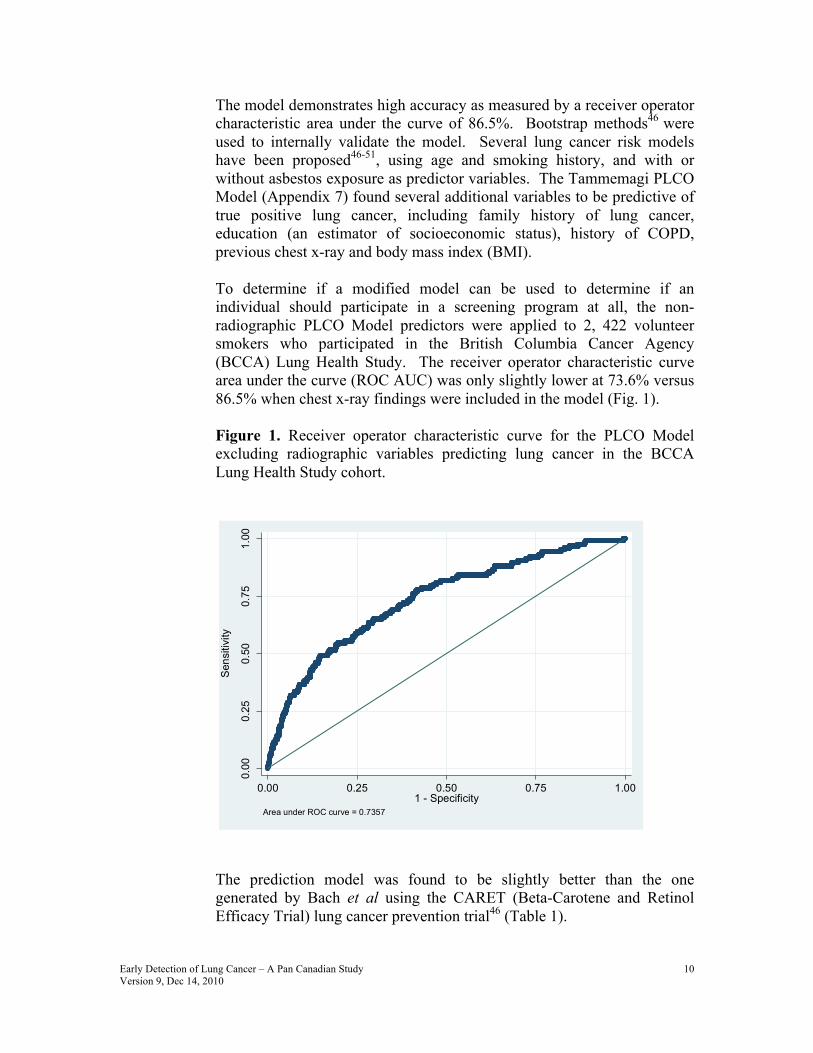
The model demonstrates high accuracy as measured by a receiver operator characteristic area under the curve of 86.5%. Bootstrap methods46 were used to internally validate the model. Several lung cancer risk models have been proposed46-51, using age and smoking history, and with or without asbestos exposure as predictor variables. The Tammemagi PLCO Model (Appendix 7) found several additional variables to be predictive of true positive lung cancer, including family history of lung cancer, education (an estimator of socioeconomic status), history of COPD, previous chest x-ray and body mass index (BMI). To determine if a modified model can be used to determine if an individual should participate in a screening program at all, the non-radiographic PLCO Model predictors were applied to 2, 422 volunteer smokers who participated in the British Columbia Cancer Agency (BCCA) Lung Health Study. The receiver operator characteristic curve area under the curve (ROC AUC) was only slightly lower at 73.6% versus 86.5% when chest x-ray findings were included in the model (Fig. 1).
Figure 1. Receiver operator characteristic curve for the PLCO Model excluding radiographic variables predicting lung cancer in the BCCA Lung Health Study cohort.
The prediction model was found to be slightly better than the one generated by Bach et al using the CARET (Beta-Carotene and Retinol Efficacy Trial) lung cancer prevention trial46 (Table 1).
0.00
0.25
0.50
0.75
1.00
Sens
itivi
ty
0.00 0.25 0.50 0.75 1.001 - Specificity
Area under ROC curve = 0.7357
Early Detection of Lung Cancer – A Pan Canadian Study Version 9, Dec 14, 2010
11
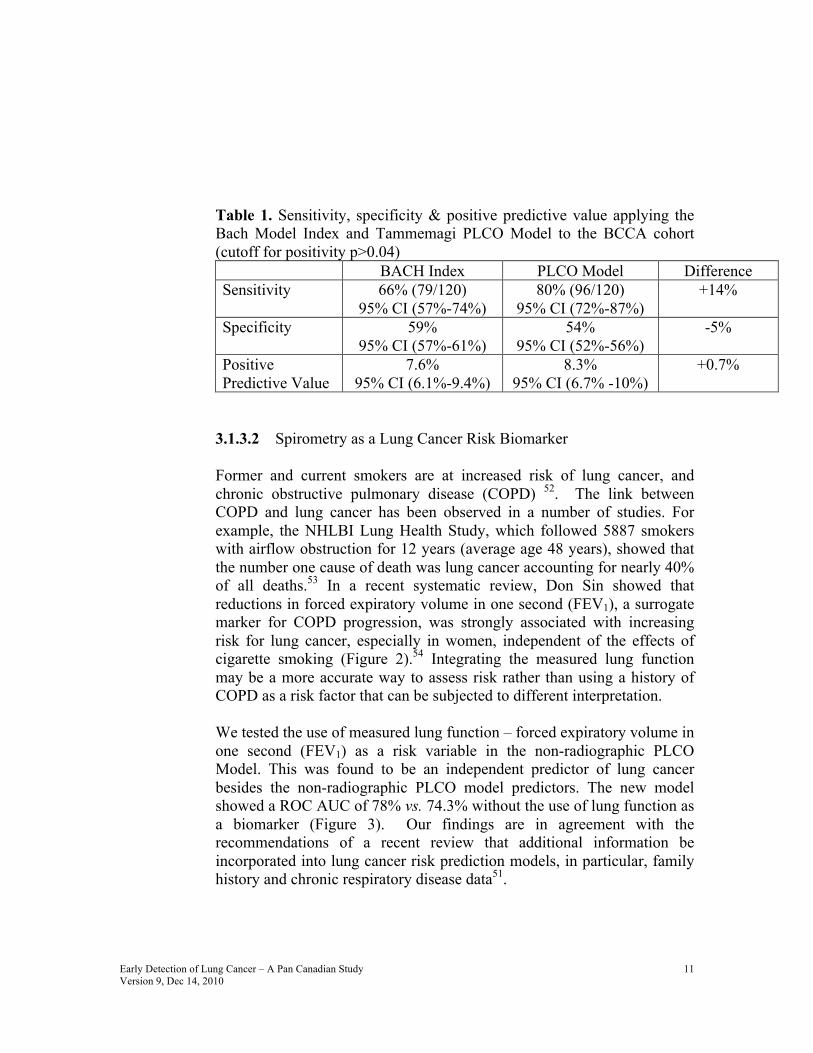
Table 1. Sensitivity, specificity & positive predictive value applying the Bach Model Index and Tammemagi PLCO Model to the BCCA cohort (cutoff for positivity p>0.04) BACH Index PLCO Model Difference Sensitivity 66% (79/120)
95% CI (57%-74%) 80% (96/120)
95% CI (72%-87%) +14%
Specificity 59% 95% CI (57%-61%)
54% 95% CI (52%-56%)
-5%
Positive Predictive Value
7.6% 95% CI (6.1%-9.4%)
8.3% 95% CI (6.7% -10%)
+0.7%
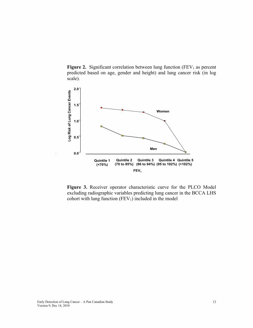
3.1.3.2 Spirometry as a Lung Cancer Risk Biomarker Former and current smokers are at increased risk of lung cancer, and chronic obstructive pulmonary disease (COPD) 52. The link between COPD and lung cancer has been observed in a number of studies. For example, the NHLBI Lung Health Study, which followed 5887 smokers with airflow obstruction for 12 years (average age 48 years), showed that the number one cause of death was lung cancer accounting for nearly 40% of all deaths.53 In a recent systematic review, Don Sin showed that reductions in forced expiratory volume in one second (FEV1), a surrogate marker for COPD progression, was strongly associated with increasing risk for lung cancer, especially in women, independent of the effects of cigarette smoking (Figure 2).54 Integrating the measured lung function may be a more accurate way to assess risk rather than using a history of COPD as a risk factor that can be subjected to different interpretation.
We tested the use of measured lung function – forced expiratory volume in one second (FEV1) as a risk variable in the non-radiographic PLCO Model. This was found to be an independent predictor of lung cancer besides the non-radiographic PLCO model predictors. The new model showed a ROC AUC of 78% vs. 74.3% without the use of lung function as a biomarker (Figure 3). Our findings are in agreement with the recommendations of a recent review that additional information be incorporated into lung cancer risk prediction models, in particular, family history and chronic respiratory disease data51.
Early Detection of Lung Cancer – A Pan Canadian Study Version 9, Dec 14, 2010
12
Figure 2. Significant correlation between lung function (FEV1 as percent predicted based on age, gender and height) and lung cancer risk (in log scale).
Quintile 1(<70%)
Log
Risk
of L
ung
Canc
er E
vent
s
0.0
0.5
1.0
1.5
2.0
Quintile 2(70 to 85%)
Quintile 3(86 to 94%)
Quintile 4(95 to 102%)
Quintile 5(>102%)
Men
Women
FEV1
Figure 3. Receiver operator characteristic curve for the PLCO Model excluding radiographic variables predicting lung cancer in the BCCA LHS cohort with lung function (FEV1) included in the model
Early Detection of Lung Cancer – A Pan Canadian Study Version 9, Dec 14, 2010
13
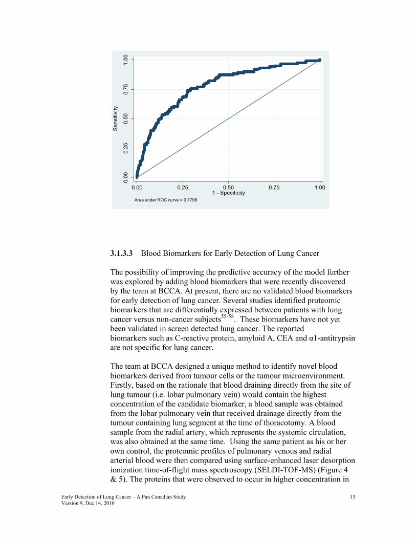
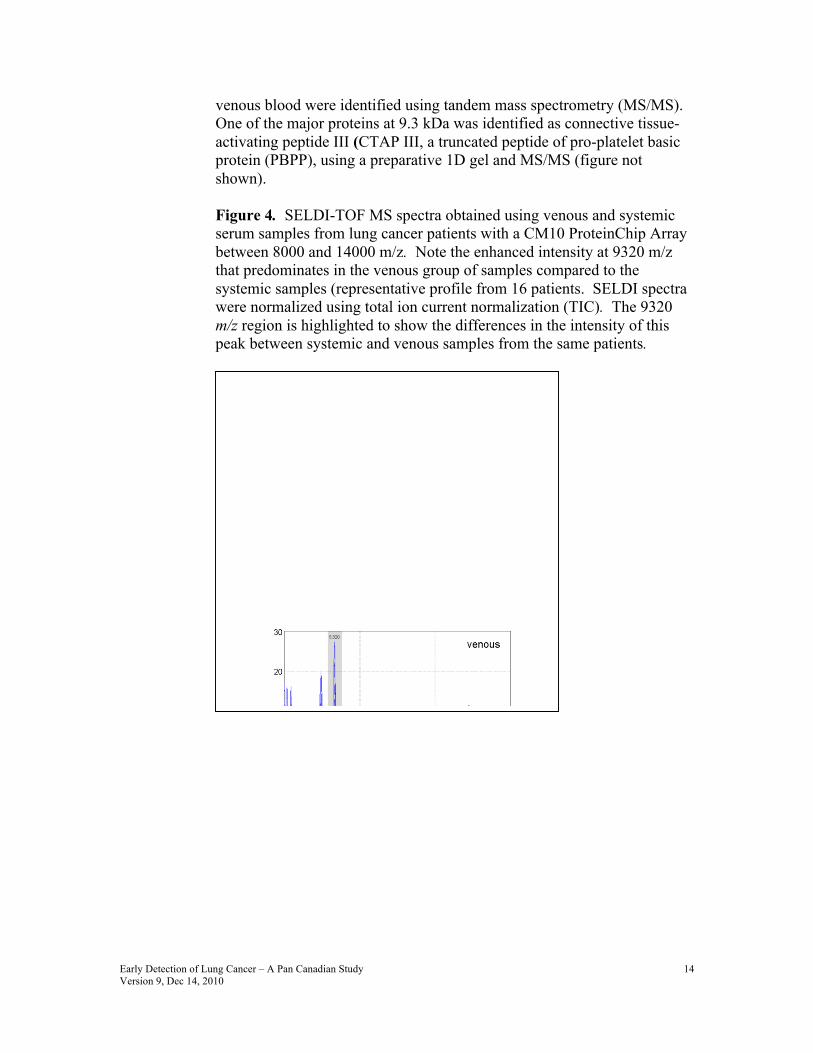
3.1.3.3 Blood Biomarkers for Early Detection of Lung Cancer The possibility of improving the predictive accuracy of the model further was explored by adding blood biomarkers that were recently discovered by the team at BCCA. At present, there are no validated blood biomarkers for early detection of lung cancer. Several studies identified proteomic biomarkers that are differentially expressed between patients with lung cancer versus non-cancer subjects55-58. These biomarkers have not yet been validated in screen detected lung cancer. The reported biomarkers such as C-reactive protein, amyloid A, CEA and α1-antitrypsin are not specific for lung cancer. The team at BCCA designed a unique method to identify novel blood biomarkers derived from tumour cells or the tumour microenvironment. Firstly, based on the rationale that blood draining directly from the site of lung tumour (i.e. lobar pulmonary vein) would contain the highest concentration of the candidate biomarker, a blood sample was obtained from the lobar pulmonary vein that received drainage directly from the tumour containing lung segment at the time of thoracotomy. A blood sample from the radial artery, which represents the systemic circulation, was also obtained at the same time. Using the same patient as his or her own control, the proteomic profiles of pulmonary venous and radial arterial blood were then compared using surface-enhanced laser desorption ionization time-of-flight mass spectroscopy (SELDI-TOF-MS) (Figure 4 & 5). The proteins that were observed to occur in higher concentration in
0.00
0.25
0.50
0.75
1.00
Sens
itivi
ty
0.00 0.25 0.50 0.75 1.001 - Specificity
Area under ROC curve = 0.7798
Early Detection of Lung Cancer – A Pan Canadian Study Version 9, Dec 14, 2010
14
venous blood were identified using tandem mass spectrometry (MS/MS). One of the major proteins at 9.3 kDa was identified as connective tissue-activating peptide III (CTAP III, a truncated peptide of pro-platelet basic protein (PBPP), using a preparative 1D gel and MS/MS (figure not shown). Figure 4. SELDI-TOF MS spectra obtained using venous and systemic serum samples from lung cancer patients with a CM10 ProteinChip Array between 8000 and 14000 m/z. Note the enhanced intensity at 9320 m/z that predominates in the venous group of samples compared to the systemic samples (representative profile from 16 patients. SELDI spectra were normalized using total ion current normalization (TIC). The 9320 m/z region is highlighted to show the differences in the intensity of this peak between systemic and venous samples from the same patients.
Early Detection of Lung Cancer – A Pan Canadian Study Version 9, Dec 14, 2010
15
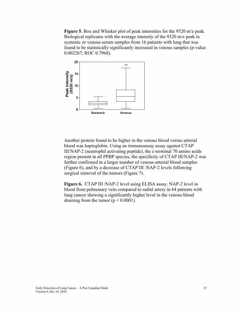
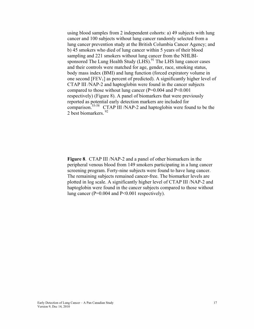
Figure 5. Box and Whisker plot of peak intensities for the 9320 m/z peak. Biological replicates with the average intensity of the 9320 m/z peak in systemic or venous serum samples from 16 patients with lung that was found to be statistically significantly increased in venous samples (p-value 0.002267; ROC 0.7968). Another protein found to be higher in the venous blood versus arterial blood was haptoglobin. Using an immunoassay assay against CTAP III/NAP-2 (neutrophil activating peptide), the c-terminal 70 amino acids region present in all PPBP species, the specificity of CTAP III/NAP-2 was further confirmed in a larger number of venous-arterial blood samples (Figure 6), and by a decrease of CTAP III /NAP-2 levels following surgical removal of the tumors (Figure 7). Figure 6. CTAP III /NAP-2 level using ELISA assay. NAP-2 level in blood from pulmonary vein compared to radial artery in 64 patients with lung cancer showing a significantly higher level in the venous blood draining from the tumor (p < 0.0001).
Systemic Venous0
5
10
15
20**
Peak
Inte
nsity
(932
0 m
/z)

Early Detection of Lung Cancer – A Pan Canadian Study Version 9, Dec 14, 2010
16
Radial Artery Pulmonary Vein
020
0040
0060
00
NAP
2 Le
vel (
ng/m
l)
P<.00001
Figure 7. Change in CTAP III /NAP-2 level in the peripheral venous blood before and after surgical resection of the tumor in 24 patients. A significantly lower CTAP III /NAP-2 level was observed after tumor removal (p=0.01).
020
0040
0060
0080
00
Before Surgery After Surgery
NAP
2 Le
vel (
ng/m
l)
P=.01
To determine the potential application of the discovered proteins as biomarkers for early lung cancer detection, we compared their concentrations in heavy smokers who did and did not develop lung cancer
Early Detection of Lung Cancer – A Pan Canadian Study Version 9, Dec 14, 2010
17
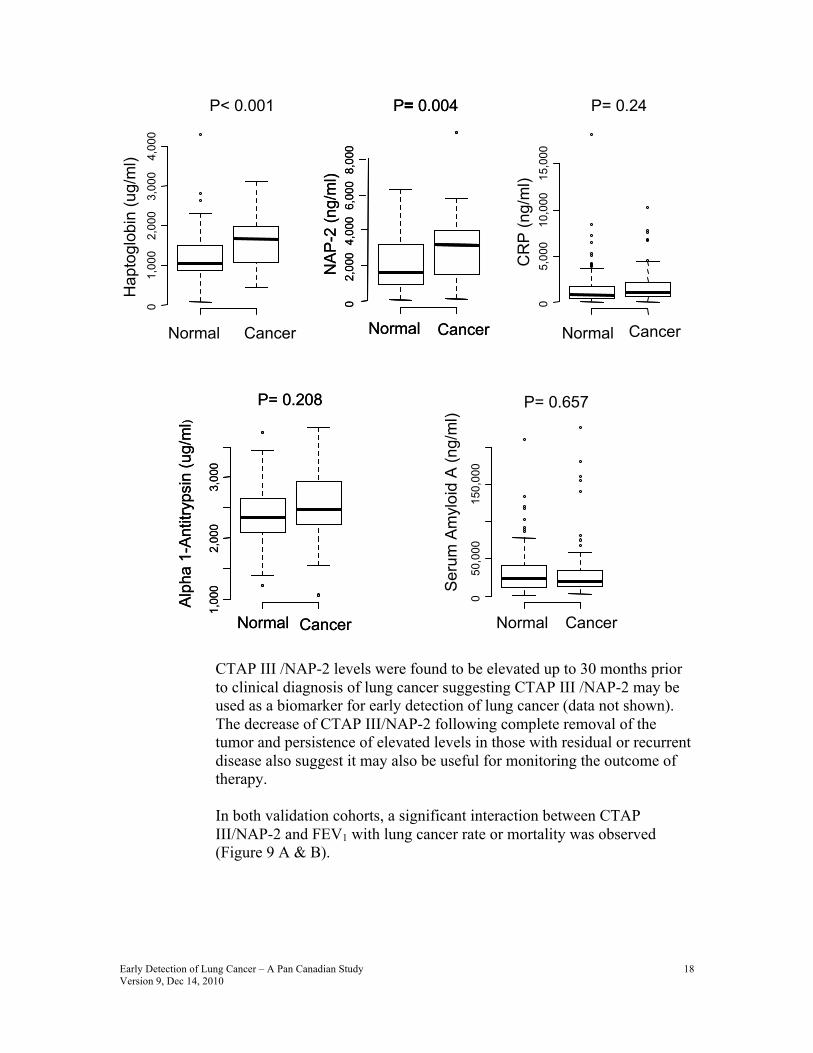
using blood samples from 2 independent cohorts: a) 49 subjects with lung cancer and 100 subjects without lung cancer randomly selected from a lung cancer prevention study at the British Columbia Cancer Agency; and b) 45 smokers who died of lung cancer within 5 years of their blood sampling and 221 smokers without lung cancer from the NHLBI-sponsored The Lung Health Study (LHS).91 The LHS lung cancer cases and their controls were matched for age, gender, race, smoking status, body mass index (BMI) and lung function (forced expiratory volume in one second [FEV1] as percent of predicted). A significantly higher level of CTAP III /NAP-2 and haptoglobin were found in the cancer subjects compared to those without lung cancer (P=0.004 and P<0.001 respectively) (Figure 8). A panel of biomarkers that were previously reported as potential early detection markers are included for comparison.55-58 CTAP III /NAP-2 and haptoglobin were found to be the 2 best biomarkers. 92
Figure 8. CTAP III /NAP-2 and a panel of other biomarkers in the peripheral venous blood from 149 smokers participating in a lung cancer screening program. Forty-nine subjects were found to have lung cancer. The remaining subjects remained cancer-free. The biomarker levels are plotted in log scale. A significantly higher level of CTAP III /NAP-2 and haptoglobin were found in the cancer subjects compared to those without lung cancer (P=0.004 and P<0.001 respectively).
Early Detection of Lung Cancer – A Pan Canadian Study Version 9, Dec 14, 2010
18
CTAP III /NAP-2 levels were found to be elevated up to 30 months prior to clinical diagnosis of lung cancer suggesting CTAP III /NAP-2 may be used as a biomarker for early detection of lung cancer (data not shown). The decrease of CTAP III/NAP-2 following complete removal of the tumor and persistence of elevated levels in those with residual or recurrent disease also suggest it may also be useful for monitoring the outcome of therapy. In both validation cohorts, a significant interaction between CTAP III/NAP-2 and FEV1 with lung cancer rate or mortality was observed (Figure 9 A & B).
Normal Cancer
01,
000
2,00
03,
000
4,00
0
P< 0.001H
apto
glob
in(u
g/m
l)
Normal Cancer
05,
000
10,0
0015
,000
P= 0.24
CR
P(n
g/m
l)
Normal Cancer0
2,00
04,
000
6,00
08,
000
P= 0.004
NAP
-2 (n
g/m
l)Normal Cancer
02,
000
4,00
06,
000
8,00
0
P= 0.004
NAP
-2 (n
g/m
l)
Normal Cancer
1,00
02,
000
3,00
0
P= 0.208
Alph
a 1-
Antit
ryps
in(u
g/m
l)
Normal Cancer
1,00
02,
000
3,00
0
P= 0.208
Alph
a 1-
Antit
ryps
in(u
g/m
l)
Normal Cancer
050
,000
150,
000
P= 0.657
Seru
m A
myl
oid
A(n
g/m
l)

Early Detection of Lung Cancer – A Pan Canadian Study Version 9, Dec 14, 2010
19
Figure 9. A Fitted Line Showing The Relationship Between The Risk Of Lung Cancer And NAP-2 As A Function Of FEV1%. A. Data from the Lung Cancer Chemoprevention. B. Study Data from the NHLBI Lung Health Study The relative risk or hazard ratio of lung cancer is shown for every 1-unit increase in NAP-2 expression (in ng/ml-logarithmic scale) as a function of FEV1% of predicted. As FEV1% decreases the risk of lung cancer is amplified for every 1 unit increase in levels of CTAP III/NAP-2.
Since lung cancer consists of 4 major cell types (squamous cell carcinoma, adenocarcinoma, small cell carcinoma and large cell carcinoma) and each type may consist of several subtypes (e.g. at least 6 sub-types in adenocarcinoma), it is perhaps unrealistic to anticipate that a single or even a panel of biomarkers could detect all lung cancers with high sensitivity and specificity when used alone. A better approach is to develop a prediction model that is similar to the highly successful Framingham cardiac risk model. In the Framingham model,87, 88 cholesterol, a blood biomarker is not used alone but rather integrated with socio-demographic factors and medical data such as age, sex, smoking, and presence/absence of diabetes and blood pressure. Using a similar approach, we combine CTAP III/NAP-2, and haptoglobin along with age, smoking and FEV1. The accuracy of the risk prediction model improved to 84% (Figure 10).
20 40 60 80 100 120 140
02
46
810
Rel
ativ
e R
isk
of L
ung
Can
cer f
orEv
ery
1-un
it in
crea
se in
NAP
-2
FEV1% of Predicted
Haz
ard
Rat
io o
f Lun
g C
ance
r Dea
th fo
rEv
ery
1-un
it in
crea
se in
NAP
-2
0 50 100 150
12
34
56
FEV1% of Predicted
Haz
ard
Rat
io o
f Lun
g C
ance
r Dea
th fo
rEv
ery
1-un
it in
crea
se in
NAP
-2
0 50 100 150
12
34
56
FEV1% of Predicted
0 50 100 150
12
34
56
FEV1% of Predicted
A B

Early Detection of Lung Cancer – A Pan Canadian Study Version 9, Dec 14, 2010
20
Figure 10. Receiver operating characteristic curve showing sensitivity and specificity of CTAP III/NAP-2 and haptoglobin in combination with age, sex and lung function (FEV1) to detect lung cancer in a high risk population. The lung cancer risk prediction by the model was improved further compared to age, sex and lung function (FEV1) alone.
In this study, we will evaluate prospectively the ability of blood biomarkers (both our own biomarkers and those published in the literature) independently and jointly to detect early (asymptomatic) lung cancer in a high risk population and their ideal implementation in a screening program. We will also evaluate whether blood biomarkers can upstage or downstage the malignancy potential of CT detected lung nodules or indicate more intensive surveillance in the face of negative examinations. In addition to protein-based markers, we will also evaluate the role of DNA-based genetic markers of lung cancer susceptibility to complement on-going large scale genome wide association studies (Toronto-IARC-Houston, US NIH). Recently the first of these papers described a nicotine acetylcholine receptor subunit polymorphism as being associated with the risk of lung cancer95,96. These genetic susceptibility markers in 6q23-25 and 15q25 and other loci will be rapidly tested in our repository and if positive, we will evaluate their ideal implementation into a screening program.
0.0 0.2 0.4 0.6 0.8 1.0
0.0
0.2
0.4
0.6
0.8
1.0
1-Specificity
Sens
itivi
ty
Area under the curve: 0.83995% confidence interval (0.765, 0.913)

Early Detection of Lung Cancer – A Pan Canadian Study Version 9, Dec 14, 2010
21
The DNA-based genetic markers being assessed are highly prevalent, low penetrant germline genetic variations. They will be selected on the basis of their ability to enhance a multi-modality approach to risk stratification. They are also crucial to the main biomarker study objectives, and are necessary for model generation. These DNA-based markers are NOT similar to the high penetrant low prevalence markers such as BRCA1, MLH1, and others in cancer genetic syndromes. The DNA-based markers in question are currently being studied in molecular epidemiologic studies. Typically, these markers have odds ratios between 1.1 and 1.5, as opposed to odds ratios of 10-30 for cancer genetic syndromes. None of these DNA-based markers have ever heen demonstrated to have an individual impact clinically, outside of multimodality models. These markers are studied in research laboratories which are not certified for clinical purposes. As such, any important findings will be communicated to participants through the usual channels of conference presentations and publications. By the same token, these research findings will not be acted on like a clinical genetic test since the result needs to be validated and interpreted in the context of a multi-variate risk assessment model. Scientific knowledge is changing rapidly. The final list of protein-based and DNA-based biomarkers to be assessed in addition to CTAP III/NAP-2 and Haptoglobin will be determined by the steering committee at the start of the third year. Measurement of biomarkers besides CTAP III/NAP-2 and Haptoglobin will depend on availability of additional funding. An amendment will be filed with each REB before measuring additional biomarkers. In addition to evaluating these markers, we understand that new biomarkers of other types will be discovered on a regular basis. We will also seek permission from participants to utilize their blood sample outside the scope of the present study and after the end of this study. This secondary optional banking will be requested specifically in the consent form. 3.1.4 Autofluorescence Bronchoscopy (AFB) AFB was originally developed by the team at BCCA37 and has since been commercialized world-wide by several endoscope companies for the detection of early lung cancer59. As discussed in 2.1.1 above, the concept of complementary screening using both spiral CT and AFB was developed based on data at BCCA in almost 1,600 subjects. In that study, 19.6% of the cancers detected were negative on spiral CT. We plan to use both spiral CT and AFB to confirm the added value of AFB in an early detection program.

Early Detection of Lung Cancer – A Pan Canadian Study Version 9, Dec 14, 2010
22
3.1.4.1 Optical Coherence Tomography (OCT) Optical coherence tomography is an optical imaging method to visualize structures below the bronchial surface (25). It is similar in principle to ultrasound. Instead of using sound waves, infrared light is used. An image is obtained from the back-scattered light. There is no associated risk from the weak infrared red light. Preliminary data even with an axial resolution of 16 µm showed that it is possible to distinguish dysplastic and in-situ carcinoma lesions from lower grade lesions (Figure 11). Further improvement to this technology using higher resolution (4 µm) and Doppler measurement of vascular density is on-going. Subjects participating in this study will allow us to further develop this non-biopsy optical imaging method to study the effect of chemopreventive interventions.
Figure 11. Optical Coherence Tomography. (A) area with metaplasia. BM= Basement, E=Epithelium (B) area with dysplasia. (C) Carcinoma in-situ with intact basement membrane. (D) Invasive cancer with loss of basement membrane.
A B
E
BM
C D
Early Detection of Lung Cancer – A Pan Canadian Study Version 9, Dec 14, 2010
23
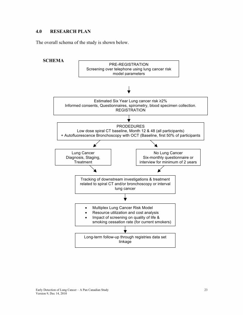
4.0 RESEARCH PLAN The overall schema of the study is shown below.
SCHEMA
PRE-REGISTRATION Screening over telephone using lung cancer risk
model parameters
Long-term follow-up through registries data set linkage
Estimated Six Year Lung cancer risk ≥2% Informed consents, Questionnaires, spirometry, blood specimen collection.
REGISTRATION
PRODEDURES Low dose spiral CT baseline, Month 12 & 48 (all participants)
+ Autofluorescence Bronchoscopy with OCT (Baseline, first 50% of participants only)
Lung Cancer Diagnosis, Staging,
Treatment
No Lung Cancer Six-monthly questionnaire or
interview for minimum of 2 years
Tracking of downstream investigations & treatment related to spiral CT and/or bronchoscopy or interval
lung cancer
• Multiplex Lung Cancer Risk Model • Resource utilization and cost analysis • Impact of screening on quality of life &
smoking cessation rate (for current smokers)
Early Detection of Lung Cancer – A Pan Canadian Study Version 9, Dec 14, 2010
24
4.1 Target Population Study Subjects
Inclusion Criteria • Women or men age 50 to 75 years. • Current or former smokers who have smoked cigarettes for 20 years or more. A former smoker is defined as one who has stopped smoking for one or more years. • An estimated 6-year lung cancer risk of ≥2% based on the risk prediction model.* • ECOG performance status 0 or 1. • Capable of providing, informed consent for screening procedures (low dose spiral CT, AFB, spirometry, blood biomarkers). * The programmer at the study center where risk scores were being calculated inadvertently forgot to divide the six-year risk by 2. This was discovered about 8 months after recruitment began. At that time, the number of lung cancers being detected was on track to satisfy our power requirements. Therefore, the risk calculator was not changed to 3 year risks. In addition, early during the study, planning was underway to extend the study with further follow-up and an additional year four screen. The extended follow-up made the ≥2% risk in 6 year of follow-up appropriate.
Exclusion Criteria
• Any medical condition, such as severe heart disease (e.g. unstable angina, chronic congestive heart failure), acute or chronic respiratory failure, bleeding disorder, that in the opinion of the investigator could jeopardize the subject’s safety during participation in the study or unlikely to benefit from screening due to shortened life-expectancy from the co-morbidities • Have been previously diagnosed with lung cancer • Have had other cancer with the exception of the following cancers which can be included in the study: non-melanomatous skin cancer, localized prostate cancer, carcinoma in situ (CIS) of the cervix, or superficial bladder cancer. Treatment of the exceptions must have ended >6 months before registration into this study • On anti-coagulant treatment such as warfarin or heparin • Known reaction to Xylocaine, salbutamol, midazolam, and alfentanil. • Pregnancy • Unwilling to have a spiral chest CT • Chest CT within 2 years • Unwilling to sign a consent
4.2 Number of Subjects A total of 2,700 subjects will be accrued over 20 months from 8 centres across Canada. Both men and women and members of all races and ethnic groups are
Early Detection of Lung Cancer – A Pan Canadian Study Version 9, Dec 14, 2010
25
eligible for this trial. The rationale for the sample size is described in section 4.7 below.
4.3 Subject Recruitment Subjects will be recruited by newspaper, TV and radio announcements
4.4 Study Procedures
4.4.1 Screening by Short Questionnaire & Registration When a potential participant indicates interest in the study in writing or by phone, a brief explanation of the study will be given by the study clerk. A lung cancer risk index will be generated using self-reported age, sex, smoking history (number of cigarettes, duration of smoking, years since smoking cessation), family history of lung cancer, education level and body mass index (from height and weight). The study inclusion and exclusion criteria will be reviewed for those with an estimated 3-year lung cancer risk ≥ 2%. An appointment will be given to come to the study site for an interview by the study coordinator for possible enrollment.
Each participating site will fax the completed Registration Eligibility Checklist to the Coordination Center (CC) in Vancouver to register all subjects. A study number will be assigned by the Project Manager. To register a participant, fax to 604/675-8098 a completed Registration Eligibility Checklist to the CC between 8 a.m. and 4:00 p.m. Pacific time, Monday through Friday. Participant ID numbers will be assigned to the participant by the CC within 24 hours.
4.4.2 Post-Registration Following informed consent, study questionnaires will be administered by a trained site study coordinator (Appendix). Spirometric measurements will be obtained according to the Canadian Chronic Obstructive Disease Study Network. A blood specimen will also be obtained according to standard protocol (Appendix). Former smoking status will be verified by urine cotinine measurement. Three questionnaires will be administered electronically on the day of the first visit using a laptop or desktop computer: (i) a study questionnaire covering socio-demographic factors, smoking, occupational exposure, family history and medical data (Appendix). (ii) Quality of life (SF-12, EQ-5D)60 and (iii) Spielberger State
Early Detection of Lung Cancer – A Pan Canadian Study Version 9, Dec 14, 2010
26
Trait Anxiety Index (Stai)61 (licensed from NCI). At the end of the session, a hard copy will be printed and stored on site. An electronic version will be sent to the Coordinating Center (CC) server in Vancouver with the subject identified by the study number only. The CC Project Manager will be notified by fax or e-mail when the data has been transferred. The CC personnel will verify the participant eligibility and completeness of the data in the questionnaires.
4.4.3 Spirometry Spirometry will be conducted using a flow-sensitive spirometer (EasyOne™ Diagnostic Spirometer, ndd Medical Technologies, Andover MA) in accordance with the American Thoracic Society recommendations62. To estimate lung function, we will use both forced expiratory volume in 1 second (FEV1) and forced vital capacity (FVC). These will be recorded in litres (L) and as a percent of predicted (% predicted) using standardized prediction equations63. Post-bronchodilator measurements will only be performed to assess the presence of a significant bronchodilator response if the FEV1 <80% predicted (Salbuatomol 200 mcg). Spirometry will be collected on the day of registration and annually for two years. Spirometry tracings identified by a study code will be transmitted by telephone line to the COLD server for quality check by Dr. Wan Tan. Satisfactory tracings will be sent to the CC server.
4.4.4 Spirometry Quality Assurance The site study coordinator who will be performing the spirometric measurements will be trained by Dr. Wan Tan through one of the existing COLD Study network sites across Canada. The spirometry tracings identified by a anonymized study code will be transmitted via a website to Dr. Tan who will grade the quality of the tracings. Spriometry of failed tracings will be asked to be repeated.
4.4.5 Low dose spiral CT All multi-center studies including the large studies in the US have been using different CT scanners, and there is vast experience with the different imaging equipment. Heterogeneity of equipment occurs throughout the clinical world. It is not problematic if it does not lead to heterogeneity of diagnosis. The imaging protocol in the NLST and the NELSON trial as well as ours define detailed data acquisition parameters that can be implemented on any CT scanner - always adhering to the thin-slice, low-dose regimen. A multi-detector row CT scanner with minimum section collimation of 1.25 mm and minimum number of data acquisition channels ≥ four will be employed. The CT scans will be performed at 120 kV, 40-50 mA, beam pitch 1.5 (1.5 x 4 detectors is also referred to as pitch 6). Low radiation dose acquisitions using less than 2 mSv effective dose will be obtained using reduced mA and a minimum gantry rotation time providing an
Early Detection of Lung Cancer – A Pan Canadian Study Version 9, Dec 14, 2010
27
average Dose Length Product less than 120 mGy cm. Images will be acquired in a single inspiratory breath hold with the subject in the supine position. Images will be reconstructed using 1.25 mm or less section thickness and 1.25 mm or less spacing. Two image reconstruction algorithms will be employed, a high spatial frequency algorithm for lung parenchyma (e.g. bone (GE) or B60 (Siemens)) and an intermediate spatial frequency algorithm for mediastinal structures (e.g. standard (GE) or B35 (Siemens)). The mediastinal reconstruction algorithm will be useful to provide lower noise images on these reduced dose images. Images will be archived to the hospital based PACS server with full annotation and stored in local site for clinical use if needed. A second image file will be saved with an anonymized study number. The file with the anonymized study number will be sent to the central study data server. Calibration scans will be performed at all participating sites using the body calibration phantoms and spatial resolution supplied with the CT scanners at each site. These calibration scans will be performed using the same technical parameters as proposed in the low dose CT protocol (kVp 120, 40 mAs, rotation time 1 second or less, 32 cm field of view reconstruction, intermediate and high spatial frequency reconstruction algorithm). Spatial resolution and image noise will be measured on the submitted images and used for standardization of each site. Reference scans will be repeated on a yearly basis and forwarded to the Vancouver coordinating center. Dr. John Mayo and Dr. John Aldrich, the VGH Radiation Protection Officer and Medical Physicist will review the data to ensure adequate scanner performance at each of the sites.
Study radiologist(s) at each of the enrolling institutions will report all study CT scans (both baseline and follow up), and will be called “designated readers”. Images will be reviewed in a dedicated workstation with appropriate illumination and ergonomics. Lung and mediastinal images will be reviewed using appropriate window and level settings as determined by the reviewing radiologist (suggested lung 1500/-750, suggested mediastinum 450/35). Up to 10 lung nodules will be identified and recorded on the radiology study work sheets indicating nodule type (solid, ground glass opacity (GGO), semisolid, peri-fissural opacity) with measurement of the long and short axis diameters. The nodule(s) will be described as well-defined, lobulated, spiculated, or demonstrating a halo, and their location as parenchymal or pleural/fissural. Patients with greater than 10 nodules will have the 10 largest nodules measured and documented. In patients with innumerable nodules and no dominant nodule, no measurements will be performed. Any other abnormalities in the lung or mediastinal tissues will be recorded. The radiologist’s visual assessment of the extent of emphysema will be recorded on a five-point scale (none, minimal, mild, moderate, or severe). The spatial distribution of emphysema will be recorded using a four point scale (upper, mid, lower, or diffuse). The presence of fully or partially calcified lung parenchymal nodules and calcified hilar or mediastinal lymph nodes will be recorded. Coronary artery calcification, non calcified enlarged mediastinal and hilar adenopathy, and chest wall, pleural and upper abdominal pathology will be

Early Detection of Lung Cancer – A Pan Canadian Study Version 9, Dec 14, 2010
28
recorded. On subsequent examinations, any change in the size of the nodules recorded at baseline will be documented plus any new nodules that become visible or larger in the interval.
4.4.6 Lung Nodule Follow-up Protocol CT scan follow up protocol will be determined by the maximum long axis diameter of the largest nodule identified. It is estimated that ~70% of the participants will have one or more nodules or GGO in the baseline CT scan. Subjects with no abnormality on the baseline exam will have a repeat scan in 12 months. For subjects with baseline scans where the largest solid nodule is less than 5 mm in diameter or GGO less than 8 mm, a follow up examination in 12 months’ time will be performed. If there are no new nodules and no growth of existing nodules on the 12-month exam, an additional CT scan will be performed at 24 months. The subject will be discharged from the study at 24 months if there is no growth of existing nodules and no development of new nodules. Subjects with any semi-solid or solid nodule 5 to 10 mm or GGO 8-10 mm will receive an additional limited or low dose full chest scan at 3 months, then the routinely scheduled scans at 12 and 24 months. Any subject with growth of an existing nodule, development of a solid component in GGO or a new nodule will receive an additional scan at 3 months with decision on successive scans or biopsy to be made at the discretion of the study physician and radiologist. A nodule that grows on two consecutive scans, a non-solid opacity showing development of a solid component and any nodule greater than 10 mm in diameter will be considered as suspicious for lung cancer or a positive scan. The lesion will be managed based on the practice patterns of the local institution. Further diagnostic procedures may include serology for cryptococosis and histoplasmosis, PET/CT imaging, CT guided transthoracic needle aspiration/core biopsy, bronchoscopy, wedge resection for diagnosis. The overall aim will be to establish a pathologic or cytologic diagnosis and perform definitive treatment. Nodule doubling time will be calculated for all growing nodules using volumetric analysis. Any other abnormality on the CT in the surrounding soft tissue of the chest and abdomen will be followed up according to standard of care in the institution as directed by the medical team and the local study radiologist.
4.4.7 Spiral CT Quality Assurance A teaching set of 20 cases selected from archives of experienced lung cancer screening radiologists will be established and reviewed by the designated reader at each center. A test set of 20 different cases will be administered to each enrolling centre to document an acceptable level of inter-observer agreement prior to commencing study screening. Conferences will be held on a two monthly basis with all radiology readers in the study to discuss enrollment, difficulties with interpretation, report turn around time and to review pathologically/cytologically confirmed lung cancer cases (both prevalence and interval cancers). The first 60
Early Detection of Lung Cancer – A Pan Canadian Study Version 9, Dec 14, 2010
29
cases in each center will be independently interpreted by a review chest radiologist (Dr. Nestor Muller) and a chest radiology fellow under his supervision working by consensus and without knowledge of the management plan of the patient. The scans will be first reviewed without computer aided diagnosis (CAD) software and then compare with the highlighted areas using CAD. Clinically significant discrepancies will be recorded and reviewed with follow up discussion with the initial reading radiologist by videoconference or in person. Videoconferences will be held on a monthly basis with all bronchoscopists to review pathologically/cytologically confirmed lung cancer cases (both prevalence and interval cancers).
4.4.8 Autofluorescence Bronchoscopy and Biopsy AFB is a technique that enables the bronchoscopist to identify abnormal tissue from high grade dysplasia to frank malignancy using the alteration in tissue fluorescence that accompanies this differentiation. Identifying invasive malignancy with traditional white light bronchoscopy (WLB) is usually easy whereas changes of severe dysplasia or carcinoma in situ are often much more visually subtle or inapparent by WLB even in experienced hands41. Autofluorescence bronchoscopy has demonstrated detection of dysplasia, carcinoma in situ and early invasive cancers not visible by standard WLB techniques41. The labour-intensive nature of AFB, coupled with the minimally invasiveness and cost of this procedure in addition to spiral CT lend itself to a practical compromise. We have planned that the first 50% (n=1250) of individuals undergoing screening without lesions suspicious of lung cancer on spiral CT will also have AFB. Those with suspicious lesion detected by CT will be managed based on the practice patterns of the local institution which may include a diagnostic bronchoscopy (see section 3.4.5 above). With 1250 individuals without lesion suspicious of lung cancer on CT, there is adequate power to determine if AFB can detect lung cancers that are missed by spiral CT (see section 4.7). Further, this number of individuals affords us some flexibility in the event that some sites are slower to procure the necessary equipment or train personnel for AFB. In this event, the centers who are already set up to perform AFB can take on a higher fraction of patients undergoing AFB, and subtle differences across sites will be adjusted for in the subsequent analysis.
Within 4 weeks after spiral CT, autofluorescence and white light bronchoscopy will be performed under conscious sedation and topical anesthesia to the upper airway after being NPO (nothing by mouth) for a minimum of six hours prior to the procedure. A Health Canada approved clinical device (e.g. Pentax SAFE 3000, Novadaq Onco-LIFE) will be used. Complete airway inspection will be performed using both autofluorescence and white light techniques. AFB will be performed first, followed by the white light bronchoscopy (WLB) exam.
Early Detection of Lung Cancer – A Pan Canadian Study Version 9, Dec 14, 2010
30
Alternatively, if the device allows, simultaneous white-light and fluorescence examination can be performed. Care is taken to identify and record areas that are suspicious for or clearly demonstrate abnormality. Trauma to the mucosa either by the bronchoscope tip or by suctioning needs to be avoided as this can obscure the imaging under the autofluorescence system. Bronchial biopsies will be obtained from areas with abnormal fluorescence suspicious of severe dysplasia or worse pathology. Location of the biopsies obtained will be recorded on a procedural paper record as well as in the dictation of the procedure. Any area suspicious for carcinoma in-situ or invasive cancer will be recorded in case report form and biopsied for histopathological diagnosis. The bronchoscopic procedure will be recorded in digital form using the participant’s anonymized study ID, and sent to the central data bank. Diagnosis of dysplasia, carcinoma in-situ or invasive carcinoma will be reviewed. The Pathologists in this study will include: Leaders: Ming-Sound Tsao (Toronto) & External Consultant: Adi Gazdar Site Pathologists: Vancouver: Diana Ionescu, John English Calgary: Stefan Urbanski Hamilton: JC Cutz Ottawa: Harman Sekhon Quebec City (Laval): Christian Couture Halifax: Zhaolin Xu Newfoundland: Dan Fontaine Dr. Tsao and Dr. Gazdar are current members of the Pathology Panel of International Association for the Study of Lung Cancer (IASLC). Once the study is approved, a teleconference among site pathologists will be conducted to work out the details of pathology diagnostic criteria and data submission. The criteria and a set of images representative of squamous pre-neoplastic lesions and carcinoma in-situ as defined in the 1999 WHO/IASLC classification64 of pulmonary/pleural tumours will be prepared and emailed to each participating pathologists. The lung cancers are as defined in the 2004 WHO Classification of Lung, Mediastinum and Heart neoplasm.93,94 The biopsies will be processed as routine surgical pathology sample and assigned to the site pathologist for the study. The pathology report will be faxed immediately to the Central Office (604/675-8098). The site pathologist will either submit electronic images of the diagnostic lesions for e-review and archiving or send one representative diagnostic HE slide of the lesion to Dr. Tsao and Dr. Gazdar for review 4.4.9 Follow-up of Abnormal Bronchial Biopsies

Early Detection of Lung Cancer – A Pan Canadian Study Version 9, Dec 14, 2010
31
Carcinoma in-situ or invasive cancer will be managed based on the standard clinical practice patterns of the local institution. Currently, there is no established clinical guideline for follow-up or treatment of dysplasia. Follow-up bronchoscopy and biopsy will be at the discretion of the site endoscopist in discussion with the participant.
4.4.10 AF Bronchoscopy Quality Assurance A teaching set of 20 cases selected from archival pathologically or cytologically confirmed cases will be established and reviewed by the designated bronchoscopists at each center. A test set of 20 different cases will be administered to each enrolling centre to document an acceptable level of inter-observer agreement prior to commencing study screening. Centers without prior experience with AF bronchoscopy will have their endoscopists visit BCCA to observe the procedure. Alternatively, Dr. Lam or Dr. McWilliams will visit the site and perform bronchscopies together to ensure uniformity in grading abnormality. Videoconferences will be held on a monthly basis with all bronchoscopists to review pathologically/cytologically confirmed lung cancer cases (both prevalence and interval cancers). The first 30 cases in each center will be independently interpreted by Dr. Tom Sutedja (Free University, Netherlands) without knowledge of the management plan of the patient. Clinically significant discrepancies will be recorded and reviewed with follow up discussion with the initial reading endoscopist by videoconference or in person.
4.4.11 Blood Specimen Blood samples will be collected on the day of registration after signing informed consent with the option of collecting blood annually for two years. For subjects found to have lung cancer, a blood specimen will be obtained prior to treatment and 3 to 6 months post treatment (for those who will be given treatment with curative intent) to determine whether the biomarkers decrease after treatment with curative intent. Blood samples will be drawn without regard to fasting status or time of day, although the time of day and approximate time of last meal will be recorded on the sample collection form.
A Standard Operating Procedures Manual will be supplied to all clinical research coordinators with specific details. In summary, blood sampling kits will be assembled at Dr. Geoff Liu’s laboratory and distributed to each site to ensure the specimens are collected in the proper tubes. Based on discussions with the large US NIH PLCO Repository, CTRNet (Canada), and discussions with Canadian and US oncology cooperative groups, the following strategy wil be implemented. Similar strategies have been
Early Detection of Lung Cancer – A Pan Canadian Study Version 9, Dec 14, 2010
32
implemented for the PLCO biorepository, as well as those within large cooperative groups. Initially, one 5 mL sample (obtained from a 6mL Lavender K2EDTA tube) will be drawn from the patients. This sample has been shown to contain contaminating substances from epithelial cells. The sample will be aliquoted and stored for quality control purposes (destructive quality control tests in Years 2 and 3). There is a great deal of potential problems with using the initial specimen for anything other than DNA-based markers, and this first sample is drawn mainly for quality control purposes. At present, there is no consensus as to which preservative, if any, will be most optimal for each protein-based biomarker evaluation in this study. As such, we will obtain both serum samples and plasma samples using different preservatives. One 9 mL blood sample (obtained from a 10 cc Red uncoated blood tube) will be drawn and processed into serum and clot. One 9 ml blood sample obtained from a 10 cc yellow-top ACD tube and one 9 ml blood sample obtained from a lavender-top 10 mL potassium EDTA tube will be drawn and processed into plasma for the biomarkers measurements. The buffy coat-red cell component will be stored for the DNA-based marker assessments. The time between blood collection and storage will be recorded. Samples that are more than 2 hours between collections, processing and storage will be repeated. A unique identifier will be associated with each specimen and linked to the patient data in the CC database management office. The samples will be stored at -76oC, batched and shipped every two months via express mail on dry ice to the consortium Biospecimen Respository at Princess Margaret Hospital (PMH): Applied Molecular Profiling Laboratory Lung Cancer Screening Consortium Biospecimen Repository c/o Dr. Geoffrey Liu Princess Margaret Hospital Room 7-124, 610 University Avenue Toronto, Ontario M5G 2M9
Upon receipt at the PMH laboratory, samples will be logged into an electronic database and stored.
4.4.12 Selection Of Blood Biomarkers For Measurement There are a number of putative lung cancer biomarkers at various stages of development but none has been approved for clinical use. We will work closely with the NCI Early Detection Research Network (EDRN) and other organizations such as the Canary Foundation to be kept informed regarding clinical biomarker development. In our proposed study, the blood biomarkers are scheduled to be
Early Detection of Lung Cancer – A Pan Canadian Study Version 9, Dec 14, 2010
33
tested in Year 3. We will have ample time to monitor the progress of development of other biomarkers. Prior to embarking on studying biomarkers in Year 3, the steering committee along with the Scientific Advisory Board will review the data in the literature (CancerLit, MedLine, PubMed) and those presented in major conferences before making a final decision regarding measurements of additional biomarkers. A special feature of our study is that we will be evaluating the incremental benefit of biomarkers in our prediction model and not just examining the test performance of the biomarkers as a stand-alone test. The criteria we will be using to evaluate potential biomarkers will be those published by Pepe et al.90 Namely: I - “exploratory” or discovery e.g. in tissues II - biomarker detection in people with clinically evident disease with quality control e.g. age, sex, race, within day & between day variation III - Performance in preclinical disease IV - Performance in prospective screening study V - Performance in large scale population study In addition to the study design, the published findings will be weighted by a variety of factors such as sample size, validity of comparison group(s) and replication of study results. 4.4.13 Blood Biomarkers Measurement The final list of protein and DNA-based biomarkers to be evaluated will be determined by committee in Year 3. Planned analyses include: NAP2/Haptoglobin One aliquot (0.5 ml) of plasma sample from each participant will be designated for blood biomarker studies and sent out on dry ice via overnight courier from the PMH Biospecimen repository laboratory to Dr. Sin’s laboratory at:
Don Sin, MD, FRCPC iCapture Center St. Paul’s Hospital, 8/F, B Wing, 1081 Burrard Street Vancouver BC V6Z 1Y6. Blood protein biomarkers will be measured using the SearchLight Proteome Array™ system (Pierce Biotechnology Inc, Rockford, IL). This is a highly sensitive chemiluminescent multiplexed sandwich enzyme-linked immunoassay (ELISA) analyzer that allows quantitative measurements of multiple analytes
simultaneously 65,66. All assays will be performed according to the manufacturer’s recommendation. Briefly, the samples are first diluted with SearchLight sample diluent to a concentration that is most appropriate for the

Early Detection of Lung Cancer – A Pan Canadian Study Version 9, Dec 14, 2010
34
biomarker to achieve levels within the dynamic range. The diluted samples are then transferred to special plates, which are pre-spotted with different capture antibodies per well. Within each well, the samples undergo an ELISA reaction, generating a chemiluminescent signal for each biomarker, which is then captured by a commercially-available 16-bit cooled CCD camera. The captured signal is interpreted by array software, which then compares the intensity of the spots for each unknown sample with the values generated by the standard curve. This allows for the calculation of the exact value for each biomarker per sample.
The concentrations of CTAP III/NAP-2 (connective tissue activating peptide/neutrophil activating peptide-2), and haptoglobin in plasma will be measured using commercially prepared ELISA kits in accordance with the manufacturer’s instructions. The lower detection limit of CTAP III/NAP-2 is 0.015 ng/ml, and for haptoglobin is 3.13 ng/ml (Immunology Consultants Laboratory, Newberg, OR).
DNA-based markers We will extract DNA for genetic polymorphism analysis of nicotine acetylecholine receptor subunit alpha and other DNA-based markers (growth factors, angiogenesis factors, inflammatory/immunologic factors, DNA repair pathways) previously associated with lung cancer risk and apply this to the screening setting. DNA extraction will utilize Puregene and Qiagen kits, and the DNA will be returned to the PMH repository after extraction. The quality of DNA will allow for genome-wide scanning and copy number variation analysis. Sequenom (mass spec), Taqman, array, and next generation sequencing methods will be utilized for determination of polymorphic variations.
4.4.14 Outcome Evaluation The outcomes of interest in this study are: • the number of lung cancer cases detected by the early detection test procedures (spiral CT and AF bronchoscopy • the number of interval lung cancer cases • stage distribution of the lung cancers • prevalence of lung nodules and differences in distribution across Canada • rate of detection of other incidental significant treatable diseases • type and costs of downstream investigation and treatment related to abnormalities found by the screening procedures whether the final diagnosis is lung cancer or not • potential physical and psychosocial impact on the participants • adverse events (morbidity related to bronchoscopy, biopsies, surgery or other treatments) • identify logistics/barriers for an early detection program
Early Detection of Lung Cancer – A Pan Canadian Study Version 9, Dec 14, 2010
35
The participants will be followed regularly at 6 monthly intervals for two years either by telephone or personal visits, and details of all outpatient visits and use of allied health services related to lung cancer diagnosis or treatment will be obtained. Additional information regarding development of lung cancer or death from lung cancer beyond 2 years will be obtained from Cancer Registries and Death Registries. Change in smoking status will be monitored annually using urinary cotinine.
Diagnostic procedures related to the early detection procedures will be tracked by the site study coordinator. The lung cancer cases will be evaluated by the Steering Committee with External Advisors. The histology cell type, TNM stage, treatment procedures, length of hospital stay, type of hospital ward (Intensive Care, step-down unit, general ward), physician visits etc. will be tracked by the site study coordinator and submitted to the CC database. For lung cancer cases (both prevalent and interval) that are not managed by the site investigators, the site coordinator will obtain the procedure and pathology reports as well as the tissue blocks from the outside facility for review. The provincial cancer registries will be used as a final back up to our notification procedures
4.4.15 Quality of Life Assessment (QOL) To assess potential psychosocial consequences of lung cancer screening and risk notification, we will measure global QOL. We will use the SF-12 Physical and Mental Component Scales60,61 and EQ-5D to determine the participants’ QOL at each assessment. The SF-12 scale is a generic measure of health status that was designed to be a shorter alternative to the SF-36 to reduce the burden on the participants. The test–retest reliability coefficient is 0.89 for the Physical Component Scale (PCS) and 0.76 for the Mental Component Scale (MCS). Scores on the SF-12 are standardized (i.e., mean = 50 and SD = 10)
60, with a higher
score indicating better QOL.
To evaluate potential anxiety induced by the results of the screening tests, we will use the Spielberger State Trait Anxiety Index (Stai) licensed from NCI. The questionnaires will be applied at baseline, 1month after the CT and bronchoscopy reports have been received by the participants, 1 month after any additional follow-up CT scan and at the annual follow-ups. Currently, it will be possible to compare QOL before and after screening and all groups will be getting spirometry, phlebotomy for biomarkers, and CT, and half of the study sample will additionally be getting the AFB. No control group without any screening is evaluated. The following contrasts will be evaluated:
Early Detection of Lung Cancer – A Pan Canadian Study Version 9, Dec 14, 2010
36
QOL following Spirometry, Phlebotomy, CT screening versus Spirometry, Phlebotomy, CT + AFB screening will estimate the incremental impact of AFB. QOL before and after screening with Spirometry, Phlebotomy, CT will estimate the impact of this intervention, using each individual matched to himself/herself as the control comparison. QOL before and after Spirometry, Phlebotomy, CT & AFB will estimate the impact of this intervention, using each individual matched to himself/herself as the control comparison. 4.4.16 Smoking Cessation Program for Current Smokers and Monitoring
for Smoking Resumption Similar to the PLCO study, every current smoker will be provided at the very minimum, a brochure such as Clear Horizons, which goes into details about how to stop smoking after a brief counseling by the study personnel. When sufficient numbers exist, we will also offer group counseling similar to the Fresh Start program. Resumption of smoking among former smokers will be monitored using urine cotinine monitoring at baseline and the annual visits.
4.4.17 Study Monitoring
4.4.17.1 Data Management This study will report clinical and imaging data using the CC Clinical Remote Data Capture web-based application. The CC record will be the database of record for the protocol and subject to REB audit. All site users will be trained to use the system and will comply with the Tri-council privacy and confidentiality policies. 4.4.17.2 Case Report Forms Participant data will be collected using protocol-specific case report forms (CRF) developed from the consortium. The approved CRFs will be used to create the electronic CRF (e-CRF). Site staff will enter data into the e-CRF for transmission to CC according to pre-established consortium standards and procedures. Amended CRFs will be submitted to respective REBs for review and approval.
4.4.17.3 Source Documents A source document is any document, form, or record where specific subjects’ data are first recorded. Among many other items, source documents include:
Early Detection of Lung Cancer – A Pan Canadian Study Version 9, Dec 14, 2010
37
• Study questionnaires • Inpatient and outpatient medical records • Progress notes • Consults • Nursing notes • Pathology reports • Radiology reports • Medicine/radiation administration records • Surgical reports • Laboratory reports • Protocol or study road maps • Appointment books • Subject diaries/calendars
4.4.17.4 Record Retention Clinical records for all participants, including CRFs, all source documentation (containing evidence to study eligibility, history and physical findings, laboratory data, results of consultations, etc.), as well as REB records and other regulatory documentation will be retained by the Site Lead Investigator in a secure storage facility in compliance with HIPAA, Health Canada regulations and guidances. The records for all studies will be maintained for 25 years after the completion of the research.
4.4.18 Institutional Review Board/Review of Ethics Board Approval Prior to initiating the study, the site Lead Investigator must obtain written approval to conduct the study from the appropriate IRB/REB. Should changes to the study become necessary, protocol amendments will be submitted to the IRB/REB prior to implementation.
4.4.19 Informed Consent All potential study participants will be given a copy of the REB-approved Informed Consent to review. The investigator/study coordinator will explain all aspects of the study in lay language and answer all questions regarding the study. If the participant decides to participate in the study, he/she will be asked to sign the Informed Consent document. Subjects who refuse to participate or who withdraw from the study will be treated without prejudice. Participants must be provided the option to allow the use of blood samples and tissues obtained during testing, operative procedures, or other standard medical practices for further research purposes. A separate signature area is required to allow participants to opt out of allowing tissue to be used for further research.

Early Detection of Lung Cancer – A Pan Canadian Study Version 9, Dec 14, 2010
38
The informed consent document must be reviewed and approved by the REB at each study site at which the protocol will be implemented prior to study initiation. Any subsequent changes to the informed consent must be approved by each institution’s REB for approval prior to initiation. 4.4.20 Clinical Trial Agreement • Each Collaborator shall agree to permit use of the Multi-Party Data from the clinical trial by any other Collaborator solely to the extent necessary to allow said other Collaborator to develop or obtain regulatory approval. • Data and Results and Raw Data developed under a collaborative agreement will be made available exclusively to consortium parties. All data made available will comply with HIPAA regulations. Any manuscripts, abstracts or press releases reporting the results of this clinical trial must be approved by the Steering Committee.
4.5 To develop the most effective means of integrating and testing novel biomarkers in lung cancer risk assessment algorithms. In anticipation of the potential incorporation of biomarkers into a screening strategy, M Tammemagi will carry out Monte Carlo Markov simulation modeling67,68 to identify the optimal requirements for multiplex screening. Methods for deciding how to combine screen tests, whether in series or in parallel, have been described69 and will be utilized. At the end of Year 4, we expect to have models describing the most efficient means of integrating biomarker(s) into the screening algorithm. The added benefit of spirometry, autofluorescence bronchoscopy and blood biomarkers over and above LDCT will be evaluated in silico.
TreeAge Pro software (TreeAge Software Inc., Williamstown, MA) for decision analysis and Monte Carlo simulation will be used to model expected outcome differences given various screening parameters and multiplex combinations. Model parameters will be estimated from values reported in the literature and from results obtained from the planned study regarding the specific biomarkers of interest. In particular, sensitivity and specificity of imaging screening and biomarker are required and the correlation between the two methods will have to be determined. Sensitivity analysis will be carried out to investigate ranges in which true values might lie70. 4.6 Cost Analysis and Model Generation This study will develop a decision analytic framework for: (a) determining the costs and cost-effectiveness of novel lung cancer screening technologies; and (b) setting priorities in lung cancer screening research.

Early Detection of Lung Cancer – A Pan Canadian Study Version 9, Dec 14, 2010
39
The health economics literature is devoting an increasing amount of attention to the transferability of economic evidence. In this study, we will be collecting economic data from seven sites across Canada, which are situated in Provinces with slightly different health system models. Evidence suggests that there may be some variation in the cost-effectiveness of interventions between Provinces, but that this variation will not be as pronounced as, say, between Canada and the US. In this study, we will follow the recommendations of the International Society for Pharmacoeconomics and Outcomes Research (ISPOR) Task Force on Transferability.
(http://www.ispor.org/councils/documents/ISPOR_Report_Good_Practices_Econ_Data_Transferability0308.pdf).
Statistical Analysis of Multi-Centre Studies. Prior to statistical modeling, simple descriptive statistics will be used to examine key differences between sites in incremental costs and effects. Site point estimates, with confidence intervals, will be reported. Heterogeneity will be explored using statistical analysis (e.g. test of interaction, multivariate regression). Subsequent statistical modeling will use fixed effects models based on the seven participating centers.
Transferability and Decision-Analytic Models. The ISPOR taskforce concluded that prices and baseline risk probably need to be jurisdiction specific, whereas intervention effect and clinical practice patterns (resource use) may be more generalizable. We will use statistical analysis (where possible) and expert judgment to test these propositions in the context of the lung cancer screening study. Sensitivity Analysis (both one-way and multi-way) will be used to test the effects of varying key parameters within the cost-effectiveness model – a standard technique for determining the implications of jurisdiction specific values on model results.
Interpreting Study Results Outside of Canada. In writing up study results, we will offer our team’s opinion on whether the setting for our study is sufficiently similar to other jurisdictions (e.g. the US, Europe etc.).
The analyzes will be from the health system perspective. The actual costs of screening will be tracked for each participant. Costs will include all medical care and health system resource costs regardless of relationship to lung cancer using provincial medical plan costing. The costs of hospital episodes and ambulatory care visits will be estimated from professional fee codes, diagnostic and procedure codes, hospital length of stay information and hotel costs based on unit costs appropriate for a typical Canadian institution undertaking screening studies. This part of the study will be performed under the supervision of Drs. Stuart Peacock, Bill Evans and Natasha Leighl.
Computer simulation models will be used to: • assess the potential benefits of the screening interventions under investigation; • conduct a health economic evaluation of the lung cancer screening protocol developed for this study; and
Early Detection of Lung Cancer – A Pan Canadian Study Version 9, Dec 14, 2010
40
• conduct economic analyses of priorities for future lung cancer screening.
4.6.1 Background The introduction of any public health intervention requires robust health economic analysis to clearly identify the benefits and costs of the proposed program. How alternative screening strategies employing established or emerging technology might affect a given population can be modeled using stochastic methods. Such simulation models have, for example, been used to explore the relative value of established screening and diagnostic tests on life expectancy, colorectal cancer incidence, and mortality in Canada71,72. The implications of new screening technologies for lung cancer as part of a population based screening program can also be simulated using such models. Information from the clinical trial will be collected to establish the performance characteristics of novel screening technologies, which will be used to model the effectiveness and cost-effectiveness of the screening intervention. Even as the evidence regarding the sensitivity and specificity of novel screening technologies is emerging, techniques can be applied to model this uncertainty and aid judgments of their likely utility. Models rely on assumptions not only of the screening tests, but regarding the natural history of lung cancer, the effectiveness of surveillance guidelines, physician and patient adherence to recommended guidelines, and the costs of tests and procedures. These assumptions relating to model parameters can be incorporated into the model. An important benefit of modeling is the ability to vary the model assumptions to assess the relative importance of each of the model’s parameters in determining important public health outcomes (known as sensitivity analysis). Simulation models are therefore valuable tools that can guide decision-makers as they can be used to evaluate the effectiveness and cost-effectiveness of alternative screening strategies and investments in basic research.
Sensitivity analysis is especially important in this context because of parameter uncertainty: uncertainty in a probabilistic sense around the costs and benefits of novel screening technologies. Traditionally, sensitivity analysis involves defining plausible ranges of values for parameters in the cost-effectiveness model, justifying the selected ranges, and varying one (or a small subset) of the model parameters at a time, (whilst holding other parameters constant) to determine its impact on cost-effectiveness73. This approach is known as Deterministic Sensitivity Analysis. There are several drawbacks with this approach, in particular its inability to handle interactions between variables in the model74. However, with the recent development of Probabilistic Sensitivity Analysis (PSA) many of these difficulties have been overcome. PSA is capable of handling interactions between variables in the model; non-linear relationships such as discounting; and providing a more complete assessment of uncertainty associated with all

Early Detection of Lung Cancer – A Pan Canadian Study Version 9, Dec 14, 2010
41
variables. It is also more likely to produce an unbiased estimate of the mean costs and benefits of novel lung cancer screening technologies.
Equally important in this context, PSA allows a Bayesian interpretation of results and which can be used to set research priorities. Historically, a number of alternative methods have been proposed for prioritizing areas of research. These include burden of disease metrics and measures of the expected ‘payback’ from research. An important limitation of these approaches is that they characterize research merely as a means to achieving changes in clinical practice. Critically, they do not view research as an information generation process (information which can be used to reduce uncertainty about what is appropriate in clinical practice). Bayesian frameworks based on PSA use a decision analytic framework to prioritize further research by identifying those areas in which gathering additional information, and hence reducing uncertainty, would be of most value75,76. It follows logic of Expected Value of Perfect Information (EVPI) analysis which has its foundations in statistical decision theory77 and has been used in a number of health applications78-81. EVPI analysis recognizes that decision-making about adopting a new technology is a binary process (adopt the technology or reject it). The central idea behind EVPI is that parameter uncertainty results in decision uncertainty, which has a calculable expected cost. At any point in time, parameter uncertainty is a function of the available evidence base, including expert opinion, randomized and observational data. This uncertainty can be reduced by further research. A major function of lung cancer screening research is to reduce parameter uncertainty, and thus decision uncertainty, and thereby reduce the expected loss that arises from making decisions under uncertainty. For example, the identification of risk susceptibility genes will reduce uncertainty in outcomes from screening because screening will be targeted with respect to a patient’s genotype. EVPI can then be used to estimate the value of research into identifying these susceptibility genes (as well as for technologies which reduce uncertainty in screening outcomes). 4.6.2 Methods The trial will include a health economic evaluation of screening using spiral CT and AF bronchoscopy compared to usual practice which is currently no screening. The PLCO and NELSON randomized trials have a non-screening group for comparison with a screening test such as chest X-ray (PLCO) or spiral CT (NLST). We can also make use of the data that is forthcoming from the US NLST study that uses chest X-ray as control. The first element of the analysis will be to model the decision problem in a way that reflects the key features of the lung cancer technologies being compared. The second element is populating the model with estimates of variable parameters and adequately reflecting the uncertainty surrounding these estimates. The latter is important because key tasks of the
Early Detection of Lung Cancer – A Pan Canadian Study Version 9, Dec 14, 2010
42
model, in terms of prioritizing future research, are dependent on adequately characterizing uncertainty.
The health economic evaluation will be undertaken as an integral component of the trial to allow for the prospective collection of high-quality cost data, and the direct incorporation of the trial outcome data into the analysis. It will be conducted using two alternative perspectives, for example, that of the BC Provincial Health Services Authority (PHSA), and a societal perspective, following established guidelines70. The former includes only direct costs borne by a public payer such as PHSA, whilst the latter includes both direct and indirect costs (such as travel and time costs incurred by participants attending screening tests). Direct costs will be estimated through a combination of routine utilization data sources, published price tariffs, and interviews with/surveys of clinical, laboratory and administrative staff. Indirect costs will be estimated through surveys of the trial participants, who will be asked about time and travel costs, out-of-pocket medical expenditures, time off work, etc. (An example to illustrate the type of types of questions the participants will be asked questionnaire is attached in the appendix). The questionnaire will be administered to a sub-set of the participants in each center (300 in total)
Incremental cost-effectiveness ratios (ICERs) for the cost per case of lung cancer detected will be estimated for each arm using outcomes data from our study and the trials conducted outside of Canada (e.g. PLCO, NLST and NELSON). PSA will be performed to test key assumptions, and cost-effectiveness acceptability curves will be used to examine uncertainty in ICER estimates82. However, since these ICERs are based on intermediate measures of health outcomes (lung cancer cases detected), patient level simulation modelling techniques will also be used to estimate the cost per Quality Adjusted Life Year (QALY) gained for each arm of the trial83. This will allow analysis of the long-term impact of novel lung cancer screening on morbidity and mortality compared to current practice. Data from the trial will be augmented with existing BCCA and Vancouver General Hospital (VGH) lung cancer datasets to model long-term costs and morality associated with each arm. QALY estimates will be derived from the literature and patient surveys at the BCCA and the VGH (using established utility instruments such as the EQ-5D or SF-12). Sensitivity analysis will be performed to test assumptions about the discount rate, and cost-effectiveness acceptability curves generated. PSA will be performed through Monte Carlo simulation based on ranges and probability distributions for key parameters. Plausible ranges will be determined by reviewing the literature, consulting with lung cancer experts, and using specified confidence intervals around the mean for stochastic data (in accordance with established guidelines74. EVPI will be estimated from PSA results80. EVPI will be used to identify the maximum societal return to additional information, and clinical decision problems which should be regarded as priorities for further research. The value of information associated with particular uncertain parameters within the decision model will also be established using Expected Value of Partial
Early Detection of Lung Cancer – A Pan Canadian Study Version 9, Dec 14, 2010
43
Perfect Information or EVPPI. This provides a useful means of focusing research priorities on those aspects of uncertainty where more information would be most valuable. A review of relevant simulation modeling and cost-effectiveness methodologies and data needs will be made in Year 3. Years 4-5 will focus on data acquisition, model building and PSA. EVPI analysis will be conducted in Year 5 to determine priorities for future lung cancer screening research or program implementation dependent on findings of the NLST and NELSON trials.
The main outcomes of this part of the study will be to identify: the cost-effectiveness of novel lung cancer screening technologies; and an analytical framework which supports decision-making in maximizing health gains from lung cancer screening programs, and from future screening research or program implementation. The value of incorporating novel technologies into population-based screening strategies will help guide the development of lung cancer screening programs nationally and internationally.
4.7 Statistical Considerations Statistical analyses will start with descriptive statistics and univariate analyses. Differences in distributions will be evaluated by t-test, Fisher’s exact test and nonparametric test of trend for continuous, categorical and ordinal variables, respectively. Odds ratios and confidence intervals prepared by multiple logistic regression will be used to evaluate associations and prepare predictive models84. Regression diagnostics and model fit will be evaluated. The accuracy of model classification will be evaluated by receiver operator characteristic area under the curve (ROC AUC). Statistical differences in the ROC AUC of complete and nested models including and excluding study variables such as FEV1 from pulmonary function test and biomarker results will be evaluated using the Stata commands roctab and roccomp. Model ability to explain data variation will be estimated by adjusted pseudo-R2. Internal validation of models will be carried out using bootstrap re-sampling methods85.
4.7.1 Rationale for Sample Size
The study power calculations are based on hypothesis testing regarding the associations between biomarker and lung cancer, poor pulmonary function and lung cancer, and whether LDCT plus AFB detected significantly more lung cancers than by CT alone.
Twenty-five hundred smokers will undergo low dose spiral CT, spirometry and blood biomarker studies. The US NCI Lung Screening Study (LSS), the BCCA data, and a risk prediction model based on U.S. NCI Prostate Lung Colorectal Ovarian Cancer

Early Detection of Lung Cancer – A Pan Canadian Study Version 9, Dec 14, 2010
44
Screening Trial data were used to predict expected number of lung cancer cases in the proposed study. Carcinoma in-situ (CIS) cases were excluded from this model projection as the PLCO study did not perform autofluorescence bronchoscopy. The LSS was the U.S. NCI sponsored pilot and feasibility study for the full National Lung Screening Trial. It consisted of heavy or long-term smokers with 1658 individuals randomized to chest radiography (CXR) and 1660 randomized to low dose computed tomography (LDCT) in six PLCO study sites. Twenty and 40 invasive lung cancers (stage I to IV) were detected in the CXR and LDCT arms, respectively, during the study follow-up period. Conservatively, the average lung cancer risk in the two addition years of follow-up in our study cohort is expected to be 2.4% and the expected number of invasive lung cancer cases is 60 (stage I to IV). The pilot study at the BCCA using both spiral CT and AF bronchoscopy showed that among 1,594 smokers 50-74 years of age, 20% of the tumors were Stage 0 (carcinoma in situ, CIS) and 50% were Stage IA. Including 20% additional anticipated stage 0 (CIS) cases, the number of lung cancers is expected to be 72.
The sample size and power calculations were prepared using PS Power and Sample Size Calculations software (version 2.1.30, February 2003)86 and Stata/IC 10.0 (StataCorp, College Station, TX). For the purposes of sample size/power calculation, we can assume that those with the lowest quartile of FEV1 (spirometry) values are exposed and those with the highest quartile of serum biomarker values are exposed (estimated from control values, which best reflect population values). This yields p2 = 0.25 in controls, and with an odds ratio of two, a p1 for cases (proportion of cases exposed) of 0.41. All study subjects will be tested for biomarkers. Given 72 cases and 2428 controls and the preceding values, the estimated study power is estimated to be 81. The Stata power calculation printout follows:
. sampsi0.41.0.25,n1(72) n2(2428) Estimated power for two-sample comparison of proportions Test Ho: p1=p2, where p1 is the proportion in population 1 and p2 is the proportion in population 2 Assumptions: alpha = 0.0500 (two-sided) p1 = 0.4100 p2 = 0.2500 sample size n1 = 72 n2 = 2428 n2/n1 = 33.72 Estimated power: power = 0.8066

Early Detection of Lung Cancer – A Pan Canadian Study Version 9, Dec 14, 2010
45
We recently carried out an analysis of 2413 individuals screened for lung cancer at the BCCA. A logistic regression model was used to predict lung cancer and predictor variables included family history of lung cancer (rellung), body mass index (bmicurr), education in 7 levels (educat), smoking duration (duration1) , pack-years smoked (pack-years) and FEV1 from spirometry comparing the lowest quartile (FEV1≤75%) to the remaining three quartiles (fev1di75). The results are presented in Table 1, below. The outcome variable lung cancer is named cancerl7. These preliminary findings indicate that FEV1 is an independent predictor of lung cancer and the odds ratio comparing the lowest quartile to the remaining was 2.81 (95% CI 1.89-4.17, p < 0.001). These findings suggest that the odds ratio that we may expect is greater than 2.0, and that a sample of 2500 will be adequate to demonstrate significance for FEV1.
Table 2. Logistic regression model of individuals screened for lung cancer in the BCCA Logistic regression Number of obs = 2413
LR chi2(6) = 125.16 Prob > chi2 = 0.0000 Log likelihood = -432.06641 Pseudo R2 = 0.1265 ------------------------------------------------------------------------------ cancerl7 | Odds Ratio Std. Err. z P>|z| [95% Conf. Interval] -------------+---------------------------------------------------------------- rellung | 1.635945 .4182294 1.93 0.054 .9911948 2.700091 bmicurr | .8674295 .0306253 -4.03 0.000 .8094346 .9295796 educat | .9125505 .0595563 -1.40 0.161 .8029796 1.037073 duration1 | 1.061477 .0145997 4.34 0.000 1.033244 1.090481 pack_years | 1.011358 .004484 2.55 0.011 1.002608 1.020185 fev1di75 | 2.807919 .5669533 5.11 0.000 1.890239 4.17112 ------------------------------------------------------------------------------
The rationale for evaluating AFB comes from biologic reasoning and past research data that indicates that AFB can detect centrally located early lung cancers with greater sensitivity than LDCT. Pooled data from five studies based on 18,028 individuals indicate that LDCT screening has a sensitivity of 0.76818, 22, 26-28, 31, 97-100. Data from the BCCA study and a study carried out in Roswell Park Cancer Center in Buffalo, New York29-31, which screened for lung cancer using both CT & AFB in high risk individuals (current or former smokers) found that of all cancers detected, 20% were found by AFB screening alone and were missed by CT screening (14 out of 64 lung cancers, pooled data from the two studies). Sensitivity of combined CT & AFB was estimated to be 97%.
In the planned study with AFB and LDCT applied to 1250 individuals, it is estimated that approximately 36 lung cancers will be detected. If AFB detects 20% lung cancer cases in additional to those that are detected by LDCT, this study will have 91% power to demonstrate that this difference is >5%. Five percent was used as a comparison, because it was argued that a difference of less than 5% might not be useful in clinical or public health practice. A one-sided statistical test was used
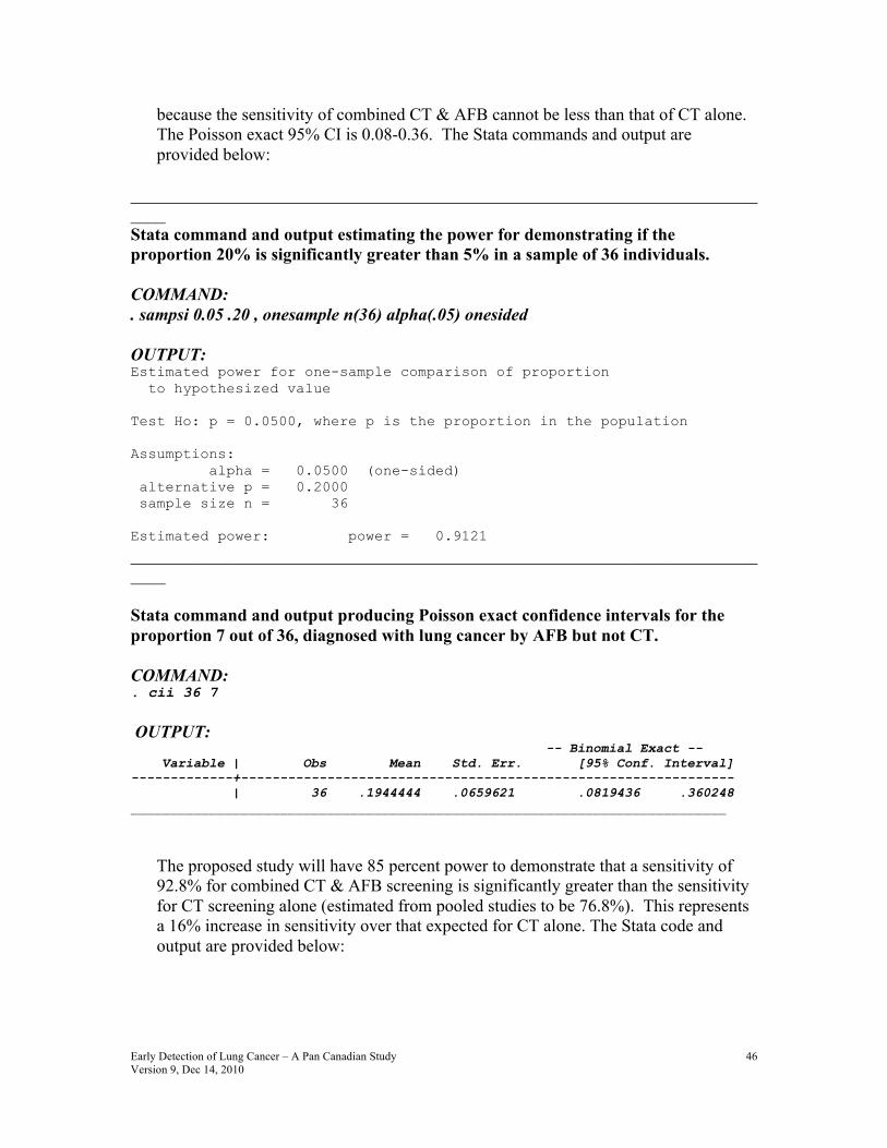
Early Detection of Lung Cancer – A Pan Canadian Study Version 9, Dec 14, 2010
46
because the sensitivity of combined CT & AFB cannot be less than that of CT alone. The Poisson exact 95% CI is 0.08-0.36. The Stata commands and output are provided below:
____________________________________________________________________________ Stata command and output estimating the power for demonstrating if the proportion 20% is significantly greater than 5% in a sample of 36 individuals. COMMAND: . sampsi 0.05 .20 , onesample n(36) alpha(.05) onesided OUTPUT: Estimated power for one-sample comparison of proportion to hypothesized value Test Ho: p = 0.0500, where p is the proportion in the population Assumptions: alpha = 0.0500 (one-sided) alternative p = 0.2000 sample size n = 36 Estimated power: power = 0.9121
____________________________________________________________________________ Stata command and output producing Poisson exact confidence intervals for the proportion 7 out of 36, diagnosed with lung cancer by AFB but not CT. COMMAND: . cii 36 7 OUTPUT: -- Binomial Exact -- Variable | Obs Mean Std. Err. [95% Conf. Interval] -------------+--------------------------------------------------------------- | 36 .1944444 .0659621 .0819436 .360248 ____________________________________________________________________________
The proposed study will have 85 percent power to demonstrate that a sensitivity of 92.8% for combined CT & AFB screening is significantly greater than the sensitivity for CT screening alone (estimated from pooled studies to be 76.8%). This represents a 16% increase in sensitivity over that expected for CT alone. The Stata code and output are provided below:
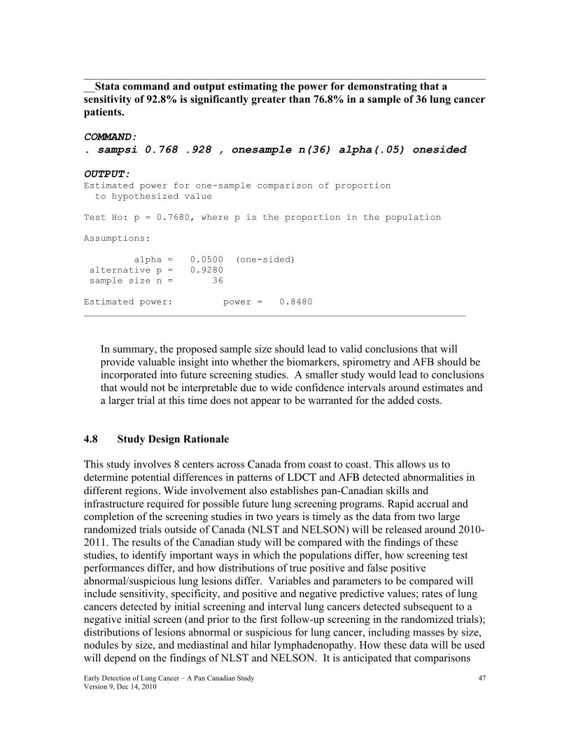
Early Detection of Lung Cancer – A Pan Canadian Study Version 9, Dec 14, 2010
47
__________________________________________________________________________Stata command and output estimating the power for demonstrating that a sensitivity of 92.8% is significantly greater than 76.8% in a sample of 36 lung cancer patients. COMMAND: . sampsi 0.768 .928 , onesample n(36) alpha(.05) onesided OUTPUT: Estimated power for one-sample comparison of proportion to hypothesized value Test Ho: p = 0.7680, where p is the proportion in the population Assumptions: alpha = 0.0500 (one-sided) alternative p = 0.9280 sample size n = 36 Estimated power: power = 0.8480 ____________________________________________________________________________
In summary, the proposed sample size should lead to valid conclusions that will provide valuable insight into whether the biomarkers, spirometry and AFB should be incorporated into future screening studies. A smaller study would lead to conclusions that would not be interpretable due to wide confidence intervals around estimates and a larger trial at this time does not appear to be warranted for the added costs.
4.8 Study Design Rationale
This study involves 8 centers across Canada from coast to coast. This allows us to determine potential differences in patterns of LDCT and AFB detected abnormalities in different regions. Wide involvement also establishes pan-Canadian skills and infrastructure required for possible future lung screening programs. Rapid accrual and completion of the screening studies in two years is timely as the data from two large randomized trials outside of Canada (NLST and NELSON) will be released around 2010-2011. The results of the Canadian study will be compared with the findings of these studies, to identify important ways in which the populations differ, how screening test performances differ, and how distributions of true positive and false positive abnormal/suspicious lung lesions differ. Variables and parameters to be compared will include sensitivity, specificity, and positive and negative predictive values; rates of lung cancers detected by initial screening and interval lung cancers detected subsequent to a negative initial screen (and prior to the first follow-up screening in the randomized trials); distributions of lesions abnormal or suspicious for lung cancer, including masses by size, nodules by size, and mediastinal and hilar lymphadenopathy. How these data will be used will depend on the findings of NLST and NELSON. It is anticipated that comparisons
Early Detection of Lung Cancer – A Pan Canadian Study Version 9, Dec 14, 2010
48
will be most useful when our planned study is completed or approaching completion to ensure stable statistics. It is unlikely that NLST or NELSON findings will result in major modifications of our planned study midstream, as such changes in study protocol may introduce biases and inefficiencies (loss of estimate precision and study power), unless it entails supplementation of the existing study.” 5.0 TIMELINE Key Milestones • Month 4: completion of REB approval, hiring of study personnel, quality control
check for site chest radiologists and endoscopists, data server and network interface between BCCA and partner sites.
• Month 12: completion of recruitment, questionnaires administration, spirometry, blood specimen collection, spiral CT in 1,000 subjects and autofluorescence bronchoscopy in 500 subjects.
• Month 24: completion of recruitment, questionnaires administration, spirometry, blood specimen collection, spiral CT 2,500 subjects and autofluorescence bronchoscopy in 1,250 subjects.
• Month 36: Completion of blood biomarker measurements. • Month 37: If the randomized clinical trials (NLST and NELSON) showed a mortality
reduction benefit of screening, initiate discussion with Provincial and Federal Health Ministries to implement screening program in Canada.
• Month 48: Completion minimumof 2 years of follow-up and tracking of health care utilization and costs.
• Month 54: Completion of Terry Fox lung cancer risk assessment model development. • Month 54: If randomized trials outside of Canada are negative, we will apply to
funding agencies to design and conduct an alternative randomized clinical trial using our multi-modal screening strategy in Canada alone or in conjunction with other countries.
• Month 60: Completion of Year 4 LDCT • Month 84: Delivery of final report and a position paper on the costs and quality of life
implications of a publicly delivered early lung cancer detection program in Canada.
6.0 IMPACT OF PROJECT Sensitive technologies that can detect lung cancer down to the sub-millimeter range are already available. However, the critical issue in lung cancer screening is to how to apply technologies selectively to individuals at highest risk so that the benefits would outweigh the potential risks and to enable affordable delivery of the screening program at the population level within the context of a public health care system. To illustrate the potential importance of a lung cancer risk assessment model, consider the lung cancer prevalence of 2.2% among people 50 to 75 years of age and a 30 pack-years smoking history. Without any risk assessment model, 46 people would have to be screened to find one person with lung cancer. Using socio-demographic factors, smoking and medical data in our risk assessment model, we need to screen only 23 people to find one cancer. With the addition of lung function (FEV1) as a biomarker such that the model has a

Early Detection of Lung Cancer – A Pan Canadian Study Version 9, Dec 14, 2010
49
sensitivity and specificity of 75%, we potentially need to screen only 15 people to find one cancer. If the model can be improved further to a sensitivity and specificity of 80% by the addition of a blood biomarker, we need to screen only 12 people to find one cancer. In addition, because the disease prevalence is higher using the risk assessment model, the positive predictive value of spiral CT will also be higher resulting in significantly fewer unnecessary downstream investigations or treatment.
The study will lead to capacity building to develop the specialized expertise to mount a Canada-wide lung cancer screening program. For example, population penetration strategies to large scale screening will be developed. Radiologists skilled in interpretation of CT scans & biopsy of small lung nodules and chest physicians skilled in performing autofluorescence bronchoscopy as well as minimally invasive treatment methods will become available. The data generated from this project will be of critical importance for determining health care policy regarding lung cancer screening in Canada once the results of the two randomized studies outside of Canada become available. While our Canadian study will not provide information on mortality benefit, we will be able to identify the key elements such as resource implication and costs to allow an informed decision on lung cancer screening when the data from these studies matures in the next few years. If the randomized trials outside of Canada show a mortality reduction benefit of screening, the participants in the study who are found to have early lung cancer and have received treatment would have already benefited from the clinical trial. Expansion of provincially based early detection programs across Canada using the seven centers in this study as nuclei to transfer the clinical knowledge that will be gained will benefit the general population similar to what has been achieved in cervical and breast cancer screening. If the randomized trials outside of Canada are negative, the multi-modal risk assessment model and screening strategy developed in the Terry Fox project would set the stage to design an alternative randomized clinical trial using our multi-modal screening strategy in Canada & elsewhere.
7.0 PROJECT MANAGEMENT 7.1 Organizational Aspects
The overall operational organization of this Team is illustrated in Table 3.
Table 3. Project Organization Project Co-Directors: Stephen Lam (BCCA) Ming-Sound Tsao (PMH)
Site Site Lead
Investigator(s) Radiologist Bronchoscopists

Early Detection of Lung Cancer – A Pan Canadian Study Version 9, Dec 14, 2010
50
BCCA University of BC
Annette McWilliams
John Mayo Annette McWilliams Stephen Lam
Foothills Hospital, U of Calgary
Alain Tremblay Paul Burrowes Alain Tremblay Paul MacEachern
Juravinski Cancer Center, McMaster University
John Goffin Lori Stewart Serge Puksa Allan McLellan
PMH, U of Toronto Heidi Roberts Heidi Roberts Kam Soghrati John Thenganatt
Ottawa Hospital Regional Cancer Centre, U of Ottawa
Garth Nicholas & Glen Goss
Jean M Seely Kayvan Amjadi
Hôpital Laval & U de Laval
Simon Martel & Francis Laberge
Michel Gingras Simon Martel Francis Laberge
QEII HSC, Dalhousie U
Michael Johnston Daria Manos Michael Johnston
Newfoundland Memorial University
Rick Bhatia Scott Harris George Fox Joe Lockyer
Lung Cancer Risk Modeling: MartinTammemagi (Brock University) Don Sin (iCapture Center, UBC) Geoffrey Liu (PMH) Health Economics & QOL: Stuart Peacock (BCCA)
Natasha Leighl (PMH) Bill Evans (Juravinski Cancer Center) MartinTammemagi (Brock University) Quality Assurance: Radiology: Dr. Nestor Muller (Vancouver General Hospital, UBC) Spirometry: Dr. Wan Tan (iCapture Center, UBC) AF Bronchoscopy: Dr. Tom Sutedja (Free University, Amsterdam) Pathology: Dr. Ming Tsao (PMH) Biomarkers Development: Don Sin (iCapture Center, UBC) Geoffrey Liu (PMH) Reference Pathologists: Ming-Sound Tsao (PMH) Adi Gazdar (UT Southwestern Medical Center) Scientific Advisory Board: Mark Elwood (Chair) Partnership Committee: Frances Shepherd (Chair)

Early Detection of Lung Cancer – A Pan Canadian Study Version 9, Dec 14, 2010
51
The Co-Directors are internationally renowned Canadian lung cancer researchers holding senior leadership and scientific positions at their respective institutions. The Team has invited an international Scientific Advisory Board (SAB) of experienced leaders in the field chaired by Dr. Mark Elwood. Dr. Mark Elwood is an epidemiologist and Vice-President for Family and Community Oncology at the BC Cancer Agency, and a clinical professor at UBC. He has extensive experience in the implementation and assessment of cancer screening programs, involving cervical, breast, oral and colorectal cancer, and melanoma, as well as lung cancer. He is a member of the Advisory Council on Cancer Control of the Canadian Partnership against Cancer. Dr. Adi Gazdar is W. Ray Wallace Distinguished Chair in Molecular Oncology and Professor of Pathology at UT Southwestern Medical Center in Dallas, Texas. He is a world renowned lung pathologist and a member of the NCI Early Detection Research Network lung cancer biomarkers group. He has served on many review panels such as the pathology review panel of the I-ELCAP lung cancer screening study at Cornell University. Dr. Sandy McEwen, Director, Department of Oncologic Imaging, Cross Cancer Institute, University of Alberta. Dr. McEwan is a world leader in oncologic imaging. Dr. Nestor Muller is Professor and Head of the Radiology Department at the University of British Columbia. Dr. Muller is a world-leader in chest radiology. With stellar scientific credentials and management experience in large-scale cancer research programs, this SAB is ideal to provide strategic counsel and ensure the success and maximal impact of the study. The SAB will monitor progress against timelines/milestones and advise on potential emerging areas of opportunity. The Team’s Executive Committee (EC) will be responsible for the overall scientific direction of the Team; it will include the Co-Directors, the site lead investigators, and the project manager. The EC is responsible for strategic planning and monitoring of progress to ensure the study will be completed in a timely manner. The Partnership Committee (PC) will be led by Dr. F. Shepherd, with significant representation from Lung Cancer Canada (LCC) including Ms. Dallas Petroff, CEO of Lung Cancer Canada, and Mrs. Roz Brodsky, a patient advocate, as well as Directors Y. Ung, John McLaughlin (Ontario), M. Johnston (Halifax), and S. Lam (BC).
7.2 Team Members & Role Project Co-directors:
Stephen Lam MD, FRCPC is Professor of Medicine at the University of British Columbia, Chair of the Lung Tumor Group and Director of the MDS-Rix Early Lung Cancer Detection Program at the BC Cancer Agency. His research interest is in early detection, treatment and chemoprevention of lung cancer. He is a co-inventor of the autofluorescence bronchoscopy device. In 1999, he was awarded the Friesen-Rygiel Prize by the Canadian Medical Discoveries Funds Inc. for this invention. In 2002, he was awarded the Gustav Killian Medal by the World Association for Bronchology for his work in early lung cancer. He has published 137 peer reviewed manuscripts and 13 patents. He is co-director of the Genome Canada Pharmacogenomics of Non-small Lung cancer team project and co-PI of an NIH program project grant in chemoprevention of lung cancer. He is highly experienced in leading multi-center

Early Detection of Lung Cancer – A Pan Canadian Study Version 9, Dec 14, 2010
52
team projects. Ming-Sound Tsao, MD, FRCPC is the M. Qasim Choksi Chair in Lung Cancer Translational Research at the Princess Margaret Hospital/University of Toronto. He is Professor of Laboratory Medicine and Pathobiology, Senior Scientist of Ontario Cancer Institute and Pathologist at the University Health Network (UHN). Dr. Tsao has is the leader of Translational Research Program in lung cancer and a co-leader (with Dr. Heidi Roberts) of the Lung Cancer Early Detection Program at the UHN. He is internationally well known in the field of lung cancer biomarkers for early detection, prognostication and prediction of response to anti epidermal growth factor receptor targeted therapy. He is the current Chair of the Correlative Science and Tumor Biology Committee of the National Cancer Institute of Canada Clinical Trials Group. Dr. Tsao has published 190 peer-reviewed manuscript and >5000 lifetime citation.
Lung Cancer Risk Model and Biomarkers: Martin Tammemagi Ph.D is Associate Professor, Faculty of Applied Health Sciences, Department of Community Health Sciences, Brock University. He has worked with the NIH Prostate, Lung, Colorectal and Ovarian Cancer screening trial since 1998 and the National Lung Cancer Screening Trial since its inception in 2000. He has developed a predictive regression model that utilizes socio-demographic factors, smoking exposure, medical and radiographic data from over 12,000 individuals with abnormal suspicious chest radiographs to predict true vs. false positive lung cancer screens in the PLCO study. He will develop the Terry Fox Multi-modal Lung Cancer Risk Model along with Drs. Sin and Liu. Don Sin MD, FRCPC, MPH is Associate Professor of Medicine at the University of British Columbia, Canadian Research Chair & Michael Smith Foundation/GlaxoSmithKline Professor in Chronic Obstructive Pulmonary Diseases. One of his major research interests is in the inflammatory link between lung cancer and COPD. He will supervise measurements of blood biomarkers in the project. Geoffrey Liu MD, FRCPC is the Alan B. Brown Chair of Molecular Genomics at the Princess Margaret Hospital, Assistant Professor at the University of Toronto, and a lung molecular epidemiologist. His major interests are in the role of biomarkers in the development and prognosis of aerodigestive cancers, including lung cancer. His laboratory provides the primary blood-based tissue repository of all aerodigestive cancers (head and neck, esophageal, lung, gastric cancers) and associated epidemiological and clinical data attached to all PMH patients. His laboratory also serves as the primary blood-based tissue repository for the PMH lung cancer screening program. Health Economics Modeling & Quality of Life Assessment:
Stuart Peacock Ph.D Stuart Peacock is a Senior Scientist in Health Economics at the BC Cancer Agency and an Assistant Professor, Health Care and Epidemiology, UBC. Stuart holds a Michael Smith Foundation for Health Research Scholar Award. His research interest is in the economics of cancer and cancer genetics, priority setting methods, health-related quality of life, and health econometrics. He has worked with over 60 health agencies in Canada, the UK, Australia, New Zealand, and the Pacific Islands, and has acted as a consultant for the World Health Organization. His role in the project will be modeling the cost and cost-effectiveness of the screening interventions as well as the impact of screening on the quality of life of the
Early Detection of Lung Cancer – A Pan Canadian Study Version 9, Dec 14, 2010
53
participants. Bill Evans MD, FRCPC is a medical oncologist and health care administrator. Throughout his career he has been active in lung cancer care and research and has chaired the provincial lung cancer disease site group in Ontario for over a decade. The Lung DSG develops lung cancer practice guidelines that are used by practitioners to guide their practice and for funding decisions on new drugs and technologies (PET). He has chaired the NCIC's Working Group in Economic Analysis since its inception. Working with the Health Analysis Modeling Group at Statistics Canada, he developed a microsimulation model of lung cancer, which has been used to estimate the lifetime costs of lung cancer and to determine the cost-effectiveness of new treatment approaches. His role in the project will be to provide advice on the resource utilization and cost data to be used in modeling the cost and cost-effectiveness of the screening interventions. Natasha Leighl MD, FRCPC is a medical oncologist at the Princess Margaret Hospital, Toronto. She has special clinical interest is in lung cancer and targeted therapy, and academic interest in quality of life issues and economic of cancer therapy, and is the new Chair of the NCIC CTG Working Group in Economic Analysis. Her role in the project will be to provide advice on the resource utilization and cost data to be used in modeling the cost and cost-effectiveness of the screening interventions.
Chest Radiology: John Mayo MD, FRCPC is Professor of Radiology, University of British Columbia and Head of Radiology at the Vancouver General Hospital. He has been a co-investigator in several NCI sponsored chemoprevention trials at BCCA that includes the use of spiral CT for detection of early lung lesions. He and his thoracic surgical colleagues pioneered the microcoil technique for localization of small lung nodules for biopsy and removal. Heidi Roberts MD, FRCPC is Associate Professor of Radiology, University of Toronto, site Director for Medical Imaging, Women’s College Hospital and Principal Investigator, Lung Cancer Screening Study, Princess Margaret Hospital. She has extensive experience in lung cancer screening using spiral CT and is a member of the I-ELCAP consortium. Dr. Mayo and Dr. Roberts are both highly experienced in interpretation of screening spiral CT and management of screen detected lung abnormalities. In addition to reviewing the CTs in their own sites, they will provide their expertise to the rest of the chest radiology team in the project. The other team members are: Michel Gingras MD, CPSQ, FRCPC Professeur adjoint de radiologie diagnostique, Université de Sherbrooke and Radiologiste, Chef de service, Département d’imagerie médicale, Hôpital Laval; Lori K. Stewart MD, FRCPC Assistant Clinical Professor, Department of Radiology & Director of CT Imaging, Department of Diagnostic Imaging, Hamilton Health Sciences-Henderson Hospital, Daria Manos MD, FRCPC a thoracic radiologist at the QEII Health Sciences Centre in Halifax Nova Scotia and on faculty with the Dalhousie University Medical School; Jean Seely MDCM, FRCPC Associate Professor, University of Ottawa and Head of the Division of Thoracic Imaging at the Ottawa Hospital; and Paul Burrowes MD, FRCPC Clinical Associate Professor, University of Calgary and Director, Department of Diagnostic Imaging, Foothills Medical Centre. Autofluorescence Bronchoscopy:
Early Detection of Lung Cancer – A Pan Canadian Study Version 9, Dec 14, 2010
54
Stephen Lam MD, FRCPC developed autofluorescence bronchoscopy. He was the PI in two multi-center trials leading to worldwide regulatory approval of autofluorescence bronchoscopy. Alain Tremblay MDCM, FRCPC is Clinical Associate Professor, Departments of Medicine & Oncology, University of Calgary. He is an internationally recognized expert in Interventional Pulmonology. He will be joined by Paul MacEachern MD, FRCPC in July 2008. Annette McWilliams MBBS, FRACP, FRCPC is Clinical Assistant Professor, Department of Medicine, University of British Columbia & Vancouver General Hospital In it For Life Clinician Scientist. She has been the co-PI of several US NCI sponsored lung cancer chemoprevention projects. She is highly experienced in autofluorescence bronchoscopy and endobronchial therapy of early lung cancer. Serge Puska MD, FRCPC and Allan Mclellan MD, FRCPC are Associate Clinical Professor of Medicine, McMaster University and Respirologist at the Henderson General Hospital. Simon Martel CPSQ, FRCPC is Professeur agrégé de clinique à la Faculté de médecine de l’université Laval. Kam Soghrati MD, FRCPC Staff Respirologist at Scarborough General Hospital and clinical faculty at the University of Toronto. During his fellowship, he has received special training in autofluorescence bronchoscopy with Dr. Lam in Vancouver. Kayvan Amjadi MD, FRCPC is Assistant Professor of Medicine at University of Ottawa and a Respirologist at Ottawa Civic Hospital. Michael Johnston MD, FRCSC is Professor of Surgery at Dalhousie University and Adjunct Professor, Department of Surgery, University of Toronto. He is a thoracic oncologist highly experienced in clinical trials. The endoscopy team will ensure AF bronchoscopies are done in a timely manner and participants with abnormal bronchial biopsies or abnormal spiral CT are managed according to standard clinical practice. Site Lead Investigators.
Drs. Heidi Roberts, Michael Johnston, Annette McWilliams, Simon Martel and Alain Tremblay have been described above. John Goffin MD, FRCPC is Assistant Professor, Department of Medicine, McMaster University. He is a thoracic medical oncologist involved with clinical trials and trials methodology. Glen Goss MD, FRCPC is Professor and Head, Medical Oncology Division, University of Ottawa. He is the incoming Chair of Lung Cancer site of NCIC CTG. Garth Nicholas MD, FRCPC, MS is a Medical Oncologist treating lung cancer at the Ottawa Hospital. He has graduate-level training in Statistics and Clinical Epidemiology. He is Chair of the NCIC CTG Clinical Research Evaluation Committee. Francis Laberge MD, CPSQ, FRCPC is Directeur, Centre de Pneumologie, Hôpital Laval. The site lead investigators will be responsible for obtaining REB approval, oversee subject recruitment, study procedures, record keeping, data transfer to the central coordinating centre, and management of screen detected lung cancers. They will be part of the executive committee to address any issues that may arise from the study and participate in regular tele- or video-conferences.
7.3 Integration and Communications

Early Detection of Lung Cancer – A Pan Canadian Study Version 9, Dec 14, 2010
55
A Team Website updated weekly by the Central Coordination Center, will communicate research activities, progress, and achievements of the Team. It will also allow message posting for internal communication of the Team. Monthly Teleconference will serve as the forum for updating progress, identifying barriers in subject recruitment and exchange of results, material and techniques. Executive Committee Meetings will be held quarterly to monitor progress, resource allocation and all reporting responsibilities. An annual meeting of the team will be held along with other investigators in the Terry Fox Research Institute sponsored projects to exchange ideas.
7.4 Monitoring Of Milestones Lung cancer screening is not yet a publicly funded activity. Historical accrual rates for lung cancer screening are available in BCCA and PMH only. In the last few years, an average of 1,000 subjects per year (~83 per month) participated in the PMH program and 400 to 500 per year (33 to 42 per month) attended the BCCA program. Our goal is to recruit 2,500 subjects over 24 months. With 8 centers, an average of 13 subjects needs to be enrolled each month per center. Even though 5 of the 8 sites have not specifically conducted a lung cancer screening trial, they are all experienced in other lung studies. With an active advertising campaign and an anticipated high acceptance rate similar to the NLST study in the US, we believe 13 subjects per month per center is a realistic figure.
Accrual will be monitored continuously. Weekly inputting of data and plotting accrual will establish individual site and overall progress. Monthly teleconference of the study coordinators in the network will be held to address recruitment/retention issues and how best to overcome them. Variations in accrual are expected. For example, December and August are usually the lower points due to holidays. Quarterly accrual rates, cumulative accrual rates and trend are important parameters the steering committee will be monitoring closely to detect significant deviation from the target enrollment. Once a site has reached its accrual goal, the steering committee will decide if enrollment can be continued in that site to make up for the short-fall in other sites if necessary.
8.0 REFERENCES
1. Jemal A, Siegel R, Ward E, Murray T, Xu J, EJ, Thun MJ. Cancer statistics, 2007. CA Cancer J Clin 2007; 57:43-66.
2. Canadian Cancer Statistics, 2007. Available at http://www.cancer.ca 3. Greenlee RT, Hill-Harmon MB, Murray T, Thun M. Cancer Statistics 2001. CA
Cancer J Clin 2001;51:15-36.

Early Detection of Lung Cancer – A Pan Canadian Study Version 9, Dec 14, 2010
56
4. Cortese D, Pairolero P, Bergsralh E, Woolner L, Uhlenhopp M, Piehler J,Sanderson D, Bernatz P, Williams D, Taylor W, Payne W, Fontana R. Roentgenographically Occult Lung Cancer: A Ten-Year Experience. Journal of Thoracic and Cardiovascular Surgery 1983;86:373-380.
5. Saito Y, Nagamoto N, Ota S, Sato M, Sagawa M, Kamma K, Takahashi S, Usuda K, Endo C, Imai T, Fujimura S. Results of Surgical Treatment for Roentgenographically Occult Bronchogenic Squamous Cell Carcinoma. Journal of Thoracic and Cardiovascular Surgery 1992;104:401-407.
6. Rami-Porta R, Ball D, Crowley J et al. The IASLC lung cancer staging project: Proposals for the revision of the T descriptors in the forthcoming (seventh) edition of the TNM classification of lung cancer. J Thorac Oncol 2007; 2:593-602
7. Peto R, Darby S, Deo H, et al. Smoking Cessation, and Lung Cancer in the UK Since 1950: Combination of National Statistics with Two Case-Control Studies. Br Med J 2000;321:323-329.
8. Halpern MT, Gillespie BW, Warner KE. :Patterns of Absolute Risk of Lung Cancer Mortality in Former Smokers. J Natl Cancer Inst 1993; 85:457-464.
9. Tong L, Spitz MR, Fueger JJ, Amos CA. Lung Cancer in Former Smokers. Cancer 1996;78:1004-1010.
10. Canadian Tobacco Use, 2006. Available at http://www.hc-sc.gc.ca 11. Flehinger BJ, Melamed MR, Zaman MB, Heelan RT, Perchick WB. Martini N.
Early lung cancer detection: results of the initial (prevalence) radiologic and cytologic screening the Memorial Sloan-Kettering study. Am Rev Respir Dis, 1984;130:555-560.
12. Fontana RS, Sanderson DR, Taylor WF, Woolner LB, Miller WE, Muhm JR, Uhlenhopp MA. Early lung cancer detection: results of the initial (prevalence) radiologic and cytologic screening in the Mayo Clinic study. Am Rev Respir Dis, 1984;130:561-565.
13. Frost JK, Ball WC, Levin ML, Tockman MS, Baker RR, Carter D, Eggleston JC, Erozan YS, Gupta PK, Khouri NF. Early lung cancer detection: results of the initial (prevalence) radiologic and cytologic screening in the Johns-Hopkins study. Am Rev Respir Dis, 1984;130:549-554.
14. Kubik A, Parkin DJ, Khlat M, Erban J, Polak J, Adamec M. Lack of benefit from semi-annual screening for cancer of the lung: followup report of a randomised controlled trial on population of high-risk males in Czechoslovakia. Int J Cancer, 1990;45:26-33.
15. Marcus PM, Bergstralh EJ, Zweig MH, Harris A, Offord KP, Fontana RS. Extended lung cancer incidence follow-up in the Mayo Lung Project and overdiagnosis. J Natl Cancer Inst 2006;98:748-56
16. Sone S, Li F, Yang Z-G, Takashima S, Maruyama Y, Hasegawa M, Wang J-C, Kawakami S, Honda T. Characteristics of small lung cancers invisible on conventional chest radiography and detected by population based screening using spiral CT. Br J Radiol, 2000;73:137-145.
17. Henschke C, McCauley D, Yankelevitz D, Naidich D, McGuinness G, Miettinen O, Libby D, Pasmantier M, Koizumi J, Altorki N, Smith J. Early Lung Cancer Action Project: overall design and findings from baseline screening. Lancet, 1999;354:99-105.

Early Detection of Lung Cancer – A Pan Canadian Study Version 9, Dec 14, 2010
57
18. Henschke C, Naidich D, Yankelevitz D, McGuinness G, McCauley D, Smith J, Libby D, Pasmantier M, Vazquez M, Koizumi J, Flieder D, Altorki N, Miettinen O. Early Lung Cancer Action Project: Initial findings on repeat screening. Cancer, 2001;92(1):153-159.
19. Henschke C, Yankelvitz D, libby DM, et al. Survival of patients with Stage I lung cancer detected on CT screening. New Engl J Med, 2006;355:1763-1771.
20. Kaneko M, Eguchi K, Ohmatsu H, Kakinuma R, Naruke T, Suemasu K, Moriyama N. Peripheral lung cancer: Screening and detection with low-dose spiral CT versus radiography. Radiology, 1996;210:798-802.
21. Sobue T, Moriyama N, Kaneko M, Kusumoto M, Kobayashi T, Tsuchiya R. Kakinuma R, Ohmatsu H, Nagai K, Nishiyama H, Matsui E, Eguchi K. Screening For Lung Cancer With Low-Dose Helical Computed Tomography: Anti-Lung Cancer Association Project. Journal Of Clinical Oncology, 2002;20(4):911-920.
22. Sone S, Yang Z-G, Honda T, Maruyama Y, Takashima S, Hasegawa M, Kawakami S, Kubo K, Haniuda M, Yamanda T. Results of three-year mass screening program for lung cancer using mobile low-dose spiral computed tomography scanner. Br J Cancer, 2001;84(1):25-32.
23. Swensen SJ, Jett JR, Sloan JA, Midthun DE, Hartman TE, Sykes AM, Aughenbaugh GL, Zink FE, Hillman SL, Noetzel GR, Marks RS, Clyton AC, Pairolero PC. Screening for Lung Cancer with Low-Dose Spiral Computed Tomography. Am J Respir Crit Care Med, 2002;165:508-513.
24. Gohagan JK, Marcus PM, Fagerstrom RM, Pinsky PF, Kramer BS, Prorok PC, Ascher S, Bailey W, Brewer B, Church T, Engelhard D, Ford M, et al. Final results of the Lung Screening Study, a randomized feasibility study of spiral CT versus chest X-ray screening for lung cancer. Lung Cancer 2005;47:9-15.
25. Swensen S, Jett J, Hartman T, Midthun D, Sloan J, Sukes AM, Aughenbaugh G, Clemens M. Lung Cancer Screening with CT: Mayo Clinic Experience. Radiology 2003;226:756-761.
26. Diederich S, Wormanns D, Semik M, Thomas M, Lenzen H, Roos N, Heindel W. Screening for early lung cancer with low-dose spiral CT: prevalence in 817 asymptomatic smokers. Radiology, 2002;222(3):773-781.
27. Nawa T, Nakagawa T, Kusano S, Kawasaki Y, Sugawara Y, Nakata H. Lung cancer screening using low-dose spiral CT: results of baseline and 1-year follow-up studies. Chest, 2002;122(1):15-20.
28. Pastorino U, Bellomi M, Landoni C, De Fiori E, Arnaldo P, Picchio M, Pelosi G, Boyle P, Fazio F. Early lung-cancer detection with spiral CT and positron emission tomography in heavy smokers:2-year results. The Lancet, 2003;362:593-597.
29. McWilliams A, Mayo J, MacDonald S, leRiche J, Palcic B, Szabo E, Lam S. Lung Cancer Screening: A Different Paradigm. American Journal of Respiratory and Critical Care Medicine 2003;168:1167–1173.
30. McWilliams AM, Mayo JR, Ahn MI, MacDonald SLS, Lam S. Lung Cancer Screening Using Multi-Slice Thin-Section Computed Tomography and Autofluorescence Bronchoscopy. J Thorac Oncol 2006; 1(1):61-68.

Early Detection of Lung Cancer – A Pan Canadian Study Version 9, Dec 14, 2010
58
31. Loewen G, Natarajan N, Tan D, Nava E, Klippenstein D, Mahoney M, Cummings M, Reid M. Autofluorescence bronchoscopy for lung cancer surveillance based on risk assessment.Thorax, 2007; 62(4):335-340.
32. Aberle DR, Black WC, Goldin JG, Patz EF, Gareen I, Gatsonis C. Experimental Design and Outcomes of the National Lung Screening trial (NLST):A Multicenter Randomized Controlled trial of Helical CT vs Chest-X-Ray for Lung Cancer Screening. Am J Resp Crit Care Med 2003;167(7): A736. Further information in: http://www.cancer.gov/nlst
33. van Iersel CA, de Koning HJ, Draisma G, et al. Risk-based selection from the general population in a screening trial: Selection criteria, recruitment and power for the Dutch-Belgian randomised lung cancer multi-slice CT screening trial (NELSON). Int J Cancer 2007; 120:868-874
34. Roberts HC, Patsios D, Paul NS et al. Lung cancer screening with low-dose computed tomography: Canadian Experience. CARJ 2007;58:225-235.
35. MacMahon H, Austin J, Gamsu G, Herold C, Jett J, Naidich D, patz E, Swensen S. Guidelines for Management of Small Pulmonary Nodules Detected on CT Scans: A Statement from the Fleischner Society. Radiology, 2005;237:395-400.
36. Janssen-Heijnen ML, Coeburgh JW. Trends in incidence and prognosis of the histological subtypes of lung cancer in North America, Australia, New Zealand and Europe. Lung Cancer, 2001;31:123-137.
37. Lam S, Kennedy T, Unger M, Miller YE et al : Localization of bronchial intraepithelial neoplastic lesions by fluorescence bronchoscopy Chest 1998;113: 696-702.
38. Ernst, Armin MD; Simoff, Michael J MD; Mathur, Praveen N MD; Yung, Rex C MD ; Beamis, John F Jr MD : D-Light Autofluorescence in the Detection of Premalignant Airway Changes: A Multicenter Trial. J Bronchol 2005; 12:133-138.
39. Hirsch FR, Prindiville SA, Miller YE, Franklin WA, Dempsey EC, Murphy JR, Bunn PA Jr, Kennedy TC. Fluorescence versus white-light bronchoscopy for detection of preneoplastic lesions: a randomized study. J Natl Cancer Inst 2001; 93(18):1385-1391.
40. Häussinger K, Becker H, Stanzel F, et al: Autofluorescence bronchoscopy with white light bronchoscopy compared with white light bronchoscopy alone for the detection of precancerous lesions: a European randomised controlled multicentre trial: Thorax 2005; 60:496-503.
41. Lam S, McWilliams A. The Role of Autofluorescence Bronchoscopy in Diagnosis of Early Lung Cancer. In: IASLC Textbook of Prevention and Early Detection of Lung Cancer. Eds. FR Hirsch, PA Bunn Jr, H Kato, JL Mulshine. (2005).
42. Bach PB, Katten MW, Thornquist MD, Kris MG, Tate RC, Barnett MJ, Hsieh LJ, Begg CB. Variations in lung cancer risk among smokers. J Natl Cancer Inst 2003;95:470-8.
43. Gohagan JK, Prorok PC, Hayes RB, Kramer BS. The Prostate, Lung, Colorectal and Ovarian (PLCO) Cancer Screening Trial of the National Cancer Institute: history, organization, and status. Control Clin Trials 2000;21:251S-72S.
44. Church TR. Chest radiography as the comparison for spiral CT in the National Lung Screening Trial. Acad Radiol. 2003;10(6):713-5.

Early Detection of Lung Cancer – A Pan Canadian Study Version 9, Dec 14, 2010
59
45. Prorok PC, Andriole GL, Bresalier RS, Buys SS, Chia D, Crawford ED, Fogel R, Gelmann EP, Gilbert F, Hasson MA, Hayes RB, Johnson CC, et al. Design of the Prostate, Lung, Colorectal and Ovarian (PLCO) Cancer Screening Trial. Control Clin Trials 2000;21:273S-309S.
46. Efron B, Tibshirani, R. An Introduction to the Bootstraped. New York: Chapman & Hall, 1993.
47. Bach PB, Kattan MW, Thornquist MD, Kris MG, Tate RC, Barnett MJ, Hsieh LJ, Begg CB. Variations in lung cancer risk among smokers. J Natl Cancer Inst 2003;95:470-8.
48. Cassidy A, Myles JP, Liloglou T, Duffy SW, Field JK. Defining high-risk individuals in a population-based molecular-epidemiological study of lung cancer. Int J Oncol 2006;28:1295-301.
49. Prindiville SA, Byers T, Hirsch FR, Franklin WA, Miller YE, Vu KO, Wolf HJ, Baron AE, Shroyer KR, Zeng C, Kennedy TC, Bunn PA. Sputum cytological atypia as a predictor of incident lung cancer in a cohort of heavy smokers with airflow obstruction. Cancer Epidemiol Biomarkers Prev 2003;12:987-93.
50. Spitz MR, Hong WK, Amos CI, Wu X, Schabath MB, Dong Q, Shete S, Etzel CJ. A risk model for prediction of lung cancer. J Natl Cancer Inst 2007;99:715-26.
51. Cassidy A, Duffy SW, Myles JP, Liloglou T, Field JK. Lung cancer risk prediction: a tool for early detection. Int J Cancer 2007;120:1-6.
52. Ezzati M, Lopez AD. Estimates of global mortality attributable to smoking in 2000. Lancet. 2003; 362:847-52.
53. Anthonisen NR, Skeans MA, Wise RA, Manfreda J, Kanner RE, Connett JE. The effects of a smoking cessation intervention on 14.5-year mortality: a randomized clinical trial. Ann Intern Med 2005;142(4):233-9.
54. Wasswa-Kintu S, Gan WQ, Man SF, Pare PD, Sin DD. Relationship between reduced forced expiratory volume in one second and the risk of lung cancer: a systematic review and meta-analysis. Thorax. 2005;60:570-5.
55. Benson MD, Eyanson S, Fineberg NS. Serum amyloid A in carcinoma of the lung. Cancer 1986;57:1783-1787.
56. Khan N, Cromer CJ, Campa M, Patz Jr EF. Clinical utility of serum amyloid A and macrophage migration inhibitory factor as serum biomarkers for the detection of nonsmall cell lung carcinoma. Cancer 2004;101:379-384.
57. Patz EF, Campa MJ, Gottlin EB, Husmartseva I, Guan XR. Panel of serum biomarkers for the diagnosis of lung cancer. J Clin Oncol 2007; 25:5578-5583.
58. Gao WM, Kuick R, Orchekowski RP, Misek DE, Qiu J, Greenberg AK, et al. Distinctive serum protein profiles involving abundant proteins in lung cancer patients based upon antibody microarray analysis. BMC Cancer. 2005;5:110.
59. Lam S. Detection of Preneoplastic Lesions. In: Roth JA, Cox, J.D., Hong, W.K. Lung Cancer, 4th ed. Cambridge, MA: Blackwell Scientific Publications Inc., 2007.
60. Jenkinson C, Layte R, Jenkinson D, et al. A shorter form health survey: can the SF-12 replicate results from the SF-36 in longitudinal studies? J Public Health Med 1997; 19:179-186.
61. Spielberger CD, Gorsuch RL, Lushene RE. Test manual for the State-Trait Anxiety Inventory. Palo Alto, CA: Consulting Psychologists Press, 1970.
Early Detection of Lung Cancer – A Pan Canadian Study Version 9, Dec 14, 2010
60
62. American Thoracic Society. Standardization of spirometry--1987 update. Am Rev Respir Dis. 1987 Nov;136(5):1285-1298.
63. Crapo RO, Morris AH, Gardner RM. Reference spirometric values using techniques and equipment that meet ATS recommendations. Am Rev Respir Dis. 1981 Jun;123(6):659-664.
64. Travis, W. D., Colby, T. V., Corrin, B., Shimosato, Y., and Brambilla, E. Histologic and graphical text slides for the histological typing of lung and pleural tumors. World Health Organization pathology panel: World Health Organization. International Classification of tumors, pp. 5: Berlin: Springer Verlag, 1999.
65. Huang RP, Huang R, Fan Y, Lin Y. Simultaneous detection of multiple cytokines from conditioned media and patient's sera by an antibody-based protein array system. Anal Biochem 2001;294(1):55-62.
66. Lash GE, Scaife PJ, Innes BA, Otun HA, Robson SC, Searle RF, et al. Comparison of three multiplex cytokine analysis systems: Luminex, SearchLight and FAST Quant. J Immunol Methods 2006;309(1-2):205-8.
67. Hurley SF, Matthews JP. The Quit Benefits Model: a Markov model for assessing the health benefits and health care cost savings of quitting smoking. Cost Eff Resour Alloc 2007;5:2.
68. Mahadevia PJ, Fleisher LA, Frick KD, Eng J, Goodman SN, Powe NR. Lung cancer screening with helical computed tomography in older adult smokers: a decision and cost-effectiveness analysis. Jama 2003;289:313-22.
69. Sullivan Pepe M. The Statistical Evaluation of Medical Tests for Classification and Predictioned. New York, NY: Oxford University Press, 2003.
70. Petitti D. Meta-Analysis, Decision Analysis, and Cost-Effectiveness Analysis: Methods for Quantitative Synthesis in Medicine, 2nd ed. New York: Oxford University Press, 2000.
71. Phillips, N. and A. J. Coldman. "Computer simulation of the effect of different colorectal cancer screening strategies for British Columbia." BC Medical Journal 2003;45(6): 254-258.
72. Flanagan, W. M., C. Le Petit, et al. "Potential impact of population-based colorectal cancer screening in Canada." Chronic Dis Can 2003; 24(4): 81-8.
73. Gold MR., Siegel JE., Russell LB., Weinstein MC. (Eds.). Cost-effectiveness in health and medicine. Oxford University Press, New York 1996.
74. Drummond M, Sculpher M, Torrance G, O’Brien B, Stoddart G (2005). Methods for the Economic Evaluation of Health Care Programmes (3rd ed.) OUP: Oxford.
75. Heitjan DF, Li H. Bayesian estimation of cost-effectiveness: an importance-sampling approach. Health Economics 2004;13(2):191-8. 87.
76. Claxton K, Sculpher M, Drummond M. A rational framework for decision making by the National Institute For Clinical Excellence (NICE). Lancet 2002;360(9334):711-5.
77. Schlaiffer R. Probability and Statistics for Business Decisions. New York: McGraw-Hill, 1958.
78. Claxton, K and Posnett, J. An economic approach to clinical trial design and research priority-setting. Health Economics. 1996;5:513-524.
Early Detection of Lung Cancer – A Pan Canadian Study Version 9, Dec 14, 2010
61
79. Claxton, K. The irrelevance of inference: a decision-making approach to the stochastic evaluation of health care technologies. Journal of Health Economics 1999;18:342-64.
80. Claxton, K, Neuman, PJ, Araki, SS et al. The value of information: an application to a policy model of Alzheimers disease. International Journal of Technology Assessment in Health Care 2001;17:38-55.
81. Fenwick E, Claxton K, Sculpher M, Briggs A. Improving the efficiency and relevance of health technology assessment: The role of decision analytic modelling. Centre for Health Economics Discussion Paper 179. York: Centre for Health Economics, University of York, 2000.
82. O’Brien B., Briggs A. Analysis of uncertainty in health care cost-effectiveness studies: an introduction to statistical issues and methods. Statistical Methods in Medical Research 2002, II, 455-468.
83. Williams AH. Economics of coronary artery bypass grafting. British Medical Journal 1985; 291: 326-329.
84. Hosmer DW, Jr, Lemeshow S. Applied Logistic Regression. 2nd Edition ed New York: John Wiley & Sons, Inc.; 1999 Wiley Series in Probability and Statistics).
85. Efron B, Tibshirani R. An Introduction to the Bootstrap New York: Chapman & Hall; 1993.
86. Dupont WD, Plummer WD: "Power and Sample Size Calculations: A Review and Computer Program", Controlled Clinical Trials 1990; 11:116-28.
87. Kannel WB, McGee D, Gordon T. A general cardiovascular risk profile: the Framingham study. Am J Cardiol 1976;38:46-51.
88. Grundy SM, Balady GJ, Cirqui MH, Fletcher G, Greenland P, Hiratzka LE et al. Primary prevention of coronary heart disease: guidance from the Framingham. Circulation 1998; 97:1876-87.
89. Chapman CJ, Murray A, McElveen JE, Sahin U et al. Autoantibodies in lung cancer – possibilities for early detection and subsequent care. Thorax (on line 11 October 2007. doi:10.1136/thx.2007.083592).
90. Pepe MS, Etzioni R, Feng Z, Potter JD, Thompson ML, Thornquist M, Winget M, Yasui Y. J Natl Cancer Inst 2001; 93:1054-61.
91. Anthonisen NR, Skeans MA, Wise RA, Manfreda J, Kanner RE, Connett JE; Lung Health Study Research Group. The effects of a smoking cessation intervention on 14.5-year mortality: a randomized clinical trial. Ann Intern Med. 2005;142:233-9.
92. Yee J, Sadar M, Kuzyk M, Sin DD, Li X, Rattenbury J, McWilliams AM, Man SFP, Lam S. Novel Plasma Biomarkers for Lung Cancer Detection. (Submitted).
93. Franklin WA. Diagnosis of Lung Cancer: Pathology of Invasive and Preinvasive Neoplasia. Chest 2000;117;80-89.
94. WHO Classification of Tumours. Pathology and Genetics, Tumours of lung, pleura, thymus and heart. W. D. Travis, E. Brambilla, H. K. Müller-Hermelink, C.C. Harris (Eds). IARCPress Lyon, 2004.
95. Hung RJ et al. A susceptibility locus for lung cancer maps to nicotinic acetylcholine receptor subunit genes on 15q25. Nature 2008; 452:633-7.
96. Amos CI et al. Genome-wide association scan of tag SNPs identifies a susceptibility locus for lung cancer at 15q25.1. 2008; doi:10.1038/ng.109.

Early Detection of Lung Cancer – A Pan Canadian Study Version 9, Dec 14, 2010
62
97. Henschke CI, McCauley DI, Yankelevitz DF, et al. Early lung cancer action project: a summary of the findings on baseline screening. The oncologist 2001;6(2):147-52.
98. Henschke CI, Yankelevitz DF, Libby DM, et al. Early lung cancer action project: annual screening using single-slice helical CT. Annals of the New York Academy of Sciences 2001;952:124-34.
99. Diederich S, Thomas M, Semik M, et al. Screening for early lung cancer with low-dose spiral computed tomography: results of annual follow-up examinations in asymptomatic smokers. European radiology 2004;14(4):691-702.
100. Black C, de Verteuil R, Walker S, et al. Population screening for lung cancer using computed tomography, is there evidence of clinical effectiveness? A systematic review of the literature. Thorax 2007;62(2):131-8.

Early Detection of Lung Cancer – A Pan Canadian Study Version 9, Dec 14, 2010
63
APPENDIX
1. Initial contact short questionnaire 2. Registration eligibility check list 3. Study questionnaire 4. Quality of life questionnaires 5. Study consent form 6. Optional tissue banking consent form 7. Lung cancer risk prediction model algorithm