Embed Size (px)
Citation preview

Progression From IgD� IgM� to Isotype-Switched B Cells Is SiteSpecific during Coronavirus-Induced Encephalomyelitis
Timothy W. Phares,a Krista D. DiSano,a,b Stephen A. Stohlman,a Cornelia C. Bergmanna
Department of Neurosciences, Lerner Research Institute, Cleveland Clinic Foundation, Cleveland, Ohio, USAa; School of Biomedical Sciences, Kent State University, Kent,Ohio, USAb
ABSTRACT
Various infections in the central nervous system (CNS) trigger B cell accumulation; however, the relative dynamics between viralreplication and alterations in distinct B cell subsets are largely unknown. Using a glia-tropic coronavirus infection, which is ini-tiated in the brain but rapidly spreads to and predominantly persists in the spinal cord, this study characterizes longitudinalchanges in B cell subsets at both infected anatomical sites. The phase of T cell-dependent, antibody-independent control of infec-tious virus was associated with a similar recruitment of naive/early-activated IgD� IgM� B cells into both the brain and spinalcord. This population was progressively replaced by CD138� IgD� IgM� B cells, isotype-switched CD138� IgD� IgM� memoryB cells (Bmem), and CD138� antibody-secreting cells (ASC). A more rapid transition to Bmem and ASC in spinal cord than inbrain was associated with higher levels of persisting viral RNA and transcripts encoding factors promoting B cell migration, dif-ferentiation, and survival. The results demonstrate that naive/early-activated B cells are recruited early during coronavirus CNSinfection but are subsequently replaced by more differentiated B cells. Furthermore, viral persistence, even at low levels, is adriving force for accumulation of isotype-switched Bmem and ASC.
IMPORTANCE
Acute and chronic human CNS infections are associated with an accumulation of heterogeneous B cell subsets; however, theirinfluence on viral load and disease is unclear. Using a glia-tropic coronavirus model, we demonstrate that the accumulation of Bcells ranging from early-activated to isotype-switched differentiation stages is both temporally and spatially orchestrated.Acutely infected brains and spinal cords indiscriminately recruit a homogeneous population of early-activated B cells, which isprogressively replaced by diverse, more differentiated subsets. The latter process is accelerated by elevated proinflammatory re-sponses associated with viral persistence. The results imply that early-recruited B cells do not have antiviral function but maycontribute to the inflammatory environment or act as antigen-presenting cells. Moreover, CNS viral persistence is a driving forcepromoting differentiated B cells with protective potential.
Central nervous system (CNS) inflammation during microbialinfections, autoimmunity, or spinal cord injury is associated
with recruitment of various B cell subsets, including antibody-secreting cells (ASC) (1–5). In cases of acute encephalitis, B celland antibody (Ab) accumulation is transient; however, humoralresponses persist during chronic CNS diseases such as subacutesclerosing panencephalitis and multiple sclerosis (MS) (6–8).However, the mechanisms driving the accumulation of various Bcells as well as their phenotype, role, and precursor relationshipsto ASC are poorly defined. In patients with subacute sclerosingpanencephalitis, the majority of oligoclonal Ab bands are measlesvirus specific, suggesting that persisting viral antigen drives localhumoral responses (6, 9), yet their role is difficult to assess. A largeproportion of CNS-localized ASC in Sindbis virus and neu-rotropic coronavirus infection models is also virus specific andcorrelated with protection (2, 4, 10).
One mechanism thought to promote local CNS B cell differen-tiation and Ab production involves the formation of ectopic fol-licle-like structures, as described previously for neuroborreliosisand MS (11–13). Ectopic follicle formation in the CNS duringmicrobial or autoimmune inflammation is supported by the con-stitutive and induced expression of several factors regulating B cellresponses in lymphoid organs. Among these factors are thechemokines CXCL13, CCL19, and CCL21, which guide B cell mi-gration within lymph nodes, as well as CXCL9, CXCL10, and
CXCL12, which are implicated in ASC trafficking (3, 14–16).Moreover, factors involved in both B cell differentiation, such asinterleukin-6 (IL-6), IL-10, and IL-21, as well as B cell survival,namely, B cell-activating factor of the tumor necrosis factor (TNF)family (BAFF) and a proliferation-inducing ligand (APRIL), arealso upregulated during virus- or autoantigen-induced CNS in-flammation (3, 15, 17–19). Although CXCL13 is implicated in theformation of ectopic follicle-like structures in the CNS (11–13,16), there is no evidence for ectopic lymphoid follicles duringSindbis virus infection, despite the expression of CXCL13 andCCL19 and the presence of various B cell subsets within the CNS(2, 15). Increasing proportions of isotype-switched memory Bcells (Bmem) and ASC during Sindbis virus CNS persistence thussuggested that B cell subset alterations toward a more differenti-ated phenotype may reflect their egress into circulation from pe-ripheral maturation sites and survival in the CNS (2). Early B cell
Received 25 March 2014 Accepted 21 May 2014
Published ahead of print 28 May 2014
Editor: S. Perlman
Address correspondence to Cornelia C. Bergmann, [email protected].
Copyright © 2014, American Society for Microbiology. All Rights Reserved.
doi:10.1128/JVI.00861-14
August 2014 Volume 88 Number 16 Journal of Virology p. 8853– 8867 jvi.asm.org 8853
on February 17, 2018 by guest
http://jvi.asm.org/
Dow
nloaded from

accumulation with an increasing proportion of ASC during viralpersistence is also evident during glia-tropic coronavirus infection(3, 4, 20). Moreover, in this model, direct ASC recruitment fromthe periphery was implicated by CXCR3-dependent ASC accumu-lation within the CNS, subsequent to peak peripheral expansion(20). The gradual downregulation of major histocompatibilitycomplex (MHC) class II on ASC further suggested ongoing localCNS differentiation of plasmablasts or preferential survival ofmore differentiated ASC (10).
Infection with the glia-tropic coronavirus strain JHMV wasthus used to elucidate how a differential viral load and/or theinflammatory milieu affects the progression of humoral responsesat distinct sites within the CNS. JHMV replication is initiated inthe brain, but the virus rapidly spreads to and predominantly per-sists in the spinal cord (21–23). T cells control infectious virus inthe CNS within 2 weeks independent of humoral immunity; how-ever, persisting viral RNA is controlled by ASC (24, 25). While Bcells are recruited during acute infection, ASC do not emerge inthe CNS until day 14 postinfection (p.i.), increase in numberssignificantly by day 21, and remain at declining numbers throughpersistence (3, 23). An essential role of sustained CNS Ab produc-tion in ongoing protection (24, 25) was confirmed in infectedCXCR3-deficient mice, which exhibit impaired humoral re-sponses in the CNS but not the periphery (20).
The data reported here demonstrate that B cells recruited to theCNS early during acute inflammation display a naive/early-acti-vated IgD� IgM� phenotype. A gradual loss of IgD expression andincreased numbers of CD138� ASC and IgD� CD138� Bmem in-dicated replacement by more differentiated B cells as infectionprogressed into persistence. This transition was more rapid androbust in the spinal cord than in the brain, despite similar num-bers of early-recruited B cells. These studies suggest that naive/early-activated B cells migrate indiscriminately to sites of acuteinflammation as bystanders but are replaced by more differenti-ated B cells derived from peripheral germinal centers. During thistransition, site-specific accumulation and survival of Bmem andASC within the CNS are clearly dictated by the magnitude of viralpersistence, which in turn drives ongoing inflammatory re-sponses, including factors promoting migration and survival ofdifferentiated isotype-switched B cells.
MATERIALS AND METHODSMice, virus infection, and virus titers. C57BL/6 mice were purchasedfrom the National Cancer Institute (Frederick, MD). All mice werehoused under pathogen-free conditions at an accredited facility at theCleveland Clinic Lerner Research Institute. Mice were infected at 6 to 7weeks of age by intracranial injection into the left cerebral hemispherewith 1,000 PFU of the J.2.2v-1 monoclonal Ab (MAb)-derived glia-tropicJHMV variant in 30 �l sterile phosphate-buffered saline (PBS) (23, 26).Notably, intracranial injection of sterile PBS alone does not elicit an in-crease in the number of infiltrating CD45hi cells by flow cytometry 5 dayslater compared to naive mice. All animal experiments were performed inaccordance with guidelines approved by the Cleveland Clinic LernerResearch Institute Institutional Animal Care and Use Committee. Vi-rus titers within the brain and spinal cord were determined in clarifiedsupernatants by a plaque assay using the murine delayed brain tumorastrocytoma (DBT) cell line, as described previously (26). Plaqueswere counted after 48 h of incubation at 37°C, and titers were calcu-lated per mg tissue. Typical weights of brain and spinal cord were397 � 10 mg and 80 � 2 mg, respectively.
CNS Ab. Virus-specific immunoglobulin within the CNS was detectedin clarified spinal cord or brain supernatants by an enzyme-linked immu-
nosorbent assay (ELISA), as described previously (20). Briefly, 96-wellplates were coated with 100 �l of a serum-free supernatant derived fromJMHV-infected DBT cells and incubated overnight at 4°C. Plates werewashed with PBS-Tween 20, and nonspecific binding was blocked with10% fetal calf serum in PBS overnight at 4°C. Samples were added andincubated overnight at 4°C. After washes, bound IgM and IgG2a weredetected by using biotinylated goat anti-mouse IgM (Jackson Immuno-Research, West Grove, PA) or goat anti-mouse IgG2a (Southern Biotech,Birmingham, AL). Secondary Ab was detected by using streptavidinhorseradish peroxidase (BD Bioscience) followed by 3,3=,5,5=-tetrameth-ylbenzidine (TMB reagent set; BD Bioscience). Optical densities were readat 450 nm on a SpectraMax Mz microplate reader (Molecular Devices,Sunnyvale, CA). Data are expressed as arbitrary units/mg tissue, where 1arbitrary unit equals an absorbance of 0.1. Levels were calculated by usingthe following formula: (absorbance) � dilution factor � volume of clar-ified brain or spinal cord homogenate. Background levels from naive micewere subtracted.
Blood-brain barrier permeability. Blood-brain barrier (BBB) perme-ability was assessed by using sodium fluorescein (NaF) to detect fluid-phase shifts between the circulation and CNS, as described previously(27). Briefly, mice received 100 �l of 10% NaF in PBS intraperitoneally,and cardiac blood was collected 10 min later. Mice were transcardiallyperfused with 10 ml of PBS, and spinal cord and brain were removed.Tissues were homogenized in PBS, and NaF content in clarified superna-tants was measured on a SpectraMax Mz microplate reader by using stan-dards ranging from 125 to 4,000 �g. The NaF content in the CNS super-natant was normalized to serum NaF content by using the followingformula: (mg fluorescent brain tissue/mg of protein)/(mg fluorescent se-ra/�l of blood). Data are expressed as fold increases in fluorescence in thebrain or spinal cord, with the levels from uninfected mice being set at avalue of 1.
Flow cytometry and fluorescence-activated cell sorting (FACS).Brains and spinal cords from groups of 6 to 8 mice perfused with PBS werehomogenized in ice-cold Tenbroeck grinders in Dulbecco’s PBS. Mono-nuclear cells were recovered from the 30%–70% interface of a Percoll stepgradient (Pharmacia, Piscataway, NJ) following centrifugation at 850 � gfor 30 min at 4°C, as detailed previously (28). Single-cell suspensions fromcervical lymph nodes (CLN) were prepared as described previously (28).Phenotypic analysis of pooled cells was performed by staining with MAbsspecific for CD19 (MAb 1D3), CD45 (30-F11), CD138 (281-2), IgD (11-26), IgG2a/b (R2-40) (all from BD Bioscience), and IgM (eB121-15F9)(eBioscience). For surface and intracellular detection of IgG2a/b, cellswere stained with biotin-labeled anti-IgG2a/b Ab and allophycocya-nin-conjugated streptavidin. Cells were then permeabilized with Cyto-fix/Cytoperm reagent (BD Bioscience), stained with fluorescein iso-thiocyanate-labeled anti-IgG2a/b Ab, and analyzed on a BD FACSAriainstrument (BD, Mountain View, CA) using FlowJo 10 software (TreeStar, Ashland, OR).
For RNA expression in CNS-derived B cell subsets, pooled spinalcords (n � 6 to 8) were digested with collagenase and purified by using aBD FACSAria instrument. In brief, spinal cords were finely minced with arazor blade and digested in 5 ml of RPMI supplemented with 10% fetalcalf serum, 250 �l of collagenase D (100 mg/ml) (Roche Diagnostics,Indianapolis, IN), and 50 �l of DNase I (1 mg/ml) (Roche Diagnostics)for 40 min at 37°C. Collagenase and DNase I activities were terminated bythe addition of 500 �l of 0.1 M EDTA (pH 7.2) at 37°C for 5 min. Follow-ing centrifugation, cells were washed with RPMI supplemented with 25mM HEPES, and mononuclear cells were recovered from the 30%–70%interface of a Percoll gradient as described above. CD19� IgD� spinalcord-derived B cells at day 7 p.i. were compared to CD19� IgD� B cellsisolated from pooled CLN of naive mice or infected mice at day 7 p.i.CD19� CD138� ASC and CD19� IgD� CD138� Bmem from spinal cordswere purified at day 21 p.i. A minimum of 1 � 105 cells were collected perpooled sample and frozen in 400 �l TRIzol (Invitrogen, Carlsbad, CA) at
Phares et al.
8854 jvi.asm.org Journal of Virology
on February 17, 2018 by guest
http://jvi.asm.org/
Dow
nloaded from
�80°C for subsequent RNA extraction and PCR analysis, as describedpreviously (29).
Gene expression analysis. Snap-frozen brains or spinal cords fromindividual mice (n � 6 to 7) were placed into TRIzol (Invitrogen, GrandIsland, NY) and homogenized by using a TissueLyser and stainless steelbeads (Qiagen, Valencia, CA). RNA was extracted according to the man-ufacturer’s instructions. DNA contamination was removed by DNase Itreatment for 30 min at 37°C (DNA-free kit; Ambion, Austin, TX), andcDNA was synthesized by using Moloney murine leukemia virus (M-MLV) reverse transcriptase (Invitrogen), oligo(dT) primers (Promega,Madison, WI), and random primers (Promega), as detailed previously(30). Quantitative real-time PCR was performed by using 40 ng of cDNAand SYBR green master mix (Applied Biosystems, Foster City, CA) induplicate or triplicate on a 7500 Fast real-time PCR system (Applied Bio-systems). PCR conditions were 10 min at 95°C followed by 40 cycles at95°C for 15 s, 60°C for 30 s, and 72°C for 30 s. Primers used for transcriptsencoding glyceraldehyde-3-phosphate dehydrogenase (GAPDH), JHMVnucleocapsid, IL-6, IL-10, IL-21, APRIL, CXCL9, CXCL10, CXCL12,BAFF receptor (BAFF-R), B cell maturation antigen (BCMA), and trans-membrane activator and calcium modulator and cyclophilin ligand inter-actor (TACI) were previously described (3, 31). GAPDH, gamma inter-feron (IFN-�), BAFF, CCR7, CCL19, CCL21, CXCR3, CXCR4, CXCR5,CXCL13, CD38, IgD, � heavy chain, and activation-induced cytidinedeaminase (AID) mRNA levels were determined by using Applied Biosys-tems gene expression arrays with Universal TaqMan Fast master mix andTaqMan primers (Applied Biosystems). Primer and probe sequences for light chain and sphingosine-1-phosphate 1 (S1P1) mRNA detection weredescribed previously (27, 32). PCR conditions were 20 s at 95°C followedby 40 cycles at 95°C for 3 s and 60°C for 30 s. Transcript levels werecalculated relative to the levels of the housekeeping gene GAPDH by usingthe formula 2[CT(GAPDH) � CT(target gene)] � 1,000, where CT represents thethreshold cycle at which the fluorescent signal becomes significantlyhigher than that of the background.
Statistical analysis. Gene transcripts in whole spinal cord or brain areexpressed as the means � standard errors of the means (SEM) of valuesobtained from at least 6 individual mice from 2 separate experiments, eachcomprising 3 to 4 mice per time point. Flow cytometric analysis data areexpressed as the means � SEM of values from 2 separate experiments,each comprising pooled samples from groups of 3 to 4 mice per time pointper experiment. In all cases, a P value of 0.05 was considered significant,as determined by an unpaired t test. Graphs were plotted and statisticswere assessed by using GraphPad Prism 4.0 software.
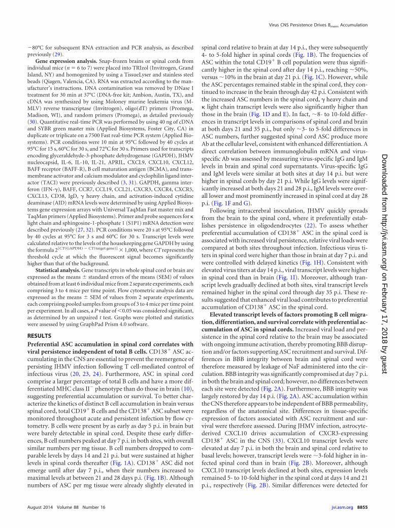
RESULTSPreferential ASC accumulation in spinal cord correlates withviral persistence independent of total B cells. CD138� ASC ac-cumulating in the CNS are essential to prevent the reemergence ofpersisting JHMV infection following T cell-mediated control ofinfectious virus (20, 23, 24). Furthermore, ASC in spinal cordcomprise a larger percentage of total B cells and have a more dif-ferentiated MHC class II� phenotype than do those in brain (10),suggesting preferential accumulation or survival. To better char-acterize the kinetics of distinct B cell accumulation in brain versusspinal cord, total CD19� B cells and the CD138� ASC subset weremonitored throughout acute and persistent infection by flow cy-tometry. B cells were present by as early as day 5 p.i. in brain butwere barely detectable in spinal cord. Despite these early differ-ences, B cell numbers peaked at day 7 p.i. in both sites, with overallsimilar numbers per mg tissue. B cell numbers dropped to com-parable levels by days 14 and 21 p.i. but were sustained at higherlevels in spinal cords thereafter (Fig. 1A). CD138� ASC did notemerge until after day 7 p.i., when their numbers increased tomaximal levels at between 21 and 28 days p.i. (Fig. 1B). Althoughnumbers of ASC per mg tissue were already slightly elevated in
spinal cord relative to brain at day 14 p.i., they were subsequently4- to 5-fold higher in spinal cords (Fig. 1B). The frequencies ofASC within the total CD19� B cell population were thus signifi-cantly higher in the spinal cord after day 14 p.i., reaching �50%,versus �10% in the brain at day 21 p.i. (Fig. 1C). However, whilethe ASC percentages remained stable in the spinal cord, they con-tinued to increase in the brain through day 42 p.i. Consistent withthe increased ASC numbers in the spinal cord, � heavy chain and light chain transcript levels were also significantly higher thanthose in the brain (Fig. 1D and E). In fact, �8- to 10-fold differ-ences in transcript levels in comparisons of spinal cord and brainat both days 21 and 35 p.i., but only �3- to 5-fold differences inASC numbers, further suggested spinal cord ASC produce moreAb at the cellular level, consistent with enhanced differentiation. Adirect correlation between immunoglobulin mRNA and virus-specific Ab was assessed by measuring virus-specific IgG and IgMlevels in brain and spinal cord supernatants. Virus-specific IgGand IgM levels were similar at both sites at day 14 p.i. but werehigher in spinal cords by day 21 p.i. While IgG levels were signif-icantly increased at both days 21 and 28 p.i., IgM levels were over-all lower and most prominently increased in spinal cord at day 28p.i. (Fig. 1F and G).
Following intracerebral inoculation, JHMV quickly spreadsfrom the brain to the spinal cord, where it preferentially estab-lishes persistence in oligodendrocytes (22). To assess whetherpreferential accumulation of CD138� ASC in the spinal cord isassociated with increased viral persistence, relative viral loads werecompared at both sites throughout infection. Infectious virus ti-ters in spinal cord were higher than those in brain at day 7 p.i. andwere controlled with delayed kinetics (Fig. 1H). Consistent withelevated virus titers at day 14 p.i., viral transcript levels were higherin spinal cord than in brain (Fig. 1I). Moreover, although tran-script levels gradually declined at both sites, viral transcript levelsremained higher in the spinal cord through day 35 p.i. These re-sults suggested that enhanced viral load contributes to preferentialaccumulation of CD138� ASC in the spinal cord.
Elevated transcript levels of factors promoting B cell migra-tion, differentiation, and survival correlate with preferential ac-cumulation of ASC in spinal cords. Increased viral load and per-sistence in the spinal cord relative to the brain may be associatedwith ongoing immune activation, thereby promoting BBB disrup-tion and/or factors supporting ASC recruitment and survival. Dif-ferences in BBB integrity between brain and spinal cord weretherefore measured by leakage of NaF administered into the cir-culation. BBB integrity was significantly compromised at day 7 p.i.in both the brain and spinal cord; however, no differences betweeneach site were detected (Fig. 2A). Furthermore, BBB integrity waslargely restored by day 14 p.i. (Fig. 2A). ASC accumulation withinthe CNS therefore appears to be independent of BBB permeability,regardless of the anatomical site. Differences in tissue-specificexpression of factors associated with ASC recruitment and sur-vival were therefore assessed. During JHMV infection, astrocyte-derived CXCL10 drives accumulation of CXCR3-expressingCD138� ASC in the CNS (33). CXCL10 transcript levels wereelevated at day 7 p.i. in both the brain and spinal cord relative tobasal levels; however, transcript levels were �3-fold higher in in-fected spinal cord than in brain (Fig. 2B). Moreover, althoughCXCL10 transcript levels declined at both sites, expression levelsremained 5- to 10-fold higher in the spinal cord at days 14 and 21p.i., respectively (Fig. 2B). Similar differences were detected for
Virus CNS Persistence Drives Bmem Accumulation
August 2014 Volume 88 Number 16 jvi.asm.org 8855
on February 17, 2018 by guest
http://jvi.asm.org/
Dow
nloaded from

the IFN-�-dependent expression of the CXCR3 ligand CXCL9(Fig. 2C). Consistent with IFN-�-mediated CXCL9 and CXCL10upregulation in the CNS during JHMV infection (3), IFN-� tran-script levels were significantly higher in the spinal cord than in thebrain (Fig. 2D). These data suggest that enhanced CXCL10 expres-sion in spinal cord relative to that in brain promotes infiltration ofCD138� ASC egressing from lymph nodes at days 14 and 21 p.i.
In addition to chemotaxis, the expression of APRIL and BAFF,key cytokines that promote B cell survival (34–38), may enhancefrequencies of CD138� ASC in the spinal cord. Notably, basal
levels of BAFF transcripts were already �4-fold higher in spinalcord than in brain. At day 7 p.i., BAFF transcript levels were in-creased �4- and 2-fold in brain and spinal cord, respectively (Fig.2E), consistent with IFN-�-mediated regulation (3, 19). BAFFtranscript levels subsequently decreased to background levels inthe brain and also gradually declined in spinal cords (Fig. 2E).Overall, BAFF mRNA in the brain remained below basal levels inthe spinal cord (Fig. 2E). In contrast to differential basal BAFFexpression, APRIL transcript levels in naive mice were similar inboth sites and were significantly upregulated only in the spinal
FIG 1 Preferential accumulation of ASC in the spinal cord versus the brain is associated with increased viral loads. (A and B) Numbers of total CD19� B cells(A) or CD19� CD138� ASC (B) were determined by flow cytometry. Data are expressed as the mean number of CD19� B cells (A) or CD138� ASC (B) per mgof tissue � SEM and represent two independent experiments, each comprising pooled brains or spinal cords of 6 to 8 mice per time point. (C) Percentages ofCD138� ASC within total CD19� B cells calculated as the means � SEM from data presented in panels A and B. (D and E) Relative transcript levels of � heavychain and light chain in brains and spinal cords of naive and infected mice assessed by real-time PCR. Data depict the means � SEM relative to GAPDH mRNAlevels for at least 6 to 7 individual mice derived from two independent experiments with at least 3 mice per time point per experiment. Transcript levels at day 7p.i. are expressed as means � SEMs relative to GAPDH mRNA levels and were as follows: 0.17 � 0.08 in brain and 0.26 � 0.14 in spinal cord for � heavy chain(D) and 0.8 � 0.2 in brain and 0.9 � 0.4 in spinal cord for light chain (E). (F and G) Virus-specific IgG2a and IgM levels in clarified supernatants from brainand spinal cord homogenates at the indicated time points were assessed by an ELISA. Arbitrary units reflect Ab levels converted to mg of tissue from 3 to 4 miceper time point. (H) Virus titers in brain or spinal cord supernatants were determined by a plaque assay and are expressed as mean PFU per mg of tissue � SEM.Data are from two independent experiments with 5 to 10 total mice per time point. (I) Relative transcript levels of viral RNA in brain or spinal cord assessed byreal-time PCR. Data depict the means � SEM relative to GAPDH mRNA levels for 6 to 7 total mice per time point derived from two independent experiments.Significant differences between naive and infected brain or naive and infected spinal cord are indicated (*, P 0.05; **, P 0.005; ***, P 0.001). Significantdifferences between brain and spinal cord at a given time point are indicated (#, P 0.05; ##, P 0.005; ###, P 0.001). In all cases, a P value of 0.05 wasconsidered significant, as determined by an unpaired t test. BD, below the detection limit.
Phares et al.
8856 jvi.asm.org Journal of Virology
on February 17, 2018 by guest
http://jvi.asm.org/
Dow
nloaded from
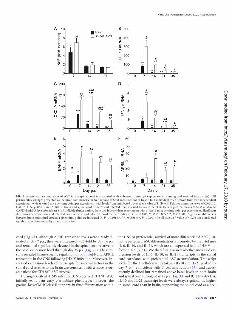
cord (Fig. 2F). Although APRIL transcript levels were already el-evated at day 7 p.i., they were increased �25-fold by day 14 p.i.and remained significantly elevated in the spinal cord relative tothe basal expression level through day 35 p.i. (Fig. 2F). These re-sults revealed tissue-specific regulation of both BAFF and APRILtranscripts in the CNS following JHMV infection. Moreover, in-creased expression levels of transcripts for survival factors in thespinal cord relative to the brain are consistent with a more favor-able niche for CD138� ASC survival.
During persistent JHMV infection, CNS-derived CD138� ASCinitially exhibit an early plasmablast phenotype; however, thegradual loss of MHC class II supports in situ differentiation within
the CNS or preferential survival of more differentiated ASC (10).In the periphery, ASC differentiation is promoted by the cytokinesIL-6, IL-10, and IL-21, which are all expressed in the JHMV-in-fected CNS (3, 31). We therefore assessed whether increased ex-pression levels of IL-6, IL-10, or IL-21 transcripts in the spinalcord correlated with preferential ASC accumulation. Transcriptlevels for the T cell-derived cytokines IL-10 and IL-21 peaked byday 7 p.i., coincident with T cell infiltration (39), and subse-quently declined but remained above basal levels in both brainand spinal cord through day 21 p.i. (Fig. 3A and B). Nevertheless,IL-10 and IL-21 transcript levels were always significantly higherin spinal cord than in brain, supporting the spinal cord as a pre-
FIG 2 Preferential accumulation of ASC in the spinal cord is associated with enhanced transcript expression of homing and survival factors. (A) BBBpermeability changes presented as the mean fold increase in NaF uptake � SEM measured for at least 6 to 8 individual mice derived from two independentexperiments with at least 3 mice per time point per experiment, with levels from uninfected mice set at a value of 1. (B to F) Relative transcript levels of CXCL10,CXCL9, IFN-�, BAFF, and APRIL in brain and spinal cord of naive and infected mice assessed by real-time PCR. Data depict the means � SEM relative toGAPDH mRNA levels for at least 6 to 7 individual mice derived from two independent experiments with at least 3 mice per time point per experiment. Significantdifferences between naive and infected brain or naive and infected spinal cord are indicated (*, P 0.05; **, P 0.005; ***, P 0.001). Significant differencesbetween brain and spinal cord at a given time point are indicated (#, P 0.05; ##, P 0.005; ###, P 0.001). In all cases, a P value of 0.05 was consideredsignificant, as determined by an unpaired t test.
Virus CNS Persistence Drives Bmem Accumulation
August 2014 Volume 88 Number 16 jvi.asm.org 8857
on February 17, 2018 by guest
http://jvi.asm.org/
Dow
nloaded from

ferred site of potential ASC differentiation in situ. Notably, basallevels of IL-6 transcripts were already �3-fold higher in spinalcord than in brain (Fig. 3C). While IL-6 transcript levels peaked atday 7 p.i., with expression levels being significantly higher in thespinal cord (Fig. 3), they declined to near-basal levels by day 21p.i., suggesting a limited contribution of IL-6 to ASC differentia-tion within the CNS during JHMV persistence.
Decline in levels of early-activated B cells coincides with iso-type-switched Bmem accumulation. The comparable total CD19�
B cell numbers in brain and spinal cord throughout infection (Fig.1A), yet preferential accumulation of CD138� ASC in spinal
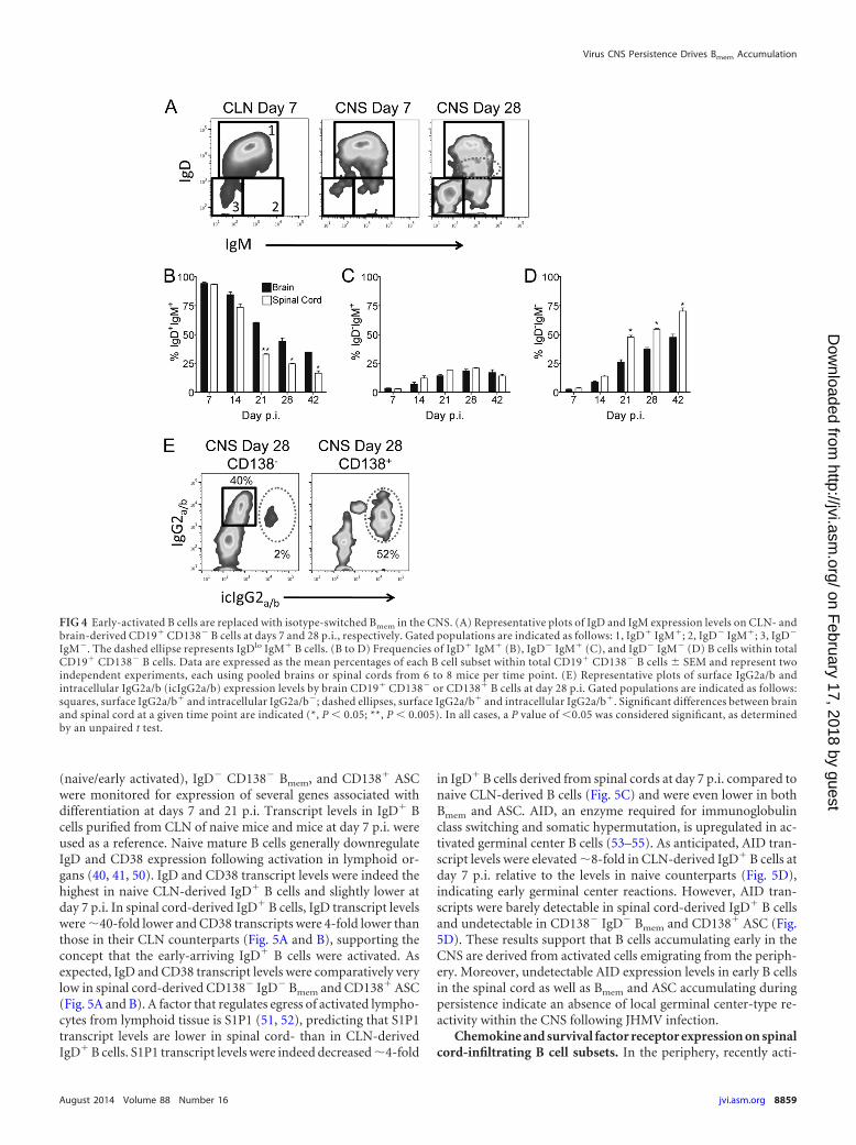
cords, were consistent with an unbiased recruitment of “by-stander” B cells to both sites early during infection. To character-ize the phenotype of the CD138� B cells and potential site-specificalterations in their composition, we assessed surface expression ofthe B cell receptors IgD and IgM. At day 7 p.i., �90% of B cells inboth the brain and spinal cord were IgD� IgM� (Fig. 4A and B),consistent with a naive or early-activated phenotype. Althoughfrequencies of IgD� IgM� B cells declined only slowly by day 14p.i., they continued to drop more rapidly in the spinal cord there-after, constituting only �50% of those in the brain by day 21 p.i.(Fig. 4B). As activated IgD� IgM� B cells in spleen and lymphnodes are characterized by a downregulation of IgD and sustainedIgM expression (40, 41), we monitored IgD expression on CNS-derived B cells over time. Increasing populations of IgDlo IgM�
(Fig. 4A, dashed ellipse) and IgD� IgM� cells by day 28 p.i. relativeto those at day 7 p.i. (Fig. 4A) indicated downregulation of IgD.The frequencies of IgDlo IgM� B cells increased to similar levels inboth sites (data not shown), consistent with unbiased ongoingrecruitment of newly activated B cells from the periphery and/orlocal activation of already CNS-infiltrated IgD� IgM� B cells. Asimilar increase in the frequency of IgD� IgM� B cells to �20% inboth sites (Fig. 4C) yet the enhanced decline of IgD� IgM� B cellsin spinal cord argued against a direct precursor relationship be-tween these CNS populations. With time, the CD138� B cells alsocomprised an increasing population lacking both IgD and IgM(Fig. 4D), thus representing isotype-switched antigen-experi-enced Bmem. Although the frequencies of IgD� IgM� Bmem in-creased at a similar rate to �10% and �15% at both sites by day 14p.i., accumulation of Bmem was accelerated in spinal cord relativeto brain by day 21 p.i. (Fig. 4D). The percentages of isotype-switched CD138� Bmem further increased in both sites but re-mained higher in spinal cord out to day 42 p.i. (Fig. 4D). Overall,the decline in IgD� IgM� B cells correlated with the increase in thefrequency of Bmem in both brain and spinal cord but was faster inspinal cord, as noted for ASC.
The majority of virus-specific ASC within the CNS followingJHMV infection secrete IgG2a/b (4). The proportion of isotype-switched IgD� IgM� Bmem in CD138� B cells (�55%) (Fig. 4D)approximated that of IgG2a/b surface-expressing cells (Fig. 4E),demonstrating that Bmem in the CNS largely express IgG2a/b. Tofurther confirm the characteristic phenotype of Bmem in express-ing surface IgG but limited intracellular IgG, both surface IgG andintracellular IgG were compared in CD138� B cells and CD138�
ASC (2, 42–44). Intracellular IgG2a/b was detected in 5% ofCD138� B cells but �55% of CD138� ASC (Fig. 4E), demonstrat-ing that non-Ab-secreting isotype-switched Bmem can localize tothe CNS, similarly to ASC. Taken together, the data indicate thatnaive/early-activated IgD� IgM� B cells are recruited to brain andspinal cord at similar frequencies early during acute JHMVinfection but are progressively replaced by Bmem and ASC. Thistransition is most prominent at between days 14 and 21 p.i.,coinciding with the general time of lymph node germinal cen-ter formation following infection or immunization (45–49).Furthermore, this process is accelerated at the site of enhancedviral persistence and inflammation, supporting the conceptthat differentiated B cell accumulation is driven by localchemokines and survival factors.
B cells accumulating early in the spinal cord have an acti-vated phenotype. To further characterize the differentiation stateof CNS-localized B cell subsets, spinal cord-derived IgD� B cells
FIG 3 Transcript expression levels of cytokines promoting ASC differentia-tion are higher in the spinal cord. Relative transcript levels of IL-10, IL-21, andIL-6 in brains and spinal cords of naive and infected mice were assessed byreal-time PCR. Data depict the means � SEM relative to GAPDH mRNA levelsfor at least 6 to 7 individual mice derived from two independent experimentswith at least 3 mice per time point per experiment. Significant differencesbetween naive and infected brain or naive and infected spinal cord are indi-cated (*, P 0.05; **, P 0.005; ***, P 0.001). Significant differencesbetween brain and spinal cord at a given time point are indicated (#, P 0.05;##, P 0.005; ###, P 0.001). In all cases, a P value of 0.05 was consideredsignificant, as determined by an unpaired t test.
Phares et al.
8858 jvi.asm.org Journal of Virology
on February 17, 2018 by guest
http://jvi.asm.org/
Dow
nloaded from
(naive/early activated), IgD� CD138� Bmem, and CD138� ASCwere monitored for expression of several genes associated withdifferentiation at days 7 and 21 p.i. Transcript levels in IgD� Bcells purified from CLN of naive mice and mice at day 7 p.i. wereused as a reference. Naive mature B cells generally downregulateIgD and CD38 expression following activation in lymphoid or-gans (40, 41, 50). IgD and CD38 transcript levels were indeed thehighest in naive CLN-derived IgD� B cells and slightly lower atday 7 p.i. In spinal cord-derived IgD� B cells, IgD transcript levelswere �40-fold lower and CD38 transcripts were 4-fold lower thanthose in their CLN counterparts (Fig. 5A and B), supporting theconcept that the early-arriving IgD� B cells were activated. Asexpected, IgD and CD38 transcript levels were comparatively verylow in spinal cord-derived CD138� IgD� Bmem and CD138� ASC(Fig. 5A and B). A factor that regulates egress of activated lympho-cytes from lymphoid tissue is S1P1 (51, 52), predicting that S1P1transcript levels are lower in spinal cord- than in CLN-derivedIgD� B cells. S1P1 transcript levels were indeed decreased �4-fold
in IgD� B cells derived from spinal cords at day 7 p.i. compared tonaive CLN-derived B cells (Fig. 5C) and were even lower in bothBmem and ASC. AID, an enzyme required for immunoglobulinclass switching and somatic hypermutation, is upregulated in ac-tivated germinal center B cells (53–55). As anticipated, AID tran-script levels were elevated �8-fold in CLN-derived IgD� B cells atday 7 p.i. relative to the levels in naive counterparts (Fig. 5D),indicating early germinal center reactions. However, AID tran-scripts were barely detectable in spinal cord-derived IgD� B cellsand undetectable in CD138� IgD� Bmem and CD138� ASC (Fig.5D). These results support that B cells accumulating early in theCNS are derived from activated cells emigrating from the periph-ery. Moreover, undetectable AID expression levels in early B cellsin the spinal cord as well as Bmem and ASC accumulating duringpersistence indicate an absence of local germinal center-type re-activity within the CNS following JHMV infection.
Chemokine and survival factor receptor expression on spinalcord-infiltrating B cell subsets. In the periphery, recently acti-
FIG 4 Early-activated B cells are replaced with isotype-switched Bmem in the CNS. (A) Representative plots of IgD and IgM expression levels on CLN- andbrain-derived CD19� CD138� B cells at days 7 and 28 p.i., respectively. Gated populations are indicated as follows: 1, IgD� IgM�; 2, IgD� IgM�; 3, IgD�
IgM�. The dashed ellipse represents IgDlo IgM� B cells. (B to D) Frequencies of IgD� IgM� (B), IgD� IgM� (C), and IgD� IgM� (D) B cells within totalCD19� CD138� B cells. Data are expressed as the mean percentages of each B cell subset within total CD19� CD138� B cells � SEM and represent twoindependent experiments, each using pooled brains or spinal cords from 6 to 8 mice per time point. (E) Representative plots of surface IgG2a/b andintracellular IgG2a/b (icIgG2a/b) expression levels by brain CD19� CD138� or CD138� B cells at day 28 p.i. Gated populations are indicated as follows:squares, surface IgG2a/b� and intracellular IgG2a/b�; dashed ellipses, surface IgG2a/b� and intracellular IgG2a/b�. Significant differences between brainand spinal cord at a given time point are indicated (*, P 0.05; **, P 0.005). In all cases, a P value of 0.05 was considered significant, as determinedby an unpaired t test.
Virus CNS Persistence Drives Bmem Accumulation
August 2014 Volume 88 Number 16 jvi.asm.org 8859
on February 17, 2018 by guest
http://jvi.asm.org/
Dow
nloaded from
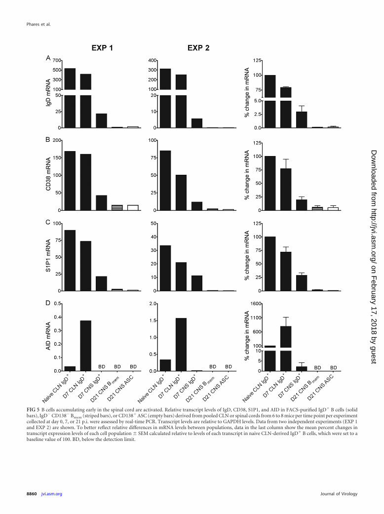
FIG 5 B cells accumulating early in the spinal cord are activated. Relative transcript levels of IgD, CD38, S1P1, and AID in FACS-purified IgD� B cells (solidbars), IgD� CD138� Bmem (striped bars), or CD138� ASC (empty bars) derived from pooled CLN or spinal cords from 6 to 8 mice per time point per experimentcollected at day 0, 7, or 21 p.i. were assessed by real-time PCR. Transcript levels are relative to GAPDH levels. Data from two independent experiments (EXP 1and EXP 2) are shown. To better reflect relative differences in mRNA levels between populations, data in the last column show the mean percent changes intranscript expression levels of each cell population � SEM calculated relative to levels of each transcript in naive CLN-derived IgD� B cells, which were set to abaseline value of 100. BD, below the detection limit.
Phares et al.
8860 jvi.asm.org Journal of Virology
on February 17, 2018 by guest
http://jvi.asm.org/
Dow
nloaded from
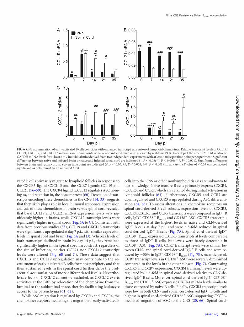
vated B cells primarily migrate to lymphoid follicles in response tothe CXCR5 ligand CXCL13 and the CCR7 ligands CCL19 andCCL21 (56–59). The CXCR4 ligand CXCL12 regulates ASC hom-ing to, and retention in, the bone marrow (60). Detection of tran-scripts encoding these chemokines in the CNS (14, 33) suggeststhat they likely play a role in local humoral responses. Expressionanalysis of these chemokines in brain versus spinal cord revealedthat basal CCL19 and CCL21 mRNA expression levels were sig-nificantly higher in brains, while CXCL12 transcript levels weresignificantly higher in spinal cords (Fig. 6A to C). Consistent withdata from previous studies (33), CCL19 and CXCL13 transcriptswere significantly upregulated at day 7 p.i., with similar expressionlevels in spinal cord and brain (Fig. 6A and D). Whereas levels ofboth transcripts declined in brain by day 14 p.i., they remainedsignificantly higher in the spinal cord. In contrast, regardless ofthe site of infection, neither CCL21 nor CXCL12 transcriptlevels were altered (Fig. 6B and C). These data suggest thatCXCL13 and CCL19 upregulation may contribute to the re-cruitment of early-activated B cells from the periphery and thattheir sustained levels in the spinal cord further drive the pref-erential accumulation of more differentiated B cells. Neverthe-less, effects of CXCL12 cannot be excluded, as CXCL12 exertsactivities at the BBB by relocation of the chemokine from theluminal to the subluminal space, thereby facilitating leukocyteaccess to the parenchyma (61, 62).
While ASC migration is regulated by CXCR3 and CXCR4, thechemokine receptors mediating the migration of early-activated B
cells into the CNS or other nonlymphoid tissues are unknown toour knowledge. Naive mature B cells primarily express CXCR4,CXCR5, and CCR7, which are retained during initial activation inlymphoid follicles (63). Furthermore, CXCR5 and CCR7 aredownregulated and CXCR3 is upregulated during ASC differenti-ation (64, 65). To assess alterations in chemokine receptors onspinal cord-derived B cell subsets, expression levels of CXCR3,CXCR4, CXCR5, and CCR7 transcripts were compared in IgD� Bcells, IgD� CD138� Bmem, and CD138� ASC. CXCR5 transcriptswere expressed at the highest levels in naive and CLN-derivedIgD� B cells at day 7 p.i. and were �5-fold reduced in spinalcord-derived IgD� B cells (Fig. 7A). Spinal cord-derived IgD�
CD138� Bmem expressed CXCR5 transcripts at levels comparableto those of IgD� B cells, but levels were barely detectable inCD138� ASC (Fig. 7A). CCR7 transcript levels were similar be-tween CLN- and spinal cord-derived IgD� B cells and were re-duced by �50% in IgD� CD138� Bmem (Fig. 7B). As anticipated,CCR7 transcript levels in CD138� ASC were severely diminishedcompared to the levels in the other subsets (Fig. 7). ContrastingCXCR5 and CCR7 expression, CXCR4 transcript levels were up-regulated by �5-fold in spinal cord-derived relative to CLN-de-rived IgD� B cells. Moreover, spinal cord-derived IgD� CD138�
Bmem and CD138� ASC expressed CXCR4 mRNA levels similar tothose expressed by naive B cells. Finally, CXCR3 transcript levelswere low in both CLN- and spinal cord-derived IgD� B cells andhighest in spinal cord-derived CD138� ASC, supporting CXCR3-mediated migration of ASC to the CNS (20, 66). Spinal cord-
FIG 6 CNS accumulation of early-activated B cells coincides with enhanced transcript expression of lymphoid chemokines. Relative transcript levels of CCL19,CCL21, CXCL12, and CXCL13 in brains and spinal cords of naive and infected mice were assessed by real-time PCR. Data depict the means � SEM relative toGAPDH mRNA levels for at least 6 to 7 individual mice derived from two independent experiments with at least 3 mice per time point per experiment. Significantdifferences between naive and infected brain or naive and infected spinal cord are indicated (*, P 0.05; **, P 0.005; ***, P 0.001). Significant differencesbetween brain and spinal cord at a given time point are indicated (#, P 0.05; ##, P 0.005; ###, P 0.001). In all cases, a P value of 0.05 was consideredsignificant, as determined by an unpaired t test.
Virus CNS Persistence Drives Bmem Accumulation
August 2014 Volume 88 Number 16 jvi.asm.org 8861
on February 17, 2018 by guest
http://jvi.asm.org/
Dow
nloaded from

FIG 7 Transcript expression levels of chemokine receptors on spinal cord-derived B cell subsets. Relative transcript levels of CXCR5, CCR7, CXCR4, and CXCR3in FACS-purified IgD� B cells (solid bars), IgD� CD138� Bmem (striped bars), or CD138� ASC (empty bars) from pooled CLN or spinal cords of 6 to 8 mice pertime point per experiment collected at day 0, 7, or 21 p.i. were assessed by real-time PCR. Transcript levels are relative to GAPDH levels. Two independentexperiments (EXP 1 and EXP 2) are shown. To better reflect relative differences in mRNA levels between populations, data in the last column show the meanpercent changes in transcript expression levels of each cell population � SEM calculated relative to levels of each transcript in naive CLN-derived IgD� B cells,which were set to a baseline value of 100. BD, below the detection limit.
Phares et al.
8862 jvi.asm.org Journal of Virology
on February 17, 2018 by guest
http://jvi.asm.org/
Dow
nloaded from

derived IgD� CD138� Bmem also expressed CXCR3 mRNA albeitat lower levels than in ASC. CCL19/CCL21/CCR7 and/orCXCL12/CXCR4 interactions may thus promote the accumula-tion of early-activated IgD� B cells, while Bmem trafficking may bemore diversely regulated by responsiveness to CCR7, CXCR4, andCXCR3 signaling.
Distinct B cell subsets are also characterized by differential ex-pression of the B cell survival receptors BCMA, TACI, andBAFF-R. Naive B cells predominantly express BAFF-R, whileCD138� ASC upregulate BCMA and TACI expressions (34, 67–71). Analysis of respective transcripts in B cell subsets in CLN andthe spinal cord during JHMV infection revealed that the BAFF-R
expression level was highest in CLN-derived IgD� B cells, whilelevels in all three spinal cord-derived populations were �80%lower (Fig. 8A). On the contrary, spinal cord-derived CD138�
ASC had �500- and 5-fold-higher BCMA and TACI transcriptlevels, respectively, than did naive CLN-derived IgD� B cells (Fig.8B and C). Although the BCMA expression level was low in IgD�
B cells irrespective of their tissue origin, transcript levels for bothBAFF-R and TACI were reduced in spinal cord-derived IgD� Bcells relative to CLN-derived IgD� B cells (Fig. 8). Bmem revealedan intermediate phenotype, expressing all 3 transcripts at moder-ate levels. These data suggest that BAFF promotes survival ofearly-activated IgD� B cells within the CNS, while Bmem survival
FIG 8 Transcript expression of survival factor receptors in spinal cord-derived B cell subsets. Relative transcript levels of BAFF-R, BCMA, and TACI inFACS-purified IgD� B cells (solid bars), IgD� CD138� Bmem (striped bars), or CD138� ASC (empty bars) from pooled CLN or spinal cords of 6 to 8 micecollected per time point per experiment at day 0, 7, or 21 p.i. were assessed by real-time PCR. Transcript levels are relative to GAPDH levels. Two independentexperiments (EXP 1 and EXP 2) are shown. To better reflect relative differences in mRNA levels between populations, data in the last column show the meanpercent changes in transcript expression levels of each cell population � SEM calculated relative to levels of each transcript in naive CLN-derived IgD� B cells,which were set to a baseline value of 100. BD, below the detection limit.
Virus CNS Persistence Drives Bmem Accumulation
August 2014 Volume 88 Number 16 jvi.asm.org 8863
on February 17, 2018 by guest
http://jvi.asm.org/
Dow
nloaded from

may be regulated by either APRIL or BAFF. Furthermore, the dataare consistent with CD138� ASC receiving TACI- and/or BCMA-dependent survival signals via APRIL engagement.
DISCUSSION
The naive CNS is largely devoid of B cells, but heterogeneous B cellsubsets accumulate in cerebrospinal fluid (CSF) during acute andchronic CNS infections (1, 72). However, there is little informa-tion on the forces driving CNS B cell lineage accumulation, theirheterogeneity, or their function over time. The majority of CSF Bcells exhibit an isotype-switched memory phenotype distinct fromthe dominant naive phenotype in circulation, independent of thedisease diagnosis (1). Furthermore, oligoclonal IgG in the CSFduring chronic infection as well as MS is consistent with local ASCenrichment (12, 73–75). B cells, including ASC, are also promi-nent in the CNS during experimental murine infections (2, 10, 20,76, 77), where they appear to play a protective role (24, 76–79).While CNS-resident ASC may also provide local immunity duringhuman CNS infections (80–83), the influence of other B cell sub-sets on disease progression in distinct neuroinflammatory infec-tions is unclear. The characterization of B cell accumulation indistinct models is thus critical to reveal potential commonalities,precursor relationships, and humoral-based targets to manipulatedistinct B cell functions.
Using the glia-tropic JHMV model of viral encephalomyelitis,here we demonstrate that B cells accumulating in the CNS evolveprogressively from an early-activated IgD� IgM� phenotype to amore differentiated phenotype comprising IgM� and IgG� Bmem
as well as ASC. Moreover, although overall recruitment kineticsand numbers of total B cells were similar in brain and spinal cordthroughout infection, loss of IgD expression, coincident with in-creased numbers of Bmem and CD138� ASC, was accelerated andmore robust in spinal cord after initial T cell-mediated viral con-trol. Early recruitment of IgD� IgM� B cells was also noted forSindbis virus infection (2), suggesting that the mobilization ofnaive or early-activated B cells prior to the detection of virus-specific ASC in the periphery may be common to a number of viralCNS infections. The data for both models are also consistent witha gradual replacement of early IgD� B cells in the CNS with moredifferentiated B cells at the time of peripheral germinal centerreactions. Our results also indicate that the magnitude of ongoinginflammatory responses, directly correlating with the extent ofviral load, is a dominant force driving preferential recruitmentand survival of Bmem and ASC at a given anatomical site within theCNS. In this regard, APRIL expression in the spinal cord relativeto the brain was not only increased but sustained, indicating dis-tinct site-specific regulation of survival factors in the CNS. Up-regulation of APRIL by specific Toll-like receptor (TLR) ligands(84, 85) suggests that enhanced APRIL expression in spinal cordmay be attributed to distinct TLR activation. Regardless, elevatedviral persistence and tissue damage in the spinal cord relativeto brain likely promotes ongoing inflammatory responses andthereby preferential accumulation of more differentiated B cells.
Whether the infiltrating IgD� IgM� B cells constitute “earlymemory” B cells, as indicated during neuroinflammatory diseasesin humans (1), is difficult to assess, as murine Bmem do not expressCD27 (86). Nevertheless, CNS IgD� IgM� B cells are presumablyactivated by innate signals or viral antigen in lymphoid tissue, asnaive B cells do not access the human CNS. Modest degrees ofactivation and migratory behavior to distal sites are supported by
the decreased expression levels of IgD, CD38, CXCR5, and S1P1mRNAs in spinal cord-derived relative to CLN-derived popula-tions in our JHMV model. The chemokines recruiting IgD� Bcells to the CNS are unknown. However, the CCR7 ligands CCL19and CCL21 are supported as candidates based on the sustainedexpression of CCR7 and CXCR4 transcripts by IgD� B cells in ourmodel and the detection of CCR7 on CNS-derived CD19� B cellsduring Sindbis virus infection (15). CNS Bmem expressed a diverserange of receptors promoting migration, including CCR7,CXCR4, and CXCR3, whereas ASC predominantly expressedCXCR3 and CXCR4. The elevated CXCR3 transcript levels inCNS-derived Bmem relative to early-activated IgD� B cells implythat Bmem may respond to CXCR3-mediated signaling, similar toCD138� ASC during JHMV infection (20, 66).
A potential precursor relationship of early-infiltrating B cells tomore differentiated B cells accumulating during CNS virus persis-tence remains unclear. Although ectopic follicle-like structuressustained by CXCL13 and CCL19/CCL21 have been associatedwith local B cell differentiation during MS and neuroborreliosis(11, 12, 87, 88), limited or undetectable AID expression in IgD� Bcells, Bmem, or ASC accumulating within the JHMV-infected CNSargued against local germinal center formation. This is supportedby the absence of B cell clusters by histological examination ofspinal cords during persistence (10, 20, 33). Similarly, there wasno evidence for ectopic follicle formation during Sindbis virusinfection (2). The progressive accumulation of more differenti-ated B cell phenotypes in these two infections is thus more likelydriven by the preferential survival of ASC and Bmem over IgD� Bcells. Regardless, during persistent JHMV infection, the gradualdownregulation of MHC class II on ASC supports their ongoingfocal differentiation (10). It thus remains possible that ASC andBmem continue to proliferate and differentiate locally within theCNS. In this context, it is of interest to note that Bmem may convertto ASC in the presence of CXCL10, thus potentially contributingto long-lived ASC even in the absence of antigen restimulation(89).
While ASC appear to mediate protective functions by control-ling persistent virus, the functions of heterologous B cell subsetswithin the infected CNS are unknown. Besides serving as precur-sors to more differentiated Bmem or ASC, they may participate asantigen-presenting cells (APC) for T cell activation or exert pro-tective functions via cytokine secretion (90–96). In the JHMVmodel, B cell-deficient and B cell-sufficient, yet Ab-deficient, miceprovided no evidence for a role of B cells in APC function (24, 25).Nevertheless, B cells play an Ab-independent role as APC in graftrejection and the experimental allergic encephalomyelitis modelof MS (96–98), thereby playing detrimental roles. Taken together,both JHMV and Sindbis virus infection models demonstrate com-monalities in B cell subset recruitment and dynamics. Moreover,this is the first study to reveal that virus load drives enrichment ofBmem and antiviral ASC in a nonlymphoid organ.
ACKNOWLEDGMENT
This work was supported by U.S. National Institutes of Health grantsNS086299 and AI047249.
REFERENCES1. Cepok S, von Geldern G, Grummel V, Hochgesand S, Celik H, Hartung
H, Hemmer B. 2006. Accumulation of class switched IgD-IgM- memoryB cells in the cerebrospinal fluid during neuroinflammation. J. Neuroim-munol. 180:33–39. http://dx.doi.org/10.1016/j.jneuroim.2006.06.031.
Phares et al.
8864 jvi.asm.org Journal of Virology
on February 17, 2018 by guest
http://jvi.asm.org/
Dow
nloaded from

2. Metcalf TU, Griffin DE. 2011. Alphavirus-induced encephalomyelitis:antibody-secreting cells and viral clearance from the nervous system. J.Virol. 85:11490 –11501. http://dx.doi.org/10.1128/JVI.05379-11.
3. Phares TW, Marques CP, Stohlman SA, Hinton DR, Bergmann CC.2011. Factors supporting intrathecal humoral responses following viralencephalomyelitis. J. Virol. 85:2589 –2598. http://dx.doi.org/10.1128/JVI.02260-10.
4. Tschen SI, Bergmann CC, Ramakrishna C, Morales S, Atkinson R,Stohlman SA. 2002. Recruitment kinetics and composition of anti-body-secreting cells within the central nervous system following viralencephalomyelitis. J. Immunol. 168:2922–2929. http://dx.doi.org/10.4049/jimmunol.168.6.2922.
5. Ankeny DP, Guan Z, Popovich PG. 2009. B cells produce pathogenicantibodies and impair recovery after spinal cord injury in mice. J. Clin.Invest. 119:2990 –2999. http://dx.doi.org/10.1172/JCI39780.
6. Pohl-Koppe A, Kaiser R, Meulen VT, Liebert UG. 1995. Antibodyreactivity to individual structural proteins of measles virus in the CSF ofSSPE and MS patients. Clin. Diagn. Virol. 4:135–147. http://dx.doi.org/10.1016/0928-0197(95)00006-T.
7. Norrby E, Link H. 1977. The relationship between measles virus-specificantibodies and oligoclonal IgG in the cerebrospinal fluid from patientswith subacute sclerosing panencephalitis and multiple sclerosis. ActaNeurol. Scand. Suppl. 63:161–171.
8. Owens GP, Ritchie AM, Burgoon MP, Williamson RA, Corboy JR,Gilden DH. 2003. Single-cell repertoire analysis demonstrates that clonalexpansion is a prominent feature of the B cell response in multiple sclero-sis cerebrospinal fluid. J. Immunol. 171:2725–2733. http://dx.doi.org/10.4049/jimmunol.171.5.2725.
9. Burgoon MP, Caldas YA, Keays KM, Yu X, Gilden DH, Owens GP.2006. Recombinant antibodies generated from both clonal and less abun-dant plasma cell immunoglobulin G sequences in subacute sclerosing pa-nencephalitis brain are directed against measles virus. J. Neurovirol. 12:398 – 402. http://dx.doi.org/10.1080/13550280600957414.
10. Tschen SI, Stohlman SA, Ramakrishna C, Hinton DR, Atkinson RD,Bergmann CC. 2006. CNS viral infection diverts homing of antibody-secreting cells from lymphoid organs to the CNS. Eur. J. Immunol. 36:603– 612. http://dx.doi.org/10.1002/eji.200535123.
11. Serafini B, Rosicarelli B, Magliozzi R, Stigliano E, Aloisi F. 2004.Detection of ectopic B-cell follicles with germinal centers in the meningesof patients with secondary progressive multiple sclerosis. Brain Pathol.14:164 –174. http://dx.doi.org/10.1111/j.1750-3639.2004.tb00049.x.
12. Narayan K, Dail D, Li L, Cadavid D, Amrute S, Fitzgerald-Bocarsly P,Pachner AR. 2005. The nervous system as ectopic germinal center:CXCL13 and IgG in Lyme neuroborreliosis. Ann. Neurol. 57:813– 823.http://dx.doi.org/10.1002/ana.20486.
13. Corcione A, Aloisi F, Serafini B, Capello E, Mancardi GL, Pistoia V,Uccelli A. 2005. B-cell differentiation in the CNS of patients withmultiple sclerosis. Autoimmun. Rev. 4:549 –554. http://dx.doi.org/10.1016/j.autrev.2005.04.012.
14. Lalor SJ, Segal BM. 2010. Lymphoid chemokines in the CNS. J. Neuro-immunol. 224:56 – 61. http://dx.doi.org/10.1016/j.jneuroim.2010.05.017.
15. Metcalf TU, Baxter VK, Nilaratanakul V, Griffin DE. 2013. Recruitmentand retention of B cells in the central nervous system in response to alpha-virus encephalomyelitis. J. Virol. 87:2420 –2429. http://dx.doi.org/10.1128/JVI.01769-12.
16. Kowarik MC, Cepok S, Sellner J, Grummel V, Weber MS, Korn T,Berthele A, Hemmer B. 2012. CXCL13 is the major determinant for B cellrecruitment to the CSF during neuroinflammation. J. Neuroinflamma-tion 9:93. http://dx.doi.org/10.1186/1742-2094-9-93.
17. Magliozzi R, Columba-Cabezas S, Serafini B, Aloisi F. 2004. Intracere-bral expression of CXCL13 and BAFF is accompanied by formation oflymphoid follicle-like structures in the meninges of mice with relapsingexperimental autoimmune encephalomyelitis. J. Neuroimmunol. 148:11–23. http://dx.doi.org/10.1016/j.jneuroim.2003.10.056.
18. Thangarajh M, Masterman T, Hillert J, Moerk S, Jonsson R. 2007. Aproliferation-inducing ligand (APRIL) is expressed by astrocytes and isincreased in multiple sclerosis. Scand. J. Immunol. 65:92–98. http://dx.doi.org/10.1111/j.1365-3083.2006.01867.x.
19. Krumbholz M, Theil D, Derfuss T, Rosenwald A, Schrader F,Monoranu CM, Kalled SL, Hess DM, Serafini B, Aloisi F, Wekerle H,Hohlfeld R, Meinl E. 2005. BAFF is produced by astrocytes and up-regulated in multiple sclerosis lesions and primary central nervous system
lymphoma. J. Exp. Med. 201:195–200. http://dx.doi.org/10.1084/jem.20041674.
20. Marques CP, Kapil P, Hinton DR, Hindinger C, Nutt SL, RansohoffRM, Phares TW, Stohlman SA, Bergmann CC. 2011. CXCR3-dependent plasma blast migration to the central nervous system duringviral encephalomyelitis. J. Virol. 85:6136 – 6147. http://dx.doi.org/10.1128/JVI.00202-11.
21. Wang FI, Hinton DR, Gilmore W, Trousdale MD, Fleming JO. 1992.Sequential infection of glial cells by the murine hepatitis virus JHM strain(MHV-4) leads to a characteristic distribution of demyelination. Lab. In-vest. 66:744 –754.
22. Parra B, Hinton DR, Marten NW, Bergmann CC, Lin MT, Yang CS,Stohlman SA. 1999. IFN-gamma is required for viral clearance from cen-tral nervous system oligodendroglia. J. Immunol. 162:1641–1647.
23. Bergmann CC, Lane TE, Stohlman SA. 2006. Coronavirus infection ofthe central nervous system: host-virus stand-off. Nat. Rev. Microbiol.4:121–132. http://dx.doi.org/10.1038/nrmicro1343.
24. Ramakrishna C, Bergmann CC, Atkinson R, Stohlman SA. 2003.Control of central nervous system viral persistence by neutralizingantibody. J. Virol. 77:4670 – 4678. http://dx.doi.org/10.1128/JVI.77.8.4670-4678.2003.
25. Ramakrishna C, Stohlman SA, Atkinson RD, Shlomchik MJ, BergmannCC. 2002. Mechanisms of central nervous system viral persistence: thecritical role of antibody and B cells. J. Immunol. 168:1204 –1211. http://dx.doi.org/10.4049/jimmunol.168.3.1204.
26. Fleming JO, Trousdale MD, el-Zaatari FA, Stohlman SA, Weiner LP.1986. Pathogenicity of antigenic variants of murine coronavirus JHM se-lected with monoclonal antibodies. J. Virol. 58:869 – 875.
27. Phares TW, Kean RB, Mikheeva T, Hooper DC. 2006. Regional differ-ences in blood-brain barrier permeability changes and inflammation inthe apathogenic clearance of virus from the central nervous system. J.Immunol. 176:7666 –7675. http://dx.doi.org/10.4049/jimmunol.176.12.7666.
28. Bergmann CC, Altman JD, Hinton D, Stohlman SA. 1999. Invertedimmunodominance and impaired cytolytic function of CD8� T cells dur-ing viral persistence in the central nervous system. J. Immunol. 163:3379 –3387.
29. Ireland DD, Stohlman SA, Hinton DR, Kapil P, Silverman RH, Atkin-son RA, Bergmann CC. 2009. RNase L mediated protection from virusinduced demyelination. PLoS Pathog. 5:e1000602. http://dx.doi.org/10.1371/journal.ppat.1000602.
30. de Aquino MT, Puntambekar SS, Savarin C, Bergmann CC, Phares TW,Hinton DR, Stohlman SA. 2013. Role of CD25(�) CD4(�) T cells inacute and persistent coronavirus infection of the central nervous system.Virology 447:112–120. http://dx.doi.org/10.1016/j.virol.2013.08.030.
31. Puntambekar SS, Bergmann CC, Savarin C, Karp CL, Phares TW, ParraGI, Hinton DR, Stohlman SA. 2011. Shifting hierarchies of interleukin-10-producing T cell populations in the central nervous system duringacute and persistent viral encephalomyelitis. J. Virol. 85:6702– 6713. http://dx.doi.org/10.1128/JVI.00200-11.
32. Matloubian M, Lo CG, Cinamon G, Lesneski MJ, Xu Y, Brinkmann V,Allende ML, Proia RL, Cyster JG. 2004. Lymphocyte egress from thymusand peripheral lymphoid organs is dependent on S1P receptor 1. Nature427:355–360. http://dx.doi.org/10.1038/nature02284.
33. Phares TW, Stohlman SA, Hinton DR, Bergmann CC. 2013. Astrocyte-derived CXCL10 drives accumulation of antibody-secreting cells in thecentral nervous system during viral encephalomyelitis. J. Virol. 87:3382–3392. http://dx.doi.org/10.1128/JVI.03307-12.
34. Benson MJ, Dillon SR, Castigli E, Geha RS, Xu S, Lam KP, Noelle RJ.2008. Cutting edge: the dependence of plasma cells and independence ofmemory B cells on BAFF and APRIL. J. Immunol. 180:3655–3659. http://dx.doi.org/10.4049/jimmunol.180.6.3655.
35. Bossen C, Cachero TG, Tardivel A, Ingold K, Willen L, Dobles M, ScottML, Maquelin A, Belnoue E, Siegrist CA, Chevrier S, Acha-Orbea H,Leung H, Mackay F, Tschopp J, Schneider P. 2008. TACI, unlikeBAFF-R, is solely activated by oligomeric BAFF and APRIL to supportsurvival of activated B cells and plasmablasts. Blood 111:1004 –1012. http://dx.doi.org/10.1182/blood-2007-09-110874.
36. Belnoue E, Pihlgren M, McGaha TL, Tougne C, Rochat AF, Bossen C,Schneider P, Huard B, Lambert PH, Siegrist CA. 2008. APRIL is criticalfor plasmablast survival in the bone marrow and poorly expressed byearly-life bone marrow stromal cells. Blood 111:2755–2764. http://dx.doi.org/10.1182/blood-2007-09-110858.
Virus CNS Persistence Drives Bmem Accumulation
August 2014 Volume 88 Number 16 jvi.asm.org 8865
on February 17, 2018 by guest
http://jvi.asm.org/
Dow
nloaded from
37. Cassese G, Arce S, Hauser AE, Lehnert K, Moewes B, Mostarac M,Muehlinghaus G, Szyska M, Radbruch A, Manz RA. 2003. Plasma cellsurvival is mediated by synergistic effects of cytokines and adhesion-dependent signals. J. Immunol. 171:1684 –1690. http://dx.doi.org/10.4049/jimmunol.171.4.1684.
38. Rolink AG, Tschopp J, Schneider P, Melchers F. 2002. BAFF is a survivaland maturation factor for mouse B cells. Eur. J. Immunol. 32:2004 –2010.http://dx.doi.org/10.1002/1521-4141(200207)32:72004::AID-IMMU2004�3.0.CO;2-5.
39. Savarin C, Stohlman SA, Atkinson R, Ransohoff RM, Bergmann CC.2010. Monocytes regulate T cell migration through the glia limitans dur-ing acute viral encephalitis. J. Virol. 84:4878 – 4888. http://dx.doi.org/10.1128/JVI.00051-10.
40. Bourgois A, Kitajima K, Hunter IR, Askonas BA. 1977. Surface immu-noglobulins of lipopolysaccharide-stimulated spleen cells. The behavior ofIgM, IgD and IgG. Eur. J. Immunol. 7:151–153.
41. Monroe JG, Havran WL, Cambier JC. 1983. B lymphocyte activation:entry into cell cycle is accompanied by decreased expression of IgD butnot IgM. Eur. J. Immunol. 13:208 –213. http://dx.doi.org/10.1002/eji.1830130306.
42. Arpin C, Dechanet J, Van Kooten C, Merville P, Grouard G, Briere F,Banchereau J, Liu YJ. 1995. Generation of memory B cells and plasmacells in vitro. Science 268:720 –722. http://dx.doi.org/10.1126/science.7537388.
43. Tangye SG, Avery DT, Hodgkin PD. 2003. A division-linked mech-anism for the rapid generation of Ig-secreting cells from human mem-ory B cells. J. Immunol. 170:261–269. http://dx.doi.org/10.4049/jimmunol.170.1.261.
44. Blink EJ, Light A, Kallies A, Nutt SL, Hodgkin PD, Tarlinton DM. 2005.Early appearance of germinal center-derived memory B cells and plasmacells in blood after primary immunization. J. Exp. Med. 201:545–554.http://dx.doi.org/10.1084/jem.20042060.
45. Jacob J, Kassir R, Kelsoe G. 1991. In situ studies of the primary immuneresponse to (4-hydroxy-3-nitrophenyl)acetyl. I. The architecture and dy-namics of responding cell populations. J. Exp. Med. 173:1165–1175.
46. MacLennan IC. 1994. Germinal centers. Annu. Rev. Immunol. 12:117–139. http://dx.doi.org/10.1146/annurev.iy.12.040194.001001.
47. Rasheed MA, Latner DR, Aubert RD, Gourley T, Spolski R, Davis CW,Langley WA, Ha SJ, Ye L, Sarkar S, Kalia V, Konieczny BT, LeonardWJ, Ahmed R. 2013. Interleukin-21 is a critical cytokine for the genera-tion of virus-specific long-lived plasma cells. J. Virol. 87:7737–7746. http://dx.doi.org/10.1128/JVI.00063-13.
48. Olson MR, McDermott DS, Varga SM. 2012. The initial draining lymphnode primes the bulk of the CD8 T cell response and influences memory Tcell trafficking after a systemic viral infection. PLoS Pathog. 8:e1003054.http://dx.doi.org/10.1371/journal.ppat.1003054.
49. Boyden AW, Legge KL, Waldschmidt TJ. 2012. Pulmonary infectionwith influenza A virus induces site-specific germinal center and T follicu-lar helper cell responses. PLoS One 7:e40733. http://dx.doi.org/10.1371/journal.pone.0040733.
50. Oliver AM, Martin F, Kearney JF. 1997. Mouse CD38 is down-regulatedon germinal center B cells and mature plasma cells. J. Immunol. 158:1108 –1115.
51. Chiba K, Matsuyuki H, Maeda Y, Sugahara K. 2006. Role of sphingosine1-phosphate receptor type 1 in lymphocyte egress from secondary lym-phoid tissues and thymus. Cell. Mol. Immunol. 3:11–19. http://www.cmi.ustc.edu.cn/3/1/11.pdf.
52. Schwab SR, Cyster JG. 2007. Finding a way out: lymphocyte egress fromlymphoid organs. Nat. Immunol. 8:1295–1301. http://dx.doi.org/10.1038/ni1545.
53. Keim C, Kazadi D, Rothschild G, Basu U. 2013. Regulation of AID, theB-cell genome mutator. Genes Dev. 27:1–17. http://dx.doi.org/10.1101/gad.200014.112.
54. Maul RW, Gearhart PJ. 2010. AID and somatic hypermutation. Adv. Im-munol. 105:159–191. http://dx.doi.org/10.1016/S0065-2776(10)05006-6.
55. Muramatsu M, Kinoshita K, Fagarasan S, Yamada S, Shinkai Y, HonjoT. 2000. Class switch recombination and hypermutation require activa-tion-induced cytidine deaminase (AID), a potential RNA editing enzyme.Cell 102:553–563. http://dx.doi.org/10.1016/S0092-8674(00)00078-7.
56. Okada T, Ngo VN, Ekland EH, Forster R, Lipp M, Littman DR, CysterJG. 2002. Chemokine requirements for B cell entry to lymph nodes andPeyer’s patches. J. Exp. Med. 196:65–75. http://dx.doi.org/10.1084/jem.20020201.
57. Forster R, Schubel A, Breitfeld D, Kremmer E, Renner-Muller I, WolfE, Lipp M. 1999. CCR7 coordinates the primary immune response byestablishing functional microenvironments in secondary lymphoid or-gans. Cell 99:23–33. http://dx.doi.org/10.1016/S0092-8674(00)80059-8.
58. Ansel KM, Ngo VN, Hyman PL, Luther SA, Forster R, Sedgwick JD,Browning JL, Lipp M, Cyster JG. 2000. A chemokine-driven positivefeedback loop organizes lymphoid follicles. Nature 406:309 –314. http://dx.doi.org/10.1038/35018581.
59. Forster R, Mattis AE, Kremmer E, Wolf E, Brem G, Lipp M. 1996. Aputative chemokine receptor, BLR1, directs B cell migration to definedlymphoid organs and specific anatomic compartments of the spleen. Cell87:1037–1047. http://dx.doi.org/10.1016/S0092-8674(00)81798-5.
60. Cyster JG. 2003. Homing of antibody secreting cells. Immunol. Rev. 194:48 – 60. http://dx.doi.org/10.1034/j.1600-065X.2003.00041.x.
61. Cruz-Orengo L, Holman DW, Dorsey D, Zhou L, Zhang P, Wright M,McCandless EE, Patel JR, Luker GD, Littman DR, Russell JH, Klein RS.2011. CXCR7 influences leukocyte entry into the CNS parenchyma bycontrolling abluminal CXCL12 abundance during autoimmunity. J. Exp.Med. 208:327–339. http://dx.doi.org/10.1084/jem.20102010.
62. McCandless EE, Budde M, Lees JR, Dorsey D, Lyng E, Klein RS. 2009.IL-1R signaling within the central nervous system regulates CXCL12 ex-pression at the blood-brain barrier and disease severity during experimen-tal autoimmune encephalomyelitis. J. Immunol. 183:613– 620. http://dx.doi.org/10.4049/jimmunol.0802258.
63. Cyster JG. 2005. Chemokines, sphingosine-1-phosphate, and cell migra-tion in secondary lymphoid organs. Annu. Rev. Immunol. 23:127–159.http://dx.doi.org/10.1146/annurev.immunol.23.021704.115628.
64. Pereira JP, Kelly LM, Cyster JG. 2010. Finding the right niche: B-cellmigration in the early phases of T-dependent antibody responses. Int.Immunol. 22:413– 419. http://dx.doi.org/10.1093/intimm/dxq047.
65. Goodnow CC, Vinuesa CG, Randall KL, Mackay F, Brink R. 2010.Control systems and decision making for antibody production. Nat. Im-munol. 11:681– 688. http://dx.doi.org/10.1038/ni.1900.
66. Gil-Cruz C, Perez-Shibayama C, Firner S, Waisman A, Bechmann I,Thiel V, Cervantes-Barragan L, Ludewig B. 2012. T helper cell- andCD40-dependent germline IgM prevents chronic virus-induced demyeli-nating disease. Proc. Natl. Acad. Sci. U. S. A. 109:1233–1238. http://dx.doi.org/10.1073/pnas.1115154109.
67. Mackay F, Schneider P, Rennert P, Browning J. 2003. BAFF and APRIL:a tutorial on B cell survival. Annu. Rev. Immunol. 21:231–264. http://dx.doi.org/10.1146/annurev.immunol.21.120601.141152.
68. Rauch M, Tussiwand R, Bosco N, Rolink AG. 2009. Crucial role for BAFF-BAFF-R signaling in the survival and maintenance of mature B cells. PLoSOne 4:e5456. http://dx.doi.org/10.1371/journal.pone.0005456.
69. Shulga-Morskaya S, Dobles M, Walsh ME, Ng LG, MacKay F, Rao SP,Kalled SL, Scott ML. 2004. B cell-activating factor belonging to the TNFfamily acts through separate receptors to support B cell survival and Tcell-independent antibody formation. J. Immunol. 173:2331–2341. http://dx.doi.org/10.4049/jimmunol.173.4.2331.
70. Mantchev GT, Cortesao CS, Rebrovich M, Cascalho M, Bram RJ. 2007.TACI is required for efficient plasma cell differentiation in response toT-independent type 2 antigens. J. Immunol. 179:2282–2288. http://dx.doi.org/10.4049/jimmunol.179.4.2282.
71. O’Connor BP, Raman VS, Erickson LD, Cook WJ, Weaver LK, AhonenC, Lin LL, Mantchev GT, Bram RJ, Noelle RJ. 2004. BCMA is essentialfor the survival of long-lived bone marrow plasma cells. J. Exp. Med.199:91–98. http://dx.doi.org/10.1084/jem.20031330.
72. Corcione A, Casazza S, Ferretti E, Giunti D, Zappia E, Pistorio A,Gambini C, Mancardi GL, Uccelli A, Pistoia V. 2004. Recapitulation ofB cell differentiation in the central nervous system of patients with multi-ple sclerosis. Proc. Natl. Acad. Sci. U. S. A. 101:11064 –11069. http://dx.doi.org/10.1073/pnas.0402455101.
73. Skoldenberg B, Kalimo K, Carlstrom A, Forsgren M, Halonen P. 1981.Herpes simplex encephalitis: a serological follow-up study. Synthesis ofherpes simplex virus immunoglobulin M, A, and G antibodies and devel-opment of oligoclonal immunoglobulin G in the central nervous system.Acta Neurol. Scand. 63:273–285.
74. Schultze D, Weder B, Cassinotti P, Vitek L, Krausse K, Fierz W. 2004.Diagnostic significance of intrathecally produced herpes simplex and vari-zella-zoster virus-specific antibodies in central nervous system infections.Swiss Med. Wkly. 134:700 –704. http://www.smw.ch/docs/pdf200x/2004/47/smw-10766.pdf.
75. Burke DS, Nisalak A, Lorsomrudee W, Ussery MA, Laorpongse T. 1985.
Phares et al.
8866 jvi.asm.org Journal of Virology
on February 17, 2018 by guest
http://jvi.asm.org/
Dow
nloaded from

Virus-specific antibody-producing cells in blood and cerebrospinal fluidin acute Japanese encephalitis. J. Med. Virol. 17:283–292. http://dx.doi.org/10.1002/jmv.1890170310.
76. Griffin D, Levine B, Tyor W, Ubol S, Despres P. 1997. The role ofantibody in recovery from alphavirus encephalitis. Immunol. Rev. 159:155–161. http://dx.doi.org/10.1111/j.1600-065X.1997.tb01013.x.
77. Hooper DC, Phares TW, Fabis MJ, Roy A. 2009. The production ofantibody by invading B cells is required for the clearance of rabies virusfrom the central nervous system. PLoS Negl. Trop. Dis. 3:e535. http://dx.doi.org/10.1371/journal.pntd.0000535.
78. Fragkoudis R, Ballany CM, Boyd A, Fazakerley JK. 2008. In SemlikiForest virus encephalitis, antibody rapidly clears infectious virus and isrequired to eliminate viral material from the brain, but is not required togenerate lesions of demyelination. J. Gen. Virol. 89:2565–2568. http://dx.doi.org/10.1099/vir.0.2008/002238-0.
79. Lee H, Sunden Y, Ochiai K, Umemura T. 2011. Experimental intrace-rebral vaccination protects mouse from a neurotropic virus by attractingantibody secreting cells to the CNS. Immunol. Lett. 139:102–109. http://dx.doi.org/10.1016/j.imlet.2011.05.008.
80. Thakare JP, Gore MM, Risbud AR, Banerjee K, Ghosh SN. 1991.Detection of virus specific IgG subclasses in Japanese encephalitis patients.Indian J. Med. Res. 93:271–276.
81. Kaiser R, Dorries R, Luer W, Poser S, Pohle HD, Felgenhauer K, terMeulen V. 1989. Analysis of oligoclonal antibody bands against individualHIV structural proteins in the CSF of patients infected with HIV. J. Neu-rol. 236:157–160. http://dx.doi.org/10.1007/BF00314332.
82. Ryzhova E, Aye P, Harvey T, Cao W, Lackner A, Gonzalez-Scarano F.2009. Intrathecal humoral responses are inversely associated with the fre-quency of simian immunodeficiency virus macrophage-tropic variants inthe central nervous system. J. Virol. 83:8282– 8288. http://dx.doi.org/10.1128/JVI.00235-09.
83. Puccioni-Sohler M, Rios M, Bianco C, Zhu SW, Oliveira C, Novis SA,Pombo-de-Oliveira MS. 1999. An inverse correlation of HTLV-I viralload in CSF and intrathecal synthesis of HTLV-I antibodies in TSP/HAM.Neurology 53:1335–1339. http://dx.doi.org/10.1212/WNL.53.6.1335.
84. Bombardieri M, Kam NW, Brentano F, Choi K, Filer A, Kyburz D,McInnes IB, Gay S, Buckley C, Pitzalis C. 2011. A BAFF/APRIL-dependent TLR3-stimulated pathway enhances the capacity of rheuma-toid synovial fibroblasts to induce AID expression and Ig class-switchingin B cells. Ann. Rheum. Dis. 70:1857–1865. http://dx.doi.org/10.1136/ard.2011.150219.
85. Hardenberg G, Planelles L, Schwarte CM, van Bostelen L, Le Huong T,Hahne M, Medema JP. 2007. Specific TLR ligands regulate APRIL secre-tion by dendritic cells in a PKR-dependent manner. Eur. J. Immunol.37:2900 –2911. http://dx.doi.org/10.1002/eji.200737210.
86. Xiao Y, Hendriks J, Langerak P, Jacobs H, Borst J. 2004. CD27 is
acquired by primed B cells at the centroblast stage and promotes germinalcenter formation. J. Immunol. 172:7432–7441. http://dx.doi.org/10.4049/jimmunol.172.12.7432.
87. Franciotta D, Salvetti M, Lolli F, Serafini B, Aloisi F. 2008. B cells andmultiple sclerosis. Lancet Neurol. 7:852– 858. http://dx.doi.org/10.1016/S1474-4422(08)70192-3.
88. Owens GP, Bennett JL, Gilden DH, Burgoon MP. 2006. The B cellresponse in multiple sclerosis. Neurol. Res. 28:236 –244. http://dx.doi.org/10.1179/016164106X98099.
89. Xu W, Joo H, Clayton S, Dullaers M, Herve MC, Blankenship D, De LaMorena MT, Balderas R, Picard C, Casanova JL, Pascual V, Oh S,Banchereau J. 2012. Macrophages induce differentiation of plasma cellsthrough CXCL10/IP-10. J. Exp. Med. 209:1813–1823. http://dx.doi.org/10.1084/jem.20112142.
90. Vadasz Z, Haj T, Kessel A, Toubi E. 2013. B-regulatory cells in autoim-munity and immune mediated inflammation. FEBS Lett. 587:2074 –2078.http://dx.doi.org/10.1016/j.febslet.2013.05.023.
91. Kalampokis I, Yoshizaki A, Tedder TF. 2013. IL-10-producing regula-tory B cells (B10 cells) in autoimmune disease. Arthritis Res. Ther.15(Suppl 1):S1. http://dx.doi.org/10.1186/ar3907.
92. Constant S, Schweitzer N, West J, Ranney P, Bottomly K. 1995. Blymphocytes can be competent antigen-presenting cells for primingCD4� T cells to protein antigens in vivo. J. Immunol. 155:3734 –3741.
93. Lanzavecchia A, Bove S. 1985. Specific B lymphocytes efficiently pick up,process and present antigen to T cells. Behring Inst. Mitt. 1985:82– 87.
94. Lanzavecchia A. 1985. Antigen-specific interaction between T and B cells.Nature 314:537–539. http://dx.doi.org/10.1038/314537a0.
95. van der Veen RC, Trotter JL, Kapp JA. 1992. Immune processing of prote-olipid protein by subsets of antigen-presenting spleen cells. J. Neuroimmunol.38:139–146. http://dx.doi.org/10.1016/0165-5728(92)90098-6.
96. Pierson ER, Stromnes IM, Goverman JM. 2014. B cells promote induc-tion of experimental autoimmune encephalomyelitis by facilitating reac-tivation of T cells in the central nervous system. J. Immunol. 192:929 –939.http://dx.doi.org/10.4049/jimmunol.1302171.
97. Zeng Q, Ng YH, Singh T, Jiang K, Sheriff KA, Ippolito R, Zahalka S, LiQ, Randhawa P, Hoffman RA, Ramaswami B, Lund FE, Chalasani G.2014. B cells mediate chronic allograft rejection independently of antibodyproduction. J. Clin. Invest. 124:1052–1056. http://dx.doi.org/10.1172/JCI70084.
98. Molnarfi N, Schulze-Topphoff U, Weber MS, Patarroyo JC,Prod’homme T, Varrin-Doyer M, Shetty A, Linington C, Slavin AJ,Hidalgo J, Jenne DE, Wekerle H, Sobel RA, Bernard CC, Shlomchik MJ,Zamvil SS. 2013. MHC class II-dependent B cell APC function is requiredfor induction of CNS autoimmunity independent of myelin-specific anti-bodies. J. Exp. Med. 210:2921–2937. http://dx.doi.org/10.1084/jem.20130699.
Virus CNS Persistence Drives Bmem Accumulation
August 2014 Volume 88 Number 16 jvi.asm.org 8867
on February 17, 2018 by guest
http://jvi.asm.org/
Dow
nloaded from