Embed Size (px)
Citation preview

LUND UNIVERSITY
PO Box 117221 00 Lund+46 46-222 00 00
Progress Report 1995-1996
Edner, Hans
1996
Link to publication
Citation for published version (APA):Edner, H. (1996). Progress Report 1995-1996. (Lund Reports in Atomic Physics; Vol. LRAP-228). AtomicPhysics, Department of Physics, Lund University.
General rightsUnless other specific re-use rights are stated the following general rights apply:Copyright and moral rights for the publications made accessible in the public portal are retained by the authorsand/or other copyright owners and it is a condition of accessing publications that users recognise and abide by thelegal requirements associated with these rights. • Users may download and print one copy of any publication from the public portal for the purpose of private studyor research. • You may not further distribute the material or use it for any profit-making activity or commercial gain • You may freely distribute the URL identifying the publication in the public portal
Read more about Creative commons licenses: https://creativecommons.org/licenses/Take down policyIf you believe that this document breaches copyright please contact us providing details, and we will removeaccess to the work immediately and investigate your claim.
Progress Report
1995-1996
Editor: Hans Edner
Lund Reports on Atomic Physics LRAP-228
Division of Atomic Physics Lund Institute of Technology (L TH)
Box 118 S-221 00 Lund
Sweden

Table of Contents
Introduction ................................................................................................................... 1 Staff ..................................................................................................................... 5
I Basic Atomic Physics ............................................................................................. 11 A High-order harmonic generation ................................................................... 12
A 1 Fundamental studies of high-order harmonics and optimisation of the harmonic source .. . . ............. .. . . . . . . .. . . . . . . . . . . . . . . . . 12
A2 Towards attosecond pulse generation using high-order harmonics .... 15 B X-ray laser related investigations ........................................... : ...................... 17 C Generation of hard X-rays from laser-produced plasmas .............................. 19 D Clusters in intense laser fields ...................................................................... 21 E Time-resolved laser spectroscopy in the short-wavelength spectral region .... 22 F Theoretical atomic physics ........................................................................... 24 References ......................................................................................................... 25
II Applied Optics and Quantum Electronics ............................................................ 31 A Soft X -ray sources and applications .............................................................. 31
A 1 Liquid-target laser-plasma source ..................................................... 31 A2 Applications ...................................................................................... 33
B Non-intrusive scanning near-field microscopy .............................................. 35 C Photon echoes in rare-earth-ion-doped crystals,
time-domain optical storage and processing ................................................. 36 C1 Erasure of photon-echo data .............................................................. 36 C2 Homogeneous dephasing processes in
rare-earth-ion-doped inorganic crystals ............................................. 39 C3 International co-operation ................................................................. 39
References ......................................................................................................... 40
ill Environmental Monitoring ................................................................................... 42 A Lidar measurements of atmospheric gases .................................................... 42 B Laser-induced fluorescence of vegetation ..................................................... 45 C Monitoring of the working environment ....................................................... 46 D Diode laser spectroscopy .............................................................................. 47
01 Tomographic imaging of an oxygen flow .......................................... 4R 02 Vapour pressure measurements,
a tool for water activity determinations in solutions .......................... 4R 03 High resolution absorption measurements ......................................... 49
References ......................................................................................................... 50

IV Laser Applications in Medicine and Biology ........................................................ 53 A Tissue diagnostics using laser-induced fluorescence ..................................... 54
A 1 Experimental diagnostics .................................................................. 54 A2 Clinical studies ................................................................................. 55
B Tissue optical properties ............................................................................... 56 C Diffuse light reflection and transmission
with applications to medical diagnostics ....................................................... 58 D Photodynamic therapy .................................................................................. 61 E Laser-induced heat treatment ........................................................................ 61 F Raman spectroscopy ..................................................................................... 62 G Time-resolved transillumination studies in plant leaves ................................ 63 H Analytical chemistry .................................................................................... 64 References . .. . . . . . . ... . . .. . . . . . .. . . . . . . . . . . . . . .. ... . . . . . . . .. . . . . . . . . . ... . . . . . .. . . . .. . .. . . .. . . . . . . . . . . . . . . . . . . . . . .. . 66
V Industrial Applications .......................................................................................... 71 A The physics of electric breakdown ................................................................ 71
A I Spatially and temporally resolved studies of the electron density in liquid streamers by emission spectroscopy ........................ 72
A2 Shadow technique imaging of prebreakdown streamers .................... 74 A3 Spectroscopic measurements on a circuit-breaker arc ........................ 75
B Optical spectroscopy of paper ....................................................................... 76 B 1 The propagation of light in paper ...................................................... 76 B2 Paper fluorescence ............................................................................ 77
References ......................................................................................................... 78
VI Teaching Programme ............................................................................................ 80 A Undergraduate teaching ................................................................................ 80
A I Basic courses .................................................................................... 81 A2 Specialised courses ........................................................................... 82 A3 Master's projects ............................................................................... 83
B Graduate teaching ......................................................................................... 84

Introduction
Introduction
The Division of Atomic Physics, Lund Institute of Technology (L TH), is responsible for basic physics teaching in all engineering disciplines and for specialised teaching in Optics, Atomic Physics, Spectroscopy, Laser Physics and Non-linear Optics. Research activities of the Division are mainly carried out in the fields of basic and applied optical spectroscopy, largely based on the use of lasers. The Division is also one of seven divisions comprising the Physics Department, Lund University. Since 19XO, biennial progress reports have been issued within the series Lund Reports on Atomic Physics (LRAP). Our latest report, covering 1993-94 was LRAP-172, preceded by the reports LRAP-20, LRAP-43, LRAP-85, LRAP-90, LRAP-119 and LRAP-144. The present report describes the activities of our division during the calendar years 1995 and 1996. The research programme consists of a number of basic and applied projects some of which are pursued jointly leading to mutual benefits.
Research at the Division of Atomic Physics takes place in a multi-disciplinary atmosphere, in which informal collaborations with external scientists and industry form an important part. The division is part of the Lund Laser Centre (LLC), which based on a long informal existence, was officially established at the Lund University on March 28, 1995, directly under the Rectorate of Lund University. Further members are the Division of Combustion Physics (Prof. Marcus Alden), the Atomic Spectroscopy Division (Profs I. Martinson/Se. Johansson) and the Chemical Dynamics Division at the Chemical Centre (Prof. Villy Sundstrom). The Lund University Medical Laser Centre also is part of the LLC and two further umbrella organisations, the Combustion Centre and the Environmental Measurement Techniques Centre, are associated members. The Board of the LLC have members from the Technical, Natural Sciences and Medical Faculties of the Lund University. The chairman of the board, Prof. Bengt E.Y. Svensson, is appointed by the rector as is the LLC director, S. Svanberg.
Using the Lund Laser Centre as a platform an application was sent to the European Community, resulting in the acceptance of the LLC under the Access to Large Scale Facilities Scheme. A three-year grant, starting January 1, 1996 is available to support the LLC in receiving European researchers for experiments in Lund, following a referee and selection procedure. The LLC is part of a cluster of Large Scale Facilities which also includes LENS - University of Florence, LOA - Ecole Polytechnique, Palaiseau, MaxBorn Institute, Berlin and ULF-FORTH, Heraklion. Accepted researchers are supported with travel and subsistence costs. The scheme has increased our international interaction even further.
In December 1996, Anne L 'Huillier, earlier affiliated with the CEN-Saclay, was appointed professor of Atomic Physics at the Division of Atomic Physics. We much welcome Anne, a world-renowned authority in the field of high-power laser-matter interaction, particularly high-harmonics generation, to our division and wish her all success.

Progress Report 1995-1996, Div. of Atomic Physics, LTH, Lund, Sweden
At the High-Power Laser Facility, which is operated by the Atomic Physics Division, a vigorous research programme is being pursued, coordinated by Dr. Claes-Goran Wahlstrom. The facility was inaugurated at the end of 1992 and the equipment, spearheaded by a terawatt chirped-pulse amplification titanium sapphire system is constantly being upgraded. A 10 MSEK grant from the Knut and Alice Wallenberg Foundation was received in 1995 for such purposes. The facility is the main experimental resource for our basic atomic physics program, and is also used for applications. The division collaborates with National and European visitors with approved scientific projects. The Division has participated in three European networks (Human Capital and Mobility) with intense activities at the Facility in Lund. One network is continuing and further applications are filed. High harmonics have been studied extensively. Optimisation of the generation with regard to the atomic response and phase matching has been pursued and coherence properties have been investigated. Within our X-ray laser research programme, which has been pursued together with Max-Planck lnstitut fiir Quantenoptik, spectroscopy of highly ionised laser-produced species has been performed with search for gain. The Division arranged the 5th International Conference on X-ray Lasers in Lund, June 10-14, 1996 with about 140 participants from the whole world.
Broadband X-rays are produced by focusing on rotating solid targets. The properties of the radiation are being studied and radiological applications are being investigated, including gated X-ray viewing for suppressing scattered radiation.
Theoretical atomic physics is mostly centred around high-harmonic processes and the possibility to generate attosecond pulses.
Extensive research activities concerning time-resolved laser spectroscopy in the VUV region have also been pursued, using four-wave mixing or Raman shifting in the generation process. Rydberg sequences in free atoms have been investigated, and resonance lines in atoms and ions, observed by the Hubble Space Telescope, have been studied. Atomic beams or laser-produced plasmas have been used in the experiments. Further, XUV spectroscopy using low harmonics has been pursued.
A debris-free, laser-produced plasma x-ray source has been further developed and used in lithography experiments. X-ray microscopy development is being pursued in collaboration with U niversitat Gottingen.
Photon echoes are being used for the investigation of relaxation processes in rare-earthion-doped crystals at liquid helium temperatures and are being tested as a means of optical storage and processing. Different all-optical operations are being implemented using photon-echo techniques.
Applied molecular spectroscopy at the Division of Atomic Physics includes atmospheric remote sensing using differential absorption lidar monitoring of atmospheric pollutants and fluorescence lidar studies of vegetation. Apart from monitoring of industrial effluents, the atmospheric work is focused on geophysical gas emissions from mining, geothermal and volcanic activities. During the last two years an extensive reconstruction of our mobile laser radar system has been pursued. New electronics and computers have been introduced, and the Lab View platform has been used for system steering and data evaluation. A new routine for automatic wind velocity measurement based on image
2

Introduction
correlation in video recorded plumes has been developed. IR laser technology based on optical parametric oscillators is being implemented for hydrocarbon monitoring. Diode laser spectroscopy for applied gas monitoring is being pursued with the frequency modulation technique. Further, a project on working environment studies using optical techniques, in particular diode laser particle monitoring and gas correlation passive in1aging has developed well with several collaboration partners.
The research activities within the Lund University Medical Laser Centre have further developed during the last two years. A main part of the research deals with malignant tumour detection and treatment. A core group consisting of 8 physicists and 3 physicians is now located together at the Physics Department ensuring a close and daily interaction. Members from this group also participate in a large number of projects at other departments and clinics. Particularly active clinical departments in this collaboration are Oncology, Dermatology, ENT, Surgery, Urology and Pathology at the Lund University Hospital. A joint study of colon cancer has been performed at the Endoscopy Unit of the Karolinska Hospital. Photodynamic treatment has now been firmly established in Lund with treatment of hundreds of tumours. The use of the haem precursor ALA, applied topically on the lesion or administered orally, has meant a breakthrough in the clinical application. A clinical study for basal cell carcinomas is now almost completed and a follow up phase is entered. Apart from assessing the therapeutical results, fluorescence and Doppler perfusion imaging are used to obtain insight into the processes involved. In parallel with the clinical work, studies of new sensitisers are performed on animals. Work on the cell level involving two-photon and confocal fluorescence microscopy is also being pursued to increase our understanding of photodynamic imaging and treatment. A further new aspect of our medical work is laser-induced hyperthermia, which is being studied through both theoretical modelling and animal tumour treatment.
In order to detect deeper lesions we are developing techniques for tissue transillumination. The long-term goal of this research is to achieve an optical mammographic method for screening without the use of ionising radiation. Promising results have been obtained with techniques varying from terawatt laser-induced white-light illumination to diode-laser time-resolved spectroscopy. The work, which also includes phase-modulation spectroscopy, is being carried out in a collaboration with the Department of Diagnostic Radiology in Lund.
Emission spectroscopy has proved to be a powerful technique for industrial monitoring of pyrometallurgical processes involving copper and steel. Fluorescence and scattering spectroscopy are employed for characterisation of paper and pulp. Optical and laser techniques are also utilised in another industrial project, in which the insulating properties of oils are being studied. Laser-induced breakdown in the insulating fluid is used to trigger discharges in high-voltage devices. The project, which is supported by ABB, is aimed at an increased understanding of the origins of electric breakdown.
In our report series "Lund Reports on Atomic Physics" (LRAP), material which is not published in international journals is presented. The reports include master's dissertations, doctoral theses and special investigations. So far 209 papers have appeared in this series. At the end of the period covered by this Progress Report the staff of the Division of Atomic Physics totalled about 65. It is through the dedicated work of all the research, teaching and support staff that the accomplishments reported here have been made possible.
3

Progress Report 1995-1996, Div. of Atomic Physics, LTH, Lund, Sweden
We are very grateful for the support of a large number of funding agencies, in particular the European Community, the Swedish Natural Sciences Research Council (NFR), the Swedish Research Council for the Engineering Sciences (TFR), the Swedish Board for Technical and Industrial Development (NUTEK), the Swedish Space Board (RS), the Swedish Cancer Society (RmC), the Swedish Medical Research Council (MFR), the Council for Planning and Coordination of Research (FRN), the Knut and Alice Wallenberg Foundation (KA W) and the Crafoord Foundation.
Special thanks are due to Dr Hans Edner, who has invested a great deal of time, patience and skill in serving as the editor of this progress report.
Sune Svanberg Head of the Division of Atomic Physics
4

STAFF
Head of Division: Prof. Sune Svanberg Emeritus Prof. Lennart Minnhagen
Deputy Heads: Prof. Anne L'Huillier Prof. Willy Persson
Adjunct Professor: Prof. Lennart Malmqvist
University Lecturers and Senior Researchers: Docent Stefan Andersson-Engels Dr. Stig Borgstrom, deceased Febr. 4, 1996 Dr. Hans Edner Docent Lars Engstrom Docent Hans Hertz Docent Gilbert Jonsson Docent Goran Jonsson Docent Stefan Kroll Docent Rune Kullberg Docent Hans Lundberg Dr. Anders Persson Dr Sven-Goran Pettersson Docent Nina Reistad Dr. Anders Sunesson, ABB Docent Claes-Goran Wahlstrom
Post-doctoral Researchers: Dr. Carlo Altucci Dr. Jonas Johansson Dr. Laurence Kiernan, TMR Postdoc. Fellow Dr. Dimitris Lappas Dr. Corrado de Lisio Dr. Ian Mercer, HCM Postdoc. Fellow Dr. Eric Mevel, HCM Postdoc. Fellow Dr. Antonio Pifferi Dr. Jiirgen Steingruber, HCM Postdoc. Fellow
Research Engineer: Jorgen Carlsson, M.Sc.
Graduate Students: Mats Andersson, Tech. Lie. 96-09-27 Viacheslav Avetisov, PhD 96-01-25 Roger Berg, PhD 95-11-17 Magnus Berglund, M.Sc.
5
Introduction

Progress Report 1995-1996, Div. of Atomic Physics, LTH, Lund, Sweden
Hans Buska, M.Sc. (TFL) Peter Barmann, M.Sc. Ulf Elman, Tech. Lie. 96-04-17 Mette Gaardeh, Tech. Lie. 96-10-22 Bo Gallec, M.Sc. (IVL) Matthias Gratz, Tech. Lie. 96-12-21 Ulf Gustafsson, M.Sc. Per Jonsson, PhD 95-01-20 Peter Kauranen PhD 95-12-15 Claes af Klinteberg, M.Sc. Charlotta Lindquist, M.Sc. Luo Caiyan, PhD 96-05-10 Luo Baozhu, M.Sc. Claire Lynga, M.Sc. Lars Malmqvist, PhD 96-12-20 Annika Nilsson, M.Sc. Ingrid Rokahr, Tech. Lie. 95-10-20 Lars Rymell, Tech. Lie. 95-01-27 Tomas Starzcewski, M.Sc. Christian Sturesson, Tech. Lie. 96-1 0-0X Carl Tillman, PhD 96-12-19 Svante Wallind, M.Sc. (OPSIS) Petter Weibring, M.Sc. Wilhelm Wendte, M.Sc. (SEMTECH) Raoul Zerne, PhD 96-05-31
a) External student, Address: TFL, Danderydsv 95, 182 65 Djursholm b) Also University of Copenhagen c) External student, Address: Swedish Environmental Research Institute,
P.O. Box 470R6, S-402 58 Goteborg d) External student, Address: OPSIS AB, Ideon, S-223 70 Lund. e) External student, Address: SEMTECH AB, Ideon, S-223 70 Lund.
Medical Researchers stationed at the Atomic Physics Division: David L. Liu, M.D. Henrik Nilsson, M.Sc. Katarina Svanberg, M.D., PhD. Ingrid Wang, PhD. student
Master's Students: Daniel Akenine Peter Alsholm Karin Amnehagen Christer Andersson Magnus Bengtsson Joakim Bood Hugo Carlsson Anne Dederichs Mats Fagerstrom
6

Joachim Garmer Marcus Gustavsson Anna Goransson Johan Holm Magnus Holmberg Johan Jason Helene Karlsson Christoffer Lindheimer Johan Mattsson Bo Nilson Lars-Gunnar Nilsson Bjorn Persson Asa Persson Mikael Palsson Dag Stalhandske Johannes Swartling Helene Tagesson Kent Wallin Mats Wictor UlfWistrom Anders Akesson Marten Obring
Technical Staff: Ake Bergquist Maria Jonsson Berti! Hermansson Jan Hultqvist Gunnel Mattsson Kerstin Nilsson Lennart Nilsson Andrea Nord Jan Olsson Georg Romerius Goran Werner
Collaborating visitors (1 week- 1 year): M.Sc. Carlo Altucci (University of Napoli) Dr. Marco Bellini (LENS, Firenze) Dr. Uldis Berzinsh (University of Latvia, Riga) Dr. Bertrand Carre (CEA Saclay) Dr. Romualdas Danielius (Vilnius University, Vilnius) Dr. Stefan Hunsche (FOM-lnstitut, Amsterdam) Prof. Theodor Hansch (MPQ, Miinich) Dr. Gisbert Holzer (University of Jena) Doc Elisabeth Rachlew-Kallne (KTH, Stockholm) M.Sc. Pascal Salieres (CEA Saclay) Dr. Valdas Sirutkaitis (Vilnius University, Vilnius) Dr. Jiirgen Thieme (Universitat Gottingen)
7
Introduction

Progress Report 1995-1996, Div. of Atomic Physics, LTH, Lund, Sweden
Ph.D. Theses Per Jonsson
Roger Berg
Peter Kauranen
Viacheslav A vetisov
Caiyan Luo
Raol Zeme
Carl Tillman
Lars Malmqvist
Licenciate Degrees Lars Rymell
Ingrid Rokahr
Carl Tillman
Peter Barman
Ulf Elman
<}5-0 1-20
<}5-11-17
SJS-12-15
96-01-25
96-05-10
96-05-31
%-12-19
%-12-20
95-01-27
<}5-10-20
95-12-22
SJ6-0 1-30
%-04-17
8
Large Scale Atomic Calculations using Variational Methods, LRAP-167 Laser-Based Cancer Diagnostics and Therapy-Tissue Optics Considerations, LRAP-184 Near-infrared Diode Laser FrequencyModulation Spectroscopy for HighSensitivity Gas Analysis, LRAP-185 High-Sensitivity High-Resolution Diode Laser Spectroscopy in the Near-Infrared Region, LRAP-189 Time-resolved and Frequency-Resolved Laser Spectroscopy in Free and Perturbed atoms, LRAP-194 Time-Resolved Studies of Atoms and Ions in the Short Wavelength Region, LRAP-195 Development and Characterisation of a Laser Based Hard X-Ray Source, LRAP-204 New Laser-Based Techniques for Nanometer Lithography and Microscopy, LRAP-206
Debris-free Laser-plasma Soft X-Ray Source for Microscopy, LRAP-170 Fluorescence Spectroscopy Applied to Microscopy and to Clinical Tumour Identification, LRAP-182 Characterisation and Applications of Hard X-Rays from a Laser-Produced Plasma Source, LRAP-188 Spectroscopic investigation of streamers in a dielectric liquid, LRAP-1 S!l Photon echo based all-optical data storage and processing, LRAP-1<)2

Mats Andersson 96-09-27
Christian Sturesson 96-10-08
Mette Gaarde 96-10-22
Matthias Gratz 96-12-21
9
Introduction
Development of Lidar-Techniques for Environmental Studies, LRAP-202 Theoretical and Biological Aspects of Laser-Induced Heat Treatment m Medicine, LRAP-203 High-order Sum- and Difference Frequency Mixing in a Strong Two Color Laser Field, LRAP-205 Hard X-rays from a Laser-Produced Plasma: Source Characterization and Applications LRAP-208
Progress Report 1995-1996, Div. of Atomic Physics, LTH, Lund, Sweden
Basic Atomic Physics
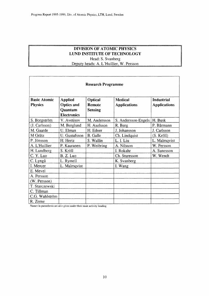
S. Borgstrom (J. Carlsson) M. Gaarde M Gratz P. Jonsson A. L'Huillier H. Lundberg C. Y. Luo c. Lynga I. Mercer E. Mevel A. Persson (W. Persson) T. Starczewski C. Tillman C.G. Wahlstrom R. Zerne
DIVISION OF ATOMIC PHYSICS LUND INSTITUTE OF TECHNOLOGY
Head: S. Svanberg Deputy heads: A. L'Huillier, W. Persson
Research Programme
Applied Optical Medical Optics and Remote Applications Quantum Sensing Electronics V. Avetisov M. Andersson S. Andersson-Engels M. Berglund H. Axelsson R. Berg U. Elman H. Edner J. Johansson U. Gustafsson B. Galle Ch. Lindquist H. Hertz S. Wallin L. I. Liu P. Kauranen P. Weibring A. Nilsson S. Kroll I. Rokahr B. Z. Luo Ch. Sturesson L. Rymell K. Svanberg L. Malmqvist I. Wang
Names m parenthesis are also gJVe.n under theu mam acttvtty headmg
10
Industrial Applications
H. Busk P. Barmann J. Carlsson cs. Kroll) L. Malmqvist W. Persson A. Sunesson W. Wendt

I Basic Atomic Physics
I Basic Atomic Physics
A central part of the research at the Division during the past 15 years, has been within basic atomic physics and fundamental aspects of laser-matter interactions. Initially, the main emphasis was on atomic hyperfine structures, isotope shifts and radiative lifetimes. These properties were investigated using time-resolved and high-resolution laser spectroscopy in the visible or near-UV spectral region, as well as various theoretical methods. With the establishment of the Lund High-Power Laser Facility in 1992, a change in the direction of our research took place. Since then, most of our efforts in basic atomic physics have been directed towards ultra-intense laser-matter interactions, while the continued work in laser spectroscopy has focused on investigations in the VUV and XUV spectral regions. In both these cases, the experimental work has extensively utilised the new resources provided by the establishment of the high-power laser facility.
Being a part of the Lund Laser Centre, the high-power laser facility became open in January I YY6 to European users through the European Community TMR Programme "Access to Large-Scale Facilities". This has contributed in a very positive and stimulating way to the international atmosphere in the basic atomic physics group.
The high-power laser facility consists of three major laser systems, all operating at 10Hz repetition rate but with very different pulse durations. The first, which is frequently referred to as the Lund VUV system, is a narrow-bandwidth, tunable system with pulse duration in the nanosecond range. It is designed to be used for pulsed laser spectroscopy in the UV and VUV spectral ranges. The second system is a mode-locked, Q-switched Nd:YAG laser, which gives pulses in the picosecond range. It is frequently used to pump a short-pulse dye laser, followed by a solid-state power amplifier. In combination with high-order harmonic generation in gas jets, this system provides tunable short-pulse radiation in the XUV spectral range. In this configuration, it is referred to as the Lund XUV system. Finally, the third and largest system is the femtosecond terawatt laser. This is based on chirped-pulse amplification in titanium-doped sapphire, and provides 110 fs pulses of terawatt power. During the past two years, we have begun a major upgrading of the laser facility, and the terawatt system in particular. Through this upgrade, which is financed by the Knut and Alice Wallenberg Foundation, we expect to achieve a substantial increase in peak power during the coming year.
Most of our work using the femtosecond terawatt laser and part of the work with the picosecond laser are described in Sections A-C of this chapter. This work has been devoted to the study of high-order harmonic generation in gases and plasmas, X-ray laser related investigations and to the generation and applications of hard X-rays from laserproduced plasmas. The femtosecond laser has also been used in a study of multiphoton excitation and fragmentation of c60 molecules, presented in Section D.
Time-resolved laser spectroscopy in the short-wavelength region (UV NUV /XUV) has been pursued through work with the nanosecond VUV laser system as well as with the picosecond XUV laser system. A number of investigations of atomic and molecular excited states have been performed. Most of these have been of astrophysical interest and
II

Progress Report 1995-1996, Div. of Atomic Physics, LTH, Lund, Sweden
some directly linked to observations made by the Hubble Space Telescope. This work is described in Section E.
The activities in theoretical atomic physics have also changed direction. The work devoted to theoretical investigations on oscillator strengths, radiative lifetimes, hyperfine splittings and isotope shifts, which are presented in Section F, has been reduced. The main theoretical effort has instead been focused on the dynamics of atoms in intense laser fields and the propagation of short optical pulses in various media (Section A).
Most of the work in the group has been presented at international conferences on atomic physics, astrophysics, spectroscopy, strong-field interactions and quantum electronics [ 1-43]. During the period, three MSc projects [44-46], three Licentiate theses [47-49] and four PhD theses [50-53] have been completed and successfully defended.
A High-order harmonic generation
Our research on the generation and application of high-order harmonics is currently the main research activity within the basic atomic physics group [Al-A20]. Several projects have been performed over the last two years, leading to many publications. We have two main goals. The first is to understand the fundamental properties of high-order harmonics and to optimise its performances in view of applications. The second is to investigate the possibility of producing pulses of extremely short duration (attosecond) using harmonics.
Al Fundamental studies of high-order harmonics and optimisation of the harmonic source
Harmonic generation in molecular gases Claire Lyngd, Anne L'Huillier and Claes-Goran Wahlstrom
In order to gain increased insight into the dependency on the type of nonlinear medium used in high-order harmonic generation, we have performed a systematic study in several systems: rare gases (Ar, Xe), diatomic molecules (N2, H2, 0 2, CO) and polyatomic molecules (SF6, NzO, COz, CH4, C3Hg) [A 19]. The harmonics were generated using the fundamental (800 nm) and the second harmonic (400 nm) of the femtosecond terawatt laser, at an intensity of about 2x ]()14 W/cm2. The atomic and molecular species were selected in order to span a very large range of ionisation energies, (static) polarisabilities, ionisation and dissociation probabilities, and, generally speaking, molecular structures, with and without a permanent dipole moment. Our results show that, in general, the harmonic spectra do not depend to any large degree on the generating gas. The harmonic "plateau" is wider for those with a high ionisation potential than for those with a low ionisation potential. To our surprise, the rare gases (argon and xenon) remain the most efficient species, although some of the molecules studied exhibit much higher (static) polarisabilities.
12

I Basic Atomic Physics
Harmonics as an intense source in the XUV range Carlo Altucci, Demetris Lappas, Anne L'Huillier, Corrado de Lisio, Tomas Starczewski and Claes-Goran Wahlstrom
Our aim is to obtain focused peak intensities in the XUV high enough to induce nonlinear processes in this short-wavelength region. For example, we hope, within the near future, to generate the 3rd harmonic of the 21st harmonic (33 eV) of the Ti:S laser, with Xe as generating medium for the 3rd harmonic, and Ar for the 21st. This project poses several interesting problems, regarding the optimisation of the generation of the 21" harmonic, the focusing of the generated short-wavelength radiation and the interaction of intense XUV radiation with free atoms. Experimentally, we have optimised the number of photons emitted at the 21st harmonic frequency. The study of molecular gases, described above, was part of this investigation. Other studies have been performed to optimise the excitation wavelength [14], the gas density [A16] and the geometrical conditions. We have also developed ways of focusing the harmonic radiation without temporally stretching the pulse, using multilayer mirrors. Focal spot sizes below 10 j.lm in diameter have been obtained. We have established a collaboration with the Department of Theoretical Physics (U. von Barth and R. van Leeuwen) in Lund, and the University of Wiirzburg (E. K. U. Gross), in Germany, the aim of which is to provide a good theoretical description of nonlinear processes in intense XUV fields, using time-dependent, densityfunctional theory.
High-order sum- and difference frequency mixing Mette Gaarde, Maciej Lewenstein*, Anne L'Huillier, Anders Persson and Claes-Goran Wahlstrom *Visiting scientist
The aim of this project, which includes both experimental and theoretical aspects, was to generate continuously tunable XUV light by mixing the radiation from an intense
790 nm
femtosecond Ti:S laser with the tunable radiation from an Optical Parametric Generator (OPG). The experiment was
4--lil-*"- performed in collaboration with the Service des Photons, Atomes et Molecules in Saclay, BS
DC To XUV· spectrometer France. The OPG of the High-Power Laser Diaph.
Fixed delay line
Variable delay line ......... t
475-600 nm
Fig Al. Experimental set-up for highorder sum- and difference-frequency mixing. The light from the femtosecond Ti:S laser is mixed in the {?as jet with the tunable output from an OPG.
Facility was moved to Saclay and the experiment was performed with the femtosecond laser system of the DRECAM facility in Saclay. Tunable radiation from 15 to 70 e V was produced through frequencymixing processes involving absorption or emission of one or two photons from the OPG. The tunability range covered was up to 70% of the total spectrum. The sum-frequency processes were found to be more efficient than the difference-frequency processes at low intensity and less efficient at high intensity [A 17]. To understand the latter result, we developed a theoretical
13

Progress Report 1995-1996, Div. of Atomic Physics, LTH, Lund, Sweden
approach to two-colour frequency mixing, combining the response of individual atoms to a two-colour radiation field, and propagation effects [A9]. We were able to satisfactorily reproduce the experimental results and explain the reduced efficiency of the sumfrequency processes at high intensity. Indeed, the phase variation, as a function of intensity, was found to be more rapid for sum-frequency processes than for harmonic generation or difference-frequency processes, leading, in the first case, to deterioration of phase-matching and reduced efficiency.
Harmonics as a source for interferometry experiments Carlo Altucci, Mette Gaarde, Anne L'Huillier, Claire Lynga, Claes-Goran Wahlstrom, Raoul Zerne, Marco Bellini* and T. W. Hiinsch* *Visiting scientists
An interesting application, utilising the coherence of the harmonic source, is XUV interferometry, e.g. for plasma diagnostics. Such interferometry experiments would be extremely simplified if one could split the laser beam into two beams, and generate independent but phase-locked harmonic sources, rather than splitting the generated harmonic beam, which requires complex XUV beam splitters. We have therefore performed an experimental study to investigate the locking of the phase of highorder harmonics, generated in gases in intense laser fields, with the phase of the fundamental field [A20]. The experiment consisted of separating a laser beam into two parallel beams focused at different locations under the nozzle of a gas jet, therefore Fig. A2. Far-field interference pattern producing two independent sources of created by overlapping, in space, two harmonic radiation, and of studying the beams of the 13th harmonic, generated interference pattern in the far field. We used independently at different places in a the picosecond mode-locked laser system xenon gas jet. focused in a jet of xenon atoms. We found that the generated harmonics, from the 7th to the 17th, were indeed phase-locked with the fundamental, with a good and robust fringe visibility. An example of a recorded far-field pattern is shown in Fig. A2. Detailed calculations distinguish between moderately high harmonic orders, generated at modest laser intensities, and harmonics of very high orders, generated at much higher laser intensities. The moderately high harmonic orders exhibit good temporal coherence properties and phase-locking with the fundamental field. The very high-order harmonics exhibit a significant frequency chirp, and phase-locking is predicted to be possible only if the two laser beams have exactly the same intensity.
14

I Basic Atomic Physics
A2 Towards attosecond pulse generation using high-order harmonics
Theoretical studies of attosecond pulse trains using high-order harmonics Anne L'Huillier, Mette Gaarde, Philippe Antoine* and Maciej Lewenstein* *Visitin~; scientists
The harmonic spectra show a characteristic behaviour, first a decrease in the efficiency for the low-order harmonics, followed by a broad plateau of almost constant conversion efficiency, ending with a sharp cut-off. After filtering away the first and last harmonics, the spectra look like a "comb" of peaks with constant amplitude, equally spaced in frequency. If the harmonics are all in phase at a given time (i.e. phase locked), the radiation emitted from the medium consists of a train of pulses separated by half the laser period, of extremely short duration, about 100 attoseconds (1 as = I0-18 s). There is a clear analogy here with mode-locked lasers, where axial modes oscillating in a laser cavity are locked in phase, leading to the production of trains of short pulses. This is an appealing idea, because it could provide a means of producing extremely short light pulses, much shorter than any ever produced before. The attosecond time scale is that of the electronic motion in atoms or molecules. It might then be possible to look at atomic processes in time, instead of looking at them in the frequency domain. In a theoretical study [AX], we analysed the problem of the production of attosecond pulse trains, both in the single-atom response, using the quantum-mechanical formulation of the quasi-classical interpretation [A3] and taking into account propagation in the nonlinear medium. We showed that, although the harmonics in the plateau region are not strictly speaking phase locked, the time-dependent single-atom emission consists of a train of ultrashort pulses, with two dominant pulses per half cycle, corresponding to the two main trajectories giving rise to harmonic emission. Under certain geometrical conditions, only one of these two contributions becomes phase matched, leading to macroscopic trains of ultrashort pulses, with one pulse per half cycle.
Generation of isolated attosecond pulses Carlo Altucci, Mette Gaarde, Anne L'Huillier, Claire Lynga, Ian Mercer, Claes-Goran Wahlstrom, Philippe Antoine*, Maciej Lewenstein* *Visiting scientists
The generation of high-order harmonic radiation is extremely sensitive to the degree of ellipticity of the pumping laser radiation [AlO, A4]. If one can generate a laser pulse that is linearly polarised during one optical cycle only and elliptically polarised otherwise, the harmonics, mostly generated by linearly polarised light, would be emitted during this interval, and thus a single, ultrashort (attosecond) optical pulse would be produced. In a theoretical study, we have investigated the possibility of modulating the polarisation of the fundamental field and hence to generate attosecond pulses, using two cross-polarised laser pulses, with slightly different frequencies. We found that the ratio between the two frequencies had to be as high as 6% and the laser pulses as short as 10 fs in order to obtain an isolated attosecond pulse. We therefore plan to investigate an alternative, but related approach, theoretically as well as experimentally. This approach consists of using two frequency-chirped, cross-polarised pulses, separated by a time delay which is shorter than the pulse duration. This should enable the generation of single attosecond pulses, without having to use extremely short incident laser pulses.
15

Progress Report 1995-1996, Div. of Atomic Physics, LTH, Lund, Sweden
Spatial mode control of high-order harmonics Anne L'Huillier, Ian Mercer, Eric Mevel, Claes-Goran Wahlstrom, Raoul Zerne and Philippe Antoine* *Visiting scientist
In an experimental study, supported by theoretical simulations, we have used the sensitivity of harmonic generation to the degree of polarisation of the laser beam in order to continuously control the harmonic emission in space [A 18]. This experiment can be viewed as the first step towards the generation of attosecond pulses. Instead of modulating the ellipticity of the fundamental field in time, we modulated it in space. This was achieved using a birefringent focusing lens, with the axis at 45 degrees to the laser polarisation. This lens produces two focii, one for horizontally polarised light and one for vertically polarised light. Each of these fields, close to the focus, has a phase which depends quadratically on the radial coordinate - i.e. on the distance from the optical axis. However, the magnitude of this phase is different for the two fields because of the different distances from their respective focii. This results in a phase difference, and consequently an ellipticity, which varies strongly in space. By introducing, in addition, a variable phase shift between the two components of the laser field with a Babinet compensator (effectively a birefringent material of variable thickness), before the focusing lens, the ellipticity distribution can be continuously controlled. We demonstrated that it was possible to control the angular emission of the harmonics and obtain beam profiles ranging from Gaussian to annular, or even to several rings.
16
Fig. AJ. Examples of two far-field spatial intensity distributions of the 13th harmonic. The different images represent different settings of the Babinet compensator, and illustrate the ability to control the spatial mode of high-order harmonics.

I Basic Atomic Physics
B X-ray laser related investigations Stig Borgstrom1, Ulf Litzen", Anders Persson, Tomas Starczewski, Jurgen Steingruber, Sune Svanberg and Claes-Goran Wahlstrom
1deceased 'Atomic Spectroscopy Division
We have continued to investigate different routes to compact, table-top, X-ray lasers. During the past two years, we have concentrated on a number of optical-field ionisation schemes in gas targets. We have also pursued a number of issues of more basic interest to X-ray laser research.
Laser direction
0'' 3s-2p
300 350 400 450 500 550 600 650 Wavelength (A)
Fig. Bl. Axial oxygen spectrum. Geometry of the nozzle as seen from below (inset).
Axial view Transverse view
3 tubes 3 tubes
320 340 360 380 400 320 340 360 380 400
Wavelength cAl
Fig. B2. Axial and transverse oxygen spectra at different plasma lengths.
In one study, we have investigated the possibility of obtaining population inversion with respect to the ground states in o· and 0 2+, in an effort to reproduce results published by Chichkov et al. [Phys. Rev. A 52, 1629 (1995)]. The terawatt laser was focused into a pulsed oxygen jet. The pulsed gas nozzle consisted of an arrangement of tubes, as illustrated in Fig. B 1. By moving the nozzle perpendicularly to the laser beam, the length of the laser-produced plasma could be varied.
Fig. B 1 shows a spectrum from 29 to 66 nm. The gain line candidate (02+ line at 37.4 nm) is clearly seen to dominate the spectrum. To investigate the question of gain, we observed the plasma simultaneously on-axis
and perpendicular to that direction (transversely) for different plasma lengths and gas pressures. The results show the same nonlinear enhancement with plasma length of the lines in both directions of observation (Fig. B2). This indicates that we are observing a volume effect rather than gain [26]. Another scheme, demonstrated by Lemoff etal. [Phys. Rev. Lett. 74, 1574 (1995)], where electrons and ions are produced by tunnelling ionisation and accelerated in a circularly polarised laser field was investigated using xenon as the target gas. Comparisons were made using linearly and circularly polarised laser radiation. However, with the laser-pulse characteristics available at the time of our experiment (pulse duration 150 fs, pulse energy onto the target 100 mJ), gain could not be observed [26].
17

Prugress Report 1995-1996, Div. of Atomic Physics, LTH, Lund, Sweden
Saturable absorber In order to gain a better understanding of the
.::::::1---+--1+--~-+--""'-.Ml importance of laser prepulses in optimizing
Vacuum chamber
the plasma production needed to obtain lasing, we systematically studied the influence of femtosecond laser prepulses on
"t':J'"1¥-c:::::::ttt::::~::t::t=~~:J;i~. the soft X-ray emission from solid target \\ M2 plasmas [B3].
-+-Variable optical The laser beam was directed, at normal M5 M4 '+' delay
Fig. B3. Experimental setup for prepulse dependence experiments.
incidence, onto a target consisting of polished aluminum or vanadium disks. The laser system itself produces prepulses which are difficult to eliminate completely. Under normal laser operation at the time of the experiments, the prepulse-to-main-pulse ratio was of the order of 1 x 10⁻⁴. The exact
number and relative amplitudes of the prepulses in the train depended on the actual laser adjustment, but the most common configuration comprised three distinguishable prepulses with energies of the same order of magnitude. These prepulses were separated by 11 ns in time (the round-trip time of the regenerative amplifier) and the last of the prepulses arrived 11 ns ahead of the main pulse. These prepulses could be eliminated by inserting a saturable-absorber cell in the laser beam after pulse compression. A small fraction of the beam could also be split off at the edge and allowed to travel a shorter distance through a system of smaller mirrors, thus arriving before the main pulse, creating a so-called artificial prepulse (Fig. B3). Comparisons were made between spectra obtained with an inherent laser prepulse, with a clean laser pulse without any prepulses, and with a clean laser pulse with an artificially added prepulse.
The X-ray emission was also studied as a function of the delay time between the prepulse and the main pulse. In order to increase the reproducibility, the emission from the laserproduced plasma was recorded spectrally integrated, but spatially resolved by use of a pinhole and a back-side illuminated, X-ray-sensitive CCD chip (Fig. B4).
HXXl
~ a)
8 ~
b)
.... -·--------------..6.
6 0 500 -~
.s ~ ~
5 10
Prepulse-to·main-pulse ratio Prepulse delay (ns)
Fig. B4. Spectrally and spatially integrated X -ray yields from laser-irradiated aluminum: a) versus the prepulse-to-main-pulse ratio (for different delays); b) versus the time delay between the prepulse and the main pulse (for two different pre pulse intensities).
1R

I Basic Atomic Physics
C Generation of hard X-rays from laser-produced plasmas Matthias Gratz, Gisbert HOlzer*, Laurence Kiernan, Ian Mercer, Arne Nykiinen**, Sune Svanberg, Carl Tillman and Claes-Goran Wahlstrom
* Visiting scientist, ** MSc student
Hard X-rays are now routinely produced in an experimental set-up based on a laserproduced plasma. We use pulses from the terawatt laser system, which are focused on a solid metal target and thereby create the plasma. Electrons are accelerated in the plasma and generate X-rays upon their interaction with the target material. Hard X-rays with energies up to the MeV region can be generated due to the ultrashort duration and the ultrahigh intensity of the laser pulses. Ablation from the plasma was observed as a function of various parameters [Cl ]. The experiments have been focused on both the characterisation of this unique source of hard X-rays and its various applications. The plasma emits radiation extending from the far infrared to the MeV -region. The hard X-rays are emitted · a time period of a few picoseconds. Studies were carried out in order to determine the X-ray source size, which was measured to be 30-60 pm in diameter (See Fig. Cl). Spectroscopic investigations have been performed using a variety of techniques: diffractive crystal spectrometers [C2], conventional single-photon counting detectors [C3], CCD arrays and K-edge absorption filters. The spectral -~ investigations have revealed both charac- ~ teristic radiation emission from the target ~ element (See Fig. C2 a) and a Bremsstrah- -~
c lung continuum extending up to the ] MeV-region (See Fig. C2 b).
·150 ·100 -50 0 50 100 !50 Position, !Jm
These properties allow for various applications. The small source size is favourable for X-ray imaging since it enables the recording of sharp images [C4], which are Fig. CJ. Pinhole image of the X-ray
source with an intensity profile. a)
~ ~ N
> ~13 "' :2 .-,
0 ~-+--r--+--~-+--~-+--~~ 52 56 60 64 6R
I~ ~
1Q·7
JO·'
. ' ' ' ...
JO·' +------+------+------~-------1 0 100 200 300 4()(
Photon energy, keY Photon energy, ke V
Fig. C2. Spectra from a tantalum target recorded with a germanium detector showing in particular (a) a Lorentzian fit in the energy region corresponding to characteristic X -ray emission and (b) the continuum radiation.
19

Progress Report 1995-1996, Div. of Atomic Physics, LTH, Lund, Sweden
Fig. C3. Differential image of two rat stomachs administered with different contrast agents. Cerium (left) appears dark because of its strong non-differential absorption, while gadolinium (right) appears light due to the differential absorption.
necessary in, for example, mammography. Differential imaging in the spectral domain
was also shown to be a possible application of this X-ray source [C5,C6]. Two images were recorded using two different target elements, with their characteristic emission on either side of the K-absorption edge in a chosen contrast agent, i.e. gadolinium and tantalum targets in combination with a gadolinium contrast agent. A third image was calculated by division, pixel by pixel, of the two recorded images, showing the occurrence of contrast agent (See Fig. C3). The ultrashort X-ray pulses give the opportunity to use time-gated imaging as a method to improve the image quality for a constant absorbed dose, by means of scatter reduction. An ultrafast detector, capable of separating the scattered from the ballistic (non-scattered) photons, in combination with this X-ray source may allow for a dose
reduction factor of up to 10, depending on tissue thickness and photon energy. Preliminary investigations were done, using one- dimensional, time-resolved imaging with an X-ray streak camera. The image quality was shown to increase for a constant absorbed dose with the use of this technique (See Fig. C4) [C7]. The biological effect of ultra-intense X-rays must be investigated before this kind of radiation can used for medical applications in vivo_ A preliminary study was made of the survival of cell cultures being exposed to this ultra-intense X-ray source [C8]. There were no indications that the degree of biological damage was larger compared to the case where standard tubes were used_
. nogate . ~A.~_,.W< ,. .,. ,,.,
•' ., :-··~~CM'<.i(.," ' . ' .
lead object shadow Fig. C4. Time-resolved line imaging of a lead object through 15 em of water, with corre.1ponding object images for various gating times. The contrast is improved by a factor of about 6, compared to imaging without gating.
20

I Basic Atomic Physics
D Clusters in intense laser fields Sfefan Hunsche*, Anne L'Huillier, Tomas Starczewski, Sune Svanberg and Claes-Goran Wahlstrom
*Visiting scientist
In addition to the main topics described above, as part of a joint project within the EU programme Access to Large Scale Facilities, we have studied femtosecond multiphotonmultiplasmon excitation and fragmentation of C6o molecules [Dl], using a newly constructed ion-time-of-flight spectrometer. Our results led to the conclusion that ionization and fragmentation of c60 by ultrashort high-intensity laser pulses occurs predominantly via plasmon excitation.
Laser: Ti-Sapphire CPA 1 0 Hz, 200 mJ, 100 fs
A=790 nm I 395 nm (SHG)
Fig. Dl: Experimental setup for the time-of-flight mass spectroscopy of ionised C6Q and charged fragments.
104 hv :::3.14 eV ac Hf
D c. "' d c,:~}.+
; 103 0 c • """" w'
~ '0 ] 102
1rf >-c N-f ..s
101 101
1<fl 20 50 100 50 100
Laser Intensity (arb.u.)
Fig. D2: Intensity-dependent ion yield for C60 and its fragments. Left: frequency-doubled excitation; Right: fundamental laser frequency. Straight lines: Fits of the c;o and c;; data according to a power
law.
21

Progress Report 1995-1996, Div. of Atomic Physics, LTH, Lund, Sweden
E Time-resolved laser spectroscopy in the short-wavelength spectral region
Uldis Berzinsh*, Luo Caiyan, Hans Lundberg, Sune Svanberg and Raoul Zerne *Visiting scientist
Radiative properties of free atoms depend sensitively on the atomic wavefunctions and thus experimentally determined natural lifetimes and oscillator strengths are useful for testing theoretical calculations. Further, astrophysical determinations of stellar abundances rely on the availability of accurate atomic radiative data. With the Hubble Space Telescope (HST) the VUV spectral region has become accessible at high resolution and the need for matching atomic data has strongly increased. The Lund laser spectroscopy group has during the last few years focused its activities on studying some problems, where the experimental difficulties earlier have prevented a solution. VUV transitions must be excited with short-wavelength radiation available at the High Power Laser Facility, and the time resolution demands must be met by using fast excitation and detection schemes. Further, many of the elements of interest evaporate as molecules and must be dissociated in a discharge, in a laser-produced plasma or by multi-photon dissociation. Frequently, ions rather than atoms are of interest. An overview of useful experimental techniques and recent Lund results are presented in Ref. [E 1].
The non-metallic p3 ground configuration elements P and Bi and the p4 configuration elements S and Te were studied during the last two years. Although Bi largely evaporates as molecules a sufficient fraction of free atoms were obtained in an atomic beam to allow the measurement of lifetimes for 10 states and the evaluation of revised oscillator strengths of 28 bismuth lines [E2]. Stimulated Raman shifting was used to reach the short-
Ablation Laser
VUV Laser System
Computer Transient recorder
SIDE VIEW
VUV monochromator
Fig. El. Experimental set-up for time-resolved laser spectroscopy on atoms in a laserproduced plasma.
22

:; ~ 1.0
.!;;:· ~ 0.8
.s 0.6
OA
02
Recombmationlight
6
Time().ts)
LIF
10 12
Fig. E2. Recording of recombination light and laser-induced fluorescence from a sulphur plasma. The detection wavelength is 126 nm corresponding to fluorescence from the 7s 3 S 1 state.
I Basic Atomic Physics
wavelength transitions. In our recent Te study, which has now been published [E3], differential heating of a sealed-off resonance cell was employed to obtain a useful atomic fraction. Stepwise excitation was used to reach high-lying states.
Free sulphur or phosphorus atoms were obtained by thermal evaporation of the elements and subsequent photo-dissociation of molecules in the focus of a spectroscopic laser beam, tuned to two-photon resonances of low-lying states [E4,E5]. Alternatively, free atoms were created by forming a laserproduced plasma by using a pulsed laser beam focused on the surface of PbS powder in a vibrating container. Free atoms in the
recombining plasma plume were then excited by VUV radiation produced by four-wave mixing of dye laser beams in krypton gas [E6]. The experimental set-up and the recombination light and the laser-induced fluorescence spike at a delay of 10 ~s after the ablation are shown in Figs E 1 and E2. Decay curves with increasingly longer decay constant for more highly excited sulphur states are shown in Fig. E3. The experimental data were compared with theoretical calculations including a new Superstructure calculation by E. Biemont, Universite de Liege. Improved absorption oscillator strengths yielded the new solar abundance value of 5.49 for P [E5].
Laser pulse -------------------
0 50 100 150 200
Time (ns) Fig. E3. Time-resolved decay curves form high-/yin[? sulphur states. The shape of the exciting laser pulse is also included.
Recently performed lifetime measurements for excited states of Pt II and Pd II allowing the interpretation of the HST spectra recorded for the chemically peculiar star X Lupii have now been published jointly with NASA researchers [E7 ,ER].
Stepwise or VUV laser excitation was also used to reach long Rydberg series in Yb, for which natural lifetimes and Lande g1 factors were measured [E9,E10]. The admixture of doubly excited states leads to drastic effects allowing a detailed study of such configuration interaction, which can conveniently be described by Multi-Channel Quantum-Defect Theory (MQDT). Similar studies for Pb are in progress.
At the end of this section we would also like to announce the publication of some work discussed in our previous Progress Report [Ell-E14].
23

Progress Report 1995-1996, Div. of Atomic Physics, LTH, Lund, Sweden
F Theoretical atomic physics Jorgen Carlsson, Per Jonsson and Lennart Sturesson
Most of the work of the atomic theory group has been carried out in close collaboration with groups in the USA and Europe. Joint projects have also been carried out with the Department of Theoretical Chemisty at Lund University.
A new transition probability program has been developed within the multiconfiguration Hartree-Fock (MCHF) formalism [Fl]. This program, allowing for the use of nonorthogonal orbitals, has proven to be capable of providing transition probabilities in light atoms with an estimated uncertainty of a few parts in a thousand [F2]. Using the new program, the transition probability has been calculated for the sodium resonance transition [F3]. The obtained value is in excellent agreement with the most recent experimental values, resolving a longstanding discrepancy between theory and experiment for this transition.
The new technique for calculating transition probabilities has been generalised to the fully relativistic case, and multiconfiguration Dirac-Fock calculations (MCDF) are in progress for the resonance lines in Cu, Ag and Au [F4]. In connection with these calculations, a new configuration generator has been developed for the MCDF program [F5]. The MCDF method has also been applied to hyperfine structure calculations with good results [F6].
Together with the theory group at the Free University of Brussels, a program has been developed for computing very weak hyperfine induced transitions. These transitions are of potential interest for electron density diagnostics in plasmas of very low density. The program has been applied to transitions in two- and four-electron systems [F7].
The isotope shift project, started in 1994, has been continued, and very accurate shifts have been calculated for transitions in light atoms [F8, F9]. Isotope shifts are very sensitive to electron correlation effects and represent a true challenge for any computational method.
To promote interdisciplinary studies, the atomic theory group arranged a two-day international workshop on large-scale atomic calculations; applications to astrophysics and nuclear structure. Among our educational activities the book on Computational Atomic Structure [FlO], which has recently been completed, should be mentioned.
24

I Basic Atomic Physics
References
[I] P. Jl)nsson, A. Ynnerman, C. Froese Fischer, M. Godefroid and J. Olsen. "Accurate MCHF calculations of oscillator strengths and hyperfine structures in Na !", International Workshop on Large-Scale Atomic Calulations; Applications to Astrophysics and Nuclear Structure, (Lund, 18-19 January, 1995).
[2] A. Aboussaid, M. Godefroid, T. Brage and P. Jonsson, "Hyperfine quenching of Js22s 2p 3po in C l/1 and N fV ". 5th EPS Conference on Atomic <md Molecular Physics, (Edinburgh 3-7 April, 1995).
[3] J. Carlsson, P. Jonsson, C. Froese Fischer <md M. Godefroid, "Systematic MCHF calculations of isotope sh1jts in liJ?ht atoms", International Workshop on Large-Scale Atomic Calulations: Applications to Astrophysics and Nuclear Structure, (Lund, 18-19 January, 1995)
[4] S. Kohlweyer, G. D. Tsakiris, C.-G. Wahlstrom, C. Tillrmm and I. Mercer, "Erzeugunf? von Harmonischen Hochintensiver Laserstrahlung an FestkOrperoberfiiichen". Deutsche Physikalische Gesellschaft, Innsbruck, Austria, February 27 - March 3, 1995.
[5] A. Aboussaid, M. Godefroid, T. Brage, C. Froese Fischer and P. Jonsson, "MCHF calculations of hyperfine-induced transitions in the Breit-Pauli approximation", European Research Conference on Relativistic Effects in Heavy-Element Chemistry and Physics - Relativistic Quantum theory of Many-Electron Systems, (Castelvecchio Pascoli 30, March- 4 April 1995).
[o] E. Mevel, J. Larsson, M. B. Gaarde, R. Zerne, A. L'Huillier, C.-G. Wahlstrom <md S. Sv<mberg, "Time-resolved laser spectroscopy on helium in the XUV spectral region", Fifth Europe<m Conference on Atomic and Molecular Physics (ECAMP), Edinburgh, 3-7 April 1995.
[7] P. Erman, A. Karawajczyk, E. Rachlew-Killlne, E. Mevel, R. Zerne, A. L'Huillier and C.-G. Wahlstrom, "A new valence state in NO studied by high-resolution higher-order harmonics laser speuroscopy", Fifth European Conference on Atomic and Molecular Physics (ECAMP). Edinburgh, U.K. Apri13-7, 1995.
[8] C.-G. Wahlstrom, "Harmonic generation and its experimental limitations", Fifth Europe<m Conference on Atomic and Molecular Physics (ECAMP), Edinburgh, U.K. April 3-7, 1995 (Invited).
[9] Luo Caiyan, U. Berzinsh, J. Larsson, M.B. Gaarde, R. Zerne, and S. Svanberg, "Time-resolved VUV laser spectroscopy on free atoms", Fifth European Conference on Atomic and Molecular Physics (ECAMP), Edinburgh, U.K. April3-7, 1995.
[10] M. Gratz, C. Tillman, I. Mercer, S. Sv<mberg, "X-ray genertion for medical applications from laser-produced plasmas", Conference on Laser Ablation. Strasbourg, May 22 -26, 1995.
[II] U. Berzinsh, M. B. Gaarde, J. Lm-sson, A. L'Huillier H. Lundberg, Luo Caiyan, E. Mevel, S. Svanberg, C.-G. Wahlstrom and R. Zerne, "Time-resolved laser spectroscopy on atoms and ions in the VUV and XUV spectral region", 12th International Conference on Laser Spectroscopy, Capri, Italy, June 11-16, 1995.
[12] J. Carlsson, P. Jonsson, C. Froese Fischer <md M. Godefroid, "LarJ?e-scale MCHF calculations of isotope shifts and hyperfine structures in light atoms", Fifth European Conference on Atomic <md Molecular Physics (ECAMP), Edinburgh, U.K. April3-7, 1995.
[13] C.-G. Wahlstrom, "Generation and applications of high-order harmonic radiation", Proceedings of X-Ray Lasers and Applications, SPIE '95, San Diego, July 1995 (Invited).
[14] C.-G. Wahlstrom, C. Altucci, S. Borgstrom, B. Carre, M. B. Gaarde, J. Lm-sson, A. L'Huillier, C. Lyng1'l, E. Mevel, A. Persson, T. Swczewski, S. Svanberg, and R. Zerne, "Optimisation and applications of high-order harmonic generation", Proceedings of NATO Advanced Research Workshop on Super-Intense Laser Atom Physics (SILAP IV), Moscow, August 1995. (Kluwer Publishing Company, eds M. Fedorov and H. M. Muller.) (Invited).
[15] M.B. Gaarde, A. Persson, C.-G. Wahlstrom, A. L'Huillier, S. Svanberg, "Wavelength dependent studies of harmonic generation processes using an optical parametric amplifier", Generation <md Application of Ultrashort X-Ray Pulses II. Pisa, Italy 20-23 September, 1995.
[ 16] M. Gr~itz, C. Tillrmm, C.-G. Wahlstrom, S. Svanberg. G. Holzer, E. Forster, "Characterization of hard X-ray emission from /user-produced plasmas by crystal spectroscopy", Generation <md Application of Ultrashort X-Ray Pulses II. Pisa, Italy 20-23 September, 1995.
25

Progress Report 1995-1996, Div. of Atomic Physics, LTH, Lund, Sweden
[17] S. Kohlweyer, G.D. Tsakiris, C.-G. Wahlstrom, C. Tillman, I. Mercer, "Harmonic generation from solid-vacuum interface irradiated at high laser intensities", Generation and Application of Ultrashort X-Ray Pulses II. Pisa. Italy 20-23 September, 1995.
[IR] Ph. Antoine, B. Carre, P. Salieres, A. L'Huillier, "Harmonic emission with elliptical polarisation", Generation and Application of Ultrashort X-Ray Pulses II. Pisa, Italy 20-23 September, 1995.
[19] P. Salieres, A. L'Huillier, M. Lewenstein, "Coherence control of high-order harmonics", Generation :md Application of Ultrashort X-Ray Pulses II. Pisa, Italy 20-23 September, 1995.
[20] J. Steingruber. S. Borgstrom, T. Starczewski, U. Litzen, "?repulse dependence of soft X-ray spectra from plasmas created by femtosecond IR laser pulses on solids", Generation and Application of Ultrashort X-Ray Pulses II. Pisa, Italy 20-23 September, 1995.
[21] C. Tillman, I. Mercer, M. Gratz, C.-G. Wahlstrom, S. Svanberg, K. Herrlin, "Applied imaf?inR and spectral measurements using differential absorption of laser generated X-rays ". Generation :md Application of Ultrashort X-Ray Pulses II. Pi sa, Italy 20-23 September, 1995 (Invited).
[22] C. Tilhmm, M. Gratz, I. Mercer. C.-G. Wahlstrom, A. Persson, S. Svanberg, B. Erlandsson, K. Herrlin, G. Svahn, C. Olsson, H. Petterson, "Hard X-rays from laser-produced plasmas -(;eneration and applications", Rontgen Centennial, Wiirzburg, Germany, 23-27 October 1995.
[23] S. Svanberg, M. Gratz, K. Herrlin, I. Mercer, A. Persson, C. Tillman, C.-G. Wahlstr()m, "Application of ultrashort X-ray pulses to bilogical and medical imaRing", IX International Symposium on Ultrafast Processes in Spectroscopy. Trieste, Italy, October 30-November 3, 1995. (Invited.) 0. Svelto, S. De Silvresti :md G. Denardo, eds., Ultrafast Processes in Spectroscopy (Plenum, New York, 1996).
[24] V. Sirutkaitis, A. Persson, E. Gaizauskas, A. Piskarskas and S. Sv:mberg, "Efficient frequency doubling of the femtosecond terawatt power Ti:sapphire laser pulses", IX International Symposium on Ultrafast Processes in Spectroscopy. Trieste, Italy, October 30-November 3, 1995. 0. Svelto, S. De Silvresti and G. Denardo, eds., Ultrafast Processes in Spectroscopy (Plenwn, New York, 1996).
[25] M. Gratz, A. Pifferi, C. Tillman, C.-G. Wahlstrom, S. Swmberg, "Propagation of laser-produced short X-ray pulses throuRh scatterinR media: Application to X-ray imaging with temporal gating", Proceedings of 5th International Conference on X-Ray Lasers, Lund, Sweden, June 10-14, 1996. (lOP Publishing, 1996).
[26] T. Starczewski, U. Litzen, J. Steingruber, S. Borgstrom, E.E. Fill, C.-G. Wahlstrom and S. Svanberg, "Spectroscopic studies in the soft X-ray region using a femtosecond laser", Proceedings of 5th International Conference on X-Ray Lasers, Lund, Sweden, June 10-14, 1996. (lOP Publishing, 1996).
[27] J. Steingruber, S. Borgstrom, T. Starczewski, U. Litzen, "?repulse dependence of soft X-ray spectra from plasmas created by femtosecond IR laser pulses on solids", Proceedings of 5th International Conference on X-Ray Lasers, Lund, Sweden, June 10-14, 1996. (lOP Publishing, 1996).
[2~] M. Griitz, A. Pifferi, C. Tillman, C.-G. Wahlstom, S. Svanberg, "Propagation of laser-produced short X-ray pulses through scal/ering media: Application to scatter-reduced medical imaging". EQEC, Hmnburg Gennany, September 1996.
[29] A. L'Huillier, Generation and application of high-order harmonics", EQEC, Hrunburg, Germ:my September 1996. (Invited.)
[30] C. de Lisio, C. Altucci, C.-G. Wahlstrom, "High-order harmonic radiation as a high-intensity XUV source for non-resonant multiphoton processes", 82nd National Conference of the Itali<m Physical Society, Verona Italy, 23-28 September 1996.
[31] C.-G. Wahlstrom, M.B. Gaarde, A. L'Huillier, C. Lyngi't, I. Mercer, E. Mevel, R. Zerne, Ph. Antoine, M. Bellini, T.W. Hansch, "Manipulations of high-order harmonics", 7th International Conference on Multiphoton Processes, Gannisch-Partenkirchen, Genmmy, 30 September - 4 October 1996 (Invited.) P. Lmnbropoulos and H. Walther, eds., Multiphoton Processes 1996 (lOP Publishing, Bristol 1996).
[32] C. Lyngi't, A. L'Huillier, C.-G. Wahlstrom, "High-order harmonic generation in molecular gases", 7th International Conference on Multiphoton Processes, Gannisch-Partenkirchen, ICOMP VII, Germany, 30 September- 4 October 1996.
[33] S. Hunsche, T. Starczewski, A. L'Huillier, A. Persson, C.-G. Wahlstrom, B. van Linden van den Heuvell, S. Svanberg, "Femtosecond Multiphoton-multiplasmon excitation in C6o·"· 7th
26

I Basic Atomic Physics
International Conference on Multiphoton Processes, Gannisch-Partenkirchen, ICOMP VII, Germany, 30 September- 4 October 1996.
[34] A. L'Huillier, "Generation and applications of high-order harmonics: an alternative coherent XUV source", Proceedings of the Fifth International Conference on X-Ray Lasers, Lund, June 10-14, 1996, (lOP Publishing, 1996), (Invited).
[35] P. Antoine, B. Carre, M. Lewenstein, A. L'Huillier, B. Piraux and M. Gajda, "Harmonic emission with elliptical polarization". Proceedings of NATO Advanced Research Workshop on SuperIntense Laser Atom Physics (SILAP IV), Moscow, August 1995. (Kiuwer Publishing Comp<my, eds M. Fedorov and H. M. Muller).
[36] Ph. Antoine, M. Gaarde, P. Salieres, B. Carre, A. L'Huillier and M. Lewenstein, "On the phase of atomic polarization in harmonic generation processes". Proceedings of the 7lli International Conference on Multi photon Processes, Gannisch-Partenkirchen, Gennany, September 1996. P. Lrunbropoulos <md H. Walther, eds., Multiphoton Processes 1996 (lOP Publishing, Bristol 1996).
[37] Ph. Antoine, M. Gaarde, A. L'Huillier, D. Milosevic, M. Lewenstein <md P. Salieres "Attosecond pulse generated by macroscopic media", 7"' International Conference on Multiphoton Processes, Gannisch-Partenkirchen, Germany, September 1996.
[38] Ph. Antoine, A. L'Huillier and M. Lewenstein "Attosecond pulse trains using high-order harmonics", 7"' International Conference on Multiphoton Processes, G<mnisch-Partenkirchen, Gennany. September 1996.
[39] Ph. Antoine, B. ClUTe, A. L'Huillier and M. Lewenstein "Polarization of high order harmonics". 7"' International Conference on Multiphoton Processes, Gannisch-Partenkirchen, Gennany, September 1996.
[40] Ph. Antoine, P. Salieres, M. Gaarde, A. L'Huillier and M. Lewenstein "Towards time resolved attosecond spectroscopy". 7"' International Conference on Multi photon Processes, GannischPartenkirchen, Germany, September 1996.
[41] M. Gaarde, A. L'Huillier and M. Lewenstein "Theory of high-order frequency mixing in a strong two-color laser field". 7lli International Conference on Multi photon Processes, GannischPartenkirchen, Germany, September 1996.
[42] A.V. Baklanov, V.P. Maltsev, L. Karlsson, B. Lindgren, U. Sassenberg and A. Persson "Femtosecond VUV REMPJ technique", Femtochemistry: The Lausarme Conference, Sept. 4-8. 1995, Lausarme, Switzerl<md.
[43] A.V. Bakhmov, V.P. Maltsev, L. Karlsson, B. Lindgren, U. Sassenberg and A. Persson "Femtosecond ( 1 +2 )VUV REMPI and nanosecond (1 +I) REM PI applied to the study of allyl iodide photodissociation". International Symposium on Modem Problems of Laser Physics. Novosibirsk, Russia, August 28- September 2, 1995.
[44] C. Andersson, "Differential absorption using hard X-rays from a laser-produced plasma", MSc thesis, Lund Reports on Atomic Physics, LRAP-174 Lund (1995).
[45] C. Lindheimer, "Optimising hard X-ray generation from laser-produced plasmas", MSc thesis, Lund Reports on Atomic Physics, LRAP-162 Lund (1995).
[46] A. Goransson, "A new method for spectral measurement of X -rays from a laser-produced plasma using differential absorption", MSc thesis, Lund Reports on Atomic Physics, LRAP-175 Lund (1995).
[47] M. B. Gaarde, "High-order sum- and difference frequency mixing in a strong two color laser field", Lie. thesis, Lund Reports on Atomic Physics, LRAP-205, Lund (1996).
[48] M. Gratz, "Hard X-rays from a laser-produced plasma: Source characterization and applications", Lie. thesis, Lund Reports on Atomic Physics, LRAP-208, Lund (1996).
[49] C. Tillman, "Characterisation and applications of hard X-rays from a laser-produced plasma source", Lie. thesis, Lund Reports on Atomic Physics, LRAP-188, Lund (1995).
[50] P. J(insson, "Large-scale atomic calculations using variational methods", PhD thesis. Lund Reports on Atomic Physics, LRAP-167, Lund (1995).
[51] R. Zeme, "Time-resolved studies of atoms and ions in the short-wavelength region", PhD thesis, Lund Reports on Atomic Physics, LRAP-195, Lund (1996).
[52] Luo Caiyan, "Time-resolved and frequency-resolved laser spectroscopy in free and perturbed atoms", PhD thesis, Lund Reports on Atomic Physics. LRAP-194, Lund (1996).
27

Progress Report 1995-1996, Div. of Atomic Physics, LTH, Lund, Sweden
[53] C. Tillman, "Development and characterisation of a laser-based hard x-ray source", PhD thesis, Lund Reports on Atomic Physics, LRAP-204, Lund (1996).
[54] S. Svanberg, A. L'Huillier, C.-G. Wahlstrom, "Atomic physics usinR short-wave/enf{th coherent radiation". Nuclem· Instruments and Methods, to appear (invited).
[55] S. Svanberg, "HiRh-power lasers and their applications", Adv. Qmmt. Chern., to appear (invited). [56] C. Altucci, C. Beneduce, R. Bruzzese, C. de Lisio, G.S. Sorrentino, T. Starczewski, F. Vigilante,
"Characterization of pulsed ,~?as sources for intense laser field - atom interaction experiments". J. Phys. D. 29, 6ti, (1996).
[57] B. Krassig, J. Hansen, W. Persson, V. Sclunidt, "High-/ satellite states in the threshold photoelectron spectrum of argon", J. Phys. B29, L449 (1996).
[AI] Ph. Balcou, P. Salieres, K. S. Budil, T. Ditmire, M. D. Perry and A. L'Huillier, "Hi,l?h-order harmonic f{eneration in rare gases: a new source in photoionization spectroscopy", Z. Phys. D 34, 107 (1995).
[A2] P. Salieres, A. L'Huillier and M. Lewenstein, "Coherence control of high-order harmonics", Phys. Rev. Lett. 74, 3776 (1995).
[A3] M. Lewenstein, P. Salieres <md A. L'Huillier, "Phase of the atomic polarisation in high-order harmonic generation", Phys. Rev. A 52,4747 (1995).
[A4] P. Antoine, A. L'Huillier, M. Lewenstein, P. Salieres and B. Carre, "Theory of hi,l?h-order harmonic generation by elliptically polarized laser fields", Phys. Rev. A 53, 1725 (1996).
[AS] Ph. Balcou, A. L'Huillier <md D. Esc<mde, "High-order harmonic ,l?eneration processes in classical and quantum anharmonic oscillators". Phys. Rev. A 53, 3456 (1996).
[A6] Ph. Balcou, P. Salieres, A. L'Huillier and M. Lewenstein, "Generalized phase-matching conditions for high harmonics: the role of field-gradient forces", Phys. Rev. A, in press (1997).
[A7] D. Joyeux, P. Jaegle and A. L'Huillier "Doing coherent optics with soft X-ray sources" in "Current trends in Optics" vol. 3, A. Consortini ed., Academic Press, 1996, p 371.
[A8] P. Antoine, A. L'Huillier and M. Lewenstein, "Attosecond pulse trains using hi,l?h-order harmonics", Phys. Rev. Lett. 77, 1234 (1996).
[A9] M. B. Gaarde, A. L'Huillier and M. Lewenstein, "Theory of high-order sum- and difference wave-mixing processes", Phys. Rev. A 54,4236 (1996).
[AIO] P. Antoine, B. Carre, A. L'Huillier and M. Lewenstein, "Polarization of high-order harmonics", Phys. Rev. A, in press ( 1997).
[All] P. Salieres. T. Ditmire. M. D. Perry, A. L'Huillier and M. Lewenstein, "Angular distributions of hiRh-order harmonics generated by a femtosecond laser", J. Phys. B 29,4771 (1996).
[Al2] C.-G. Wahlstrom, S. Borgstrom J. Larsson and S.-G. Pettersson, "High-order harmonic ,l?eneration in laser-produced ions using a near infrared laser", Phys. Rev. A 51,585 (1995).
[Al3] J. Larsson. E. Mevel, R. Zeme, A. L'Huillier, C.-G. Wahlstrom and S. Svanberg, "Two-color time-resolved spectroscopy of helium using high-order harmonics", J. Phys. B 28, L53 (1995).
[A14] A. L'Huillier, T. Auguste, Ph. Balcou, B. Carre, P. Monot, P. Salieres, C. Altucci, M.B. Gaarde, J. Larsson, E. Mevel, T. Starczewski, S. Svanberg, C.-G. Wahlstrom R. Zeme, K.S. Budil, T. Ditmire <md M.D. Perry, "High-order harmonics: a coherent source in the XUV range", Journal of Nonlinear Optical Physics and Materials, 4, 647 (1995).
[Al5] S. Kohlweyer, G.D. Tsakiris, C.-G. Wahlstrom, C. Tillman, I. Mercer, "Harmonic generation from solid - vacuum interface irradiated at hi,l?h laser intensities", Optics Comm. 117, 431 (1995).
[A16] C. Altucci, T. Starczewski, E. Mevel, B. Carre, A. L'Huillier and C.-G. Wahlstrom, "The influence of atomic density in high-order harmonic generation", J. Opt. Soc. Am. B 13, 148 (1996).
[Al7] M. B. Gaarde, P. Antoine, A. Persson, B. Carre, A. L'Huillier, C.-G. Wahlstrom, "High-order tunable sum- and difference frequency mixing in the XUV region", J. Phys. B 29, L163 (1996).
[A18] I. Mercer, E. Mevel, R. Zeme, A. L'Huillier, P. Antoine, C.-G. Wahlstrom, "Spatial mode control of high-order harmonics", Phys. Rev. Lett. 77, 1731 (1996).
[Al9] C. Lyngtl, A. L'Huillier, C.-G. Wahlstr(im, "High-order harmonic generation in molecular gases", J. Phys. B. At. Mol. Opt. Phys. 29, 3293 (1996).
2!1
I Basic Atomic Physics
[A20] R. Zerne, C. Altucci, M. Bellini, M.B. Gaarde, T.W. Hansch, A. L'Huillier, C. Lyng!t ~md C.-G. Wahlstrom, "Are high-order harmonics phase-locked with the driving field?", submitted to Phys. Rev. Lett.
[BI] E. Fill, S. Borgstrom, J. Larsson, T. Starczewski, S. Svanberg and C.-G. Wahlstr(im, "XUV spectra of optical-field ionized plasmas", Phys. Rev. E 51,6016 (1995).
[B2] S. Borgstrom, E. Fill, T. Starczewski, J. Steingruber, S. Svanberg and C.-G. Wahlstrom, "Timeresolved X-ray spectroscopy of optical-field ionized plasmas", Laser and Particle Bemns, 13, 459 (1995).
[B3] J. Steingruber, S. Borgstrom, T. Starczewski ~md U. Litzen, "?repulse dependence of X-ray emission from plasmas created by IR femtosecond laser pulses on solids", J. Phys. B 29, L75 (1996).
[Cl] M. Gratz, C. Tillman. I. Mercer and S. Sv~mberg, "X-ray generation for medical applications from a laser-produced plasma", Appl. Surface Sci. 96-98,443 ( 1996).
[C2] G. Holzer, E. Forster, M. Gratz, C. Tillman and S. Sv~mberg, "X-ray crystal spectroscopy of subpicosecond laser-produced plasmas beyond 50 keV", J. X-ray Sci. Tech. To appear.
[C3] C. Tillman, S. Joh~msson, M. Griitz, B. Erlandsson, S. Mattson and S. Svanberg, "Spectroscopy of laser-produced plasmas in the photon energy regime above 15 keV", submitted to Nucl. lnstr. Meth.
[C4] C. Tillman, A. Persson, C.-G. Wahlstrom, S. Svanberg and K. Herrlin, "Imaging using hard Xrays from a laser-produced plasma", Appl. Phys. B 61, 333 (1995).
[C5] C. Tillman, I. Mercer, S. Svanberg, and K. Herrlin, "Elemental biological imaging by differential absorption using a laser-produced x-ray source", J. Opt. Soc. Am. B 13 209 (1996).
[C6] K. Herrlin, C. Tillman, M. Gratz, C. Olsson, H. Pettersson, G. Svahn, C.-G. Wahlstrom and S. Sv~mberg, "Contrast enhanced radiography by differential absorption, using a laser-produced Xray source". Investigative Radiography, To appear (May 1997).
[C7] M. Gratz, A. Pifferi, C.-G. Wahlstrom, S. Svanberg, "Time-gated viewing in radiology: Theoretical and experimental studies", IEEE Journal of Selected Topics, to appear.
[C8] C. Tillman, G. Grafstrom, A.-C. Jonsson, I. Mercer, S. Svanberg, S.-E. Strlmd, S. Mattsson, ~md B.-A. Jonsson, "Survival of V79-CH-cells studied in vitro after extremely high dose rate irradiation by X-rays from a laser-produced plasma", Submitted to Phys. Med. Bioi.
[Dl] S. Hunsche, T. Starczewski, A. L'Huillier, A. Persson, C.-G. Wahlstrom, B. van Linden van den Heuvell, S. Sv~mberg, "Ionization and fragmentation of C60 via multiphoton-multiplasmon excitation", Phys. Rev. Lett. 77, 1966 (1996).
[EI] U. Berzinsh, S. Svanberg, "Atomic radiative lifetimes measured by pulsed laser spectroscopy in the UV!VUV spectral region", Adv. Qmmt. Chern. To appear.
[E2] Luo Caiyan, U. Berzinsh, R. Zerne and S. Svanberg, "Determination of radiative lifetimes of neutral bismuth by time-resolved UV!VUV laser spectroscopy", Phys. Rev. A 52, 1936 (1995).
[E3] R. Zerne, U. Berzinsh and S. Svanberg, Radiative lifetimes in neutral tellurium measured by timeresolved laser spectroscopy, Z. Phys. 032, 31 (1994).
[E4] R. Zerne, Luo Caiyan, U. Berzinsh and S. Svanberg, "Oscillator strengths of sulphur 3s23p34s 3s - 3s23p34p 3p transitions measured by time resolved two-photon laser spectroscopy", Physica Scripta, to appear.
[E5] U. Berzinsh, S. Sv~mberg and E. Biemont, "Radiative l1jetimes for the 4p excited states of phosphorus and the oscillator strengths of solar lines", Astronomy and Astrophys, to appear.
[E6] U. Berzinsh, Luo Caiyan, R. Zerne, S. Svanberg and E. Biemont, "Determination of radiative lifetimes of neutral sulphur by time resolved VUV laser spectroscopy", Phys. Rev. A. to appear.
[E7] J. Larsson, R. Zerne, H. Lundberg, "Measurements of excited state lifetimes and oscillator strengths in Pt/1", J. Phys. B 29, 1895 (1996).
[E8) H. Lundberg, S.E. Joh~msson, J. Larsson, D. Lecrone, U. Litzen, S. Svanberg, G.M. Wahlgren ~md R. Zerne, "Experimental Pd II oscillator strengths and the palladium abundance in the HgMntype star X Lupi", Astrophys. Journal 469, 388 (1996).
[E9] R. Zerne, Luo Caiyan, Jiang Zh~mkui and J. Larsson, "Lande gJ factor measurements in the 6snd 1,3D2 sequences ofYb /", Z. Phys. D 37,259 (1996).
[ElO) Z. Ji~mg, X. Luo, W. Peng, W. Liu, J. Larsson, C. Guo, Y. Yu, ~md H. Yu, "Lifetimes of Rydberg levels in the perturbed 6snp 1,3 P 1 series of Yb !",Physics Lett. A 204, 49 (1995).
2Y

Progress Report 1995-1996, Div. of Atomic Physics, LTH, Lund, Sweden
[Ell]
[EI2]
[EI3]
[EI4]
[Fl]
[F2]
[F3]
[F4]
[F5]
[F6]
[F7]
[FH]
[F9]
[FlO]
R. Zerne, Luo Caiyan, Jiang Zhankui, J. Larsson rmd S. Svanberg, Determination of radiative lifetimes in the 3df04snp I PI sequence in neutral Zn by time-resolved VUV laser spectroscopy, Z. Phys. 032, 187 (1994). P. Enmm, A. Karawajczyk, E. Rachlew-Killlne, E. Mevel, R. Zerne, A. L'Huillier and C.-G. Wahlstrom, "Autoionizing widths of the NO Rydberg-valence state complex in the 11-12 eV region", Chem. Phys. Lett., 239, 6 (1995). Luo Caiyrm, A. Ekers, J. Klavins and M. Janssons, Studies of inelastic cross-section in Rb (7s) + Rb(5s) collisions, Phys. Scr. 53, 306 (1996). Luo Caiyan, S. Kroll, L. Sturesson and S. Svanberg, Observation of strongly subhomogenous linewidths using high-contrast transmission spectroscopy: Experiment and Theory, Phys. Rev. A53, 1 (1996).
J. Olsen, M. Godefroid, P. Jonsson P.A. Malmqvist and C. Froese Fischer. "Transition probability calculations for atoms using nonorthogonal orbitals". Phys. Rev. E 52, 4499 ( 1995) M. Godefroid, J. Olsen, P. Jonsson and C. Froese Fischer, "Accurate MCHF calculations of oscillator strengths in light atoms. The (8 II) line at I362 A'', Astrophysical Journal. 450, 473 (1995). P. Jonsson, A. Ynnenmm, C. Froese Fischer, M. Godefroid rmd J. Olsen, "Large-scale MCHF and Cl calculations of the transition probability and hypafine structures in the sodium resonance transition", Phys. Rev. A 53,4021 (1996). P. Jonsson, L. Sturesson, J. Carlsson and C. Froese Fischer, "Relativistic calculations of transition probabilities in Cu, Ag and Au", Work in progress (1996). L. Sturesson, P. Jonsson and C. Froese Fischer, "liGEN; A flexible program for generating lists ofjj-coupled configuration state functions", To be submitted to Comp. Physics Commun. J. Bieron, F.A. Parpia, C. Froese Fischer and P. J(insson, "Large-scale multi-configuration DiracFock calculations of hyperfine interaction constants for the nd2 levels of Sc+ and y+ ", Phys. Rev. A 51,4603 (1995). A. Aboussaid, M. Godefroid, P. Jonsson and C. Froese Fischer, "MCHF calculations of hyperfineinduced transitions in helium-like ions", Phys. Rev. A 51, 2031 (1995). J. Carlsson, P. Jonsson, M. Godefroid and C. Froese Fischer, "Accurate multiconfiguration Hartree-Fock calculations of isotope shifts inC IV and C !", J. Phys. B 28, 3729 (1995). C. Froese Fischer, P. Jonsson, M. Godefroid and J. Olsen, "Accurate MCHF calculations for atomic properties of light atoms". (ln honour of A.P. Jucys) Lithuanian Journal of Physics 35, 145 (1995). C. Froese Fischer, T. Brage ;md P. JOnsson, "Computational Atomic Physics; An MCHF approach", Ad;un Hilger, to appear.
30

II Applied Optics and Quantum Electronics
II Applied Optics and Quantum Electronics
In this chapter, three projects based on lasers and non-linear optics are presented. The first two projects, soft X-ray sources and optical radiation forces, both have a common aim: the development of compact new high-resolution microscopes for investigations of living biological matter. The third project concerns solid state spectroscopy and investigations of new concepts for optical storage and optical signal processing.
A Soft X-ray sources and applications Lars Rymell, Magnus Berglund, Lars Malmqvist, Thomas Wilhein and Hans M. Hertz
High-brightness soft X-ray sources have applications in many fields, e.g., microscopy, lithography and surface science. Large facilities, such as synchrotron sources, provide high average power. However, many applications would benefit from table-top sources having high peak power and reasonable repetition rates. This is particularly true for X-ray microscopy, where the development of a compact instrument would greatly increase the accessibility to this technology and can therefore be foreseen to have a significant impact on the development of X-ray microscopy applications.
At Liquid-target laser-plasma source
Laser plasmas are attractive table-top, soft X-ray sources due to their small size, high brightness, high spatial stability and potentially high repetition rate. However, the conventional laser plasmas with solid targets produce debris which may destroy or coat sensitive X-ray components, such as masks, multilayer optics or zone plates, positioned close to the plasma. We use microscopic liquid droplets or jets as the target for table-top laser-plasma X-ray generation. This target reduces debris production by several orders of magnitude compared with conventional targets, thus increasing the effective photon flux by a few orders of magnitude, since smaller source-component distances may be employed. Furthermore, it provides narrow-bandwidth radiation making it suitable for zone-plate or multilayer optics imaging, allows nearly 47t steradian geometric access, provides fresh target material for full-day operation without interruption and allows high-repetition-rate lasers to be used for increased average X-ray flux. The work is presented in Refs. [AlA22].
The principal experimental arrangement for the droplet-target laser-plasma soft X-ray source is shown in Fig. AI. It is described in detail in several publications, e.g., [A3]. For the demonstration of basic source characteristics, ethanol is used as the target liquid. 10-15 f!m droplets are produced inside an -1 04 mbar pressure vacuum tank by an -1 MHz vibrating capillary glass nozzle. In the first arrangement, the beam from a frequencydoubled, mode-locked, 70 mJ/pulse, 100 ps, 10 Hz Nd:Y AG laser was focused onto the
31

Progress Report !995-!996, Div.of Atomic Physics, LTH, Lund, Sweden
droplets with a FWHM focal spot diameter of approximately 12 1-1m. The high spatial stability of the continuous-liquid-jet drop-generation method used here allows each laser pulse to irradiate a single droplet with high (a few 1-1m) accuracy. This stability is essential for the efficient use of the target material and in order to reduce shot-to-shot fluctuations in the soft X-ray emission.
532;: -70 mJ 0-3 mJ
Grazing ... ~ .. ·~·--~ / monochromator
Fig. AI. Experimental arrangement for droplet-target laser-plasma X-ray generation.
An alternative method is to use microscopic liquid jets as target (produced by similar means as the droplets) as target [A7]. This method is especially suitable for liquids with low surface tension, since stable droplet generation is difficult to achieve with such liquids.
The emission spectrum is characterized with a 1 m grazing incidence monochromator equipped with a Csl-coated electron-multiplier detector. The water-window emission is dominated by C V, C VI (/c~2.8-4.0 nm) 0 VII, and 0 VIII (/c~l.S-2.2 nm) line emission. Depending on the laser parameters, the source diameter is typically 10-30 !-liD, as determined by a pinhole camera.
The emitted X-ray flux is measured with a GaAsP X-ray diode covered with suitable freestanding thin-film metal filters. We have recently improved the emitted X-ray flux as well as the brightness through the use of a small UV prepulse [ A6]. The UV prepulse is generated by frequency conversion of residual IR laser light and hits the droplet target a few ns before the main visible pulse. Detailed measurements of source size and water-window photon flux as a function of prepulse delay and energy were performed. By using the prepulse, the brightness is increased by approximately a factor 2 and the photon flux by approximately a factor 8 compared with the case when no prepulse is used. Typically >l·JOI2 photons/(sr·line·pulse) in the water window are emitted. Fig. A2 shows the brightness, size and flux as a function of prepulse energy.
Debris emission is determined by exposing witness plates positioned close to the plasma for several hours to 10 Hz laser -plasma operation [A I]. Quantitative measurements of the debris emission are performed by X-ray photoelectron spectroscopy and optical opacity measurements on the witness plates. In Table AI results from a conventional low-debris tape target are compared with several different target liquids, many of which are discussed in Sect. A2.
32

"o
~ "3 Q.
"' 5. iJ c
bl N
5 1;' .e, ;:;:
" ::< ~ 5 ~
-§, ~ d§ 0
til
3 6
£' 0
~ "3 Q. iJ .5
~ .e,
"' " 0::: c 0 0 ..c 0..
u
1000
800
600
400
200
0
---\ (a) ~
II Applied Optics and Quantum Electronics
(b) /.
Prepulse energy (mJ)
Fig. A2. X-ray flux, brightness and source size as a function of pre pulse energy for 7.5 ns time delay between pre- and main pulse.
Target type Debris emission I Ref. (pg/sr. ·pulse) I
Thin-film plastic tape 5000 AI Ethanol 5 I AI Urea solution IO A3 Ammonium hydroxide <0.01 A3 Fluorocarbon 70 [A4
Table AI. Summary of quantitative debris deposition measurements
A2 Applications
X-ray microscopy X-ray microscopy allows high-resolution imaging of samples in their natural wet environment with high resolution. Natural contrast for carbon-containing objects is provided in the water window (1..=2.3-4.4 nm).
Due to their lower attenuation in water, theN VII and N VI lines at A.= 2.5 and 2.9 nm are often better suited for microscopy than the carbon lines at 1..=3.4-4.0 nm. Furthermore, quasi-monochromatic, narrow-bandwidth, single-line emission with low continuum background is important for high-contrast imaging with zone plates due to their chromatic aberration. Figure A3 shows the emission spectrum from such a source using ammonium hydroxide droplets as the target and 600 nm Ti filters [A2]. The unfiltered flux is ~I-10 12
ph./(sr·line·pulse) and the bandwidth has been experimentally determined to be A./!11..?.700 [All]. The debris is reduced by more than 2 orders of magnitude compared with the ethanol target, making the source "debris-free". Also in Ref. A2, we show that the droplet target method can be extended to solid substances by dissolving them in a suitable liquid. This extends the range of accessible wavelengths and allows spectral tailoring of the emission.
33
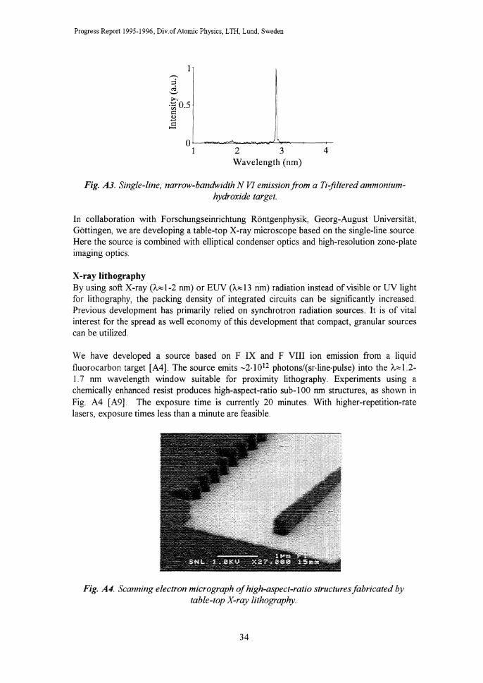
Progress Report 1995-1996, Div.of Atomic Physics, L Til, Lund, Sweden
_.-..
::i ,; '-' >. ·~0.5 ;:: ~ .s
o~--~~----~~~~~-1 2 3
Wavelength (nm) 4
Fig. A3. Single-line, narrow-bandwidth N VI emission from a Ti-jiltered ammoniumhydroxide target.
In collaboration with Forschungseinrichtung Rontgenphysik, Georg-August Universitat, Gottingen, we are developing a table-top X-ray microscope based on the single-line source. Here the source is combined with elliptical condenser optics and high-resolution zone-plate imaging optics.
X-ray lithography By using soft X-ray (A.,J-2 nm) or EUV (A-""13 nm) radiation instead ofvisible or UV light for lithography, the packing density of integrated circuits can be significantly increased. Previous development has primarily relied on synchrotron radiation sources. It is of vital interest for the spread as well economy of this development that compact, granular sources can be utilized.
We have developed a source based on F IX and F VIII ion eilllsswn from a liquid fluorocarbon target [ A4]. The source emits -2· I 012 photons/(sr·line·pulse) into the '-"" 1.2-1.7 nm wavelength window suitable for proximity lithography. Experiments using a chemically enhanced resist produces high-aspect-ratio sub- I 00 nm structures, as shown in Fig A4 [A9]. The exposure time is currently 20 minutes. With higher-repetition-rate lasers, exposure times less than a minute are feasible.
Fig. A4. Scanning electron micrograph of high-aspect-ratio structures fabricated by table-top X-ray lithography.
34

II Applied Optics and Quantum Electronics
For EUV projection lithography in the A=10-15 nm range, the emission from oxygen ions contains several suitable lines [A3, AS]. Using an 8-ns frequency-doubled Nd:YAG laser and water/methanol droplets we obtained -4·1012 photons/(sr·line·pulse) at the 2p-4d 0 VI line at A=l3.0 nm.
B Non-intrusive scanning near-field microscopy Lars Malmqvist and Hans M. Hertz
MSc student: Daniel Akenine
The non-intrusive character of classical optical microscopy is of great importance in the study of, e.g., living biological systems. Unfortunately, far-field diffraction limits the resolution of such microscopes. Near-field optical methods have demonstrated very high resolution by scanning a microscopic light source in close proximity to the studied object. However, the mechanical positioning of the probe restricts its use to mechanically accessible and smooth surfaces, limiting the method's applicability for studies of, e.g., living biological material with intervening membranes. Our work is focused on the development of a non-intrusive, near-field optical microscope. The work has been presented in Refs. [Bl-B6 and A21].
We are investigating the use of optically trapped microscopic non-linear crystals as microscopic light sources for non-intrusive, scanned-probe optical microscopy - Trapped Particle Optical Microscopy (TPOM) [B 1]. Fig. B 1 shows the experimental arrangement. A sub-1 00 nm lithium niobate or KTP crystal is optically trapped by a strongly focused A =I. 06 J..l.m N d: Y AG laser. Due to the nonlinear properties of the crystal, green light (A =532 nm) is generated when the particle is trapped close to the focus. This visible light source may be non-intrusively positioned in close proximity to the object of interest. By accurate piezoelectric scanning, a subdiffraction-limited image may be recorded
The best resolution obtained so far is <0.5
LiNbO 3
PARTICLE
Fl L TERS
CHOPPER
1 0 jew Nd:YAGj
XY2-STAGE
Lock-in amplifier
Fig. Bl. Experimental arrangement for two-colour trapped particle optical microscopy.
J..lm with an etched Si cantilever as the test object. Here van der Waals surface forces limit the minimum distance between the object and the particle due to the large refractive index of silicon With the lower refractive index of biological objects, we expect to obtain subdiffraction resolution. Theoretical calculations of the resolution indicate that 80 nm should be possible with the current particles. Further resolution improvement may be obtained by using other types of traps. This includes a novel trap based on acoustical radiation forces [B3].
35
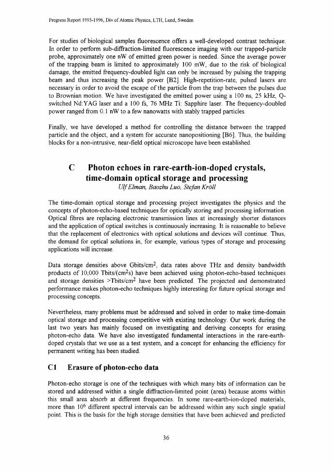
Progress Report 1995-1996, Div.of Atomic Physics, LTH, Lund, Sweden
For studies of biological samples fluorescence offers a well-developed contrast technique. In order to perform sub-diffraction-limited fluorescence imaging with our trapped-particle probe, approximately one nW of emitted green power is needed. Since the average power of the trapping beam is limited to approximately 100 mW, due to the risk of biological damage, the emitted frequency-doubled light can only be increased by pulsing the trapping beam and thus increasing the peak power [B2]. High-repetition-rate, pulsed lasers are necessary in order to avoid the escape of the particle from the trap between the pulses due to Brownian motion. We have investigated the emitted power using a 1 00 ns, 25 kHz, Qswitched Nd:Y AG laser and a 100 fs, 76 :MHz Ti: Sapphire laser. The frequency-doubled power ranged from 0.1 nW to a few nanowatts with stably trapped particles.
Finally, we have developed a method for controlling the distance between the trapped particle and the object, and a system for accurate nanopositioning [B6]. Thus, the building blocks for a non-intrusive, near-field optical microscope have been established.
C Photon echoes in rare-earth-ion-doped crystals, time-domain optical storage and processing
Ulj Elman, Baozhu Luo, Stefan Kroll
The time-domain optical storage and processing project investigates the physics and the concepts of photon-echo-based techniques for optically storing and processing information. Optical fibres are replacing electronic transmission lines at increasingly shorter distances and the application of optical switches is continuously increasing. It is reasonable to believe that the replacement of electronics with optical solutions and devices will continue. Thus, the demand for optical solutions in, for example, various types of storage and processing applications will increase.
Data storage densities above Gbits/cm2, data rates above THz and density bandwidth products of 10,000 Tbits/(cm2s) have been achieved using photon-echo-based techniques and storage densities >Tbits/cm2 have been predicted. The projected and demonstrated performance makes photon-echo techniques highly interesting for future optical storage and processing concepts.
Nevertheless, many problems must be addressed and solved in order to make time-domain optical storage and processing competitive with existing technology. Our work during the last two years has mainly focused on investigating and deriving concepts for erasing photon-echo data. We have also investigated fundamental interactions in the rare-earthdoped crystals that we use as a test system, and a concept for enhancing the efficiency for permanent writing has been studied.
Cl Erasure of photon-echo data
Photon-echo storage is one of the techniques with which many bits of information can be stored and addressed within a single diffraction-limited point (area) because atoms within this small area absorb at different frequencies. In some rare-earth-ion-doped materials, more than I o6 different spectral intervals can be addressed within any such single spatial point. This is the basis for the high storage densities that have been achieved and predicted
36

1000 ,---------,..-----;------,
>u c § 100
~ ~ iil 10 ~ w
....• ........ .. s ··-v ........................
10 100 1000 Data storage time (micro seconds)
j & Data length, 500 ns 'V Data length, 1000 ns I
Fig. CJ. Data erasure efficiency, as defined in the text, as a function of data storage time for two different data lengths. The drop in erasure efficiency is due to laser phase and frequency fluctuations.
II Applied Optics and Quantum Electronics
in these materials. In time-domain or photonecho storage, one can show that the frequency domain Fourier transform of any temporal optical input sequence is recorded in the material as a spectral redistribution of the absorbing atoms between the upper and lower states. This frequency domain Fourier transform contains information both about the amplitude and phase of the input sequence. A consequence of this is that if the spectral modulation due to a specific input sequence is added to the spectral distribution generated by a signal which is identical except that it is phase shifted 180 degrees, these two modulation patterns combine such that the spectral modulation due to the initially stored data sequence is cancelled.
The practical implementation ofthis approach has been investigated in some detail [Cl]. It has been found that, in practice, the critical issue is that the erasing sequence must be phase shifted by exactly 180 degrees with respect to the first sequence. The consequence of this is that it is necessary to put (quite stringent) requirements on the phase and frequency stability of the light source used for storing the data. Fig. C 1 shows how the data erasure efficiency decreases as a function of data storage time. The erasure efficiency is approximately equal to (the fraction of the initially stored signal remaining after erasure)-l Thus, when the erasure efficiency is equal to 100, approximately 99% of the stored data has been erased. It is clear from Fig. C 1 that for longer storage times the erasure process does not work very well. The reason for the low efficiency in the erasure process after longer storage times can be interpreted in terms offrequency drift of the light source. Furthermore we see that as the duration of the data sequence increases, the erasure efficiency again drops rapidly. The reason for this decrease in erasure efficiency has been traced to laser phase drift.
Although laser phase and frequency drift seriously impair the erasure efficiency, the observed erasure efficiency can, on the other hand, be used as a means of monitoring the laser stability. By describing the laser frequency fluctuations as a stationary stochastic process, the data erasure efficiency, as a function storage time and data sequence duration, as well as the contribution to shot-to-shot fluctuations in conventional stimulated photon echoes from laser frequency instability, were all fitted to a theoretical model with four free parameters [C2]. The fits of erasure efficiency, as a function of storage time and stimulated photon-echo shot-to-shot fluctuations as a function of the time between the second and
37
Echo suppression
1000~------------
100
10
\~-. ·----~. "'-,,
• 1'·.
10 100
·--• • •
1000
Fig. C2. Erasure efficiency versus storage time (.1) including fit to the theoretical model.

Progress Report 1995-1996, Div.of Atomic Physics, L TH, Lund, Sweden
R[AJ 60 ,--------------,
50 • •
40
30 • 20
10
10 100 1000
"(p.s)
Fig. C3. Coefficient of variation for a stimulated photon echo versus time separation between the second and third pulses.
third pulses are shown in Figs C2 and C3 for the best choice of the four parameters. The agreement with the data for the erasure efficiency as a function data sequence duration is also reasonably good. In fact, the approach presented in [C2] for describing the influence of laser frequency fluctuations is quite general and could, in principle, be applied to any non-linear optical process.
Nevertheless, laser phase and frequency fluctuations present a serious obstacle for efficient photon-echo data erasure, a circumstance which has been foreseen in the literature on the subject. The present data have clearly shown that this concern must be taken seriously. However, we believe we have solved this problem. We have proposed
a method by which the photon-echo data erasure process can be essentially unaffected by phase or frequency fluctuations in the light source [C 1]. In short, the idea is that the sequence that is to be erased is read out and then sent back into the sample after passing an "erasure loop", Fig. C4. The erasure loop phase shifts the data by 180 degrees before it is written back into the materiaL If data should still remain the procedure can be repeated to further enhance the erasure efficiency. We believe this approach can eliminate the phase and frequency stability requirements for selective photon-echo data erasure. Our work on photon-echo data erasure has been presented on various occasions [C3-C6]. A Licentiate thesis has been presented by UlfElman based on, among other things, the photon-echo data erasure work described above [C7]. The thesis also contains a more general overview of information systems, including optical systems. A Master's project investigating the possibility of enhancing the writing efficiency using a radio-frequency source to induce hyperfine transitions in the upper electronic state has also been performed [C8].
Storage ~ ... medium
Write, Data in, Read ................................................................... _,,, ... _::=----··-·- ............. ,.:
Jj --
Data out ··························· __ .,
Amplifier --------------------..------/--/ 1t phase shifter
Erasure loop
Fig. C4. Data to be erased are read out from the storage medium and sent into the erasure loop where it is phase shifted by 180 degrees, before being sent back into the sample. If the erasure process is incomplete the process can be repeated until no signal remains. A full explanation is given in Reference Cl.
38
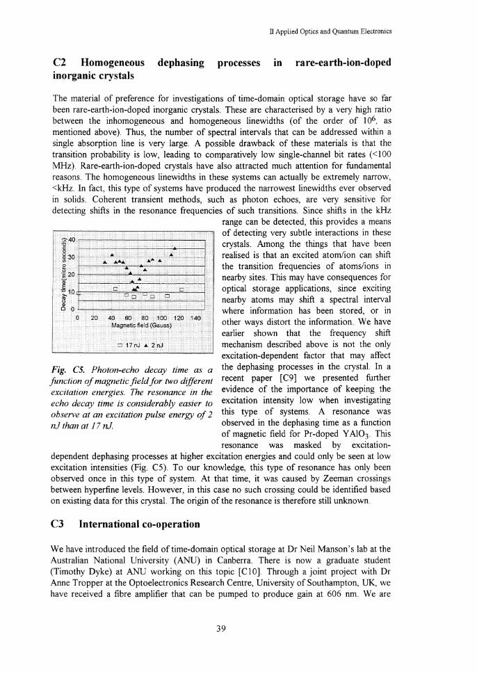
II Applied Optics and Quantum Electronics
C2 Homogeneous dephasing processes in rare-earth-ion-doped inorganic crystals
The material of preference for investigations of time-domain optical storage have so far been rare-earth-ion-doped inorganic crystals. These are characterised by a very high ratio between the inhomogeneous and homogeneous linewidths (of the order of J06, as mentioned above). Thus, the number of spectral intervals that can be addressed within a single absorption line is very large. A possible drawback of these materials is that the transition probability is low, leading to comparatively low single-channel bit rates ( < 100 MHz). Rare-earth-ion-doped crystals have also attracted much attention for fundamental reasons. The homogeneous linewidths in these systems can actually be extremely narrow, <kHz. In fact, this type of systems have produced the narrowest linewidths ever observed in solids. Coherent transient methods, such as photon echoes, are very sensitive for detecting shifts in the resonance frequencies of such transitions. Since shifts in the kHz
Fig. C5. Photon-echo decay time as a function of magnetic field for two different excitation energies. The resonance in the echo decay time is considerably easier to observe at an excitation pulse energy of 2 nJ than at 1 7 nJ.
range can be detected, this provides a means of detecting very subtle interactions in these crystals. Among the things that have been realised is that an excited atom/ion can shift the transition frequencies of atoms/ions in nearby sites. This may have consequences for optical storage applications, since exciting nearby atoms may shift a spectral interval where information has been stored, or in other ways distort the information. We have earlier shown that the frequency shift mechanism described above is not the only excitation-dependent factor that may affect the dephasing processes in the crystaL In a recent paper [C9] we presented further evidence of the importance of keeping the excitation intensity low when investigating this type of systems. A resonance was observed in the dephasing time as a function of magnetic field for Pr-doped Y AI03. This resonance was masked by excitation
dependent dephasing processes at higher excitation energies and could only be seen at low excitation intensities (Fig. C5). To our knowledge, this type of resonance has only been observed once in this type of system. At that time, it was caused by Zeeman crossings between hyperfine levels. However, in this case no such crossing could be identified based on existing data for this crystaL The origin of the resonance is therefore still unknown.
C3 International co-operation
We have introduced the field of time-domain optical storage at Dr Neil Manson's lab at the Australian National University (ANU) in Canberra. There is now a graduate student (Timothy Dyke) at ANU working on this topic [CIO]. Through a joint project with Dr Anne Tropper at the Optoelectronics Research Centre, University of Southampton, UK, we have received a fibre amplifier that can be pumped to produce gain at 606 nm. We are
39

Progress Report 1995-1996, Div.of Atomic Physics, L TH, Lund, Sweden
currently investigating the feasibility of amplifying the output from a photon-echo crystal using this fibre amplifier.
We have completed our project with Dr Rein Kaarli at the Institute of Physics at the Estonian Academy of Sciences in Tartu on programming photon-echo crystals in the frequency domain and reading them out in the time domain [Cll], and we are currently discussing a joint project on delayed-photon self-interference (JETP Lett. 58, 80 (1993)) with Rein Kaarli and members of Prof. Anton Zeilinger's group at the University of Innsbruck. A Masters student from Lund is presently performing her MSc project on spectral pulse shaping for logical operations using femto-second pulses with Dr Alexander Rebane at Prof. Urs Wild's laboratory, at ETH in Zurich.
References
[AI] L. Rymell and H.M. Hertz, "Debris-elimination in a droplet-target laser-plasma soft x-ray source", Rev. Sci. lustrum. 66,4916 (1995).
[A2] L. Rymell, M. Berglund and H.M. Hertz, "Debris-free single-line laser plasma source for microscopy", Appl. Phys. Lett. 66, 2625 (1995).
[A3] H.M. Hertz, L. Rymell, M. Berglund and L. Malmqvist, "Debris-free soft x-ray generation using a liquid droplet laser-plasma target", in Applications of Laser Plasma Radiation II, M.C. Richardsson, Ed., SPIE Vol. 2523, pp. 88-93 (Soc. Photo-Optical lustrum. Engineers, Bellingham, Washington, 1995).
[A4] L. Malmqvist, L. Rymell, and H.M. Hertz, "Droplet-target laser-plasma x-ray source for proximity lithography", Appl. Phys. Lett. 68, 2627 (1996).
[AS] L. Malmqvist, L. Rymell, and H. M. Hertz, "High-repetition-rate droplet-target for laser-plasma EUV generation", in Extreme Ultraviolet Lithography, OSA Trends in Optics and Photonics Vol. 4, G. D. Kubiak and D. R. Kania, Eds. (Optical Society of America, Washington, DC 1996) pp. 72-75.
[A6] M. Berglund, L. Rymell, and H.M. Hertz, "Ultraviolet prepulse for enhanced x-ray emission and brightness from droplet-target laser plasma", Appl. Phys. Lett. 69, 1683 ( 1996).
[A 7] L. Malmqvist, L. Rymell, M. Berglund and H.M. Hertz, "Liquid-jet target for laser-plasma soft xray generation", to appear Rev. Sci. lustrum. (Dec. 1996).
[AS] L. Rymell, M. Berglund, L. Malmqvist, and H.M. Hertz, "Liquid target for debris-free laserplasma soft x-ray generation", submitted to Proc. 5th International Conference on X-Ray Lasers, Eds. C.-G. Wahlstrom and S. Svanberg (Institute of Physics, London, 1996).
[A9] L. Malmqvist, A. L. Bogdanov, L. Montelius and H.M. Hertz, "Nanometer table-top proximity xray lithography with liquid-target laser-plasma source", submitted to 1. Vac. Science Technol.
[AIO] H.M. Hertz, L. Rymell, M. Berglund, and L. Malmqvist, "Debris-free liquid-target laser-plasma soft x-ray source for microscopy and lithography", in X-ray Microscopy and Spectromicroscopy, 1. Thieme, G. Schmahl, E. Umbach, and D. Rudolph, Eds. (Springer, Heidelberg, 1996) (invited paper).
[All] T. Wilhein, B. Niemann, L. Rymell, M. Berglund, and H.M. Hertz, "Off-axis reflecting zoneplate for high-resolution soft x-ray spectroscopy", manuscript in preparation for Opt. Lett.
[Al2] H. M. Hertz, "Debris-free liquid-target laser-plasma soft x-ray sources", X-Tech 96, Berlin, Germany (invited talk, 1996).
[A!3] L. Rymell, M. Berglund and H.M. Hertz, "Debris-free single-line source for x-ray microscopy at A.=2.88 nm ··, Euroconference 'Generation and applications of ultrashort x-ray pulses II' (Pisa, Italy, 1995).
[Al4] L. Rymell, M. Berglund, H.M. Hertz, 1. Thieme, B. Niemann, and T. Wilhein, "Laser-plasma source for table-top soft x-ray microscopy", Optics in Sweden (Stockholm. !995).
[AIS] L. Rymell, M. Berglund, and H.M. Hertz, "Debris-free laser-plasma source for x-ray microscopy", European Conference on Lasers and Electro-Optics (Hamburg. 1996).
40

[Al6]
[Al7]
[Al8]
[Al9]
[A20]
[A21]
[A22]
[Bl]
[B2]
[B3]
[B4]
[B5]
[B6]
[Cl]
[C2]
[C3]
[C4]
[C5]
[C6]
[C7]
[C8]
[C9]
[ClO]
[Cll]
IT Applied Optics and Quantum Electronics
L. Malmqvist, L. Rymell, and H.M. Hertz, "Proximity x-ray lithography using a droplet-target laser-plasma source", European Conference on Lasers and Electro-Optics (Hamburg, 1996). M. Berglund, L. Rymell, and H.M. Hertz, "Improved water-window droplet-target laser-plasma xray source by temporally tailored laser pulse", in X-ray Microscopy and Spectromicroscopy, J. Thieme, G. Schmahl, E. Umbach, and D. Rudolph, Eds. (Springer, Heidelberg, 1996). L. Rymell, M. Berglund, L. Malmqvist, and H.M. Hertz, "Characterisation of debris-free laserplasma source for x-ray microscopy", in X-ray Microscopy and Spectromicroscopy, J. Thieme, G. Schmahl, E. Umbach, and D. Rudolph, Eds. (Springer, Heidelberg, 1996). L. Malmqvist, A.L. Bogdanov, L. Montelius, and H.M. Hertz, "X-ray lithography with a liquidtarget laser-plasma source", Micro- and Nano Engineering MNE '96 (Glasgow, 1996). Lars Rymell, "Laser-plasma droplet x-ray source for microscopy", Licentiate Thesis, Lund Reports on Atomic Physics LRAP-170 (1995). Lars Malmqvist, "New laser-based techniques for nanometer lithography and microscopy", Ph.D. Thesis, Lund Reports on Atomic Physics LRAP-206 (1996). H.M. Hertz, L. Malmqvist, L. Rymell, and M. Berglund, "Laser-plasma x-ray source using liquid target", Patent application (Aprill996). H.M. Hertz, L. Malmqvist , L. Rosengren and K. Ljungberg, "Optically trapped nonlinear particles as probes for scanned near field optical microscopy", Ultramicroscopy 57, 309 ( 1995). L. Malmqvist and H.M. Hertz, "Second-harmonic generation in optically trapped nonlinear particles using pulsed lasers", Appl. Opt. 34, 3392 (1995). H.M. Hertz, "Standing-wave acoustic trap for non-intrusive positioning of microparticles", J. Appl. Phys. 78, 4845-4849 (1995). H. M. Hertz, "Optical tweezers for non-intrusive scanning near field optical microscopy", Optics in Sweden, Stockholm (invited talk, 1995). L. Malmqvist and H.M. Hertz, "Non-intrusive scanned probe microscopy using nonlinear particles trapped by a pulsed laser", International conference on Near field optics NF0-3 (Brno, 1995). Daniel Akenine, "Accurate positioning for non-intrusive near-field optical microscopy", Lund Reports on Atomic Physics LRAP-187 (1995). U Elman, B Luo and S Kroll, "Influence of laser phase and frequency fluctuations on photon echo data erasure", J. Opt. Soc. B13, 1905-1915 (1996). U Elman and S Kroll, "Statistical modelling and theoretical analysis of the influence of laser phase fluctuations on photon echo data erasure and stimulated photon echoes", Laser Phys., 6, (1996), in press. S Kroll, "Photon echo data storage, to use atomic wave functions to store and process optical data", invited seminar, Colloquium in Physics, KTH, September-95. U Elman, B Luo and S Kroll, "Photon-echo data storage and processing", poster, annual meeting of the Swedish Optical Society, Stockholm, October, 1995. U Elman, B Luo, and S Kroll, "Coherent erasure of photon echo data", contributed talk, CLE0'95. U Elman, B Luo and S Kroll, "The effect of laser and phase fluctuations on coherent addition and erasure of photon echoes", contributed talk, CLEO Europe '96 Ulf Elman, "Photon echo based all-optical data storage and processing", Licentiate thesis, Lund Reports on Atomic Physics LRAP-192, Lund, April, 1996. M Plilsson, "Radio-frequency-enhanced photon echo storage", Lund Reports on Atomic Physics LRAP-176, LTH, Lund (1996) B Luo and S Kroll, "Observation of a magnetic field induced resonance in the homogeneous dephasing time for the 3H4-1D2 transition in Pr3+:YAl03", Phys. Rev. B54, (1996) October I, in press. U Elman, T Dyke, N Akhmediev, S Kroll, N Manson, "Data erasure in stimulated photon echo optical memories", poster, IQEC · 96 B Luo, U Elman, S Kroll and R Kaarli, "Spectral programming of temporal pulse sequences using photon echoes", poster, CLEO Europe '96.
41

Progress Report 1995-1996, Div. of Atomic Physics, L TI:I, Lund Sweden
III Environmental Monitoring
Research projects in the field of environmental monitoring are directed towards development and applications of optical techniques, both laser and non-laser, to measure mostly gases and particles, but also as a tool in vegetation and water quality studies. Several of the techniques are used for remote sensing over long distances. The method most used in studies of different air pollutants is the differential absorption lidar (DIAL) technique, where a mobile DIAL system has been employed in several field campaigns. The mobile system has also been used for studies of laser-induced fluorescence from vegetation, and special techniques have been developed for multi-colour imaging. A new application of laser/optical techniques in monitoring of the working environment has been initiated. A new method has been developed for imaging of gas flows using an IR camera and gas correlation techniques. This new project is channelled through the Centre for Environmental Measurement Technology (CENTEC), which co-ordinates research and teaching in environmental sensing technology within different departments of the Lund Institute of Technology. Ultrasensitive absorption measurement methods are developed in a project utilising diode laser absorption spectroscopy and frequency modulation (FM) techniques.
During the past two-year period two PhD theses [1,2] and one Licenciate thesis [3] have been presented. Some overviews of the research have also been presented [ 4-6]. Apart from the projects described below, some activities are also pursued with the DOAS (Differential Optical Absorption Spectroscopy) technique. Earlier work on the detection of aromatic hydrocarbons [7], and the combination of DOAS measurements with PIXE (Particle-Induced X-ray Emission) analysis of aerosols [8], have now been published. New detailed studies ofthe DOAS evaluation procedure have also been performed [9,10].
A Lidar measurements of atmospheric gases Mats Andersson, Hans Edner, Sune Svanberg and Petter Wei bring
The mobile DIAL system has been used during several field campaigns for monitoring of both natural and anthropogenic gas emissions in various European regions. Earlier measurements of industrial and urban pollution concentrations and fluxes in Sweden [A 1] and in the Czech Republic [A2] are now published. The results of the campaign in Italy during September 1994, where gas fluxes from the volcanoes Etna, Stromboli and Vulcano were measured, have been analysed and published [A3-A5]. These measurements were performed with the mobile DIAL system on the aft deck of the Italian CNR research ship "Urania". The results of the active lidar technique were compared with simultaneous recordings with passive DOAS and CO SPEC (correlation spectroscopy), using the sky radiation as the light source. A further campaign of this kind is planned for the summer of 1997.
Renewed measurements of mercury concentrations in the Mt. Amiata region in Italy were performed during July 1995. This campaign was made on commission by the company Lighten AB and in co-operation with CNR-Istituto di Biofisica. Some of the results of this
42
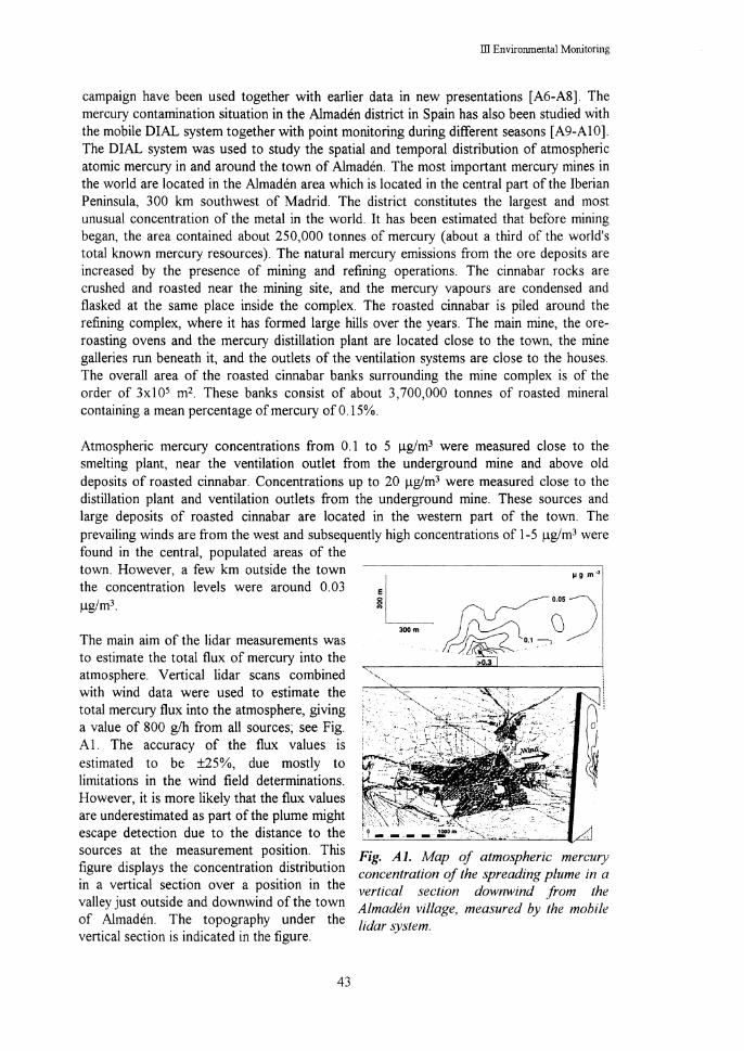
ill Environmental Monitoring
campaign have been used together with earlier data in new presentations [A6-A8]. The mercury contamination situation in the Almaden district in Spain has also been studied with the mobile DIAL system together with point monitoring during different seasons [A9-A10]. The DIAL system was used to study the spatial and temporal distribution of atmospheric atomic mercury in and around the town of Almaden. The most important mercury mines in the world are located in the Almaden area which is located in the central part of the Iberian Peninsula, 300 km southwest of Madrid. The district constitutes the largest and most unusual concentration of the metal in the world. It has been estimated that before mining began, the area contained about 250,000 tonnes of mercury (about a third of the world's total known mercury resources). The natural mercury emissions from the ore deposits are increased by the presence of mining and refining operations. The cinnabar rocks are crushed and roasted near the mining site, and the mercury vapours are condensed and flasked at the same place inside the complex. The roasted cinnabar is piled around the refining complex, where it has formed large hills over the years. The main mine, the oreroasting ovens and the mercury distillation plant are located close to the town, the mine galleries run beneath it, and the outlets of the ventilation systems are close to the houses. The overall area of the roasted cinnabar banks surrounding the mine complex is of the order of 3x10⁵ m2 These banks consist of about 3,700,000 tonnes of roasted mineral containing a mean percentage of mercury of0.15%.
Atmospheric mercury concentrations from 0.1 to 5 J..Lg/m3 were measured close to the smelting plant, near the ventilation outlet from the underground mine and above old deposits of roasted cinnabar. Concentrations up to 20 J..Lg/m3 were measured close to the distillation plant and ventilation outlets from the underground mine. These sources and large deposits of roasted cinnabar are located in the western part of the town. The prevailing winds are from the west and subsequently high concentrations of 1-5 J..Lg/m3 were found in the central, populated areas of the town. However, a few km outside the town the concentration levels were around 0.03 J..Lg/m3
The main aim of the lidar measurements was to estimate the total flux of mercury into the atmosphere. Vertical lidar scans combined with wind data were used to estimate the total mercury flux into the atmosphere, giving a value of 800 g/h from all sources; see Fig. A 1. The accuracy of the flux values is estimated to be ±25%, due mostly to limitations in the wind field determinations. However, it is more likely that the flux values are underestimated as part of the plume might escape detection due to the distance to the sources at the measurement position. This figure displays the concentration distribution in a vertical section over a position in the valley just outside and downwind of the town of Almaden. The topography under the vertical section is indicated in the figure.
43
Fig. Al. Map of atmospheric mercury concentration of the spreading plume in a vertical section downwind from the Almaden village, measured by the mobile lidar 5ystem.

Progress Report 1995-1996, Div. of Atomic Physics, LTH, Lund Sweden
The mobile differential absorption lidar (DIAL) system has recently been updated with the graphical programming language Lab VIEW in order to obtain a user-friendly system (All]. The project started in 1994 and the new lidar program was run for the first time in the summer of 1996. The software controls the lidar system and analyses measurement data. The results of the measurement are shown as maps of species concentration. Lab VIEW is a graphical programming system for data acquisition and control, data analysis and data presentation. It offers a programming methodology in which software modules called virtual instruments (Vis) are created. With Lab VIEW, images of the data flow are drawn instead of using written program code. Each VI has two different panels: the front panel and the block diagram. The front panel serves as the user interface which gives the user interactive control over the software program. However, each symbol on the front panel has a symbol in the block diagram, which shows the actual program code. Every symbol on this panel has its function written in C but it is shown as images instead of code lines. In programming, VI lines are drawn between the symbols with the mouse. To influence a signal, functional blocks can be selected from palette menus. All blocks are connected with 'wires' to pass data from one block to another.
The updated lidar system has a modular design; see Fig A2. The system computer contains four plug-in boards: one for network communication, a GPm board, and two multifunction boards, which control stepping motors, servo-motors, the PMT, and all the electronics in the receiver unit. The system computer gets inputs from the laser unit and the wind unit, analyses the detected signals and stores them on disc. The evaluation computer works as an instrument for evaluation and presentation of the measured data.
Nd:YAG-l..aset Dye-laser Laser Output Receiver Unl:
' !
~~~ Wind Monitor
I I ' I
Control Unit t.a..«er Power Supply
Lr ~ , ,.-t ----'------.
'-----f--------f liB ~ i-( II '-------'
Dye-l..aser Computer Evaluation Computer System Compuw
Fig. A2. Lidar system block diagram
44

III Environmental Monitoring
Lidar technology has proved to be a valuable tool for atmospheric studies. During the years, the technology has been developed and the measurement accuracy is today of the order of 3-10 %. Normally, lidar results are presented together with an accuracy analysis. However, this is not straightforward for flux measurements on smoke stack plumes performed by mobile lidar systems. In this application, the measurement accuracy can be as poor as 30-50%. This is due to the difficulties in estimating the wind speed around the plume. In order to improve the accuracy of flux measurements, a video graphical technology for wind speed estimations has been developed. The video graphical technique uses a frame grabber which repeatedly records images of a plume taken with a CCD camera, and by studying the plume movement the plume speed can be estimated. Preliminary measurements show an accuracy of 5-10%. The distance between the lidar system and the plume is another important parameter which should be measured. By pointing the laser beam of a lidar at the plume, the plume distance can be measured by observing the lidar echo. The theory behind the technique is based on the fact that each graph contains an immobile background and a plume which moves horizontally. The columns of a graph matrix are added, creating a vector which contains information about the contents of the graph; both background and plume information. In order to remove the background information, an average vector (which is created from a number of previously grabbed graphs) is subtracted from each vector. The remaining vector should only contain information about the plume. By considering the cross-correlation between two subsequent vectors, the plume movement from to to t 1 can be estimated. However, a great deal of work remains to be done before this technology can be said to be fully developed. Since this application is quite new, there are many parameters which must be optimised. Different optical filters will be tested in order to improve the contrast in the plume graph. In the case of an invisible plume, it is proposed that an IR camera be used to record the plume movement.
In collaboration with the Department of Mathematical Statistics the procedure for the evaluation of DIAL measurements has been investigated [A12]. A new method with a locally weighted least squares kernel regression has been shown to improve the data evaluation and accuracy estimation. The algoritms have now been incorporated in the new program.
B Laser-induced fluorescence of vegetation Mats Andersson, Hans Edner, Jonas Johansson and Sune Svanberg
Laser-induced fluorescence can be used as a tool for airborne monitoring of vegetation status and water pollutants. This work has been performed within the European projects, LASFLEUR and EUROMAR. The remote sensing group has participated in four cooperative field campaigns on remote sensing of terrestrial vegetation, in Florence ( 1990), Karlsruhe (1992), Oberpfaffenhofen (1992) and Avignon (1993), as was reported in the previous biannual report. Both point monitoring and 2-D imaging have been applied [B 1-B3]. Direct 2-D imaging was found to provide far too low a signal-to-noise ratio for airborne operation and therefore a system for 1-D push-broom scanning of images was developed. The basic principle is that the excitation laser beam is shaped into a horisontal line that can be used to scan perpendicularly to the flight direction of an aircraft. The fluorescence from this line is recorded at a high repetition rate and will form an image that
45

Progress Report 1995-1996, Div. of Atomic Physics, LTH, Lund Sweden
is successively updated. This principle was tested from a land-based vehicle and fluorescence images were scanned for several tree species [B4-B7]. The signal-to-noise ratio was found to be sufficient for a situation similar to that of an airborne measurement.
740 nm
685nm 740nm
Picea abies 60 m distance
685 nm 480 nm
lower needle side
-::::1
~ ....: c Q) 500 600 700
II I I I
500 600 700 wavelength (nm)
BOO
BOO
Fig. Bl. Scanned fluorescence images at 480, 685 and 740 nm and the ratio 685!740 for a spruce tree.
C Monitoring of the working environment Jonas Sandsten, Hans Edner and Sune Svanberg
During the last year, we have explored the principles of a powerful infrared imaging technique employing gas-correlation spectrometry. This principle made it possible for us to visualize gas flows in near realtime[Cl,C2]. Two images of the flowing gases are formed simultaneously on an IR-sensitive detector with a Cassegrainian telescope. A bandpass filter is used to enhance the contrast of the images. In front of one of the openings of the telescope is a gas cell containing the gas to be analysed and the other opening gives a direct
46

III Environmental Monitoring
IR camera with bandpass filter
Workplace with invisible gas Gas cell
Cassegrain telescope
Gas tubes
Visualisation of one specific gas
Fig. Cl. Schematic diagram of the experimental arrangement.
infrared image. The images are correlated in a computer to compensate for background, and are visualised as a movie, see Fig. C 1. Flow and concentration in two dimensions are presented in near realtime. The system is being developed primarily for the monitoring of working environments but surveying of hazardous gases is another field in which infrared imaging is useful, for example, in the remote detection of gas pipe leakage. Another industrial application would be hydrocarbon emissions from petrochemical installations. We are working towards both of the above mentioned applications. The Council of Working Life and the Swedish Space Board are supporting the research.
D Diode laser spectroscopy
Diode laser absorption spectroscopy based on frequency modulation (FM) techniques, is a powerful technique for gas concentration measurements where high sensitivity and fast response time are required. Although a variety of FM methods exist, the common underlying concept is a movement of the detection band to a high-frequency region, where the laser source (1/f) noise is avoided. The absorption signal is detected either at the same frequency as the applied modulation, at an overtone, or at an intermediate frequency. Highfrequency FM spectroscopy offers the possibility of shot-noise-limited detection at 10·7 -10·8
47
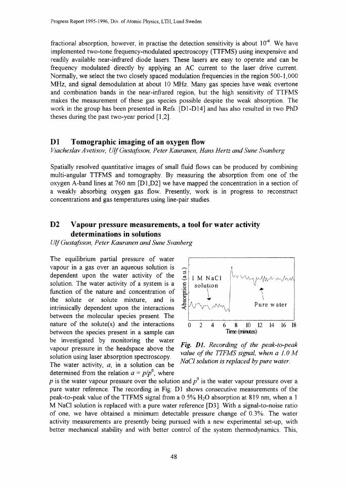
Progress Report 1995-1996, Div. of Atomic Physics, L TH, Lund Sweden
fractional absorption, however, in practise the detection sensitivity is about 10"6 We have implemented two-tone frequency-modulated spectroscopy (TTFMS) using inexpensive and readily available near-infrared diode lasers. These lasers are easy to operate and can be frequency modulated directly by applying an AC current to the laser drive current. Normally, we select the two closely spaced modulation frequencies in the region 500-1,000 MHz, and signal demodulation at about 10 MHz. Many gas species have weak overtone and combination bands in the near-infrared region, but the high sensitivity of TTFMS makes the measurement of these gas species possible despite the weak absorption. The work in the group has been presented in Refs. [D1-D14] and has also resulted in two PhD theses during the past two-year period [ 1 ,2].
Dl Tomographic imaging of an oxygen flow Viacheslav Avetisov, UlfGustafsson, Peter Kauranen, Hans Hertz and Sune Svanberg
Spatially resolved quantitative images of small fluid flows can be produced by combining multi-angular TTFMS and tomography. By measuring the absorption from one of the oxygen A-band lines at 760 nm [D1,D2] we have mapped the concentration in a section of a weakly absorbing oxygen gas flow. Presently, work is in progress to reconstruct concentrations and gas temperatures using line-pair studies.
D2 Vapour pressure measurements, a tool for water activity determinations in solutions
Ulj Gustafsson, Peter Kauranen and Sune Svanberg
The equilibrium partial pressure of water vapour in a gas over an aqueous solution is dependent upon the water activity of the solution. The water activity of a system is a function of the nature and concentration of the solute or solute mixture, and is intrinsically dependent upon the interactions between the molecular species present. The nature of the solute( s) and the interactions between the species present in a sample can be investigated by monitoring the water vapour pressure in the headspace above the solution using laser absorption spectroscopy. The water activity, a, in a solution can be determined from the relation a= plp0, where
0 2 4 6 8 10 12 14 16 18 T ll1l: (rnirues)
Fig. Dl. Recording of the peak-to-peak value of the TTFMS signal, when a 1. 0 M NaCl solution is replaced by pure water.
p is the water vapour pressure over the solution and/ is the water vapour pressure over a pure water reference. The recording in Fig. D 1 shows consecutive measurements of the peak-to-peak value of the TTFMS signal from a 0.5% H20 absorption at 819 nm, when a 1 M NaCI solution is replaced with a pure water reference [D3]. With a signal-to-noise ratio of one, we have obtained a minimum detectable pressure change of 0.3%. The water activity measurements are presently being pursued with a new experimental set-up, with better mechanical stability and with better control of the system thermodynamics. This,
48

III Enviromnental Monitoring
together with the use of the detailed TTFMS Iineshape information described in Section D3, will increase the measurement accuracy and will allow us to study very small pressure changes. The technique will be applied to the study of solution structures of biologically significant molecules, including the thermodynamic factors affecting association in solutions, e.g. the hydrophobic effect.
D3 High-resolution absorption measurements Vi ache slav A vetisov, Peter Kauranen and Ulf Gustafsson
Applying TTFMS for quantitative measurements over a wide range of concentrations, temperatures or pressures requires information about the line parameters and an adequate theory of the detected signal. Employing only the peak-topeak value of the TTFMS signal to measure the concentration of a species in a gas, may lead to large uncertamt1es when the absorption line profile is altered. We have performed extensive work (theoretically, numerically, and experimentally) aimed at a more general understanding of TTFMS lineshapes and how accurately line profile parameters, such as broadening, narrowing, line shift and line intensity, can be obtained from a least-squares fitting procedure under different experimental conditions [D4-D7]. The high measurement resolution in TTFMS allows us to resolve collisional (Dicke) narrowing. If collisional narrowing is significant, more elaborate line profiles than the computationally simple Voigt profile, such as the Galatry and the RautianSobelman profiles, provide a better fit to data and extract line parameters with higher accuracy. The influence of a small dispersion signal, appearing in the detected signal when the detection phase angle is not fully in phase with the absorption signal, and non-linear modulation response of the diode laser are included in the TTFMS theory and have been thoroughly investigated. The influence of the
1. 0 ,....__ ;::i o:i '-' <;:!
a ·v; VJ
::E ..... f:
0
0.04
"' ~ 0 "0 -~ 0.04
"
0
(a) (\ I I I I I I I I
I I I I I I I I I • , : I ;
\) \I
(b) \ t ~J v V\"-~v
R ~(\;-----~-
(c)
-------"1\j\J\~--·-~·v R
--~--------~--------./'\_,~.__r-~-.-----~-~
Frequency (a. u.)
Fig. D2. Comparison of the results from least-squares fits to experimental data. (a) TFFMS recording of an oxygen transition. (b) Residuals from a fit using Voigt (1/) and Rautian-Sobelman (R) profiles. (c) Residuals obtained when non-linear distortion is included in the TTFMS theory.
FM index on the lineshape has been evaluated and a method of accurately determining the FM index is demonstrated. The experimental investigation also includes a comparison between TTFMS and direct detection. The line parameters obtained from the two measurement methods show excellent agreement.
49

Progress Report 1995-1996, Div. of Atomic Physics, LTH, Lund Sweden
Fig. D2(a) shows a spectrum of the oxygen Rl5Q16line at 13,156 cm-1 The oxygen cell pressure was 200 torr, giving a peak absorption of 4.7%. The upper trace in Fig. D2(b) shows the residual from a least-squares fit using the Voigt profile. The characteristic signature of the residuals reveals the presence of collisional narrowing. The lower trace in Fig. D2(b) shows the residual from a Rautian-Sobelman fit. The asymmetry in the plots originates from non-linear distortion of the diode laser modulation response, and the inclusion of non-linear distortion in the TTFMS theory greatly improves quality of the fit, as the results in Fig. D2(c) show. Our study has shown that the accurate determination of line parameters of transitions recorded with TTFMS is possible. This implies that accurate quantitative measurements over a wide range of concentrations, temperatures and pressures can be performed with high-sensitivity TTFMS.
References
[I) P. Kauranen, "Near-infrared diode laser frequency-modulation spectroscopy for high-sensitivity gas analysis", PhD Thesis, Lund Institute of Technology, Lund Reports on Atomic Physics LRAP-185 (1995).
[2) G. Avetisov, "High-sensitivity high-resolution diode laser-spectroscopy in the near-infrared region", PhD Thesis, Lund Institute of Technology, Lund Reports on Atomic Physics LRAP-189 (1996).
[3) M. Andersson, "Development of lidar techniques for environmental studies", Licenciate Thesis, Lund Reports on Atomic Physics LRAP-202 (1996).
[4) H. Edner, "DIAL measurements of natural and anthropogenic gas emissions", Review of Laser Engineering 23, 142-147 (1995).
[5) S. Svanberg, ''Chemical sensing with laser spectroscopy", Sensors and Actuators B33, 1-4 (1996).
[6) M. Andersson, H. Edner, J. Johansson, S. Svanberg, E. Wallinder and P. Weibring, "Remote sensing of the environment using laser radar techniques", Invited paper for Atomic Physics Methods in Modern Research, Springer-Verlag, to be published.
[7) H. Axelsson, H. Edner, H., A. Eilard, A. Emanuelsson, B. Galle, H. Kloo, and P. Ragnarson, "Measurements of aromatic hydrocarbons with the DOAS technique", Applied Spectroscopy 49, 1254-1260 (1995).
[81 E. Swietlicki, S. Puri, H.-C. Hansson, and H. Edner, "Urban air pollution source apportionment using a combination of aerosol and gas monitoring techniques", Atmospheric Environment 30, 2795-2809 ( 1996).
[9) T. Lindstrom, ''Robust local polynomial regression with application on the DOAS filtering process", Master's project, Report LUNFD6/NFMS-5037-SE (Dept. of Mathematical Statistics 1996).
[10) T. Lindstrom, U. Holst, and H. Edner, "Robust local po~ynomial regression and statistical evaluation of DOAS measurements", Report LUTFD2/TFMS-3127-SE (Dept. of Mathematical Statistics 1996).
[All H. Edner, P. Ragnarson and E. Wallinder, "Industrial emission control using lidar techniques", Environmental Science & Technology 29, 330-337 (1995).
[A2) P. Engst, F. Janouch, J. Keder, M. Andersson, H. Edner, M. Hessman, S. Svanberg, and E. Wallinder, "What warrants the LIDAR: A short report from the air pollution control in Czech Republic", Ceskoslovensky casopis pro fyiky 45, 216-228 (1995).
[A3) H. Edner, P. Ragnarson, S. Svanberg, E. Wallinder, R. Ferrara, R. Cioni, B. Raco and G. Taddeucci, "Volcanic gas emission studies using optical remote sensing techniques", Sensors and Environmental Applications of Remote Sensing, Ed. J. Askne, pp 357-362 (Balkema, Rotterdam, 1995)
[A4) S. Svanberg, M. Andersson, P. Anderson, H. Edner, J. Johansson, R. Ferrara, E. Maserti, G. Cecchi, L. Pantani, P. Mazzinghi, L. Alberotanza, R. Cioni and T. Caltabiano, "Laser monitormg
50

ill Environmental Monitoring
of the environment", Proceedings of 12th International Conference on Laser Spectroscopy, Eds M. Inguscio, M. Allegrini and A. Sasso, pp 423-426 (World Scientific, Singapore, 1996).
[AS] M. Andersson, M. Bazzani, G. Cecchi, H. Edner, F. Meiners, L. Pantani, V. Raimondi,.S. Svanberg, D. Tirelli, M. Trambusti, G. Valmori and P. Weibring, "The SALE experiment: Ship monitoring of the volcanic emission and water quality in the southern Tyrrhenian sea", Proc. European Symposium on Satellite Sensing III, Taormina, Italy, Sept 23, 1996.
[A6] R. Ferrara, B.E. Maserti, M. Andersson, H. Edner, P. Ragnarson, and S. Svanberg, "Mercury degassing rate from mineralized areas in the Mediterranean basin··, Water, Air and Soil Pollution 93, 59-66 (1997)
[A7] R. Ferrara, B. Mazzolai, H. Edner, S. Svanberg, and E. Wallinder, "Atmospheric mercury sources in the Mt. Ami at a area", Italy, Water, Air and Soil Pollution, to be published.
[A8] R. Ferrara, B.E. Maserti, B. Mazzolai, F. DiFrancesco, H. Edner, S. Svanberg, and E. Wallinder, "Atmospheric mercury in abandoned mine structures and restored mine buildings", in Mercury contamination sites: Characterization, Risk Assessment and Remediation (Springer-Verlag Publishers, to appear).
[A9] M. Andersson, H. Edner, R. Ferrara, A. Hernandez, B.E. Maserti, P. Ragnarson, and S. Svanberg, "Atmospheric mercury emissions from the Almaden cinnabar mining area", in Advances in Atmospheric Remote Sensing with Lidar, Eds. A. Ansmann, R. Neuber, P. Rairoux, and U. Wandinger, pp 415-418 (Springer-Verlag, Berlin, 1997).
[AlO] R. Ferrara, B. E. Maserti, M. Andersson, H. Edner, P. Ragnarson, S. Svanberg, and A. Hernandez, "Atmospheric mercury concentrations and fluxes in the Almaden district (Spain)", Atmospheric Environment, to be published.
[All] M. Andersson and P. Weibring, 'A user friendly lidar system based on Lab VIEW", Lund Reports on Atomic Physics LRAP-201 (1996).
[Al2] U. Holst, 0. Hossjer, C. Bjorklund, P. Ragnarson, and H. Edner, "Locally weighted least squares kernel regression and statistical evaluation of LIDAR measurements", Environmetrics 7, 401-416 (1996).
[Bl] S. Svanberg, "Fluorescence lidar monitoring of vegetation status", Physica Scripta T58, 79-85 (1995).
[B2] H. Edner, J. Johansson, S. Svanberg, H.K. Lichtenthaler, F. Stober, C. Schindler and L.O. Bjorn, "Remote multi-colour fluorescence imaging of selected broad-leaf plants", EARSeL Advances in Remote Sensing 3, No.3, 2-14 (1995).
[B3] H. Edner, J. Johansson, P. Ragnarson, S. Svanberg and E. Wallinder, "Remote monitoring of vegetation using a fluorescence lidar system in spectrally resolving and multi-spectral imaging modes", EARSeL Advances in Remote Sensing 3, No.3, 198-206 (1995).
[B4] J. Mattsson, "A push-broom scanning system for remote fluorescence monitoring of vegetation", Master's project, Lund Reports on Atomic Physics LRAP-179 (1995).
[B5] J. Johansson, M. Andersson, H. Edner, E. Wallinder and S. Svanberg, "Remote fluorescence measurements of vegetation spectrally resolved and by multi-colour fluorescence imaging", J. Plant Physiology 148, 632-637 ( 1996)
[B6] J. Johansson, M. Andersson, H. Edner and S. Svanberg, "Remote multi-colour fluorescence imaging of vegetation", DOE/Epcot Conference on Remote Fluorosensing for Environmental Applications, Lake Buena Vista, Florida, USA, Feb 22-23, 1995
[B7] J. Johansson, M. Andersson, H. Edner and S. Svanberg, "Remote multi-colour fluorescence imaging and spectral recordings of vegetation", IGARSS'95, Firenze, Italy, July I 0-14, 1995
[CI] J. Sandsten, H. Edner, and S. Svanberg, "Gas imaging by infrared gas-correlation spectrometry", Optics Letters 21, 1945-1947 (1996).
[C2] J Sandsten, H Edner, S Svanberg, "Gas imaging using infrared gas-correlation spectroscopy", CLEO European Conference on Lasers and Electro-Optics, Hamburg, Germany, September 8-13, 1996.
[DI] P. Kauranen, H. M. Hertz and S. Svanberg, "Tomographic imaging of fluid flows by the use of two-tone frequency-modulation spectroscopy", Opt. Lett. 19, 1489-1491 (1994).
[D2] P. Kauranen, H.M. Hertz and S. Svanberg, "Two-tone frequency-modulation spectroscopy for lowabsorption tomographic imaging", CLEO-Europe CM13, Amsterdam, Netherlands (1994).
[D3] P. Kauranen, I. Harwigsson and B. Jonsson, "Relative vapour pressure measurements using a frequency-modulated tunable diode laser, a tool for water activity determinations in solutions", J. Phys. Chern. 98, 1411-1415 (1994)
51
Progress Report 1995-1996, Div. of Atomic Physics, LTH, LWld Sweden
[D4] P. Kauranen and V. G. Avetisov, "Determination of absorption line parameters using two-tone frequency-modulation spectroscopy with diode lasers", Opt. Comrnun. 106, 213-217 (1994).
[D5] G. Avetisov and P. Kauranen, "Two-tone frequency-modulation spectroscopy for quantitative measurements of gaseous species: theoretical, numerical and experimental investigation of lineshapes", Appl. Opt. 35, 4705-4723 (1996).
[D6] G. Avetisov and P. Kauranen, "High-resolution measurements using two-tone frequencymodulation spectroscopy with diode lasers", submitted to Appl. Opt. (1996).
[D7] G. Avetisov, "A least-squares fitting technique for spectral analysis of direct and frequencymodulation lineshapes", Lund Reports on Atomic Physics LRAP-186 (1995).
[DS] U. Gustafsson, "Near-infrared Raman spectroscopy using a diode laser and CCD detector for tissue diagnostics", Master's Paper, Lund Institute of Technology, Lund Reports on Atomic Physics LRAP-138 (1993).
[D9] J. Bengtsson and P. Kauranen, "Konstruktion av temperaturregulator - anvandbar vid diodlaserspektroskopi", Lund Reports on Atomic Physics LRAP-146 (1993).
[DlO] J. Bengtsson, "Construction of an extended-cavity diode laser", Lund Reports on Atomic Physics LRAP-147 (1993).
[D 1 I] G. Avetisov and P. Kauranen, "A software method for improving the performance characTeristics of a conventional digitizing oscilloscope", Lund Reports on Atomic Physics LRAP-158 (I 993).
[D12] J. Sandsten, "Optical methods for detection of very small vibrations and the design of a laser diode system using optical feedback", Master's Paper, Lund Institute of Technology, Lund Reports on Atomic Physics LRAP-163 (1994).
[DI3] P. Kauranen, V. G. Avetisov, H. M. Hertz and S. Svanberg, "Ultra sensitive gas detection using frequency modulated diode lasers", Proc. Swedish Optical Soc., Lund, Sweden (1994).
[D14] P. Kauranen and S. Svanberg, "Ultra sensitive gas detection using frequency modulated diode lasers", Proceeding for the Third International Workshop on the Physics and Modern Applications of Lasers, Cape Coast, Ghana (1994).
52

IV Laser Applications in Medicine and Biology
IV Laser Applications in Medicine and Biology
The Division of Atomic Physics has continued to play a central role in research at the Lund University Medical Laser Centre [ 1 ]. The aim of this organisation is to support interdisciplinary projects using lasers in medical research involving the Faculties of Medicine, Natural Sciences and Engineering within the University, to run courses and seminar series in related subjects, to act as a base for grant applications, and to act as a partner for other groups in multicentre research studies. The research activities within the Centre are focused on wide interdisciplinary research, involving clinicians from various clinical specialities, physicists and chemists. During the past two years, the number of research projects has further increased. In our presentations of the personnel involved in the different projects, only the people with office space at the Lund University Medical Laser Centre corridor at the Division of Atomic Physics are listed. In addition, a large number of collaborators, within and outside the Medical Laser Centre, also contribute to our work.
Our research activities are focused on tissue diagnostics using laser spectroscopic techniques and photochemical treatment of malignant tumours. Tissue diagnostic research has mainly been directed towards two fields: the early detection and identification of malignant tumours and the characterisation of cardiovascular tissue to identify degenerated areas causing decreased function. In the cardiovascular field we initiated a European collaboration that we now co-ordinate. It is supported by the BIOMED programme at the European Commission.
The first technique for tissue diagnostics investigated in this project was laser-induced fluorescence to identify diseased tissue. This project started in 1982. The native tissue autofluorescence, originating from endogenous fluorophores, as well as fluorescence from administered tumour markers, mostly porphyrins, was included in the studies. Research in this project during the past two years has been partly directed towards clinical studies with a prototype imaging system to evaluate the potential for the early detection of malignant tumours in various endoscopic applications; and partly to investigating new fluorescent substances regarding their tumour marking capabilities in experimental studies. Photodynamic therapy (PDT) of malignant tumours using a photochemical reaction involving externally administered photosensitising agents and tissue-bound molecular oxygen, has been examined in clinical trials. A large comparative study between PDT, following topical application of 8-amino levulinic acid (ALA) for photosensitisation, and cryo-therapy in the treatment of non-melanoma malignant skin tumours has been almost completed. It is interesting to notice that the PDT project has passed the pure scientific stage at the hospital. Hundreds of patients have now been treated. A potentially very interesting technique under development, for early breast tumour detection, is based on temporally resolved tissue transillumination measurements. At present we aim to fully explore the spectroscopic basis of the technique with in vivo and in vitro tissue studies. We furthermore aim to improve our basic understanding of light transport in tissue. This work includes tissue-simulating phantom studies and theoretical and numerical modelling of light distribution. Other alternative laser-based diagnostic techniques under
53

Progress Report 1995-1996, Div. of Atomic Physics, LTH, Lund, Sweden
investigation include elastic and Raman scattered light as well as IR tissue spectroscopy. We have also initiated two new projects for laboratory studies: spectroscopy at the microscopic level in two-photon fluorescence microscopy and in a confocal microscope. The concept of fluorescence imaging detection in chemical separations using electrophoresis is also being explored.
Collaborations with more than 10 other research groups have taken place during the past two years. Roger Berg defended his PhD thesis and Ingrid Rokahr and Christian Sturesson their Licenciate theses during the period [2-4]. Our recent work has also been presented in a large number of invited talks and review papers [5-11 ].
A Tissue diagnostics using laser-induced fluorescence
The potential of laser-induced fluorescence (LIF) as a tool in tissue diagnostics has been evaluated in a number of studies, pre-clinical as well as clinical. Both the endogenous fluorescence from the tissue and the fluorescence from externally administered drugs have been included in the study. Spectroscopy, using a clinically adapted mobile fluorosensor, has been performed as well as multi-colour fluorescence imaging. In the pre-clinical work, different tumour marking substances have been evaluated as well as the influence from other drugs. Fluorescence microscopy studies have been pursued as well [3, A 1-A 7].
Al Experimental diagnostics Wael Alian, Stefan Andersson-Engels, Claes af Klinteberg, David Lexin Liu, Henrik Nilsson, Katarina Svanberg, Sune Svanberg and Ingrid Wang
Several spectroscopic studies have been performed to evaluate the potential of new candidates as fluorescent tumour markers for tissue diagnostics. Photofrin, developed for photodynamic therapy (PDT), has been used for a number of years as a tumour marker in the fluorescence diagnosis of tissue. As Photofrin is not a perfect tumour marker and has the side effect of photosensitising the tissue for several weeks after administration, it would be preferable to find a better agent for marking tumours in fluorescence diagnostics. One potential candidate is meso-tetra(hydroxyphenyl)chlorin (mTHPC). This is a photodynamically active substance that is cleared fairly quickly from the body. It also shows better tumour demarcation than Photofrin [A8]. Other interesting substances for tissue diagnostics using LIF are porphyrins linked to carotene. The carotene quenches the photodynamic activity but not the fluorescence emission. Interesting results have been obtained regarding fluorescence diagnostics in animal experiments for two such drugs [A9].
In most of the work, however, yet another photosensitiser have been used. The administration of the endogenous substance 8-amino 1evulinc acid (ALA) results in a body production of the photodynamically active and highly fluorescent substance protoporphyrin IX (PpiX). This process is part of the haem cycle and gives raise to a higher PpiX level in malignant tissue than in normal tissue. PpiX can be used both for PDT and fluorescence diagnostics of tissue. Investigations have been performed to monitor the kinetics of the build-up of PpiX in a large number of organs in rats [A 10]. The fluorescence demarcation between tumour and surrounding tissue was also studied.
54

IV Laser Applications in Medicine and Biology
Other measurements have shown, that small tumours (3-5 mm in diameter) and metastatic tumours in rat liver yield a two to three times higher PpiX-related fluorescence than larger tumours. One explanation might be that the larger tumours are more necrotic and thus have a lower metabolic activity.
One drawback of using systemically administered ALA for PDT, is that increased PpiX levels can be found in the liver. We have studied the influence diazepam has on the PpiX levels in different tissue types in rats [All]. Diazepam was injected intraperitoneally 20 minutes prior to the intravenous administration of ALA. Since diazepam occupies part of the enzymatic activity in the liver, this might affect the transformation of ALA to PpiX. Preliminary results indicate diazepam has a dose dependent influence on the PpiX levels in liver tissue and colon adenocarcinoma inoculated in the liver. In the muscle, however, a low dose of diazepam (1 0 mg!kg body weight) did not affect the PpiX fluorescence, while a higher dose (40 mg/kg b.w.) resulted in a reduction of the signal. Thus, the PpiX production cannot entirely take place in the liver, but also in situ. If all the PpiX was produced in the liver and transported out to other tissues, the recordings in the muscle should reflect the results obtained in the liver. To better understand the mechanisms in the transformation of ALA to PpiX, further studies are being planned.
A2 Clinical studies Stefan Andersson-Engels, Roger Berg, Jonas Johansson, Claes af Klinteberg, Annika Nilsson, Ingrid Rokahr, Katarina Svanberg, Sune Svanberg and Ingrid Wang
Much of our recent fluorescence research on tissue diagnostics has been directed towards the clinical evaluation of the method for various tumour types and tissue localisations. Both in vivo and in vitro studies of various diseased tissues have been performed. The possibility of being able to exactly correlate the fluorescence reading with histopathology provides strong motivation for in vitro investigations in the evaluation of optimal conditions for high contrast between normal and diseased tissue [3,A12-A15]. In the clinical evaluation, in vivo clinical studies are necessary to determine the sensitivity and selectivity of a method. Photofrin in low doses (0.35 mg/kg body weight) has been used as a tumour marker in order to visualise Barrett oesophagus as well as breast and urinary tumours. Tumours could be detected at all these locations using LIF [A 16-A 18]. Clinical measurements have also been performed using orally administered ALA for tumour
.... -·;;; c ~ c Tumour
450 550 650 Wavelength (nm)
750
Fig. AI. Fluorescence spectra from a squamous cell carcinoma and the normal surrounding vocal cord, following 405 nm excitation.
marking in the ENT region [A19-A22], as well as for colon tissue [A23,A24].
The multi-colour fluorescence imaging system for tissue diagnostics has been examined in detail and evaluated in clinical studies [A25-A27]. For the diagnostic criterion, this system utilises the differences in tissue autofluorescence (in the wavelength region around 500 nm) and in the porphyrin fluorescence (above 600 nm) between normal and malignant tissue. The system demarcates regions that fulfil the cancer criterion. In the clinically adapted prototype system, suspect regions of malignant growth
55

Progress Report 1995-1996, Div. of Atomic Physics, LTH, Lund, Sweden
are displayed on a monitor almost in real time. The image is presented as an overlay on the normal white light image of the tissue. Typical fluorescence spectra for a squamous cell carcinoma and the surrounding vocal cord 4 hours after oral ALA administration, are shown in Fig. A 1. A significant difference in blue-green autofluorescence intensity around 500 nm is obvious. Also, the dual-peaked PpiX emission above 600 nm is only seen in the tumour spectrum. A white light image of the same vocal cord is shown in Fig. A2 (upper right). The multicolour imaging system indicates a suspect region on the upper part of the left vocal cord as a whitish overlaid image (lower right). To the left a similar pair of images is presented for a basal cell carcinoma following 6 hours of topical application of ALA in a cream.
Basal cell carcinoma Benign naevus
Squamous cell carcinoma of the
vocal cord
Fig. A2. White light images (above) and the same images with the multicolour imaging device indicating malignant areas as a bright overlay (below) for a basal cell carcinoma and a benign naevus, as well as for a squamous cell carci-noma on the vocal cord. The patient with the basal cell carcinoma had received ALA topi-cally in a cream before the examination, while ALA dissolved in orange juice was given orally to the other patient 3 hours prior to the examination.
B Tissue Optical Properties Annika Nilsson and Stefan Andersson-Engels
When light interacts with tissue, it is either absorbed or scattered in various proportions depending on the optical properties of the tissue. The tissue characteristics are important for all kinds of medical laser applications, in order to understand the interaction mechanisms between light and tissue. Knowledge concerning light transport in tissue is essential for dosimetry in conjunction with photodynamic tumour therapy, photothermal treatments, transillumination imaging and fluorescence diagnostics [Bl]. Therefore, it is important to be able to determine absolute values of the scattering coefficient, absorption
56

IV Laser Applications in Medicine and Biology
coefficient, and the mean cosine of the scattering angle - the g-factor - for various wavelengths as well as changes in these parameters due to induced injuries to the tissue during the previously mentioned laser treatments.
Measurements of optical properties can be done with an integrating sphere technique using for example an experimental arrangement as shown in Fig. B l. This set-up has been used to obtain the optical properties of tissue before and after photodynamic therapy, to investigate whether the light penetration changes during treatment. The results show a major increase in the absorption coefficient, causing a decrease in the treatment light penetration, which most probably reduces the effect of the treatment [B2].
I I
Optical fibre
Integrating sphere
\ Lens Sample I.
\ \ Aperture stop Aperture stop
\Lens_ _l ___ LJL ___ j__ ___ _ <{F- f=-T---rv =-~--T----
ND-filter A erture sto Sam le 3.
Photomultiplier
Power supply
Fig. BI. A set-up used for integrating sphere measurements of tissue optical properties.
ln collaboration with the Medical Laser Centre, AMC in Amsterdam, investigations on how optical properties of blood change due to heating have been performed. Blood contains one of the main light absorbers in tissue, transferring the light energy into heat, and is thus of particular interest in connection with photothermolysing forms of treatments. When the blood is slowly heated, imitating for example hyperthermia, we observe a small but distinct increase in the reduced scattering coefficient at 45-46°C and a continuous manifest increase in the absorption coefficient with increasing temperature [B3, B4]. When the blood is heated by a short laser pulse, to model for example laser treatment of port wine stains, characteristic changes in reflected and transmitted light occurred during the pulse at distinct laser energy densities delivered to the blood sample. The continuous decrease in the transmittance in the beginning of the pulse can be correlated with the increasing absorption coefficient observed when the blood was slowly heated, the distinct increase in the reflectance at medium energy densities can be correlated with coagulation of the blood and, finally, the manifest increase in both reflectance and transmittance at higher energy densities can be correlated with vaporisation of and bubble formation in the blood [B5].
It is also important to understand how the microstructure of the tissue, as for example the cell shape, governs the optical properties. The distinct increase in the reduced scattering coefficient of blood obtained at 45-46°C during slow heating [B3, B4] reported earlier (Fig. B2) was correlated with a prompt cell shape transformation of the red blood cells
57

Progress Report 1995-1996, Div. of Atomic Physics, LTH, Lund, Sweden
from the normal biconcave disc shaped cells into spheres. Light scattering computations of disc-shaped spheroids and spheres using T-matrix theory have been performed, confirming the measured distinct decrease in the forward-directed light scattering behind the increase in the reduced scattering coefficient, when disc-shaped cells tum spherical [B6].
24.5 2.2
2.1
2. ,..; (mm-1) 1.9
1.8
1.7
1.6
1.5 0
Temperature ( °C), Heat
28.4 35.3 41.8
500 1000 1500
Time (s)
48.0
2000
54.3
_._Heated blood
Non-heated blood, control
2500
Fig. B2. The reduced scattering coefficient of blood versus recording time and blood temperature (top) of a heated sample represented by filled circles. A non-heated control sample is represented by open squares. The shape of the red blood cells in certain temperature regimes is indicated below the curves.
C Diffuse light reflection and transmission with applications to medical diagnostics
Claes af Klinteberg, Charlotta Lindquist, Annika Nilsson, Stefan Andersson-Engels, and Sune Svanberg
Light diffusely reflected by or transmitted through tissue carries information about the status of blood perfusion [Cl, C2] and on the tissue itself, which can be used to diagnose tissue malignancies. Discrepancies in for example metabolism, physiological condition, vessel growth and cell structure in the malignant tissue affect the optical properties and thus the light interacting with the tissue. There are several different types of light sources being used for these applications, one of which is a simple xenon lamp (Fig. Cl), yielding continuous white light. The light can be transported through an optical fibre, either directly to the tissue (case 1 in Fig. Cl) or being imaged on the tissue sample placed in connection with an integrating sphere (case 2 in Fig. Cl ). The diffusely reflected light can then either, as in case 1, be probed by an optical fibre or, as in case 2, be completely collected by the integrating sphere. These two collection modes have been compared [C3], showing differences originating from the fact that the fibre collection mode (case 1) merely collects a fraction of the diffusely reflected light in contrast to a complete collection by the integrating sphere (case 2). The collection fraction function of the fibre was shown to be dependent on the optical properties of tissue.
58

Fibre
Spectrometer controller
Spectrometer
IV Laser Applications in Medicine and Biology
Optical integrating sphere
Tissue sample 2
0
Fig. CJ. The set-up used to measure diffusely reflected light from a tissue sample being collected either, as in case 1, by an optical fibre or, as in case 2, by an integrating sphere.
The integrating sphere collection mode has been used to characterise port wine stains in the skin, with respect to blood and melanin contents [C4]. A red coloration parameter was formed from the measured diffuse reflectance as Rdif (550 nm)/Rdif (650 nm), which was shown to be lower for port wine stains than for normal healthy skin.
Another set-up was used to measure the Water
optical properties of female breast tissue. Short pulses of white light were generated by using self-phase modulation of a high-power laser pulse focused into a cuvette filled with water. The white light pulses illuminated the tissue and the back-scattered light was recorded with time- and wavelength dispersion by a streak camera (Fig. C2). Measurements were performed in vitro on healthy and cancerous breast tissue samples [C5-C9], and in vivo on healthy female breast tissue. The reduced scattering coefficient and the absorption coefficient were determined in the wavelength range from 660 to 840 nm by fitting a solution of the diffusion equation to the experimental data. The reduced scattering Fig C2 Pulsed white-light set-up for multicoefficient was found to slightly decrease spectral measurements of the absorption from 14 cm-1 down to 11 cm-1 with increasing and reduced scattering coefficient of wavelengths, while the absorption coefficient female breast tissue in vivo or in vitro.
5Y

Progress Report 1995-1996, Div. of Atomic Physics, LTH, Lund, Sweden
was about 0.05 cm-1, with some spectral characteristics corresponding to the absorption of haemoglobin.
In optical tomography, red and near-infrared light is used to transilluminate human tissue, e.g. female breasts. As light is a non-ionising radiation, optical tomography might be an interesting alternative when it comes to screening larger populations. A tumour in the breast would be detectable due to differences in both absorption and scattering as compared to healthy breast tissue [CIO]. However, the high scattering of breast tissue makes it a difficult medium for tomography. Several techniques have been introduced to overcome this problem, among them the method of using short light pulses and timeresolved detection of the transmitted light. By gating the detector and measuring only the very first light transmitted through the tissue, the effects of the multiple scattering can be reduced, and small objects inside the tissue can be imaged [Cll, Cl2]. Another way of overcoming the high scattering is by studying diffuse-photon-density-wave propagation. When a light source is intensity-modulated, it will launch a photon-density wave that travels through the medium. While coherence at optical wavelengths quickly is lost due to the scattering, the photon density wave maintains coherence deeper into the tissue. If one light source is sinusoidally modulated, objects embedded in a strongly scattering medium can be found by measuring the demodulation and the phase shift of the photon density wave [Cl3]. If two light sources are modulated so that their two photon-density waves have opposite phase, there will be destructive interference in the plane midway between them. An object in the medium with optical properties different from the rest of the medium will affect this interference and can thus be detected and localised with high accuracy [C14-C16]. Experiments were performed in the time-domain, using an Ar-ion laser pumped mode locked Ti:Sapphire laser operating at 790 nm with 100 fs long pulses and a timeresolved single photon counting system. The principle of the measurements is displayed in Fig. C3. The tissue phantom is moved stepwise at intervals of 1 mm along the x-axis. For each sample position, two independent measurements were performed, flipping the source between two fixed positions, 30 mm apart and equidistant from the detector. The interference between the two sources can be reconstructed at the analysis level summing the two time-dispersion curves for each sample position and applying the Fourier transform. Applying a time-shift of ~t=I/2uc to one of the two curves corresponds to a phase shift of 1t at the chosen modulation frequency Uc in the frequency-domain. An absorbing or scattering cylinder was used to mimic a tumour in the tissue. The symmetry in the system was disturbed as the absorbing cylinder was closer to either of the sources. This
i 1L l Fourier ~
!nm. 1l~~q. +' 1 c,
tlt 1 = nme & Sa:~le lime J Absorber position
Fig. C3. The principle of studying the inteiferencc between diffuse photon density waves to localise inhomof?eneities, e.g. tumours.
60

IV Laser Applications in Medicine and Biology
resulted in a displacement of the plane of destructive interference from the detector plane. The curves hence show an amplitude null and a sharp phase transition by 1t as the inhomogeneity passes the midplane between the two source positions, i.e. when the system is symmetric.
D Photodynamic therapy lnf?rid Wanf?, Annika Nilsson, Claes af Klinteberf?,
Stefan Andersson-Engels, Katarina Svanberg and Sune SvanberR
Photodynamic therapy (PDT) is a tumour treatment modality using light in combination with photosensitisers administered to the patient. The lesion is illuminated with red light, exciting the photosensitiser molecules accumulated in the tissue. The excitation energy is transferred to the surrounding oxygen molecules, eventually causing tissue oxidation and destruction of the microcirculation in the tumour. This treatment procedure takes mainly place in the malignant tissue, due to its selective uptake of photosensitisers. We have performed both experimental [D 1, D2] and clinical [D3, D4] PDT studies. To achieve routine clinical use of PDT when treating basal cell carcinomas in the skin, a clinical trial is performed, comparing cryo-therapy - the conventional treatment modality of today - with PDT using topically applied ALA-induced protoporphyrin IX as photosensitiser. Eighty patients, 33 with superficial lesions (PDT: 19 patients, Cryo: 14 patients) and 47 with nodular lesions (PDT: 23 patients, Cryo: 24 patients), are included in the study. The study follows a strict protocol, starting with physical examination of the patient before the treatment session, including analysis of urine and blood samples. The superficial blood flow is monitored immediately before and after the treatment by means of a laser-Doppler imaging-technique (LDI) [D5] and pictures are taken of the lesion, with both normal and video cameras. Furthermore, laser-induced fluorescence measurements are performed in conjunction with the PDT-treatment [D6, D7]. The treatment session is then succeeded by follow-up visits after one week, four weeks, eight weeks, three months and one year, respectively, involving documentation of the lesions as previously described (including the LDI-monitoring), and urine, blood as well as biopsy sample analysis. This rigorous procedure is performed in order to evaluate and compare recurrence rate, healing time and cosmetic result of the two treatment modalities included in the trial [D8].
E Laser-induced heat treatment C. Sturesson and S. Andersson-Engels
Heat has for a long time been utilised as a therapeutic tool in medicine. Laser-induced heat treatment relies on the conversion into heat of light absorbed in the tissue, enabling treatment of various tumour diseases and vascular malformations. It is important to have knowledge of the entire temperature distribution in the tissue because the cellular response to the treatment is highly dependent on the local treatment temperature. For this purpose a numerical algorithm calculating the complete temperature distribution in laser-irradiated tissue has been developed [E 1 ]. According to well-controlled experimental measurements, the model is able to predict the dynamic temperature changes with an accuracy of Y5% in liver tissue during laser irradiation [E2]. The algorithm includes complex surface heat processes such as water evaporation. Blood perfusion rates in irradiated experimental
61

Progress Report 1995-1996, Div. of Atomic Physics, LTH, Lund, Sweden
tumours were estimated by matching a calculated temperature to an experimental temperature recording [E3].
A new treatment strategy for improving the treatment result after laser treatment of port wine stains has been proposed based on theoretical considerations, including dynamic cooling and pre-heating [E4].
A prototype of an internally water-cooled applicator including a diffusing optical fibre to be used in laser-induced thermo therapy of benign prostatic hyperplasia has been constructed (Fig. E 1) [E5]. The flexible applicator was made of Teflon tubes except for the distal outer part which was made of glass, providing a transparent medium for laser radiation and enabling efficient cooling of the surrounding tissue. For heating, laser light
Water in Water out
Fig. EJ. Design of the developed watercooled applicator
from a Nd: Y AG laser emitting at 1064 nm, which was coupled into an optical fiber with an institutionally made diffusing tip, was used. The importance of a number of treatment parameters was investigated using the thermal model [E6]. A new tissue temperature estimation method has been elaborated based on the temperature rise of the circulating cooling water. The effectiveness of the method was confirmed by in vitro experiments.
Vessel damage caused by hyperthermia alone and in conjunction with photodynamic therapy was investigated by scanning electron microscopy [E7]. It was shown that the treatment resulted in extensive damage to tumour vessels, which might be responsible for secondary tumour cell death.
F Raman Spectroscopy Sune Montdn, Markus Gustafsson, Anders Akesson, Joakim Bood, Hugo Carlsson, Stefan
Andersson-Engels
Raman spectroscopy generally provides more information about a sample than fluorescence does. Despite that, for medical diagnostics, fluorescence spectroscopy has been preferred to Raman studies. Since fluorescence signals are stronger, smaller and cheaper instrumentation can be used. However, in medical applications it is often desired that more information could be gained than is possible to extract from fluorescence, and then Raman spectroscopy seems to be an attractive alternative.
What makes Raman spectroscopy difficult is not only the weak signals. There is often a severe competition from fluorescence, so that the Raman signals are hidden. Several techniques have been used to suppress fluorescence and to extract Raman signals in the presence of fluorescence. Fluorescence can be suppressed by selecting a wavelength which brings about only weak fluorescence. Research groups in the field generally use an excitation wavelength in the near infrared. This causes both fluorescence and Raman to decrease (the Raman cross section is proportional to A""), but it is generally considered that Raman profits the most. Another way to suppress fluorescence is to take advantage of the
62
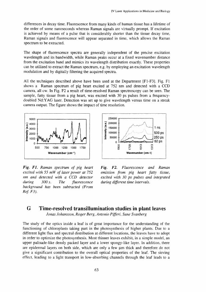
IV Laser Applications in Medicine and Biology
differences in decay time. Fluorescence from many kinds of human tissue has a lifetime of the order of some nanoseconds whereas Raman signals are virtually prompt. If excitation is achieved by means of a pulse that is considerably shorter than the tissue decay time, Raman signals and fluorescence will appear separated in time, which allows the Raman spectrum to be extracted.
The shape of fluorescence spectra are generally independent of the precise excitation wavelength and its bandwidth, while Raman peaks occur at a fixed wavenumber distance from the excitation band and mimics its wavelength distribution exactly. These properties can be utilized to extract the Raman spectrum, e.g. by employing an excitation wavelength modulation and by digitally filtering the acquired spectra.
All the techniques described above have been used at the Department [Fl-F3]. Fig. Fl shows a Raman spectrum of pig heart excited at 752 nm and detected with a CCD camera, all cw. In Fig. F2 a result of time-resolved Raman spectroscopy can be seen. The sample, fatty tissue from a pig heart, was excited with 30 ps pulses from a frequencydoubled Nd:Y AG laser. Detection was set up to give wavelength versus time on a streak camera output. The figure shows the impact of time resolution.
5000 ,--------------,
;- 4000
~ 3000 ~ ·~ 2000
~ 1000
0+--~-~~~--~-~
500 750 1000 1250 1500 1750
Wavenumber (cm-1)
Fig. Fl. Raman spectrum of pig heart excited with 55 mW of laser power at 752 nm and detected with a CCD detector during 300 s. The fluorescence background has been subtracted (From Ref F3).
250000 ......---------,
;- 200000 J ~ 150000 . .. ·. •o. i 1 ns > . ·i 1ooooo ~ 500 ps ~ 5oooo 250 ps
o, 82 ps 0 0 Ol
Ol
!?. "' "' Wavenumber (cm-1)
Fig. F2. Fluorescence and Raman emission from pig heart fatty tissue, excited with 30 ps pulses and integrated during different time intervals.
G Time-resolved transillumination studies in plant leaves Jonas Johansson, Roger Berg, Antonio Pifferi, Sune Svanberg
The study of the optics inside a leaf is of great importance for the understanding of the functioning of chloroplasts taking part in the photosynthesis of higher plants. Due to a different light flux and spectral distribution at different locations, the leaves have to adopt in order to optimize the photosynthesis. Most thinner leaves exhibit, in a simple model, an upper palisade-like densly packed layer and a lower spongy-like layer. In addition, there are epidermal layers on both side, which are only a few !lm thick and therefore do not give a significant contribution to the overall optical properties of the leaf. The sieving effect, leading to a light transport in low-absorbing channels through the leaf leads to a
63

Progress Report 1995-1996, Div. of Atomic Physics, LTH, Lund, Sweden
further heterogeniety and thus complicting the task of elucidating the optics of the leaf. The light distribution in the leaf is determined by the relative contribution of the light absorption and scattering. In a case where the absorption dominates over scattering, the light attenuation follows the Beer-Lambert law, which predicts an exponential fall-off of the light flux as a function of depth. On the other hand, if the scattereing is dominant, the light attenuation will be more determined by diffusion and the light will therefore be more evenly distributed within the leaf.
(/)
c Q) -
Phaseolus high chi
2.J ,~ ~--------~----~--~
'-.J 1 0 20 30 40 0 _J Time (ps)
Phaseolus low chi
10 20 30 40
Time (ps)
Fig. Gl. Transillumination curves at 500, 670 and 740 nm for phaseolus leaves of different chlorophyll concentrations. The apparatus response function is included in both graphs.
Different approaches for determining the optical properties of leaves have been used. Theoretical calculations have been carried out using e.g. Kubelka-Munck theory but the result has a limited value since the complicated geometry of leaves is impossible to take into acount. A more direct approach was presented, where the light flux was measured using optical fibers for different depths and angles within a leaf. This investigation gave a good result. However, the possibility of a perturbation of the system by inserting the fibers can never be ruled out. Our approach was to measure the time distribution of a 200 fs light pulse after transmitted through a leaf. The result of these measurements were correlated with a forward-feed Monte Carlo fitting procedure and data for the absorption and reduced scattering coefficients were evaluated. In addition, a simulation using these parameters then gave the light flux in a cross section of the leaves. Below examples for two different 200~m thich phaseolus leaves with high and low chlorophyll concentration, respectively, are given. Transillumination curves were recorded at wavelengths corresponding to low absorption (550 nm), high absorption (670 nm) and no absorption (740 nm).
H Analytical Chemistry Jonas Johansson
The use of spectroscopic detection methods for chromatography has become incTeasingly important in analytical chemistry. The more traditionally used UV absorption detection has for many applications been replaced by fluorescence detection mainly due to its much
64

IV Laser Applications in Medicine and Biology
higher sensitivity. A few years ago a project on fluorescence imaging detection for capillary electrophoresis was initiated. This project is a collaboration between the medical group at the Division of Atomic Physics and the Department of Technical Analytical Chemistry at Lund Institute of Technology. The project is now extended to include several different spectroscopic techniques as well as many chromatographic applications other than capillary electrophoresis.
Fluorescence imaging during chromatography is an important tool for acqumng information about separations processes [H 1, H2]. Such information is important for the understanding of new chromatographic problems and provides a fast way for optimising analytical work. A capillary gel electrophoresis separation of a DNA sequence was used as a test sample to investigate the possibilities of the imaging system. Part of the separation was followed and information about peak dispersion was extracted. In another experiment isotachophoretic preconcentration of a rhodamine sample was investigated. The dynamics of the preconcentration was obtained and compared with theoretical calculations. The actual preconcentration was found to be faster than what was predicted from theory. Investigations of gradient chromatography, double stacking preconcentration, preconcentration on frits and bubble cell detection have also been performed. Recently, a piezo-electric drop injector for capillary electrophoresis has been constructed and tested in collaboration with Dept. of Electrical Measurements in Lund. This injector is capable of ejecting drops as small as down to a few tens of a pico litre. Below are six fluorescence images shown in 2 different magnifications of the capillary after injection of 30 drops.
Fig. HI. Six consecutive fluorescence images of dansylated amino acids after drop injection into capillary. The capillary end was simultaneously imaged at 2 different magnifications as indicated in the first frame. In addition, an intensity scan through the lower part of the figure is included in each frame. The time after injection is also indicated.
65

Progress Report 1995-1996, Div. of Atomic Physics, LTH, Lund, Sweden
References
[I] C. af Klinteberg, K. Svanberg, and I. Wang (eds.), "Lund university medical laser centre, progress report 1993-94!95", (Lund University, Lund, Sweden, 1995).
[2] R. Berg, "Laser-based cancer diagnostics and therapy - Tissue optics considerations", Dissertation thesis, Lund Institute of Technology, (1995).
[3] I. Rokahr, "Fluorescence spectroscopy applied to clinical tumour identification and to microscopy", Licenciate thesis, Lund Institute of Technology, Lund Reports on Atomic Physics LRAP-182, (1995).
[4] C. Sturesson, "Thermodynamical and biological aspects of laser-induced heat treatment in medicine". Licentiate Thesis, Lund Institute of Technology, Lund Reports on Atomic Physics LRAP-203, (1996).
[5] S. Andersson-Engels,C. af Klinteberg,K. Svanberg, S. Svanberg, "In vivo fluorescence imaging for tissue diagnostics", Phys. Med, Bioi. (invited paper) (accepted) (1996).
[6] K. Svanberg and S. Svanberg, "Laser spectroscopy in medicine", to be published (1994). (Invited paper).
[7] K. Svanberg, C. af Klinteberg, A. Nilsson, I. Wang, S. Andersson-Engels and S. Svanberg, "Laser spectroscopy in tissue characterisation", Biomedical Optical Spectroscopy :md Diagnostics, Orlando, Florida, USA, (March 18-22, 1996). (Invited talk).
(8] S. Svanberg, "Tissue diagnostics using lasers", in Lasers in Medicine, eds. G. Pettit and R.W. Waynant, (Wiley, New York, in press, 1997). (Invited chapter, in press).
[9] S. Svanberg, S. Andersson-Engels, R. Berg, J. Johansson and C. af Klinteberg, "Laserbaserad medicinsk diagnostik och terapi", NUTEKs Medicintekniska konferens, Stockholm, Sweden, (April 5-6, 1995). (In Swedish).
[ 10] S. Sv:mberg, S. Andersson-Engels, R. Berg, J. Johansson and C. af Klinteberg, "Laserbaserad medicinsk diagnostik och terapi", NUTEKs Medicintekniska konferens, Stockholm, Sweden, (April 18-19, 1996). (In Swedish).
[II] S. Svanberg, "Real-world applications of laser spectroscopy", Opt. Phot. News September, 17-20 (1996).
[AI] 0. Trepte, I. Rokahr, S. Andersson-Engels and K. Carlsson, "Studies of porphyrin containing specimens using an optical spectrometer connected to a confocal scanning laser microscope", J. Microsc. 176, 238-244 (1994).
[A2] I. Rokahr, J. Carlsson and S. Andersson-Engels, "Two-photon excited fluorescence microscopy combined with spectral and time-resolved measurements for fluorophore identification". Photonics West '95, S:mJose, California, USA. (1995).
[A3) M. Obrink, "Study of two-photon excited fluorescence microscopy for spectroscopic studies of tissue", Diploma paper, Lund Institute of Technology, Lund Reports on Atomic Physics LRAP-178, (1995).
[A4] 0. Trepte, I. Rokahr and S. Andersson-Engels, "Optical spectrometer for a confocal scanning laser microscope with applications in porphyrin-containing specimens", in Three-Dimensional Microscopy: Image Acquisition and Processing II, eds. Wilson T and Cogswell CJ, Proc. SPIE 2412, 157-164, (1995).
[A5] 0. Trepte, I. Rokahr and S. Andersson-Engels, "Optical spectrometer for a confocal scanning laser microscope with applications in porphyrin-containing specimens", Photonics West '95, San Jose, California, USA, (1995). (submitted abstract).
[A6] S. Andersson-Engels, I. Rokahr and J. Carlsson, "Time- and wavelength-resolved spectroscopy in two-photon excited fluorescence microscopy", J. Microsc. 176, 195-203 (1994).
[A7] S. Andersson-Engels, J. Carlsson and I. Rokahr, "Two-photon excited fluorescence microscopy with time- and wavelength resolved detection". Ultrafast Phenomena, San Diego, California, USA. (May 28-June I, 1996).
[A8] W. Ali:m. S. Andersson-Engels, K. Svanberg and S. Svanberg, "Laser-induced fluorescence studies of meso-tetra( hydroxyphenyl)chlorin in malignant and normal tissues in rat", Br. J. Cancer 70, 880-885 (1994).
o6

IV Laser Applications in Medicine and Biology
[A9] H. Nilsson, J. Johansson, K. Svanberg, S. Svanberg, G. Jori, E. Reddi, A. Segalla, D. Gust, A.L. Moore and T.A. Moore, "Laser-induced fluorescence in malignant and normal tissue in mice injected with two different carotenoporphyrins", Br. J. C~mcer 70, 873-879 (1994).
[AlO] J. Johansson, R. Berg, K. Svanberg and S. Svanberg, "Laser-induced fluorescence studies of normal and malignant tumour tissue of rat following intravenous injection of o-amino levulinic acid", Lasers Surg. Med. 20,272-279 (1997).
[All] C. af Klinteberg, A. Pifferi, D.L. Liu, F. Bengtsson, S. Andersson-Engels and K. Svanberg, "Influence of diazepam on the accumulation of protoporphyrin IX following injection of o-amino levulinic acid in rats", 12th International Congress on Photobiology, ICP'96, Vienna, Austria, (September 1-6, 1996).
[A 12] R. Berg, "Spectrum Evaluator - a program for evaluating fluorescence spectra", Report, Lund Institute of Technology, Lund Reports on Atomic Physics LRAP-177, (1995).
[Al3] L.-G. Stri:imblad, S. Andersson-Engels, A. Brun, J. Johansson, L.G. Salford and K. Svanberg, "Peroperative recording of laser-induced fluorescence from human and rodent malignant astrocytomas", Lasers Med. Sci. (1996). (Submitted).
[Al4] J. Aganauskiene, S. Andersson-Engels, C. Blomstri:im-Lundquist, B. Olsson, U. Stenram, K. Svanberg and S. Svanberg, "Characterization of myocardial tissue utilizing laser-induced fluorescence spectroscopy", Lasers Surg. Med. (1996). (Submitted).
[Al5] K. Ekseth, I. Rokahr, E.-0. Backlund and S. Andersson-Engels, "Laser-induced fluorescence in brain tumors and normal brain studied during stereotactic biopsy", Americ~m Association of Neurosurgical Surgeons, (1995). (Submitted abstract).
[Al6] C. Stael von Holstein, A.M.K. Nilsson, S. Andersson-Engels, R. Willen, B. Walther ~md K. Svanberg, "Detection of adenocarcinoma in Barrett's oesophagus by means of laser-induced fluorescence", Gut 39,711-716 (1996).
[Al7] A.M.K. Nilsson, C. Stael von Holstein, S. Andersson-Engels, R. Willen, B. Walther and K. Svanberg, "Clinical detection studies of Barrett's metaplasia and oesophageal adenocarcinoma by means of laser-induced fluorescence", in Optical Biopsies, eds. Cubeddu R, Mordon SR and Svanberg K, Proc. SPIE 2627,49-56, (1995).
[Al8] I. Rokahr, S. Andersson-Engels, S. Svanberg, M.A. D'Hallewin, L. Baert, I. Wang and K. Svanberg, "Optical detection of human urinary bladder carcinoma utilising tissue autofluorescence and protoporphyrin IX-induced fluorescence following /ow-dose ALA instillation", in Optical Biopsies, eds. Cubeddu R, Mordon SR and Svanberg K, Proc. SPIE 2627, 2-12, (1995).
[A19] I. Wang, L. Pais Clemente, R.M.G. Pratas, E. Cardoso, M. Pais Clemente, S. Mont:'\n, S. Sv:mberg and K. Svanberg, "Fluorescence diagnostics and kinetic studies in malignant and premalignant lesions and in normal epithelium in the head and neck area utilising /ow-dose oaminolevulinic acid sensitisation", in preparation (1997). (Manuscript).
[A20] R. Rydell, K. Svanberg, J. Wennerberg, A. Elner and S. Svanberg, "Photodynamic imaging of cancer in situ and cancer of the head and neck", Head Neck 16,521 (1994).
[A21] C. Lindquist, S. Andersson-Engels, S. Svanberg, I. Wang, K. Svanberg, R. Rydell, J. Wennerberg and A. Elner, "Spectral characterisation of head and neck cancers utilising tissue autofluorescence", BiOS '96, San Jose, USA, (January 27- February 2, 1996).
[A22] C. Lindquist, S. Andersson-Engels, S. Svanberg, I. Wang, K. Svanberg, R. Rydell, J. Wennerberg and A. Elner, "Spectral characterisation of head and neck cancers utilising tissue autofluorescence", BiOS Europe '96, SPIE, Vienna, Austria, (September 7-10, 1996).
[A23] H. Messmann, P. Mlkvy, S. Montan, I. Wang, A. Nilsson, K. Svanberg, S. Svanberg, A. MacRobert and S. Bown, "Endoscopic fluorescence of gastrointestinal neoplasia after sensitisation with 5-amino laevulinic acid (ALA) or Photofrin", in 5th International Photodymunic Association Biennial Meeting, ed. Cortese DA, Proc. SPIE 2371, 506-509, (1994).
[A24] C. Lindquist. S. Montan, K. Svanberg, P. Slezak, E. Jaramillo and K. Koizumi, "Laser induced fluorescence studies on colonic mucosa in vivo, with and without ALA", BiOS Europe '96, SPIE, Vienna, Austria, (September 7-10, 1996).
[A25] S. Andersson-Engels, J. Johansson and S. Svanberg, "Medical diagnostic system based on simultaneous multi-spectral fluorescence imaging", Appl. Opt. 33, 8022-8029 (1994).
67

Progress Report 1995-1996, Div. of Atomic Physics, LTH, Lund, Sweden
[A26] S. Andersson-Engels, R. Berg, K. Svanberg and S. Svanberg, "Multi-colour fluorescence imaging in combination with photodynamic therapy of B-amino levulinic acid (ALA) sensitised skin malignancies", Bioimaging 3, 134-143 (1995).
[A27] K. Svanberg, I. Wang, S. Colleen, I. Idvall, C. Ingvar, R. Rydell, D. Jochrun, H. Diddens. S. Bown, G. Grant, S. Mont.'\n, S. Andersson-Engels and S. Svanberg, "Clinical multi-colour fluorescence imaging of malignant tumours - initial experience", Act.'t Radio!. 38, (1997). (In press).
[B1] A.M.K. Nilsson, R. Berg and S. Andersson-Engels, "Integrating sphere measurements of tissue optical properties for accurate PDT dosimetry", in Laser Interaction with Hard and Soft Tissue II, eds. Albrecht HJ, Delacret.u GP, Meier THeta/., Proc. SPIE 2323,47-57, (1994).
[B2] A.M.K. Nilsson, R. Berg and S. Andersson-Engels, ''Measurements of the optical properties of tissue in conjunction with photodynamic therapy", Appl. Opt. 34, 4609-4619 (1995).
[B3] A.M.K. Nilsson, G.W. Lucassen, W. Verkruysse, S. Andersson-Engels ~md M.J.C. v~m Gernert, "Changes in optical properties of human whole blood in vitro due to slow heating", Photochem. Photobiol. 65, 366-373 (1997).
[B4] A.M.K. Nilsson, G.W. Lucassen, W. Verkruysse, S. Andersson-Engels and M.J.C. v~m Gemert, "Optical properties of human whole blood - changes due to slow heating", in Laser-Tissue Interaction and Tissue Optics II, eds. Albrecht HJ, Delacretaz GP, Meier TH et al., Proc. SPIE 2923, 24-34, (1996).
[B5] W. Verkruysse, A.M.K. Nilsson, T.E. Milner, J.F. Beek, G.W. Lucassen and M.J.C. van Gemert, "Optical absorption of blood depends on temperature during a 0.5 ms laser pulse at 586 nm", Photochem. Photobiol. (1997). (Submitted to).
[B6] P. Alsholm, "Light scattering by individual and groups of spheroidal particles", Diploma Paper, Lund Institute of Technology, Lund Reports on Atomic Physics LRAP-200, (1996).
[C1] D.L. Liu, K. Svanberg, I. Wang, S. Andersson-Engels and S. Svanberg, "Laser Doppler perfusion imaging: New technique for determination of perfusion and reperfusion of splanchnic organs and tumor tissue", Lasers Surg. Med. 20, 473-479 ( 1997).
[C2] S. Andersson-Engels, R. Berg, L.-G. Lindberg, P.A. Oberg and S. Svanberg, "Quantification of blood flow using time-resolved reflected light", BiOS Europe '94, Lille, France, (September 6-10, 1994 ).
[C3] A. Persson, "Optical diagnostics of tissue pathologies based on elastic scattering properties", Diploma Paper, Lund Institute of Technology, Lund Reports on Atomic Physics LRAP-169, (1996).
[C4] D. Heinrich, "In vivo Untersuchungen von Gewebe durch Detektion diffusen Lichtes im sichtbaren und nahen infraroten Spektralbereich", Diploma Paper, Lund Institute of Technology, Lund Reports on Atomic Physics LRAP-196, (1996). (In German).
[C5] R. Berg, S. Andersson-Engels, C. af Klinteberg, S. Svanberg and 0. Jarlman, "Optical imaging for medical diagnostics using femtosecond white light", in Advances in Optical Imaging and Photon Migration, ed. Alfano RR, Proc. OSA 21, 126-128, (1994).
[C6] C. af Klinteberg, R. Berg, C. Lindquist, S. Andersson-Engels and S. Svanberg, "Diffusely scattered femtosecond white light examination of breast tissue in vitro and in vivo", in Photon Propagation in Tissues, eds. Chance B, Delpy DT and Muller GJ, Proc. SPIE 2626, 149-157, ( 1995).
[C7] 0. Jarlman, R. Berg, S. Andersson-Engels, S. Svanberg and H. Pettersson, "Time-resolved white light transillumination for optical imaging", Act.'t Radio!. 38, 185-189 (1997).
[C8] S. Andersson-Engels, R. Berg, J. Carlsson, J. Johansson, C. af Klinteberg, C. Lindquist, L. Malmqvist, W. Persson, A. Pifferi, S. Svanberg, L.O. Bjorn, P. Hellentin and 0. Jarlman, "Modelling of the propagation of light in turbid media from femtosecond pulse transmission measurements", Ultrafast Phenomena, s~m Diego, California, USA, (May 28-June I, 1996).
[C9] C. af Klinteberg, C. Lindquist, A. Pifferi, R. Berg, S. Andersson-Engels and S. Svanberg, "Diffusely scattered femtosecond white light examination of breast tissue", in Trends in Optics and Photonics, ed. Cubeddu R, Proc. Optical Society of America 6, 30-35, (1996). (Invited paper).
[CIO] M.S. Patterson, S. Andersson-Engels, B.C. Wilson ~md E. Osei, "Absorption spectroscopy in tissue-simulating materials: a theoretical and experimental study of photon paths", Appl. Opt. 34, 22-30 (1995).
68
IV Laser Applications in Medicine and Biology
[C11] R. Berg, S. Andersson-Engels, 0. Jarlman and S. Svanberg, "Time-gated viewing studies on tissue/ike phantoms", Appl. Opt 35, 3432-3440 (1996).
[Cl2] 0. Jarlman, R. Berg, S. Andersson-Engels, S. Svanberg and H. Pettersson, "Laser transillumination of breast tissue phantoms usinR time-resolved techniques", Eur. Radio!. 6, 387-392 (1996).
[C13] H. Tagesson, "Frequency domain near-IR light studies of turbid media with means to detect breast cancer", Diploma Paper, Lund Institute of Technology, Lund Reports on Atomic Physics LRAP-180, (1995).
[Cl4] C. Lindquist, R. Berg <md S. Andersson-Engels, "Numerical diffusion modelling of interfering photon density waves for optical mammography", in Photon Transport in Highly Scattering Tissue, eds. Avrillier S, Ch<mce B, Muller GJ eta/., Proc. SPIE 2326,31-40, (1994).
[Cl5] C. Lindquist, A. Pifferi, R. Berg, S. Andersson-Engels and S. Svanberg, "Reconstruction of diffuse photon-density wave interference in turbid media from time-resolved transmittance measurements", Appl. Phys. Lett 69, 1674-1676 (1996).
[C16] C. Lindquist, A. Pifferi, S. Andersson-Engels and S. Svanberg, "Reconstruction of diffuse photon density wave interference for detection of small inhomogeneities in turbid media", in Trends in Optics and Photonics, eds. Alfano RR and Fujimoto JG, Proc. Optical Society of America 2, 148-151, (1996).
[Dl] K. Svanberg, D.L. Liu, I. Wang, S. Andersson-Engels, U. Stenram and S. Svanberg, "Photodynamic therapy using intravenous 8-amino/aevulinic acid-induced protoporphyrin IX sensitisation in experimental hepatic tumours in rats", Br. J. Cancer 74, 1526-1533 ( 1996).
[D2] D.L. Liu, I. Wang, S. Andersson-Engels, C.H. HAkansson, U. Stenram and K. Svanberg, "Intraoperative laser-induced photodynamic therapy in the treatment of experimental hepatic tumours". Eur. J. Gastroenterol. Hepatol. 7, 1073-1080 (1995).
[D3] K. Svanberg, B. Bauer, S. Andersson-Engels and S. Svanberg, "Photodynamic therapy utilizing topical 8-amino levulinic acid (ALA) in the treatment of basal cell carcinoma of the eye lids", Lasers Opthalmol. (1996). (Invited paper, Manuscript).
[D4] I. Wang, K. Svanberg, S. Andersson-Engels, R. Berg and S. Svanberg, "Photodynamic therapy of non-melanoma skin malignancies with topical 8-amino levulinic acid: diagnostic measurements", in 5th International Photodyruunic Association Biennial Meeting, ed. Cortese DA, Proc. SPIE 2371,243-252, (1994).
[D5] I. Wang, S. Andersson-Engels, G.E. Nilsson, K. Wftrdell and K. Svanberg, "Superficial bloodflow following photodynamic therapy of malignant skin tumours measured by laser Doppler imaging", Br. J. Dennatol. 136, 184-189 (1997).
[D6] C. af Klinteberg, A.M.K. Nilsson, I. W<mg, S. Andersson-Engels, S. Svanberg and K. Sv<mberg, "Laser-induced fluorescence diagnostics of basal cell carcinomas of the skin following topical ALA application", in Optical Biopsies and Microscopic Techniques, eds. Bigio lJ, Grundfest WS, Schneckenburger H eta/., Proc. SPIE 2926,32-40, (1996).
[D7] C. af Klinteberg, A.M.K. Nilsson, I. Wang, S. Andersson-Engels, K. Svanberg and S. Svanberg, "Diagnostics of basal cell carcinomas of the skin using laser-induced fluorescence following topical ALA application", 12th International Congress on Photobiology, ICP'96, Vienna, Austria, (September 1-6, 1996).
[D8] I. Wang, K. Svanberg, N. Bendsoe, C. af Klinteberg, A. Nilsson, S. Andersson-Engels and S. Sv<mberg, "Photodynamic therapy utilising topical ALA versus conventional cryo therapy of basal cell carcinomas; diagnostic measurements and treatment results", 6th Biennial Meeting of the International Photodynamic Association, Melbourne, Australia, (March 10-14, 1996).
[Ell C. Sturesson, "Theoretical modelling of the temperature distribution in laser-induced hyperthermia", Diploma Paper, Lund Institute of Technology. Lund Reports on Atomic Physics LRAP-159, (1994).
[E2] C. Sturesson and S. Andersson-Engels, "A mathematical model for predicting the temperature distribution in laser-induced hyperthermia. Experimental evaluation and applications", Phys. Med. Bioi. 40,2037-2052 (1995).
[E3] C. Sturesson, S. Andersson-Engels and S. Svanberg, "A numerical model for the temperature increase in surface applied laser induced thermo therapy with applications to tumor blood flow estimations", in Laser-induced Interstitial Thennotherapy, eds. G. Miiller and A. Roggan, pp. 157-173 (SPIE. Bellinghmn, W A, 1995).
69
Progress Report 1995-1996, Div. of Atomic Physics, LTH, Lund, Sweden
[E4] C. Sturesson and S. Andersson-Engels, "Mathematical modelling of dynamic cooling and preheating, used to increase the depth of selective damage to blood vessels in laser treatment of port wine stains", Phys. Med. Bioi. 41,413-428 (1996).
[E5] C. Sturesson and S. Andersson-Engels, "Temperature control using a water-cooled applicator. Implications for transurethral laser-induced thermo-therapy of benign prostatic hyperplasia", Med. Phys. 24, 461-470 (1997).
[E6] C. Sturesson and S. Andersson-Engels, "Theoretical analysis of transurethral laser-induced thermo therapy for treatment of benign prostatic hyperplasia. Evaluation of a water-cooled applicator", Phys. Med. Bioi. 41, 445-463 (1996).
[E7] L. Liu, S. Andersson-Engels, C. Sturesson, K. Sv<mberg, C.H. HA!umsson <md S. Sv<mberg, "Tumour vessel damage resulting from laser-induced hyperthermia alone and in combination with photodynamic therapy", Cancer Lett. Ill, 1-9 ( 1996).
[Fl] J. Bood and H. Carlsson, "Raman and infrared spectroscopy for tissue diagnostics", Diploma Paper, Lund Institute of Technology, Lund Reports on Atomic Physics LRAP-168, (1994).
[F2] A. Akesson, "Near-infrared Raman spectroscopy for tissue characterisation: adapt ion of the setup for medical use", Diploma Paper, Lund Institute of Technology, Lund Reports on Atomic Physics LRAP-198, (1996).
[F3] M. Gustafsson, "Spectroscopic studies of tissue using near-infrared Raman microscopy", Diploma Paper, Lund Institute of Technology, Lund Reports on Atomic Physics LRAP-207, (1997).
[HI] S. Nilsson, J. Johansson, M. Mecklenburg, S. Birnbaum, S. Svanberg, K.-G. Wahlund, K. Mosbach, A. Miyabayashi and P.O. Larsson, "Real-time fluorescence imaging of capillary electrophoresis: Separation of nucleic acids", J. Capillary Electrophoresis 2, 46-52 (1995).
[H2] J. Joh<msson, T. Johansson and S. Nilsson, "Real-time fluorescence imaging in analytical chemistry", in Biomedical Optoelectronics in Clinical Chemistry and Biotechnology, eds. Andersson-Engels S, Corti M, Weber HP eta/., Proc. SPIE 2629,2-9, (1995).
70

V Industrial Applications
V Industrial Applications
Industrial applications cover areas of interest for some major Swedish industries, the electrotechnical industry and the paper industry. Spectroscopic studies of electric breakdown and circuit breaker arcs are performed in cooperation with ABB Corporate Research. The studies concerning the distribution of ink in paper and fluorescence measurements of paper are performed in cooperation with paper mills within the MoDoHolmen and Stora groups.
A The Physics of Electric Breakdown Peter Barmann, Joachim Garmer*, Stefan Kroll, and Anders Sunesson
*M Sc Student
Electric breakdown is a phenomenon of great scientific and economic interest. In liquids, the understanding of the phenomena preceding electric breakdown is at best incomplete, although modern analysis methods are revealing more and more of the processes. For gases, the situation is better, although much still remains to be done. The power industry has a great interest in knowing how best to avoid electric breakdown and how to describe and control discharges in equipment.
At the Department for High Voltage Technology at ABB Corporate Research in Viisteras, a research centre within ABB, a leading manufacturer of electrotechnical equipment in the world, research in insulation systems and related topics is being performed. At the Division of Atomic Physics broad experience of optical diagnostic methods can be found. Since 1991, ABB Corporate Research and the Division of Atomic Physics have been cooperating on a project to study the physics of liquid electric breakdown and related topics. The project has initiated a new research area in Lund, and yielded new methods of analysis relevant for ABB, as well as new results for the scientific community and ABB.
Within the project laser-induced initiation of breakdown in liquids, transient currents and broadband light emission from pre-breakdown phenomena have been studied. Spectral analysis of the pre-breakdown phenomena has been performed with both temporal and spatial resolution, and it has been possible to deduce, for the first time to our knowledge, the electron density in the pre-breakdown channel as a function of time and space. It has also been possible to infer spatial characteristics of the pre-breakdown channel impossible to monitor with any other technique [A l-A6]. Imaging of the pre-breakdown process has also been performed [A 7]. The laser-initiation technique and the broadband light emission measurement techniques developed at the Division of Atomic Physics have been adopted by ABB Corporate Research and are now routinely used in Vasteras.
A project involving spectroscopic studies of circuit breakers has also been initiated. Measurements have also been performed on a circuit breaker arc in SF6 using emission spectroscopy. The results include knowledge about the arc temperature and the interruption process [A8].
71
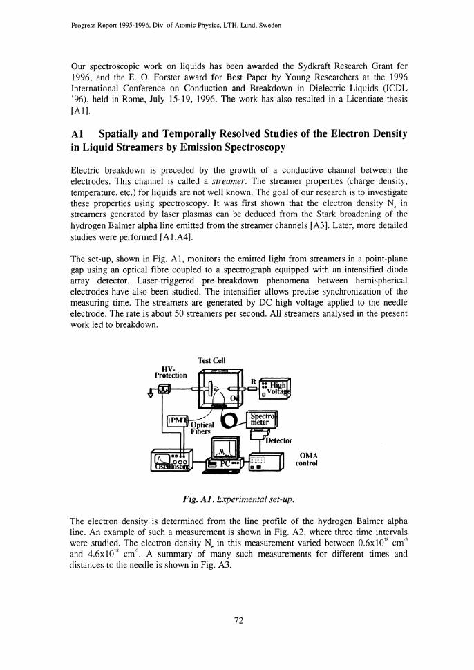
Progress Report 1995-1996, Div. of Atomic Physics, LTH, Lund, Sweden
Our spectroscopic work on liquids has been awarded the Sydkraft Research Grant for 1996, and the E. 0. Forster award for Best Paper by Young Researchers at the 1996 International Conference on Conduction and Breakdown in Dielectric Liquids (ICDL '96), held in Rome, July 15-19, 1996. The work has also resulted in a Licentiate thesis [A 1].
Al Spatially and Temporally Resolved Studies of the Electron Density in Liquid Streamers by Emission Spectroscopy
Electric breakdown is preceded by the growth of a conductive channel between the electrodes. This channel is called a streamer. The streamer properties (charge density, temperature, etc.) for liquids are not well known. The goal of our research is to investigate these properties using spectroscopy. It was first shown that the electron density N, in streamers generated by laser plasmas can be deduced from the Stark broadening of the hydrogen Balmer alpha line emitted from the streamer channels [A3]. Later, more detailed studies were performed [Al,A4].
The set-up, shown in Fig. A 1, monitors the emitted light from streamers in a point-plane gap using an optical fibre coupled to a spectrograph equipped with an intensified diode array detector. Laser-triggered pre-breakdown phenomena between hemispherical electrodes have also been studied. The intensifier allows precise synchronization of the measuring time. The streamers are generated by DC high voltage applied to the needle electrode. The rate is about 50 streamers per second. All streamers analysed in the present work led to breakdown.
Test Cell
Fig. AI. Experimental set-up.
OMA control
The electron density is determined from the line profile of the hydrogen Balmer alpha line. An example of such a measurement is shown in Fig. A2, where three time intervals were studied. The electron density N, in this measurement varied between 0.6x10¹⁸ em·' and 4.6xl018 em·'. A summary of many such measurements for different times and distances to the needle is shown in Fig. A3.
72

.£2 ~
@ 3
...... ..s 40L0~~5~00~~6~0~0 ~7-'00 400 500 600 700 400 500
Wavelength I run
V Industrial Applications
600 700
Fig. A2. Streamer spectra for three time intervals before breakdown: a) 1.7-0.71..ls, b) 0.7-02 l..lS, c) 0.3-0 l..lS before breakdown. The C2 bands between 450 and 550 nm are clearly visible and the variations in the width of the Ha line at 656 nm indicate electron densities of a) 0.6xl018 cm.3 ; b) 1.3xl018 cm.3 ; and c) 4.6xl018 cm·3 • Needle-plane distance, 10 mm and high voltage, +60 kV.
"" ~ 8
(J
co .... 8 l __.a B
Distance .. :::: EJ from needle * e ---.::r
6
l 4 --------~ ~ l'l
_g a 2 a 8 -c
(J
..!l ~
0 0 -100 -200 -300 -400 -500
Time I nanoseconds
Fig. A3. Electron density as function of distance from needle and time during the last l..lS of propagation. Needle-plane gap, 10 mm and high voltage, +57 kV.
The results for all times and distances can be summarized thus: • N, is less than 1016 cm·3 for propagation times up to ll..ls before breakdown • During the last l..lS before breakdown, N, increases to up to 1019 cm·3•
For the last l..lS the following also holds. • N, is approximately constant over time at a given spatial position, but increases towards
the streamer tip by up to a factor of four. • The Ha emission intensity increases by orders of magnitude during the last l..lS. • Higher spectral resolution measurements show that the Ha profiles contain two
components, a narrow one (instrumental linewidth-limited) corresponding to N,<l0' 6
em·' and a broader one corresponding to N,> 1018 cm·3 •
We believe that the last point means that the electron density is not uniformly distributed across the streamer cross-section and that there is some radial structure. This is also supported by the fact that the injected energy during the relevant time intervals is not sufficient to allow ionization of the complete streamer volume to the degree indicated by the higher electron density. Two hypotheses may explain the results: the high charge density region is situated at the centre of the streamer channel or at the rim of the streamer channel, see Fig. A4.
73

Progress Report 1995-1996, Div. of Atomic Physics, LTH, Lund, Sweden
Main streamer volume with low degree of ionisation and narrow hydrogen lines.
Streamer core with high degree of ionisation. Broad, shifted hydrogen lines.
Fig. A4. Possible radial structures of positive streamer. Both models would account for the detection of high electron densities while the average electron density remains low.
A2 Shadow Technique Imaging of Prebreakdown Streamers
This Master's project [A7] continued the work in an earlier project [A9] where streamer imaging technology was investigated. The technical problems encountered in the earlier work were solved and a detailed study of laser-plasma-produced bubbles in a liquid was made. It was also investigated how the bubbles in the presence of an electric field would generate streamers leading to breakdown. Earlier hypotheses about the process were tested using the images. An example of an image is shown in Fig. A5.
HV Electrode
Plasma-generated Bubble
Ground Electrode
Fig. AS. Image of laser-generated gas bubble in a liquid (in the middle) and propagating positive and negative streamers. The positive HV electrode is above the bubble and the ground electrode is below the bubble.
The bubble dynamics was tested against the Rayleigh theory. It was found that the experimental results followed the theory very well. It was also found that streamers could
74

V Industrial Applications
be imaged without any great difficulty, and the process leading to breakdown could be followed in detail. It was shown that streamers that propagate from the bubble were generated, regardless of the polarity of the gap or the position of the bubble. Normally, a positive streamer is first generated towards the lowest potential electrode. After this, a negative streamer, propagating towards the higher potential electrode, completes the process. The propagation speed of negative streamers was estimated to be 200 m/s whereas the positive streamers were too fast to follow. The study also created a database of images which will be used in future research to determine the exact cause of streamer initiation from the bubbles.
A3 Spectroscopic Measurements on a Circuit-Breaker Arc
In this project, an arc in a circuit-breaker was investigated using emission spectroscopy [A8]. The set-up was similar to that shown in Fig. Al, except that the test cell was replaced by a circuit breaker, and that the very intense arc that occurs when two contacts that conduct high power are separated was monitored. The breaker gas was SF6 and the walls were made of Teflon. The arc was studied for different times and currents during the process. Typical spectra are shown in Fig. A6, showing the four typical spectra that were obtained during the measurements.
tKhl Type I
::i Jljj ~ Type II ,g
:: ]
::i
~ ~ Type III
J ' ::i vJ:j ~ ·~ Type IV 2 .s
400 500 600 700 Wavelength/nm
Fig. A6. Typical spectra from circuit-breaker arcs drawn in SF, with Teflon walls and copper electrodes. Types I and IV occur early in the process, Type II towards the end of the process, and Type III at maximum current. Note the strong absorption bands in Type III. These correspond to absorption of the arc plasma radiation by C2 bands in the gas surrounding the arc.
The spectral analysis reveals that the arc consists of sulphur and fluorine from the filling gas, and copper and carbon from the electrodes and walls. We also intend to use spectra to deduce the arc temperature.
75

Progress Report 1995-1996, Div. of Atomic Physics, L TH, Lund, Sweden
B Optical spectroscopy of paper Hans Busk, Jorgen Carlsson, Johan Jason* Lennart Malmqvist, Magnus Nilsson,
Willy Persson and Claes-Goran Wahlstrom
*MSc student
The studies of paper described in this section are being carried out within the Paper-PrintPhysics network, consisting of the Division of atomic physics and the Division of nuclear physics at the Lund institute of technology and the Centre for imaging sciences and technologies at Halmstad university.
Bl The propagation of light in paper
Paper has a complicated inner structure, made up of fibres, fine-material and pores. With characteristic differences for different kinds of paper this structure determines how light propagates inside the paper and how the paper appears in reflected or transmitted light. For graphical paper the optical properties also have a decisive influence on how the print will appear. For an accurate description of the optical properties of paper it would be advantageous to use realistic three-dimensional models of the structure. Three-dimensional modelling is also important for studies of print quality.
Three-dimensional models of paper are being developed. The models are used in Monte Carlo simulations and compared with experimental results [B 1-B4]. The optical properties most easily measured for a paper are its reflectivity and transrnittivity. In addition to such measurements, time-resolved experiments on the propagation of light in paper have also been performed [B1]. These studies were performed using the 0.2 ps pulses from a Ti:sapphire laser system and a streak-camera. Measurements were carried out on both systematic series of well characterised sheets of paper and on ordinary newsprint.
" 10 Figure B 1 shows the reflected intensity along ... _s_ ~ a radius from the centre of a printed dot on ~ 60 - top anisotropy parameter= o.o paper. The dot has a radius of 1 00 J.!m and no = - bottom anisotropy parameter= o_g light is reflected from the part of the paper 'll 40 ~ covered by the dot. From the area outside but
~ 2:1-------+---~-~--~-~ near the dot a decrease in the reflected o 100 2oo 3oo 4oo 500 intensity is observed. This is known as optical
Radius I micrometre
Fig. BI. The normalised reflected intensity around a print dot calculated using two models which differ only in the anisotropy of the scattering.
dot gain, and makes a printed dot look larger than it is, and gives print made up of dots a higher density than the actual coverage would motivate. The reason for this is that light is scattered inside the paper. The figure shows the reflected intensity according to two different models, illustrating the influence of
the anisotropy of the scattering process. A high value, close to 1, is expected for scattering from small particles. The simplification of using isotropic scattering, corresponding to the value 0, in this case strongly underestimates the dot gain.
76
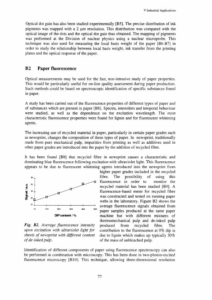
V Industrial Applications
Optical dot gain has also been studied experimentally [B5]. The precise distribution of ink pigments was mapped with a 2 11m resolution. This distribution was compared with the optical image of the dots and the optical dot gain thus obtained. The mapping of pigments was performed at the Division of nuclear physics using a nuclear microprobe. This technique was also used for measuring the local basis weight of the paper [B6-B7] in order to study the relationship between local basis weight, ink transfer from the printing plates and the optical response of the paper.
B2 Paper fluorescence
Optical measurements may be used for the fast, non-intrusive study of paper properties. This would be particularly useful for on-line quality assessment during paper production. Such methods could be based on spectroscopic identification of specific substances found in paper.
A study has been carried out of the fluorescence properties of different types of paper and of substances which are present in paper [B8]. Spectra, intensities and temporal behaviour were studied, as well as the dependence on the excitation wavelength. The most characteristic fluorescence properties were found for lignin and for fluorescent whitening agents.
The increasing use of recycled material in paper, particularly in certain paper grades such as newsprint, changes the composition of these types of paper. In newsprint, traditionally made from pure mechanical pulp, impurities from printing as well as additives used in other paper grades are introduced into the paper by the addition of recycled fibre.
It has been found [BS] that recycled fibre in newsprint causes a characteristic and dominating blue fluorescence following excitation with ultraviolet light. This fluorescence appears to be due to fluorescent whitening agents introduced into the newsprint from
8
0 10 20 30 40 50
DIP content I %
Fig. B2. Average fluorescence intensity upon excitation with ultraviolet light for sheets of newsprint with different content of de-inked pulp.
higher paper grades included in the recycled fibre. The possibility of using this fluorescence in order to monitor the recycled material has been studied [B9]. A fluorescence-based meter for recycled fibre was constructed and tested on running paper webs in the laboratory. Figure B2 shows the average fluorescence signals obtained from paper samples produced at the same paper machine but with different mixtures of thermomechanical pulp and de-inked pulp produced from recycled fibre. The contribution to the fluorescence at 0% dip is due to lignin which makes up typically 30% of the mass of unbleached pulp.
Identification of different components of paper using fluorescence spectroscopy can also be performed in combination with microscopy. This has been done in two-photon-excited fluorescence microscopy [B 1 0]. This technique, allowing three-dimensional resolution
77

Progress Report 1995-1996, Div. of Atomic Physics, LTH, Lund, Sweden
similar to that of confocal microscopy, is discussed in section IV-D. It has been used to identify lignin as well as recycled fluorescent whitening agents in newsprint.
References
[AI] P. Bfumann, "Spectroscopic Investigation of Streamers in a Dielectric Liquid", Licentiate Thesis, Lund Reports on Atomic Physics, LRAP-191, ISBN 91-628-1896-1, Lund 1996.
[A2] P. Bfumann, S. Kroll, <md A. Sunesson, "Dynamic Stark Broadening for Measuring Electron Densities in Micrometer Diameter Sized Plasma Channels", Conference on L<t~ers and ElectroOptics, CLEO '95, Baltimore, May 1995.
[A3] P .Bi\nnann, A. Sunesson, ;md S. Kroll, "Spectroscopic Measurements of Streamer Filaments in Electric Breakdown in Liquid", J. Phys. D: Applied Physics 29, 1188 (1996).
[A4] P .Bi\nnann, A. Sunesson, and S. Kroll, "Spatially and Temporally Resolved Electron Density Measurements in Streamers in Dielectric Liquids", J. Phys. D, Applied Physics 30, 856-863 (1997).
[AS] P. Bi\nnann, S. Kroll, and A. Sunesson, "Spectroscopic Measurement of Streamers in Dielectric Liquids", Ninth International Symposium on High Voltage Engineering (9th ISH), Graz, Austria, August 28-September 1 1995.
[A6] P. Bi\nnann and A. Suneson, "In-situ Spectroscopic Measurements of Streamer Properties (electron density and temperature) in Dielectric Liquids", Twelfth International Conference on Conduction ;md Breakdown in Dielectric Liquids (12th ICDL), Rome, Italy, July 15-19 1996.
[A 7] J. Gartner, "Laser-Shadow Imaging of Laser-Initiated Electric Prebreakdown Events in Transformer Oil", Master's Thesis, Lund Reports on Atomic Physics, LRAP-199, Lund 1996.
[A8] A. Sunesson, "Spectroscopic Measurements on an Ablation Dominated Circuit Breaker Arc", ABB report no. SECRC/H!I'R-96/117E (1996).
[A9] D. St,'tlhandske, "Evaluation of a Method to Image a Laser-Initiated Propagating Streamer", Master's Thesis, Lund Reports on Atomic Physics, LRAP-171, Lund 1996.
[B1] J. Carlsson, P. Hellentin, L. Malmqvist, A. Persson, W. Persson ;md C-G. Wahlstrom, ''Timeresolved studies of light-propagation in paper", Applied Optics 34, 1528 (1995).
[B2] J. Carlsson, P. Hellentin, L. Malmqvist and W. Persson, "The propagation of light in paper: modelling and Monte Carlo simulations", International paper physics conference, Niagara-on-theLake, Ontario, Canada, 11-14 September 1995.
[B3] S. Andersson-Engels, R. Berg, L.O. Bjorn, J. Carlsson, P. Hellentin, 0. Jarlman, J. Johansson, C. af Klinteberg, C. Lindquist, L. Malmqvist, W. Persson, A. Pifferi and S. Svanberg, "Modelling of the propagation of light in turbid media from femtosecond pulse transmission measurements", Ultrafast Phenomena, San Diego, USA, 28 May-1 June 1996. Also in Ultrafast Phenomena X, Eds P.F. Barbara, J.G. Fujimoto, W.H. Knox and W. Zinth, Springer, Berlin 1996.
[B4] J. C<rrlsson, L. Malmqvist ;md W. Persson, "Three-dimensional modelling of paper structure: optical properties of paper and print", Submitted to TAGA Annual Technical Conference, 4-7 May 1997, Quebec, Quebec, Canada.
[B5] H. Busk, P. Kristiansson, K. Malmqvist, L. Malmqvist, M. Nilsson and W. Persson, "Optical enhancement of closely positioned printing dots", Submitted to TAGA Annual Technical Conference, 4-7 May 1997, Quebec, Quebec, Canada.
[B6] P. Kristiansson, K. Malmqvist, L. Malmqvist, C.M. Nilsson and W. Persson, "Distribution of ink in solid prints related to the structure of the paper", International Printing <md Graphic Arts Conference, Minneapolis, USA, 16-19 September 1996.
[B7] P. Kristiansson, K. Malmqvist, L. Malmqvist, C.M. Nilsson ;md W. Persson, "Measurement of basis weight of paper by ionizing radiation", International Printing and Graphic Arts Conference, Minneapolis, USA, 16-19 September 1996.
[B8] H. Bergstrom, J. Carlsson, P. Hellentin and L. Malmqvist. "SpeClra/ly and temporally resolved studies of fluorescence from newsprint and from substances occurring in newsprint", Nordic Pulp and Paper Res. J. II, 48 ( 1996).

V Industrial Applications
[B9] J. Jason, "Construction and testing of equipment using fluorescence for on-line monitoring of the distribution of recycled fibre during newsprint production", Master Thesis, Lund Reports on Atomic Physics, LRAP-193, Lund 1996.
[BIO] S. Andersson-Engels, J. Carlsson and I. Rokahr, "Two-photon excited fluorescence microscopy with time- and wavelength resolved detection", Ultrafast Phenomena, San Diego, USA, 28 May-1 June 1996. Also in Ultrafast Phenomena X, Eds P.F. Barbara, J.G. Fujimoto, W.H. Knox ;md W. Zinth, Springer, Berlin 1996.
79

Progress Report 1995-1996, Div. of Atomic Physics, LTH, Lund, Sweden
VI Teaching Programme
Professors and lecturers: Stefan Andersson-Engels, Lars Engstrom, Hans Hertz, Gilbert Jonsson, Goran Jonsson, Stefan Kroll, Rune Kullberg, Anne L'Huillier, Hans Lundberg, Willy Persson, Rolf Petersson, Sven-Goran Pettersson, Nina Reistad, Sune Svanberg, Claes-Goran Wahlstrom. Teaching assistants: Mats Andersson, Roger Berg, Magnus Berglund, Peter Bdrmann, Ulf Elman, Ulf Gustafsson, Jonas Johansson, Peter Kauranen, Claes af Klinteberg, Charlotta Lindquist, Claire Lyngd, Lars Malmqvist, Annika Nilsson, Lars Rymell, Jonas Sandsten, Tomas Starzcewski, Carl Tillman, Raoul Zerne, Petter Weibring.
A Undergraduate teaching
At the Department of Physics, basic physics teaching is provided for the Schools of Engineering Physics (F), Electrical Engineering (E), Computer Science and Technology (D), Mechanical Engineering (M), Civil Engineering (V), Fire Protection Engineering (BI) and Chemical Engineering (K). Furthermore, specialised courses in Atomic Physics, Laser Physics, Laser Technology, Advanced Optics, Atomic and Molecular Spectroscopy, and Multispectral Imaging are given. Courses not included in the regular study programmes are Holography, Radon, Medical Laser Techniques and Technical Foundation Year.
The purpose of the courses in physics is to provide knowledge about fundamental physical concepts, laws, principles, models and their application. Experimental training is an important part of the courses.
The courses in physics are based on lectures, problem-solving sessions and laboratory work. Lectures and problem-solving sessions provide a good basis in fundamental physical principles as well as an introduction to the application of these principles. Laboratory work provides experience in the design of experiments, implementation of theory, experimental methodology and the evaluation of results.
The courses contain both theory (lectures and problem solving) and laboratory practicals. The number of hours devoted to experimental work is, as a rule, about the same as the number of hours spent on theory. During experimental sessions in basic courses, the students generally work in groups of two, and each supervisor teaches four such groups, i.e. 8 students at a time. For the specialised courses, each supervisor can teach only 4 or 6 students at a time, since the amount of equipment is limited. In the courses on atomic and molecular spectroscopy and advanced optics, research equipment is used by the students in their experimental work.
A brief survey of the courses available and the attendance is given in the Table 1. In the first column, the name of the course is given, in the second the school and year, in the third the number of students, in the fourth the number of teaching hours (not including experimental instruction) and in the fifth, the number of hours of experimental work (the figures in parentheses give the number of students in each experimental group.)
l\0

VI Teaching Programme
Table 1 Courses given by the Division of Atomic Physics, 95196
Course School/year No. of Hours Hours students theory lab.
Physics course, E El 218 110 44(8)
Physics course, D Dl 91 120 428)
Physics course , M M3 105 70 28(8) Physics, basic course, V V2 110 54 22(8)
Physics, specialised course, V V4 7 20 36(8)
Physics course, K Kl 133 66 28(8)
Physics course, 81 811 38 62 22(8)
Physic, basic course Fl 96 68 40(8)
Waves F2 82 50 40(6)
Atomic Physics F3 67 42 34(4)
Laser Physics F4 28 32 16(4)
Laser Technolo_gy E4,D4,M4,K4 38 32 16(4)
Nonlinear Optics F3,F4 11 42 -
Optical Quantum Electronics F4 II 48 -Advanced Optics F4 24 36 16(4)
Atomic and Molecular Spectroscopy F3,K3,K4 40 36 30(4)
Radon and Indoor Air Quality V4 20 32 Holography 10 10 24(10)
Holography with project 18 10 24( 10)
Radon 13 20 12(8)
Technical Foundation year 52 278 416(8)
Laser Chemistry Graduate 16 38 -Computers in Measurement Systems Graduate 16 20 -Nonlinear Laser Spectroscopy Graduate 8 30 -Medical Laser Techniques 18 24 8
The total number of teaching hours is about 7000. Teaching is performed by two professors, eleven senior lecturers, one junior lecturer, sixteen graduate teaching assistants from the Division of Atomic Physics and about thirty other graduate teaching assistants from other departments at L TH. Most lecturers divide their time between teaching and research.
A 1 Basic courses
For students in the School of Engineering Physics, three courses are given. Basic course, Waves, and Atomic Physics, coupled with laboratory practicals. The Basic course comprises experimental methods, general physics, thermodynamics and geometrical optics. The course Waves makes the students well acquainted with phenomena in physical optics and acoustics. Atomic Physics provides the students with basic knowledge on the structure of atoms and molecules and their properties. The course also gives some orientation in spectroscopic methods in different energy ranges.
81

Progress Report 1995-1996, Div. of Atomic Physics, LTH, Lund, Sweden
For students in the Schools of Electrical Engineering and Computer Science and Technology the basic course Physics course for E and D is given. This comprises general physics, thermodynamics, optics, waves and modem physics combined with laboratory work.
For students in the School of Mechanical Engineering one basic course is given. It consist of general physics, optics, waves and atomic physics combined with laboratory practices.
For students in the School of Civil Engineering (course V2) and Fire Protection Engineering (course Bll) the basic courses Physics basic course for Vand Basic course for Blare given. These consist of general physics with thermodynamics and fundamental electricity combined with laboratory practices. For students in their fourth year, V 4, the Specialised course in physics is given, which is directed towards physical measuring techniques.
For students in the School of Chemical Engineering the basic course Physics course for K is given. This consists of electricity, wave physics, geometrical optics and nuclear physics combined with laboratory work.
A2 Specialised courses
The specialised courses Laser Physics and Laser Technology are designed to provide the students with knowledge concerning the physical principles of laser physics and to teach them about the most common types of lasers and their most important fields of application. In laboratory practicals the students learn to make simple adjustments and measurements with different types of lasers. This year, the courses will be followed by about RO students from the Schools of Engineering Physics (F), Electrical Engineering (E), Mechanical Engineering (M) and Computer Science, Technology (D) and Chemical Engineering (K).
The specialised course Atomic and Molecular Spectroscopy is intended to provide knowledge about modern atomic and molecular spectroscopy with special emphasis on technical applications. About 40 students follow this course. Together with the laser physics course, this course forms the natural introduction to graduate studies at the Division.
Courses in Holo~:raphy and Holo~:raphy with projects are also available to those interested in photography, imaging techniques and optical measurements. The course starts with lectures in geometrical optics and wave optics and, together with laboratory sessions, the fundamentals of holography and related topics are discussed and different types of holograms are made.
A specialised course in Advanced Optics has been established at the Division. This course, emphasising Fourier optics, interferometry, fibre optics, holography and phase-conjugation techniques, was given for the first time in the autumn of 1985.
The specialised course Medical Laser Techniques was also given in the academic year 1995/96.
The graduate course in Non-linear Optics was in the spring 1994 for the first time also open for the undergraduate students as an elective course. It is now given biannually for undergraduate and graduate student. This theoretical course is based on the first five
82

VI Teaching Progrrunme
chapters in R.W. Boyd's book "Nonlinear Optics" and treats non-linear susceptibilities, its symmetry properties and quantum-mechanical derivation, the wave-equation description of non-linear optical interactions and non-linear optics in two-level systems.
A course in Radon was given for the first time in 1992. Measuring techniques as well as physiological aspects are included in the course.
A new course in Multispectral Imaging has been developed. It deals with the extraction of physical and chemical information from images. The course treats the topic, ranging from X-rays to microwaves, from astronomy to microscopy. Four advanced laboratory excercises are included in the course, which attracted some 30 students on the first occasion it was given.
A3 Master's Projects
Several undergraduate students are performing their Master's projects within the Atomic Physics Division. Below, those who completed their projects during the present period are listed, together with the title of their dissertations.
Daniel Akenine
Peter Alsholm
Christer Andersson
Oivind Andersson
Magnus Bengtsson Lars-Gunnar Nilsson
Alex Contis
Mats Fagerstrom
Joachim Garmer
Marcus Gustafsson
Anna Goransson
Daniela Heinrich
Magnus Holmberg
Accurate positioning for nonintrusive near-field optical microscopy, LRAP-187
Light scattering by individual and groups of spheroidal particles, LRAP-200
Differential imaging using hard X -rays from a laserproduced plasma, LRAP-174
A method for atomic spectroscopy of highly charged ions in the Pm isoelectronic sequence, LRAP-181
Development and evaluation of a laser-induced fluorescence experiment for the detection of trace compounds in water using a tuneable UV-OPO system, LRAP-197
Design of a WWW database server for atomic spectroscopy data, LRAP-190
Spectroscopic studies of microwave plasmas at atmospheric pressure, LRAP-183
Laser shadow imaging of laser-initiated electric prebreakdown events in transformer oil, LRAP-199
Spectroscopic studies of tissue using near-infrared Raman microscopy, LRAP-207
A new method for spectral measurements ofX-raysfrom a laser-produced plasma using differential absorption, LRAP-175
In vivo Untersuchungen von Gewebe durch Detektion diffusen Lichtes im sichtbaren und nahen infraroten Spektra/bereich, LRAP-196
Fly ash contributions to opacity, theory and experiment
X3

Progress Report 1995-1996, Div. of Atomic Physics, LTH, Lund, Sweden
Johan Jason
Johan Mattsson
Bo Nilsson
Bjorn Persson
Mikael Paisson
Goran Sandberg Karin Arnnehagen
Dag Stalhandske
Helene Tagesson
Kent Wallin
Anders Akesson
Marten Obrink
B
Construction and testinR of equipment usinR fluorescence for on-line monitoring of the distribution of recycled fibre during newsprint production, LRAP-193
A push-broom scanning system for remote fluorescence monitoring of vegetation, LRAP-179
Biophysical and computational analysis of protein structure and stability, LRAP-209
Erzeugung von diffraktiven Strukturen in PMMA
Radio-frequency-enhanced photon echo storage, LRAP-176
Technique for plume velocity determination using image correlation, LRAP-173
Evaluation of a method to image a laser-initiated propagating streamer, LRAP-171
Frequency domain near-1R light studies of turbid media with means to detect breast cancer, LRAP-180
Experimental and theoretical studies of a neodymiumdouble-clad fiber laser
Near-infrared Raman spectroscopy for tissue charactersation: Adaption of the set-up for medical use, LRAP-198
Study of two-photon excited fluorescence microscopy for spectroscopic studies of tissue, LRAP-178
Graduate teaching
A graduate course in Quantum Mechanics was given in 1995 in collaboration with the Department of Mathematical Physics.
A new course in Super Intense Laser Atomic Physics was given for the first time in 1995.
The graduate Non-linear Optics course was given in 1996, see section A2 in this chapter.
A course in Optical Quantum Electronics, based on the book "Lasers" by A.E. Siegman, was held in 1995 and is given biannually for graduate and undergraduate students. A graduate course on Diode Lasers was also given in 1995.
A seminar course in Laser Remote Sensing was given in 1996.
The course on Medical Laser Physics was given again in· 1995 in collaboration with the Lund University Medical Laser Centre.
X4