Embed Size (px)
Citation preview

ISBT 2015PRESENTED AT
25th Regional Congress of the International Society of Blood Transfusion
DATE
June 27 – July 1, 2015
LOCATION
London, United Kingdom
BE SURE.
PROGRAM OF ABSTRACTS

(This page intentionally blank)
.

ISBT | June 27 - July 1, 2015 | London, United Kingdom 3
ORAL PRESENTATIONS
Oral # Title & Authors Page
Monday, June 29th, 2015 - Location: Capital Suite - 2-3-4 Session: Component Storage
3C-S10-0415:00 - 15:15
Clinical Safety and Efficacy of Red Blood Cell Components Treated With the Second Generation S-303 Pathogen and Leukocyte Inactivation System - a Randomized Controlled Double-Blind Phase 3 Study in Patients Requiring Transfusion Support of Acute AnemiaV Brixner, AH Kiessling, K Madlener et al
5
Tuesday, June 30th, 2015 - Location: Capital Suite - 7-8-9 Session: Pathogen Prevention
4D-S27-0617:18 - 17:30
Inactivation of Bacteria in Apheresis Platelets with Pathogen Reduction Performed 24 Hours After InoculationSJ Wagner, RJ Benjamin, CA Hapip et al
7
POSTER PRESENTATIONSOn Display During Exhibition Hours: Poster Hall, Ground Floor and First Level Exhibition Area, ExCel, Exhibition and Convention Centre. Blood Products - 3.4 Pathogen Inactivation
Poster Walk: Monday, June 29th, 2015 from 17:30 – 18:30 hours
INTERCEPT Blood System for PLATELETS
P-247Qualification of Three U.S. Blood Centers for Use of the INTERCEPT Blood System for Platelets in 100% Plasma A Erickson, T Berry, S Brooks et al
8
P-258Pooled High Content Platelets from Reveos and Pathogen Inactivation with INTERCEPT Blood System™H Loef, KL Lejskog, IV Vedin et al
9
P-294The Assessment of Platelet Function by Thrombelastography After Transfusion of INTERCEPT Treated Platelets Reveals Satisfying ResultsG Leitner, W Rabitsch, V Kolovratova et al
10
P-297
Measurement of Nucleic Acid Modification Induced by Amotosalen and UVA Light Using a Real-Time PCR Inhibition Assay Targeting Mitochondrial DNA as a QC Assay for Pathogen Reduction of Blood ComponentsS Bakkour, D Chafets, L Wen et al
11
P-305
Influence of Pathogen Inactivation of Platelet Concentrates on Transfusion Frequency, Number of Side Effects, and Application of Pooled Platelet UnitsW Nussbaumer, M Astl, H Schennach et al
12
Table of Contents
.

4 INTERCEPT Blood System | Program of Abstracts
P-311Double Dose Platelet Concentrate from Apheresis Treated with Amotosalen and UV-A Light: First EvaluationA Castrillo, C Arcas, M Abalo et al
13
P-312
INTERCEPT Blood System Inactivates Enterococcus Faecalis, Multiple Species of Streptococcus And Serratia Liquefaciens In Platelet Components In Platelet Additive Solution and In PlasmaDF Hanson, C Goldbeck, N Patel et al
14
P-314
Pathogen Inactivation with Amotosalen and UVA Light of Routinely Produced Double Dose Buffy-Coat Concentrates Aimed for Transfusion: Preservation of Mitochondrial FunctionP Sandgren
15
P-317
Adequate Leukodepletion of Platelet Concentrates from Pools of 7 or 8 Buffy-Coats for Photochemical Pathogen Inactivation Treatment of Double Transfusion DosesM Bergsten, R Norda, B Hardarson et al
16
P-318Feasibility Evaluation of Implementing Amotosalen and UVA Treatment on Double Dose Leukoreduced Pooled Random Donor PlateletsL Amorim, ME Lopes, JF Rodrigues Paranhos et al
17
P-322
Validation of Double Dose Buffy-Coat Platelet Processing Using the INTERCEPT Dual Storage Processing Set Prepared from Seven Buffy-Coats AL Pajares Herraiz, JD Rodríguez Gambarte, MV Flores Sanz et al
18
INTERCEPT Blood System for PLASMA
P-270Photochemical Treatment of Pooled Whole Blood Derived Plasma within 19 Hours from CollectionH Isola, A Dupuis, C Naegelen et al
19
P-309 Amotosalen and Ultraviolet A Light Inactivate Zika Virus in PlasmaM Aubry, V Richard, J Green et al
20
INTERCEPT Blood System for RED BLOOD CELLS
P-298
Red Blood Cells Treated with the S-303 System for Pathogen Inactivation Demonstrate In Vitro Characteristics Suitable for Transfusion - Phase III Clinical Trial in Cardiac Surgery PatientsV Brixner, J Leibacher, H-U Pfeiffer et al
21
P-313In Vitro Evaluation of Pathogen Inactivated Apheresis RBC Using the S-303 Treatment SystemW Nussbaumer, H Volland, S Andresen et al
23
P-319
Quality Parameters of Red Blood Cells Treated with INTERCEPT Pathogen Inactivation System Using S-303: A Phase III Clinical Trial In Cardiac Surgery PatientsJ Leibacher, V Brixner, H-U Pfeiffer et al
24
P-321 Qualification of the S-303 Treatment System at Colindale Blood Centre O Agbonbhaselena, V Hicks, I Symonds et al
25

ISBT | June 27 - July 1, 2015 | London, United Kingdom 5
Clinical StudiesINTERCEPT Blood System for RED BLOOD CELLS3C-S10-04
Clinical Safety and Efficacy of Red Blood Cell Components Treated With the Second Generation S-303 Pathogen and Leukocyte Inactivation System - a Randomized Controlled Double-Blind Phase 3 Study in Patients Requiring Transfusion Support of Acute Anemia
V Brixner1, AH Kiessling2, K Madlener3, J Leibacher1, M Müller1, C Geisen1, R Henschler4, A North5, N Huang5, N Mufti5, C Ernst5, S Rico5, L Corash5, E Seifried1
1. German Red Cross Blood Donor Service Baden-Wuerttemberg-Hessen, Frankfurt am Main, Germany; 2. Department of Thoracic and Cardiovascular Surgery, Johann Wolfgang Goethe University, Frankfurt, Germany; 3. Department of Haemostaseology and Transfusion Medicine, Kerckhoff-Klinik, Bad Nauheim, Germany; 4. University Clinic Munich, Department for Transfusion Medicine, Cell Therapy and Hemostaseology, Munich, Germany; 5. Cerus Corporation, Concord, CA, USA
BACKGROUND: The second generation S-303 pathogen and leukocyte inactivation system for red blood cells (RBC) is intended to improve blood transfusion safety by reducing the risk of transfusion transmitted infections and transfusion-associated graft versus host disease.
AIMS: The clinical safety and efficacy in adult cardiovascular surgery patients requiring transfusion support for acute anemia was assessed in a randomized, double-blind, controlled, multi-center clinical trial.
METHODS: Patients undergoing coronary artery bypass grafting, and/or valve replacement or repair were randomized to receive S-303 treated or conventional RBC during a 7-day treatment period. Clinical outcomes reflective of tissue oxygenation were assessed: renal insufficiency, hepatic insufficiency; and cardiopulmonary function as assessed by the 6 Minute Walk Test. Adverse events (AE) were collected throughout the study. Immunogenicity was assessed by testing patient serum against S-303 treated RBCs using a gel card agglutination test prior to transfusion, at the end of the study (Days 28 40) and at 90 ± 5 days.
RESULTS: Eighty-seven patients in two clinical centers were enrolled, and fifty-one patients (Test 25, Control 26) who received study RBC were evaluable. A total of 73 S-303 treated RBC and 75 control RBC components were transfused. Baseline characteristics and surgical variables were comparable between groups. Overall incidence of renal insufficiency was 15.7% (Test 5, Control 3; p=0.41). None of the renal insufficiency events occurred in relationship to an acute drop in hemoglobin levels or administration of study RBC units, so a correlation of the effect of transfusion episodes to renal organ perfusion could not be established. Incidence of hepatic insufficiency was 2% (Test 1, Control 0, p=0.37). Thirty-seven patients (Test 17, Control 20) were able to perform the 6MWT at the time of first ambulation. There were no differences in the mean [SD] distance walked in meters (m) between days 0-6 (Test 44.8 m [48.6], Control 53.1 m [41.8]; 95%CI -37.0, 26.6) or at day 13 or discharge (Test 95.5 m [69.7], Control 97.7 m [51.1]; 95%CI -30.8, 50.3). Most patients in both groups (84.3%) experienced an AE. There were no statistical differences in the overall incidence of AE rates (Test 22 vs. Control 21, p=0.412), or in possibly related AEs (Test 5 vs. Control 3, p=0.24). Overall, 22 (43.1%) patients experienced a serious adverse event (SAE), with similar distribution between groups (Test 13 vs. Control 9, p=0.20). Three SAEs were considered possibly related to the transfusion of study RBC (Test 1 vs. Control 2). Five patients died during this study (Test 3 vs. Control 2, p=0.53). Deaths were not considered related to the administration of study RBC components. Observed AEs were within the expected spectrum of co-morbidity and mortality for patients of similar age and with advanced cardiovascular diseases undergoing cardiovascular surgery requiring RBC transfusion. No patients exhibited an immune response to S-303 treated RBCs.
SUMMARY/ CONCLUSIONS: Clinical safety and efficacy variables following the transfusion of S-303 treated RBC were comparable to conventional RBC. S-303 treated RBC appear to be safe to be transfused in support of acute anemia.
Continued on page 6
ORAL

6 INTERCEPT Blood System | Program of Abstracts
Clinical StudiesINTERCEPT Blood System for RED BLOOD CELLS3C-S10-04
Randomized patients with any study RBC exposure (n=51)
Test (n=25) Control (n=26) P-Value (95% CI) [1]
Baseline Variables
Age (years) 73.9 (7.7) 74.3 (6.5) 0.861 (-4.3, 3.6)
Proportion of Females 11 (44.0%) 16 (61.5%) 0.192
Body Mass Index (kg/m2) 27.8 (5.8) 26.4 (4.2) 0.317 (-1.4, 4.3)
Baseline Hgb (g/dL) 12.7 (0.8) 12.4 (1.2) 0.217 (-0.2, 0.9)
Surgical Variables from Patients ONLY with Valve Procedure Performed 10 (40.0%) 8 (30.8%)
Proportion With Valves Repaired or Replaced 10 (100%) 8 (100%) -
Aortic 6 (60.0%) 5 (62.5%) -
Mitral 5 (50.0%) 3 (37.5%) -
Tricuspid 3 (30.0%) 1 (12.5%) -
Pulmonary 0 0 -
Proportion of Bypass Pump Use 10 (100%) 8 (100%) -
Proportion of Aortic Cross Clamp Use 10 (100%) 8 (100%) -
Proportion of Cell Saver Use 4 (40.0%) 2 (25.0%) -
Surgical Variables from Patients ONLY with CABG Procedure Performed 12 (48.0%) 13 (50.0%)
Vessels Bypassed 2.9 (0.9) 2.9 (1.0) -
Grafts Placed 2.8 (0.5) 2.7 (0.8) -
Proportion of Bypass Pump Use 9 (75.0%) 10 (76.9%) -
Proportion of Aortic Cross Clamp Use 9 (75.0%) 10 (76.9%) -
Proportion of Cell Saver Use 8 (66.7%) 10 (76.9%) -
Surgical Variables from Patients with CABG and Valve Procedures Performed 3 (12.0%) 5 19.2%)
Vessels Bypassed 1.7 (0.6) 1.8 (0.8) -
Grafts Placed 1.7 (0.6) 1.4 (0.5) -
Proportion with Valves Repaired or Replaced 3 (100%) 5 (100%) -
Aortic 3 (100%) 3 (60.0%) -
Mitral 0 4 (80.0%) -
Tricuspid 0 0 -
Pulmonary 0 0 -
Proportion of Bypass Pump Use 3 (100%) 5 (100%) -
Proportion of Aortic Cross Clamp Use 3 (100%) 5 (100%) -
Proportion of Cell Saver Use 1 (33.3%) 3 (60.0%) -
Overall Surgical Variables
Overall Proportion of Bypass Pump Use 22 (88.0%) 23 (88.5%) 0.912
Overall Proportion of Aortic Cross Clamp Use 22 (88.0%) 23 (88.5%) 0.912
Overall Proportion of Cell Saver Use 13 (52.0%) 15 (57.7%) 0.781
Est Vol of Surgical Bld Loss (L) 1.57 (2.13) 1.32 (0.93) 0.63 (-0.82, 1.34)
Proportion With Surgical Complications Leading to Additional Blood Usage 1 (4.0%) 2 (7.7%) 0.631
Transfusion Variables
Number of Study RBC Units Transfused 2.9 (1.7) 2.9 (2.0) 0.87 (-1.0, 1.1)
Age of Transfused Study RBCs (days) 18.1 (8.6) 19.6 (8.1) 0.253 (-4.3, 1.1)
Est Vol of Non-Study RBCs Transfused (L) 3.17 (4.62) 1.14 (0.64) 0.625 (-8.38, 11.83)
Proportion With Platelet Exposure 7 (28.0%) 8 (30.8%) 0.91
[1] For continuous variables, the confidence intervals (CI) and P-Values for the treatment difference (T-C) in LS means are based on ANOVA (controlling for the Treatment and Cardiac Procedure performed). For categorical variables, the P-Values are based a CMH test of general association (controlling for Treatment and Cardiac Procedure performed).
ORAL

ISBT | June 27 - July 1, 2015 | London, United Kingdom 7
4D-S27-06 InactivationINTERCEPT Blood System for PLATELETS
Inactivation of Bacteria in Apheresis Platelets with Pathogen Reduction Performed 24 Hours After Inoculation
SJ Wagner1, RJ Benjamin1, CA Hapip1, N Kaelber1, A Turgeon1, A Skripchenko1 and A Stassinopoulos2
1. American Red Cross, Rockville, MD; 2. Cerus Corp., Concord, CA
BACKGROUND: Pathogen reduction methods are designed to inactivate organisms present in blood components soon after collection. Blood Center operations are improved if inactivation of apheresis platelets can be delayed until the day after collection; however fast growing bacteria can increase in concentration significantly during this time, potentially overwhelming the inactivation system.
AIMS: This study investigates whether amotosalen and UVA treatment can sterilize units (defined as no growth on culture 5-7 days after collection) contaminated with blood collection-relevant titers of either fast or slow growing organisms when inactivation is performed 24 hours after inoculation.
METHODS: Single (for K. pneumoniae and S. pyogenes) and double (for S. epidermidis and E. coli) units of platelets suspended in PAS-III (35% plasma) were collected on the Amicus separator and subsequently inoculated with K. pneumoniae (Paul Ehrlich Institue, PEI-B-P-08-01), K. pneumoniae (American Type Culture Collection, ATCC 29015), S. pyogenes PEI-B-P-20-01, E. coli PEI-B-P-19-01 and S. epidermidis PEI-B-P-06-01. After inoculation, units were stored for 24 ± 0.3 hours at 20-24ºC with agitation. The contaminated single unit and one unit from each contaminated double unit (test) were treated with amotosalen and 3 J/cm2 UVA light, followed by 4 hours incubation in a compound adsorption device (CAD) and then transferred to a final storage container. For double units, the second unit served as an untreated control. All amotosalen treated and control units were incubated at 20-24ºC with agitation for 7 days. Samples from the inoculum solutions and samples of the apheresis units were taken on Day 1 before and after inactivation, Day 2 for aerobic and anaerobic BacT/ALERT testing, and Days 5 and 7 for plate counts and aerobic and anaerobic BacT/ALERT testing. Experiments with each bacteria were performed 3 times using blood from different donors except for S. epidermidis (n=2).
RESULTS: K. pneumoniae PEI and K. pneumoniae ATCC grew 105 to 106-fold during the 24 hours between inoculation and inactivation. E. coli grew 105 to 106-fold during the 24 hours between inoculation and inactivation in two of three experiments and in one experiment, had bacteria present but no observable growth. One-hundred-fold growth was observed for S. pyogenes but no growth was observed for S. epidermidis between inoculation and inactivation. All units treated with amotosalen and UVA light were culture negative on Days 2, 5, and 7 even though bacteria were present just prior to inactivation based on plate counts or units were culture positive in non-inactivated control units. Results are summarized in Table 1.
SUMMARY/CONCLUSIONS: A 24 hour period between inoculation and pathogen reduction did not compromise the ability of amotosalen and UVA to inactivate apheresis platelet units to sterility, when contaminated with a variety of fast and slow growing organisms on the day of collection.
Table 1: Inactivation of Fast and Slow Growing Bacteria Following Growth for 24 hours in Apheresis Platelets in PAS
K. pneumoniae
(PEI) Test
K. pneumoniae
(ATCC) Test
S. pyogenes
(PEI) Test
E. coli (PEI) Test
E. coli (PEI)
Control
S. epi (PEI) Test
S. epi (PEI)
Control
Inoculum (CFU/unit range)
7-9 4-10 21-22 20-53 20-53 2-5 2-5
pre-inactivation (CFU/mL range)
6.7x10³-1.3x10⁴
1.2x10³-9.7x10³
6-176.1x10²-7.7x10³
2.9x10²-8.0x10³
<1 <1
post-inactivation (CFU/mL)
<1 <1 <1 <1 Not Tested <1 Not Tested
Day 2 positive culture 0/3 0/3 0/3 0/3 3/3 0/2 2/2
Day 5 positive culture 0/3 0/3 0/3 0/3 3/3 0/2 2/2
Day 5 count (CFU/mL range)
<1 <1 <1 <11.9x10⁸-4.0x10⁸
<15x102- 2.0x10³
Day 7 positive culture 0/3 0/3 0/3 0/3 3/3 0/2 2/2
Day 7 count (CFU/mL range)
<1 <1 <1 <12.8x10⁸-7.7x10⁸
<19.2x10⁵-1.1x10⁷
ORAL

8 INTERCEPT Blood System | Program of Abstracts
# INTERCEPT Blood System for BLOOD PRODUCT Cerus CategoryImplementationINTERCEPT Blood System for PLATELETSP-247
Qualification of Three U.S. Blood Centers for Use of the INTERCEPT Blood System for Platelets in 100% Plasma
Anna Erickson1, Travis Berry1, Scott Brooks2, Barbara Bryant2, Jose A. Cancelas3, Sharon Graminske2, Elizabeth Hartman3 Sybil Heldke2, Haley Johnson4, Marguerite Kelher5, Nicole Kimpel2, Shawna Nestheide3, F. Bernadette West4, Neeta Rugg3, Crystal Stanley4, Betsy Donnelly1
1. Cerus Corporation, Concord, CA, USA; 2. BloodCenter of Wisconsin, Inc., Milwaukee, WI, USA; 3. Hoxworth Blood Center, Cincinnati, OH, USA; 4. Belle Bonfils Memorial Blood Center; Denver, CO, USA, 5. University of Colorado, Denver, CO, USA
BACKGROUND: The INTERCEPT™ Blood System for Platelets is used in Europe for the preparation of pathogen and leukocyte inactivated platelet components (PC) for transfusion. A recent CE mark label extension allows treatment of PC in 100% plasma using the INTERCEPT platelet processing set with dual storage containers (DS set). In 2014, the FDA approved the INTERCEPT Blood System for the preparation of pathogen reduced apheresis platelets suspended in InterSol. Three U.S. Blood Centers were qualified to process INTERCEPT platelets in 100% plasma for a Phase 1 in vitro study to support label extension in the U.S. for pathogen reduction of apheresis platelets in 100% plasma.
AIMS: The purpose of this study was to qualify U.S. blood centers to produce INTERCEPT-treated platelets in 100% plasma using the DS set. The Day 5 and 7 in vitro quality of INTERCEPT platelets was compared to untreated Control and to published ranges for conventional PCs in 100% plasma.
METHODS: Apheresis PCs (3.5-8.0x10¹¹ in 288-433 mL) were collected in ACD plasma on the Trima Accel® platform. Nine PCs (2 doubles, 7 singles) were stored per manufacturer’s instructions (Control) while 32 PCs were treated with the INTERCEPT process before the end of the day after donation (Test). After treatment, Test PCs were stored under standard conditions as either singles (input dose < 7.0, n=21) or doubles (input dose ≥ 7.1, n=11) depending on the PC input dose. On Days 5 and 7 post-donation a sample was removed from each Test and Control replicate for evaluation using a full panel of in vitro function assays (Table 1).
RESULTS: After treatment, 41/43 (95%) of Test components had ≥2.5 x10¹¹ platelets/unit, and 37/43 (86%) had ≥ 3.0 x10¹¹ platelets/unit. Estimated platelet dose recovery post-treatment was 84% ±12%. Thirty-one of 32 (97%) Test PCs had Day 5 pH (22°C) ≥6.5, while 30/32 (94%) had Day 7 pH (22°C) ≥6.5. Once Trima collections were optimized for compatibility with IBS input specifications, all Test PCs maintained pH (22°C) ≥6.5 through Day 7. The mean and SD for in vitro function assays on Days 5 and 7 are listed in Table 1.
CONCLUSIONS: Over 7 days of storage, INTERCEPT platelets in 100% plasma displayed pH, ATP and morphology within published ranges known to correlate with in vivo recovery and survival and the ranges are consistent with hemostatic function for conventional platelets in 100% plasma.
Table 1: Test and Control as Compared to Published Data (Mean ±SD)
In Vitro Assay (Units)
Day 5 post-donation Day 7 post-donation
INTERCEPT Test (n=32)
Untreated Control (n=9)
Published Data (range)a
INTERCEPT Test (n=32)
Untreated Control (n=9)
Published Data (range)a
pH (22°C)b 7.12 ±0.22 7.47 ±0.07 NAc 7.03 ±0.18 7.44 ±0.08 NAc
Mean platelet volume (MPV, fL) 7.2 ±0.9 7.3 ±0.8 4.8 to 10.4 7.3 ±0.8 7.3 ±0.8 6.22 to 9.50
pCO2 (mm Hg) 30 ±7 30 ±5 19 to 43 26 ±6 28 ±3 not reported
pO2 (mm Hg) 128 ±21 124 ±15 83 to 211 132 ±12 130 ±15 not reported
HCO3 (mM) 6 ±2 12 ±2 5.7 to 16.5 4 ±2 10 ±2 not reported
Total ATP (nmol/108 platelets) 6.8 ±2.2 5.7 ±0.8 not reported 6.5 ±2.0 5.0 ±0.7 not reported
Morphology score (max 400) 324 ±55 329 ±24 165 to 400 279 ±53 323 ±34 156 to 380
Extent of Shape Change (%) 19 ±8 28 ±2 13 to 37 18 ±7 27 ±3 13 to 33
Hypotonic Shock Response (%) 46 ±17 61 ±9 34 to 86 42 ±17 55 ±9 32 to 68
Supernatant glucose (mM) 11.6 ±2.7 14.8 ±1.6 8.8 to 17.6d 7.0 ±3.2 13.3 ±1.8 8.7 to 16.7e
Supernatant lactate (mM) 13.2 ±4.0 8.5 ±0.9 3.4 to 14.5 16.7 ±5.0 11.0 ±1.0 4.7 to 16.7
CD62 (% expression) 28 ±18 12 ±8 0 to 35 26 ±12 19 ±12 0 to 45
Supernatant LDH (IU/L) 170 ±45 145 ±31 40 to 268 184 ±55 161 ±38 not reported
a. Ranges correspond to the union of ranges reported by Vassallo, 2010 and Dumont, 2013 b. pH 6.525 was the lower limit of analyzer measurement rangec. The pH criterion for the study was pH ≥6.2.d. Glucose published range of 158 to 316.8 mg/dL is equivalent to 8.8 to 17.6 mMe. Glucose published range of 156.6 to 300.6 mg/dL is equivalent to 8.7 to 16.7 mM
POSTERPOSTER

ISBT | June 27 - July 1, 2015 | London, United Kingdom 9
# INTERCEPT Blood System for BLOOD PRODUCT Cerus CategoryImplementationP-258 INTERCEPT Blood System for PLATELETS
Pooled High Content Platelets from Reveos and Pathogen Inactivation with INTERCEPT Blood System™
Helena Loef, KL Lejskog, IV Vedin, FK Knutson
Akademiska Sjukhuset, Uppsala, Sweden
BACKGROUND: The Reveos system (Terumo BCT) is a fully automated device able to process four whole blood units simultaneously into a plasma unit, a red cell unit and, one unit with white blood cells and an interim platelet unit (IPU). Multiple IPUs can be pooled to form a transfusable platelet product.
AIMS: The aim of our study was to evaluate the possibility to use pooled IPUs to form “Large Platelet Units” with a quality and volume acceptable for INTERCEPT blood systems™ (IBS) treatment for “Large Platelet Units”
METHODS: Whole blood units, 450 mL, collected in Reveos collection bags and held 17-21 hours on a bench in room temperature were processed using the Reveos 3C procedure to generate IPUs (n=110) for pooling. Before pooling each IPU rested one hour on bench and on an agitator two hours before pooling and filtration. For the pooling of IPUs we used four, six or seven to adjust PLT, Volume and Plasma content to meet INTERCEPT Blood Systems™ specifications.
RESULT: Reveos 3C generate with different settings IPUs (n=110) with a volume between 11,4 -37,6 mL (mean 22,1 mL). Pooling of seven IPUs 15-20 mL with 280 mL PAS (SSP+, Macopharma) and 140 mL Plasma shown to be optimal, (n=8) with an addition of PAS before the 2 hours agitation. Platelet: 5,46±0,49x10e11/unit (IBS 2,5-7,0x10e11/unit), Volume 396±8,3 mL (IBS 300-420 mL/unit), Plasma content 33,3% (IBS 32-47%)
CONCLUSION: Our study evaluate the possibility to use pooled IPUs to “Large Platelet Units” with a quality meeting criterions for INTERCEPT blood systems™(IBS) treatment for “Large Platelet Units” After pathogen inactivation it’s possible to split the platelet concentrate into two units, with platelet concentration higher than 2,4x10e11/dose and reduce the cost of pathogen inactivation with 50%.
POSTERPOSTER

10 INTERCEPT Blood System | Program of Abstracts
In Vitro FunctionINTERCEPT Blood System for PLATELETSP-294
The Assessment of Platelet Function by Thrombelastography After Transfusion of INTERCEPT Treated Platelets Reveals Satisfying Results
Gerda Leitner, Werner Rabitsch**, Vera Kolovratova*, Michaela Horvath*,
*University Clinic for blood group serology and transfusion medicine, **University clinic for internal medicine 1, BMT unit, Medical University, Vienna, Austria
BACKGROUND: Blood safety is a “must” in transfusion medicine. Over the last decades big efforts were made to increase blood safety and reduce the risk of transfusion transmitted infections (TTI). The means applied range from donor deferral to pathogen inactivation techniques (PI). The latest innovation in this field was the implementation of PI techniques for platelets. Nowadays bacterial contamination of platelet components (PC) is considered the most relevant risk to blood safety. Concerns arose about the function, the in vivo recovery and the possibly increased demand when using PI treated platelets. The European society of anaesthesiology (ESA) recommends among others the thrombelastography (TEG) for evaluating the need of platelet support. TEG is an in vitro test that measures viscoelastic changes of the entire clotting process. This prompted us to perform TEG before and 1h after transfusion of PI platelets in a prospective, ongoing investigation.
MATERIAL AND METHODS: At our institution the INTERCEPT Blood System is applied for the PI treatment of PCs. 21 Patients (pts) of our Bone Marrow Transplantation (BMT) unit diagnosed with either Acute Myeloid Lymphoma (12), Multiple Myeloma (3) or others (6) were included in this evaluation. We correlated the TEG results with the post-transfusion platelet counts, corrected count increments (CCI), coagulation parameters, CRP and clinical conditions. The included patients (9m/12f, median age 45yrs, range 29-66) received a median of 7 PCs (range 1-37). At this time allogeneic BMT was performed in 15 pts and autologous BMT in 4 pts. CRP was elevated in 7 pts (>10mg/dL, normal 0.5). In none of the patients the coagulation was impaired. One pt presented with 40% blasts in BM. Platelet support trigger is 10-20G/L co-triggered by clinical condition.
RESULTS: In 47 transfusion episodes the TEG was measured before and 1h after PC support. The median platelet dose transfused was 2.58x10¹¹ per unit (range 1.96-4.1). The CCI was in median 10, range 0.7-28. The majority of pts showed marked improvement in all platelet related TEG parameters, irrespective of the CCI and the 1h value. In 1 patient platelet transfusions failed to improve TEG, although CCI was 6.17. This patient was diagnosed with blast crisis in AML. None of the investigated patients experienced bleeding.
CONCLUSION: As verified by TEG measurements INTERCEPT PI inactivated platelets seem to have sufficient coagulation capacity. The measurement of the 1h CCI values may not be indicative for platelet function in all cases. Overall clinical conditions have a great influence on the success of platelet support.
POSTER

ISBT | June 27 - July 1, 2015 | London, United Kingdom 11
In Vitro FunctionINTERCEPT Blood System for PLATELETSP-297
Measurement of Nucleic Acid Modification Induced by Amotosalen and UVA Light Using a Real-Time PCR Inhibition Assay Targeting Mitochondrial DNA as a QC Assay for Pathogen Reduction of Blood Components
S Bakkour1, D Chafets1, L Wen1, G Castro2, K Dupuis2, J Green2, A Stassinopoulos2, MP Busch1,3, T-H Lee1
1. Blood Systems Research Institute, San Francisco, CA, United States of America; 2. Cerus Corporation, Concord, CA, United States of America; 3. University of California, San Francisco, CA, United States of America
BACKGROUND: The use of photochemically treated blood components to reduce the risk of transfusion-transmitted infection has been increasing rapidly. Treatment with amotosalen and UVA light results in DNA and RNA adducts blocking replication, transcription and translation. Pathogens are thus inactivated, without affecting the therapeutic efficacy of platelets and plasma. It is important to document that the treatment dose delivered resulted in sufficient nucleic acid modification to prevent breakthrough infections. Current QA approaches measure the delivered UVA light dose with illuminator sensors, relying on process validation at each center. An HPLC-based method can be used to measure the treatment dose by quantifying the percent residual amotosalen after illumination, before passage through the Compound Adsorption Device. However, a functional method directly measuring nucleic acid modification, such as long range PCR, is not currently available. Mitochondrial DNA (mtDNA) is an appropriate target since it is present in all blood products.
AIMS: The goal of this study was to measure the extent of mtDNA modification induced by amotosalen/UVA treatment of platelets and plasma, using differential amplification of short- and long-amplicon targets by real-time PCR (rtPCR).
METHODS: Apheresis platelets (N = 8) were either not treated or treated with amotosalen/UVA as mini-units (30 mL) or full units (>300 mL). A blinded panel of 10 untreated and 10 treated samples, including several duplicate aliquots, was generated and stored frozen prior to testing. DNA was extracted and amplified using short- and long-amplicon mtDNA rtPCR assays. Subsequent to completion of the blinded validation, samples from treated platelet units prepared from buffy coats for routine clinical use (N = 100) and untreated platelet units (N = 10) were tested in a blinded manner by rtPCR. To extend the applicability of the mtDNA PCR inhibition assay to apheresis and whole blood-derived plasma (N = 4), we compared two sample processing methods with or without a concentration step via centrifugation. After optimization of sample processing, a blinded panel of 100 treated plasma units prepared for clinical use and 10 untreated plasma units was tested by short- and long-amplicon mtDNA rtPCR.
RESULTS: Significant inhibition of rtPCR was found in treated samples for both long and short amplicons, increasing with amplicon size (Table 1). Treated platelets showed a greater difference in the cycle threshold (Ct) between short and long amplicons, compared to untreated platelets. For analysis of plasma, which contains lower mtDNA amounts compared to platelets, a centrifugation step prior to DNA extraction allowed an enhanced detection of unmodified mtDNA compared to modified mtDNA, resulting in improved differentiation between untreated and treated plasma. Using the enhanced detection method, treated plasma showed a greater Ct difference between short and long mtDNA amplicons compared to non-treated plasma in the blinded panel.
SUMMARY/CONCLUSIONS: A rtPCR method targeting short and long fragments in mtDNA allows the differentiation between blood products that have or have not been treated using pathogen reduction technology. This technique could be adopted as a tool to confirm the photochemical treatment process and monitor its effectiveness.
Ct73 bp Ct1065 bp ∆Ct1065 - 73
Apheresis PlateletsUntreated (N=8) 16.7 ± 0.4 17.3 ± 0.7 0.6 ± 0.4
Treated (N=5) 23.6 ± 1.0 31.8 ± 2.8 8.5 ± 2.7
Buffy coat plateletsUntreated (N=10) 17.9 ± 1.1 18.7 ± 1.4 0.8 ± 0.4
Treated (N=100) 25.1 ± 1.1 32.3 ± 1.8 7.3 ± 1.1
Plasma without spingUntreated (N=3) 28.6 ± 1.3 33.5 ± 1.8 4.9 ± 0.7
Treated (N=3) 31.9 ± 0.6 39.6 ± 0.7 7.7 ± 0.9
Plasma with spinUntreated (N=4) 21.6 ± 1.1 23.4 ±1.6 1.9 ± 1.3
Treated (N=4) 27.1± 0.8 37.6 ± 1.6 10.5 ± 1.8
PlasmaUntreated (N=10) 11.6 ± 1.7 9.4 ± 0.9 -2.2 ± 1.1
Treated (N=100) 18.4 ± 1.5 21.4 ± 1.1 3.0 ± 2.4
Values are expressed as mean ± SD.
POSTER
12 INTERCEPT Blood System | Program of Abstracts
HemovigilanceINTERCEPT Blood System for PLATELETSP-305
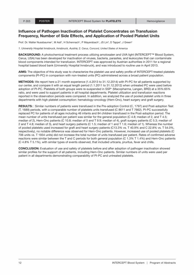
Influence of Pathogen Inactivation of Platelet Concentrates on Transfusion Frequency, Number of Side Effects, and Application of Pooled Platelet Units
Prof. Dr. Walter Nussbaumer1, M Astl1, H Schennach1, P Mayersbach1, JS Lin2, D Tappe2, J Green2
1. University Hospital Innsbruck, Innsbruck, Austria; 2. Cerus, Concord, United States of America
BACKGROUND: A photochemical treatment process utilizing amotosalen and UVA light (INTERCEPT™ Blood System, Cerus, USA) has been developed for inactivation of viruses, bacteria, parasites, and leukocytes that can contaminate blood components intended for transfusion. INTERCEPT was approved by Austrian authorities in 2011 for a regional hospital based blood bank (University Hospital Innsbruck), and was introduced to routine use in April 2013.
AIMS: The objective of this study was to characterize the platelet use and safety profile of INTERCEPT-treated platelets components (PI-PC) in comparison with non-treated units (PC) administered across a broad patient population.
METHODS: We report here a 21 month experience (1.4.2013 to 31.12.2014) with PI-PC for all patients supported by our center, and compare it with an equal length period (1.1.2011 to 31.12.2012) when untreated PC were used before adoption of PI-PC. Platelets of both groups were re-suspended in SSP+ (Macopharma, Langen, BRD) at a 35%:65% ratio, and were used to support patients in all hospital departments. Platelet utilization and transfusion reactions reported in the observation periods were compared. In addition, we analyzed the use of pooled platelet units in three departments with high platelet consumption: hematology-oncology (Hem-Onc), heart surgery and graft surgery.
RESULTS: : Similar numbers of patients were transfused in the Pre-adoption Control (C, 1797) and Post-adoption Test (T, 1689) periods, with a comparable number of platelets units transfused (C 8611 and T 7662). PI-PC successfully replaced PC for patients of all ages including 46 infants and 84 children transfused in the Post-adoption period. The mean number of units transfused per patient was similar for the general population (C 4.8; median of 2, and T 4.5; median of 2), Hem-Onc patients (C 10.8; median of 5 and T 9.9; median of 4), graft surgery patients (C 5.3; median of 2 and T 4.8; median of 3), and heart surgery patients (C 1.5; median of 1 and T 1.6; median of 1). Whereas the number of pooled platelets used increased for graft and heart surgery patients (C13.3% vs. T 40.9% and C 22.8% vs. T 54.0%, respectively), no notable difference was observed for Hem-Onc patients. However, increased use of pooled platelets (C 706 units vs. T 1934 units) did not increase the total number of units transfused per patient. Rates of confirmed adverse reactions were similar between the T and C periods for both general population (C 1.3% T 1.4%) and Hem-Onc patients (C 4.8% T 5.1%), with similar types of events observed, that included urticaria, pruritus, fever and chills.
CONCLUSION: Evaluation of use and safety of platelets before and after adoption of pathogen inactivation showed similar profiles for the support of all patients, including Hem-Onc patients. Similar numbers of units were used per patient in all departments demonstrating comparability of PI-PC and untreated platelets.
POSTER

ISBT | June 27 - July 1, 2015 | London, United Kingdom 13
# INTERCEPT Blood System for BLOOD PRODUCT Cerus CategoryIn Vitro FunctionINTERCEPT Blood System for PLATELETSP-311
Double Dose Platelet Concentrate from Apheresis Treated with Amotosalen and UV-A Light: First Evaluation
A. Castrillo, C. Arcas, M. Abalo, M. Adelantado, MI. Rodriguez
Centro de transfusión de Galicia, Santiago de Compostela, Spain
Currently the INTERCEPT platelet processing set with dual storage containers for treatment of platelet concentrates (PCs) in additive solution (AS) permits treat PCs, later they will be split en two therapeutic units, with one illumination cycle.
THE OBJECTIVE of this study was to evaluate in vitro function for 7 days of storage, following INTERCEPT treatment of double dose and also evaluate the processing efficiency.
MATERIAL AND METHODS: Apheresis PCs were collected on the Amicus device and stored overnight with continuous agitation at 22ºC. On the day after collection (day 1) the product was prepared for treatment with INTERCEPT (INTERCEPT blood system™,Cerus corp.), volume and platelet yield pre-treatment was calculated. After treatment PC was split in half and stored in 2 bags, one was supplied later another was used by study. Nine PCs were studied, they were stored under standard conditions and samples were withdrawn on day 2, 5 and 7 of storage. The cell content and the mean platelet volume (MPV) were measured using a Sysmex XT-2000i hematology analyser. The pH was measured at 22°C with a pH-meter (Crison Micro pH 2001).The phenomenon of swirling was visually assessed, giving a numerical value of 0-2 (0 = no swirling, 1 = intermediate and 2 = patent swirling). Glucose, lactate and lactate dehydrogenase (LDH) determinations were carried out with an Olympus AU400 Chemistry Analyser.
RESULTS: The average treatment dose and volume prior to split were 5.82±0.24x10¹¹ platelets and 416.11±6.25 mL per unit respectively. The final average dose per unit was 2.62 ±0.19x10¹¹ platelets and the average volume was 197.7 mL. The pH of the PCs remained stable for 7 days with a positive swirling for all PCs. In three units the glucose levels at 7 day were low. In Table 1, some parameters are shown.
CONCLUSION: INTERCEPT treated double dose PCs retained adequate in vitro platelet characteristics for 7 days storage but the platelet content is lower than our PCs standard. On the other hand this procedure has some logistic advantages and it saves cost.
Table1: PCs Parameters Throughout Storage
N=9 pH MPV (fL) Swirling (0-2)Glucose (Mmol/L)
Lactate (Mmol/L)
LDH (UI/L)
Day 1 Pre-tto 7.25±0.17 8.24±0.63 1.94±0.17 5.77±1.13 5.23±1.86 107.04±55.38
Day 2 7.20±0.14 8.31±0.68 1.94±0.17 5.06±1.06 5.09±2.12 108.78±39.44
Day 5 7.23±0.11 8.10±0.74 1.94±0.17 2.67±1.20 10.59±2.14 128.04±54.41
Day 7 7.18±0.08 8.20±0.68 1.94±0.17 0.95±0.94 13.81±1.70 133.27±45.10
POSTER
14 INTERCEPT Blood System | Program of Abstracts
InactivationINTERCEPT Blood System for PLATELETSP-312
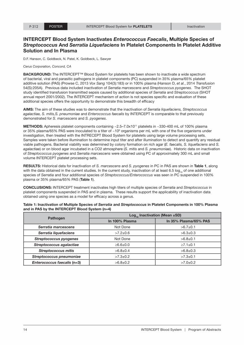
INTERCEPT Blood System Inactivates Enterococcus Faecalis, Multiple Species of Streptococcus And Serratia Liquefaciens In Platelet Components In Platelet Additive Solution and In Plasma
D.F. Hanson, C. Goldbeck, N. Patel, K. Goldbeck, L. Sawyer
Cerus Corporation, Concord, CA
BACKGROUND: The INTERCEPT™ Blood System for platelets has been shown to inactivate a wide spectrum of bacterial, viral and parasitic pathogens in platelet components (PC) suspended in 35% plasma/65% platelet additive solution (PAS) (Prowse C, 2013 Vox Sang 104(3):183) or in 100% plasma (Hanson D, et al., 2014 Transfusion 54(S):205A). Previous data included inactivation of Serratia marcescens and Streptococcus pyogenes. The SHOT study identified transfusion transmitted sepsis caused by additional species of Serratia and Streptococcus (SHOT annual report 2001/2002). The INTERCEPT mechanism of action is not species specific and evaluation of these additional species offers the opportunity to demonstrate this breadth of efficacy
AIMS: The aim of these studies was to demonstrate that the inactivation of Serratia liquefaciens, Streptococcus agalactiae, S. mitis,S. pneumoniae and Enterococcus faecalis by INTERCEPT is comparable to that previously demonstrated for S. marcescens and S. pyogenes.
METHODS: Apheresis platelet components containing ~2.5–7.0x10¹¹ platelets in ~330-400 mL of 100% plasma or 35% plasma/65% PAS were inoculated to a titer of ~106 organisms per mL with one of the five organisms under investigation, then treated with the INTRECEPT Blood System for platelets using large volume processing sets. Samples were taken before illumination to determine input titer and after illumination to detect and quantify any residual viable pathogens. Bacterial viability was determined by colony formation on rich agar (E. faecalis, S. liquefaciens and S. agalactiae) or on blood agar incubated in a CO2 atmosphere (S. mitis and S. pneumoniae). Historic data on inactivation of Streptococcus pyogenes and Serratia marcescens were obtained using PC of approximately 300 mL and small volume INTERCEPT platelet processing sets.
RESULTS: Historical data for inactivation of S. marcescens and S. pyogenes in PC in PAS are shown in Table 1, along with the data obtained in the current studies. In the current study, inactivation of at least 6.5 log10 of one additional species of Serratia and four additional species of Streptococcus/Enterococcus was seen in PC suspended in 100% plasma or 35% plasma/65% PAS (Table 1).
CONCLUSIONS: INTERCEPT treatment inactivates high titers of multiple species of Serratia and Streptococcus in platelet components suspended in PAS and in plasma. These results support the applicability of inactivation data obtained using one species as a model for efficacy across a genus.
Table 1: Inactivation of Multiple Species of Serratia and Streptococcus in Platelet Components in 100% Plasma and in PAS by the INTERCEPT Blood System (n=4)
PathogenLog10 Inactivation (Mean ±SD)
In 100% Plasma In 35% Plasma/65% PAS
Serratia marcescens Not Done >6.7±0.1
Serratia liquefaciens >7.2±0.6 >6.3±0.3
Streptococcus pyogenes Not Done >6.8±0.1
Streptococcus agalactiae ≥6.6±0.0 ≥7.1±0.1
Streptococcus mitis >6.8±0.4 >6.8±0.3
Streptococcus pneumoniae >7.3±0.2 >7.3±0.1
Enterococcus faecalis (n=3) >6.8±0.2 >7.0±0.2
POSTER

ISBT | June 27 - July 1, 2015 | London, United Kingdom 15
In Vitro FunctionP-314 INTERCEPT Blood System for PLATELETS
Pathogen Inactivation with Amotosalen and UVA Light of Routinely Produced Double Dose Buffy-Coat Concentrates Aimed for Transfusion: Preservation of Mitochondrial Function
Per Sandgren
Department of Clinical Immunology and Transfusion Medicine, Karolinska, University Hospital and Karolinska Institutet, Stockholm, Sweden
BACKGROUND: The INTERCEPT Blood System for Platelets (PLT) utilizes amotosalen (S-59) in combination with ultraviolet A (UVA) light to inactivate viruses, bacteria, protozoa and leucocytes that may contaminate PLT concentrates. Several in vitro studies have subsequently assessed the functional quality of INTERCEPT treated PLTs stored for up to 7 days after treatment. These results seem to be in line with in vivo results of INTERCEPT-treated PLTs obtained from clinical trials. However, limited data are available on the mitochondrial function of routinely produced INTERCEPT-treated double-dose (DD) buffy-coat (BC) PLT units which allows a single treatment procedure to produce two pathogen-inactivated PLT units.
AIM: The main objective of this study was to evaluate potential effects on mitochondrial function after INTERCEPT treatment on routinely produced pools of 8 BCs aimed for transfusion. Also, processing efficiency was evaluated.
MATERIALS AND METHODS: Buffy-coats (BCs) were separated from routinely collected 450 mL whole blood donations. Eight ABO matched BCs were selected, and manually pooled to undergo INTERCEPT-treatment as a unit (n=100 pools). Platelets were then aliquoted and suspended in 65% platelet additive solution (PAS) and 35% plasma. Potential changes in the mitochondrial membrane potential, a marker of pro-apoptotic events and maintenance of oxidative phosphorylation capacity, were measured. In addition, cell count, blood gas analysis, aggregates and swirling in all DD INTERCEPT-treated BC PLTs were studied preceding transfusion of 100 units.
RESULTS: The mitochondrial membrane potential determined by JC-1-labelling was well maintained in all INTERCEPT-treated units with >95% maintenance of mitochondrial function (96.36%±2.61%) after treatment (Table 1). Platelet count (232 ± 19 x 109/unit) was well above the European guideline requirements, and the pH (22°C) of all units was maintained at ~7.0 after INTERCEPT-treatment on Day 2. This is in agreement with the Council of Europe recommended range of 6.4 – 7.4. All treated PLTs showed good swirling characteristics (score = 2) indicative of well-maintained PLT morphology. No aggregates were observed in any of the units.
CONCLUSION: Our data demonstrate that photochemical pathogen inactivation of routinely produced DD-BC PLT concentrates with the INTERCEPT Blood System had no influence on the mitochondrial function in all tested units.
Table 1: Mitochondrial Function of INTERCEPT-treated DD BC PLTs is Well Maintained
Unit Parameter (n=100) Result +/- SD
Volume (mL) 191 ± 4
Platelet concentration (109/L) 1218 ± 104
Platelet Count (109/unit) 232 ± 19
MPV (fL) 9.0 ± 0.5
pH (37°) 6.934 ± 0.052
Swirl (scale 0-2) 2
Glucose (mmol/L) 6.9 ± 0.6
Lactate (mmol/L) 9.6 ± 1.2
JC-1 (R2, %) 96.36 ± 2.61
JC-1 (R4,%) 2.94 ± 1.99
POSTER

16 INTERCEPT Blood System | Program of Abstracts
In Vitro FunctionINTERCEPT Blood System for PLATELETSP-317
Adequate Leukodepletion of Platelet Concentrates from Pools of 7 or 8 Buffy-Coats for Photochemical Pathogen Inactivation Treatment of Double Transfusion Doses
Maria Bergsten1, Rut Norda1, Björn Hardarson2, Ragna Landrö2, Annica Andersson3, Mohammad Abedi4
1. Västmanalands sjukhus Västerås, Västerås, Sweden; 2. University Hospital, Reykjavík, Iceland; 3. Department of Laboratory Medicine, NU-sjukvården, Trollhättan, Sweden; 4. Department of Laboratory Medicine, Örebro University Hospital
BACKGROUND: The European requirement for White Blood Cell (WBC) count in a platelet concentrate is below 1x10⁶ per unit. It is of importance for the producing sites to have valid working processes which fulfill all requirements. The INTERCEPTTM Blood System (Cerus) utilizes amotosalen and UVA to inactivate contaminating viruses, bacteria, parasites and leukocytes in platelet concentrates and plasma. A treatment set allowing processing of a double dose of buffy-coat (BC) or apheresis platelets and further storage in two bags before transfusion is available.
AIMS: The production of double dose platelets from pools of 7 or 8 BC’s is routinely implemented in our 4 Nordic sites. We looked back at QC data on a period ranging from 2010 to 2014 demonstrating the capability to produce double dose platelets treated with INTERCEPT meeting the leukodepletion guidelines using BC pooling sets (Fresenius-Kabi) with SepacellTM PLX-5 filters (Asahi Kasei Medical).
METHODS: 7 or 8 BC’s separated on the day of donation or after overnight storage from 450-500 ml whole blood collections are sterile docked after overnight storage at 20-24°C with agitation to a pooling set with PLX-5 filter K4R7039 using the “double train” or the “octopus” method. SSP+ 280 ml (Trollhättan) or 300 ml (other sites) solution (Macopharma) is added to the pool. After soft spin centrifugation, platelets are leukodepleted through a PLX-5 filter on a Macopress Smart (Macopharma) in Trollhättan or Optipress (Fresenius Kabi) in the other sites. The double dose platelet concentrate is then treated for pathogen inactivation with INTERCEPT (amotosalen + UVA) using a Dual Storage DS set INT25. The capability of the filter to adequately leukodeplete below the required limit of 1x10⁶ white blood cells (WBC) per unit in 90% of the units is evaluated.
RESULTS: 160 platelet units were QC tested post INTERCEPT treatment (unless otherwise specified) in Örebro, 162 in Reykjavik, 606 in Trollhättan and 184 in Västerås. Table 1 shows QC data in the product split in two doses. Platelet yields were above the limit of 2 X 10¹¹ / unit in 100% of the tested units in Örebro, 99% in Reykjavik and 77% in Västerås (required 75 %, not tested in Trolhättan). WBC count was below the limit of 1x10⁶ in all of the tested units in the four sites with a maximum of 0.77x10⁶. 17 batches of pooling sets including 21 batches of filters were tested.
SUMMARY / CONCLUSIONS: The pooling and leukodepletion of 7 or 8 BC with PAS can be done with conventional pooling sets including a leukodepletion filter to produce double dose platelet concentrates subsequently treated with INTERCEPT. WBC removal is consistently meeting the guidelines.
Table 1: QC Data - Double Dose Platelet Production
Site n BC’s in the pool
Volume (ml) double dose (DD) or split
dose (SD)
Platelet count (1011 / unit) and
% >2x10¹¹
WBC count (106/unit)
Max WBC count (106/unit)
Örebro 160 7 395 + 9 (DD) 2.83 + 0.18 100% 0.08 + 0.12 0.77
Reykjavik 162 8 191+ 13 (SD) 2.79 + 0.3399% 0.03 + 0.06 0.48
Trollhättan 606 7 200 + 7 (SD) NR 0.03 + 0.04* 0.43*
Västerås 184 8 201 + 11 (SD) 2.64 + 0.3177% 0.02 + 0.05 0.3
Note: all measurements post INTERCEPT treatment except * pre INTERCEPT
NR = Not reported
POSTER

ISBT | June 27 - July 1, 2015 | London, United Kingdom 17
INTERCEPT Blood System for BLOOD PRODUCT Cerus CategoryP-318 INTERCEPT Blood System for PLATELETS Implementation
Feasibility Evaluation of Implementing Amotosalen and UVA Treatment on Double Dose Leukoreduced Pooled Random Donor Platelets
Dr. Luiz Amorim, Dr. Maria Esther Lopes, Jurema Fatima Rodrigues Paranhos, Terezinha de Miranda Ferreira
HEMORIO, Rio the Janeiro, Brazil
BACKGROUND: Optimized production methods for the production of platelet concentrates will lead to better utilization of whole blood collections, increased availability of platelet concentrates for transfusion, and significant reduction in use of disposables. Therefore, a new method was developed to prepare double dose, leukoreduced, pooled random donor platelets (RDPs). Additionally, viruses, bacteria and other micro-organisms, which are not detected by routine laboratory tests, are a concern to the Brazilian medical community in relation to the safety of transfused blood products.
AIM: The objective of this study was to determine the feasibility of implementing the INTERCEPT™ Blood System for pathogen inactivation for the double dose RDPs produced using this optimized production method. The method was assessed, and the impact on the quality of the treated products was evaluated.
METHODS: RDPs were separated from 450 mL ± 10% whole blood donations following local procedures. Within 36 hours of collection, 8 ABO-identical, whole blood-derived RDPs were pooled into a standard 600 mL transfer container (n = 12 pools), and leukoreduced by filtration. The resulting mean platelet count for the pools was 1313 ± 125 x 109/L. The double dose RDP pools were then immediately treated with 150 μM amotosalen and 3 J/cm2 UVA, split in two platelet doses for transfusion, and stored at 20-24 °C with flatbed agitation for 7 days. In vitro assays to assess platelet quality were performed on all pools on days 1 (pre-treatment), 2, 5 and 7.
RESULTS: This method for preparing double dose, leukoreduced RDP pools resulted in a 20% decrease in needed whole blood collections to produce the same number of platelet doses. Additionally, there was a 50% reduction in the use of filters and pooling systems, and a 30% reduction in the use of sterile connections. Due to reduction in pooling and centrifugation procedures labor time was reduced by 50%. Since the double dose INTERCEPT processing set was utilized, the time for producing 2 pathogen inactivated platelet doses was cut in half. For all INTERCEPT-treated RDP pools, platelet quality was well preserved during 7 days of storage, and met local and EU guidelines for blood component specifications. The mean platelet dose was 2.6 ± 0.2 x 10¹¹ per product, and further optimization is possible. The pH of the pools remained stable over 7 days of storage (mean 6.60 ± 0.17) with a platelet swirl rating of 2-3 for all pools. Additionally, all pools showed active metabolism with maintained pO2 levels (140 ± 30 mm Hg), adequate residual energy source (glucose, 190 ± 27 mg/dL), and sufficient buffering capacity.
CONCLUSIONS: INTERCEPT treatment of double dose, leukoreduced, pooled RDPs produced by the optimized manufacturing processes is feasible, and the quality of the INTERCEPT-treated platelets is well maintained. Further optimization of the platelet dose is ongoing.
POSTERPOSTER

18 INTERCEPT Blood System | Program of Abstracts
In Vitro FunctionP-322 INTERCEPT Blood System for PLATELETS
Validation of Double Dose Buffy-Coat Platelet Processing Using the INTERCEPT Dual Storage Processing Set Prepared from Seven Buffy-Coats
Angel Luis Pajares Herraiz, JD Rodríguez Gambarte, MV Flores Sanz, BE Eguía Lopez, CP Peréz Parrillas
Centro Regional de Transfusion Toledo-Guadalajara, Toledo, Spain
BACKGROUND: INTERCEPT Blood System for platelets is intended for the ex vivo preparation and storage of whole blood-derived buffy coat and apheresis platelet components and is used to inactivate a broad spectrum of pathogens as well as contaminating donor leukocytes in platelet products. The device uses amotosalen HCl (a photoactive compound) and long-wavelength ultraviolet (UVA) illumination to photochemically treat platelets
AIMS: To demonstrate that INTERCEPT platelets prepared from 7 ABO compatible whole blood derived buffy-coats met the acceptance criteria for manufacturing (Spanish and EU guidelines) and for support of patients requiring platelet transfusions according to clinical practice guidelines and standard platelet infusion methods in Spain and Europe
METHODS: Whole blood (WB) was collected into Top&Bottom bags, was overnight storage under controlled temperature and was separated into Red cells, Plasma and Buffy-coats with T-ACE II. The obtained Buffy-coats fulfilled the double dose requirements for processing set as specified in TABLE 1, and were rested for a minimum of 2 hours before pooling. Pre-selection of buffy-coats, based on platelets donor counts has been applied (minimun donor count 220x109/L). For each of the six replicates prepared, 7 ABO-matched Buffy-coat units were pooled with 280 mL of Intersol additive solution using the Octopus method. After a soft spin centrifugation platelets in intersol were extracted with a manual press and was leukoreduced through the in-line filter and were INTERCEPT treated. At the end of treatment, the platelet unit was split and INTERCEPT products were stored for 7 days under continuous agitation. Platelet concentrates were monitored on day 1, 2, 5 and 7 for the following in vitro parameters: volume; swirling; platelet counts, MPV &, WBC (Coulter LH 750); pH, pCO2 and pO2 (Gem Premier 300); glucose consumption and lactate production (Olympus 5400).
RESULTS: Platelet recovery after centrifugation was 81% in average, with Platelet content of 6.4 x 10¹¹± 0.3. Platelet and volume loss due to INTERCEPT treatment were approximately 5,5% and 4,8% respectively (TABLE 1), within the expected range of values. During the 7 days storage, platelet concentrates splited were monitored and platelet count and volume in day 2 was 3x10¹¹/unit and 186 ± 4 ml respectively. During storage the pH was stable and well maintained, within the values indicated by European and local requirements, above 6.4 through the end of shelf life. During storage we saw a little pH increase at day 5, but with no impact in platelets viability. Swirling with a minimum of 2 + is seen in all products during 7 days of storage. The platelet O2 consumption and the CO2 production are well maintained by the PL2410 platelet storage container during 7 days of storage. Platelet glucose consumption is in line with the production of lactate during the 7 days of storage. The average plasma ratio is 40%.
CONCLUSION: INTERCEPT treatment process for double dose platelets from seven Buffy-coats is able to generate platelet products that meet the acceptance criteria for manufacturing (Spanish and EU guidelines) and for support of patients requiring platelet transfusions according to clinical practice guidelines.
Table 1:
IBS-Platelets sets Platelet dose Volume (ml) Plasma content RBC content (ml) CAD time
Dual Set 2.5 - 8.0 x 10¹¹ 375 - 420 32-47 % <4 106 per mL 6 - 16 hrs
Platelets before IBS x 1011/unit
Platelet post IBS x 1011/u
Platelet loss IBS x 1011/u Platelet loss % Volume before
IBS (ml)Volume post IBS
(loss%)
6.5 6 0.3 5.5% 393.6 371.74 8%
POSTER
ISBT | June 27 - July 1, 2015 | London, United Kingdom 19
In Vitro FunctionINTERCEPT Blood System for PLASMAP-270
Photochemical Treatment of Pooled Whole Blood Derived Plasma within 19 Hours from Collection
Hervé Isola1, Arnaud Dupuis1, Christian Naegelen2, Nadine Marpaux2, Guillaume Mourey2, Catherine Ravanat1, Michel Laforet1, Christian Gachet1, Pascal Morel2
1. Etablissement Français du Sang EFS Alsace, Strasbourg; 2. Etablissement Français du Sang EFS Bourgogne Franche-Comté, Besançon
BACKGROUND: The INTERCEPT Blood System (Cerus) is a pathogen inactivation (PI) method that utilizes amotosalen and UVA to inactivate contaminating viruses, bacteria, parasites and leukocytes in platelet concentrates and plasma. Apheresis plasma treated and frozen within 18 hours (PFC-IA) is used in France.
AIMS: The objective of this study was to evaluate the quality of plasma derived from whole blood, pooled, treated with INTERCEPT, split and frozen within 18-19 hours from collection.
METHODS: 5 leukodepleted plasma units from whole blood of the same ABO group are pooled and divided into two sub-units of 650 mL wich are each pathogen inactivated. Both sites prepared 20 pools (5 O ; 15 non-O). Each pool results after PI in 2 x 3 units of >200 mL subsequently frozen. The plasma quality was evaluated before treatment (T1) and after 2 weeks (T2), 6 months (T3) and 12 months (T4, 4 parameters) of storage below -25 °C. 28 tests were carried out exploring cellular contamination, plasma proteins, coagulation factors, clotting time measurements, calibrated automated thrombin generation (CAT), fibrinolysis, coagulation and complement activation markers.
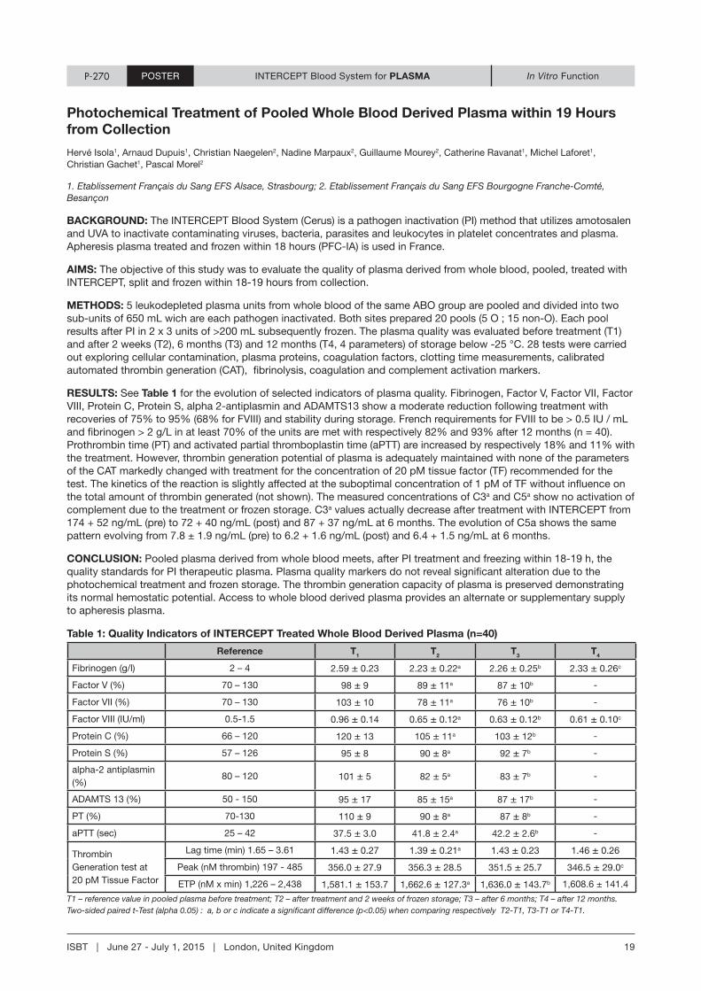
RESULTS: See Table 1 for the evolution of selected indicators of plasma quality. Fibrinogen, Factor V, Factor VII, Factor VIII, Protein C, Protein S, alpha 2-antiplasmin and ADAMTS13 show a moderate reduction following treatment with recoveries of 75% to 95% (68% for FVIII) and stability during storage. French requirements for FVIII to be > 0.5 IU / mL and fibrinogen > 2 g/L in at least 70% of the units are met with respectively 82% and 93% after 12 months (n = 40). Prothrombin time (PT) and activated partial thromboplastin time (aPTT) are increased by respectively 18% and 11% with the treatment. However, thrombin generation potential of plasma is adequately maintained with none of the parameters of the CAT markedly changed with treatment for the concentration of 20 pM tissue factor (TF) recommended for the test. The kinetics of the reaction is slightly affected at the suboptimal concentration of 1 pM of TF without influence on the total amount of thrombin generated (not shown). The measured concentrations of C3a and C5a show no activation of complement due to the treatment or frozen storage. C3a values actually decrease after treatment with INTERCEPT from 174 + 52 ng/mL (pre) to 72 + 40 ng/mL (post) and 87 + 37 ng/mL at 6 months. The evolution of C5a shows the same pattern evolving from 7.8 ± 1.9 ng/mL (pre) to 6.2 + 1.6 ng/mL (post) and 6.4 + 1.5 ng/mL at 6 months.
CONCLUSION: Pooled plasma derived from whole blood meets, after PI treatment and freezing within 18-19 h, the quality standards for PI therapeutic plasma. Plasma quality markers do not reveal significant alteration due to the photochemical treatment and frozen storage. The thrombin generation capacity of plasma is preserved demonstrating its normal hemostatic potential. Access to whole blood derived plasma provides an alternate or supplementary supply to apheresis plasma.
Table 1: Quality Indicators of INTERCEPT Treated Whole Blood Derived Plasma (n=40)
Reference T1 T2 T3 T4
Fibrinogen (g/l) 2 – 4 2.59 ± 0.23 2.23 ± 0.22a 2.26 ± 0.25b 2.33 ± 0.26c
Factor V (%) 70 – 130 98 ± 9 89 ± 11a 87 ± 10b -
Factor VII (%) 70 – 130 103 ± 10 78 ± 11a 76 ± 10b -
Factor VIII (IU/ml) 0.5-1.5 0.96 ± 0.14 0.65 ± 0.12a 0.63 ± 0.12b 0.61 ± 0.10c
Protein C (%) 66 – 120 120 ± 13 105 ± 11a 103 ± 12b -
Protein S (%) 57 – 126 95 ± 8 90 ± 8a 92 ± 7b -
alpha-2 antiplasmin (%)
80 – 120 101 ± 5 82 ± 5a 83 ± 7b -
ADAMTS 13 (%) 50 - 150 95 ± 17 85 ± 15a 87 ± 17b -
PT (%) 70-130 110 ± 9 90 ± 8a 87 ± 8b -
aPTT (sec) 25 – 42 37.5 ± 3.0 41.8 ± 2.4a 42.2 ± 2.6b -
Thrombin Generation test at 20 pM Tissue Factor
Lag time (min) 1.65 – 3.61 1.43 ± 0.27 1.39 ± 0.21a 1.43 ± 0.23 1.46 ± 0.26
Peak (nM thrombin) 197 - 485 356.0 ± 27.9 356.3 ± 28.5 351.5 ± 25.7 346.5 ± 29.0c
ETP (nM x min) 1,226 – 2,438 1,581.1 ± 153.7 1,662.6 ± 127.3a 1,636.0 ± 143.7b 1,608.6 ± 141.4
T1 – reference value in pooled plasma before treatment; T2 – after treatment and 2 weeks of frozen storage; T3 – after 6 months; T4 – after 12 months.Two-sided paired t-Test (alpha 0.05) : a, b or c indicate a significant difference (p<0.05) when comparing respectively T2-T1, T3-T1 or T4-T1.
POSTER
20 INTERCEPT Blood System | Program of Abstracts
# INTERCEPT Blood System for BLOOD PRODUCT Cerus CategoryP-309 InactivationINTERCEPT Blood System for PLASMA
Amotosalen and Ultraviolet A Light Inactivate Zika Virus in Plasma
Maite Aubry1, Vaea Richard1, Jennifer Green2, Julien Broult3, and Didier Musso1
1. Pôle de recherche et de veille sur les maladies infectieuses émergentes, Institut Louis Malardé, Tahiti, Polynésie Française; 2. Cerus Corporation, California, USA; 3. Centre de Transfusion Sanguine de la Polynésie Française, Hôpital du Taaone, Tahiti, Polynésie française
BACKGROUND: Zika virus (ZIKV) is an arthropod-borne virus (arbovirus) transmitted by the bite of infected mosquitoes. The potential for ZIKV transmission through blood transfusion was demonstrated during the largest ZIKV outbreak that occurred in French Polynesia from October 2013 to April 2014 (1): 2.8% of the blood donors, asymptomatic at the time of blood donation, were found positive using ZIKV-specific reverse transcription PCR (RT-PCR) (2). Prevention of transfusion-transmitted ZIKV infections is challenging because most of the cases are asymptomatic, and are not detected during medical questionnaire, and nucleic acid testing for ZIKV is not routinely available. Pathogen inactivation of blood products is a proactive strategy that provides the potential to reduce transfusion-transmitted diseases (3). Inactivation of arboviruses by amotosalen and ultraviolet A (UVA) illumination was previously demonstrated for chikungunya, West Nile (WNV) and dengue viruses (DENV) (4–6). We report here the efficacy of this strategy for ZIKV inactivation in plasma.
AIMS: According to the recommendations for evaluation of pathogen reduction efficacy (7), we performed a spiking experiment of plasma units with ZIKV in order to compare the viral titers and viral RNA loads before and after inactivation.
METHODS: Plasma units collected from DENV and WNV immunoglobulin G negative American donors were collected in California (USA). ZIKV was propagated on African green monkey kidney cells (VERO) and concentrated. Four plasma units were spiked with ZIKV before transfer into an INTERCEPT disposable kit (INT3102B, Cerus Corporation). Three units were inactivated with amotosalen and 3 J/cm2 UVA light for ~6 min, the fourth one was not and was the positive control. Detection of replicative ZIKV and viral titration was performed by inoculating pre- and post-inactivated plasma unit samples on VERO cells. Five serial passages of plasma unit samples were performed in order to amplify any replicative virus. Infected cells were detected by an indirect immunofluorescence (IF) assay, and viral titers were expressed in 50% tissue culture infectious dose (TCID50/mL) (8). ZIKV RNA loads were measured after each passage on cell culture. RNA quantitation was performed by RT-PCR (9) and expressed in log10 copies/mL.
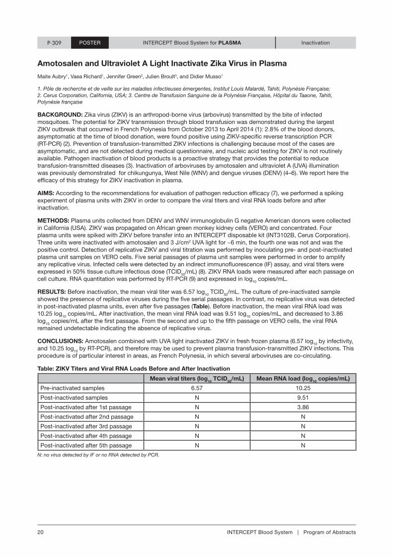
RESULTS: Before inactivation, the mean viral titer was 6.57 log10 TCID50/mL. The culture of pre-inactivated sample showed the presence of replicative viruses during the five serial passages. In contrast, no replicative virus was detected in post-inactivated plasma units, even after five passages (Table). Before inactivation, the mean viral RNA load was 10.25 log10 copies/mL. After inactivation, the mean viral RNA load was 9.51 log10 copies/mL, and decreased to 3.86 log10 copies/mL after the first passage. From the second and up to the fifth passage on VERO cells, the viral RNA remained undetectable indicating the absence of replicative virus.
CONCLUSIONS: Amotosalen combined with UVA light inactivated ZIKV in fresh frozen plasma (6.57 log10 by infectivity, and 10.25 log10 by RT-PCR), and therefore may be used to prevent plasma transfusion-transmitted ZIKV infections. This procedure is of particular interest in areas, as French Polynesia, in which several arboviruses are co-circulating.
Table: ZIKV Titers and Viral RNA Loads Before and After Inactivation
Mean viral titers (log10 TCID50/mL) Mean RNA load (log10 copies/mL)
Pre-inactivated samples 6.57 10.25
Post-inactivated samples N 9.51
Post-inactivated after 1st passage N 3.86
Post-inactivated after 2nd passage N N
Post-inactivated after 3rd passage N N
Post-inactivated after 4th passage N N
Post-inactivated after 5th passage N N
N: no virus detected by IF or no RNA detected by PCR.
POSTERPOSTER

ISBT | June 27 - July 1, 2015 | London, United Kingdom 21
Clinical StudiesINTERCEPT Blood System for RED BLOOD CELLSP-298
Red Blood Cells Treated with the S-303 System for Pathogen Inactivation Demonstrate In Vitro Characteristics Suitable for Transfusion - Phase III Clinical Trial in Cardiac Surgery Patients
V Brixner1, J Leibacher1, H-U Pfeiffer1, M Müller, M1, C Geisen1, R Henschler4, K Janetzko5, S Heldke6, N Huang7, C Ernst7, S Rico7, A Erickson7, N Mufti7, L Corash7, E Seifried1
1. German Red Cross Blood Donor Service Baden-Wuerttemberg-Hessen, Frankfurt am Main, Germany; 2. Department of Thoracic and Cardiovascular Surgery, Johann Wolfgang Goethe University, Frankfurt, Germany; 3. Department of Haemostaseology and Transfusion Medicine, Kerckhoff-Klinik, Bad Nauheim, Germany; 4. University Clinic Munich, Department for Transfusion Medicine, Cell Therapy and Hemostaseology, Munich, Germany; 5. German Red Cross Blood Donor Service Baden-Wuerttemberg-Hessen, Frankfurt am Main, Germany; 6. BloodCenter of Wisconsin, Milwaukee, WI, USA; 7. Cerus Corporation, Concord, CA, USA
BACKGROUND: The risks of red blood cell (RBC) transfusion have been greatly reduced in the past decades thanks to improvements in donor screening, good manufacturing practices, and viral marker testing. Nevertheless, threats to the blood supply remain either from known pathogens which are not tested routinely and/or from emerging pathogens. The second generation S-303 pathogen and leukocyte inactivation system was developed to reduce the risk of transfusion transmitted infections and transfusion associated graft vs. host disease.
AIMS: Evaluation of the in vitro characteristics of RBC components produced for a randomized, controlled, double-blind phase III clinical trial to assess the efficacy and safety of S-303 treated RBC components in patients requiring transfusion support for acute anemia, during or shortly after a cardiac surgery.
METHODS: Patients undergoing coronary artery bypass grafting, and/or valve replacement or repair, were randomized to receive S-303 treated (Test) or conventional (Control) RBC during a 7-day treatment period. Test and Control RBC components were either released for transfusion to patients or stored at 1°C to 6°C for 35 days. Given that the patient’s hemoglobin increase is proportional to the hemoglobin mass transfused, measuring the hemoglobin content of RBC components is considered a surrogate for the therapeutic efficacy of a RBC component. Therefore, the primary endpoint was the mean hemoglobin content per RBC component post-production (PP). A priori definition of equivalence required the 95% CI limits for the mean treatment differences to be -5 g/unit to 5 g/unit. Secondary efficacy endpoints included end-of-storage (EOS; Day 35 – Day 38) measurements of hematocrit, hemolysis, normalized ATP, plasma-free hemoglobin, and hemoglobin content.
RESULTS: A total of 774 study RBC components were produced, and 754 RBC components (389 test, 365 control) were eligible for PP hemoglobin content analysis. Mean (±SD) PP hemoglobin content per Test RBC component (53.6 ± 5.6 g/unit) was slightly lower than Control RBC (56.3 ± 6.0 g/unit), however the primary endpoint was met as the mean treatment difference (Test-Control) in hemoglobin content (95% CI -2.61 g/unit to -1.92 g/unit) was within the pre-specified equivalence margins (95% CI ±5 g/unit). There were no differences in the proportion of components having EOS hemoglobin content (≥ 40 g/component; Test 98.7%, Control 99.2%; p=0.69), or free hemoglobin (< 6 g/L; Test 100%, Control 99.6%; p=0.46), or hematocrit (50-70%; Test 99.3%, Control 99.2%; p=1.00). There were differences in the proportion of components with EOS normalized ATP (> 2 μmol/g; Test 21.8%, Control 0.90%; p<0.001) and EOS hemolysis per the Quality of Medicines (EDQM) guideline for EOS hemolysis (< 0.8%; Test 100%, Control 98.1%; p=0.02).
SUMMARY/CONCLUSIONS: S-303 treated RBC demonstrated equivalence to untreated RBC regarding hemoglobin content and met EDQM guidelines hemoglobin content, hematocrit and hemolysis. The proportion of S-303 components satisfying the EDQM guidelines for EOS hemolysis and the normalized ATP was higher for S-303 treated RBCs than conventional RBCs. S-303 treated RBCs show in vitro characteristics comparable to untreated RBC and are suitable for transfusion.
Continued on page 22
POSTER

22 INTERCEPT Blood System | Program of Abstracts
Clinical StudiesINTERCEPT Blood System for RED BLOOD CELLSP-298
Test RBC Control RBC P-Value (95% CI) [1]
Primary Endpoint
Post-Production Hemoglobin Content (g/unit)
N 389 365
Mean (SD) 53.6 (5.6) 56.3 (6.0) (-2.61, -1.92)
Satisfy EDQM (Proportion) 387 (99.5%) 365 (100%) 0.500
Secondary Endpoints
End-of-Storage Hemoglobin Content (g/unit)
N 301 261
Mean (SD) 53.1 (5.7) 55.8 (5.9) (-2.76, -1.92)
Satisfy EDQM (Proportion) 297 (98.7%) 259 (99.2%) 0.691
End of Storage Hematocrit (%)
N 301 261
Mean (SD) 60.4 (3.2) 60.9 (3.5) (-0.81, 0.12)
Satisfy EDQM (Proportion) 299 (99.3%) 259 (99.2%) 1.000
End of Storage Hemolysis (%)
N 301 261
Mean (SD) 0.28 (0.12) 0.35 (0.16) (-0.09, -0.04)
Satisfy EDQM (Proportion) 301 (100%) 256 (98.1%) 0.021
End of Storage Plasma Free Hemoglobin (g/dL)
N 263 225
Mean (SD) 1.42 (0.64) 1.79 (0.88) (-0.49, -0.23)
Satisfy EDQM (Proportion) 263 (100%) 224 (99.6%) 0.461
End of Storage Normalized ATP (μmol/g)
N 257 222
Mean (SD) 1.66 (0.44) 1.29 (0.29) (0.30, 0.44)
> 2 μmol/g (Proportion) 56 (21.8%) 2 (0.9%) <0.001
[1] 95% CIs for the mean treatment difference (Test – Control) are based on a mixed effects ANCOVA model controlling for the treatment, gender, blood type, input hematocrit, and input volume. P-Values for the proportion of components satisfying the EDQM criteria are based on Fisher’s Exact Test.
POSTER
ISBT | June 27 - July 1, 2015 | London, United Kingdom 23
# INTERCEPT Blood System for BLOOD PRODUCT Cerus CategoryP-313 In Vitro FunctionINTERCEPT Blood System for RED BLOOD CELLS
In Vitro Evaluation of Pathogen Inactivated Apheresis RBC Using the S-303 Treatment System
Walter Nussbaumer1, Heidi Volland1, Silka Andresen2, Nina Mufti3, Anna Erickson3
1. University Hospital, Innsbruck, Austria; 2. Cerus BV, Amersfoort, Netherlands; 3. Cerus Corporation, Concord, CA, USA
BACKGROUND: The Second Generation S-303 Treatment System for Red Blood Cells (RBCs) uses S-303 to crosslink nucleic acids and prevent replication of contaminating pathogens and residual leukocytes. Glutathione (GSH) is included to quench non-specific reactions. This pathogen inactivation system has shown robust pathogen inactivation for bacteria, viruses and parasites while retaining in vitro and in vivo RBC quality per AABB, FDA and EU Guidelines up to 35 days of storage.
AIMS: The purpose of this study was to assess the quality of stored S-303 treated RBCs compared to conventional (untreated) Control SAG-M RBCs collected using the Alyx apheresis system.
METHODS: Six replicates, each consisting of a double RBC apheresis units (total volume of 552±15mL), were collected using the Alyx system per manufacturer’s instructions and institutional procedures. All red cell concentrates were suspended in SAG-M and stored at 4±2°C. For each replicate Control units (268-288mL) were prepared and held at 4±2°C. Test units (265-289mL) were treated with the S-303 treatment process by combining RBC with GSH and a proprietary diluent solution followed by addition of S-303 (final concentrations: 20mM GSH and 0.2mM S-303, based on an RBC input volume of 280mL). After an 18-24h RT hold, units of S-303 treated RBC were centrifuged and the treatment solution expressed and replaced with SAG-M. S-303 treated RBCs were stored at 4±2°C for 6 weeks. Test and Control RBCs were sampled over 43 days of storage for analysis of in vitro parameters (Table 1).
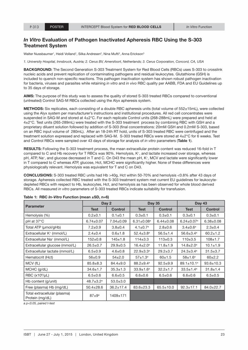
RESULTS: Following the S-303 treatment process, the mean extracellular protein content was reduced 16-fold in T compared to C and Hb recovery for T RBCs was 90%. Hemolysis, K+, and lactate increased over storage, whereas pH, ATP, Na+, and glucose decreased in T and C. On D43 the mean pH, K+, MCV and lactate were significantly lower in T compared to C whereas ATP, glucose, Hct, MCHC were significantly higher. None of these differences were physiologically relevant. Hemolysis was equivalent for T and C on D43.
CONCLUSIONS: S-303 treated RBC units had Hb >40g, Hct within 50-70% and hemolysis <0.8% after 43 days of storage. Apheresis collected RBC treated with the S-303 treatment system met current EU guidelines for leukocyte-depleted RBCs with respect to Hb, leukocytes, Hct, and hemolysis as has been observed for whole blood derived RBCs. All measured in vitro parameters of S-303 treated RBCs indicate suitability for transfusion.
Table 1: RBC In-Vitro Function (mean ±SD, n=6)
Parameter Day 2 Day 35 Day 43
Test Control Test Control Test Control
Hemolysis (%) 0.2±0.1 0.1±0.1 0.3±0.1 0.3±0.1 0.3±0.1 0.3±0.1
pH at 37°C 6.74±0.07 7.04±0.09 6.31±0.08a 6.44±0.08 6.24±0.07a 6.38±0.08
Total ATP (μmol/gHb) 7.2±0.9 3.8±0.4 4.1±0.7a 2.8±0.6 3.4±0.6a 2.3±0.4
Extracellular K+ (mmol/L) 2.4±0.4 0.6±1.8 52.4±3.8a 56.5±1.4 56.6±3.4a 60.2±1.2
Extracellular Na+ (mmol/L) 152±0.8 145±1.8 114±3.3 113±0.3 110±3.5 108±1.7
Extracellular glucose (mmol/L) 26.5±0.7 29.9±0.5 16.4±2.0a 11.8±1.9 14.8±2.0a 10.1±1.9
Extracellular lactate (mmol/L) 6.5±0.9 4.6±0.8 22.9±3.3a 29.2±3.7 24.3±3.4a 31.5±3.7
Hematocrit (Hct) 56±0.9 54±2.0 57±1.3a 60±1.5 58±1.6a 60±2.2
MCV (fL) 85.8±8.3 84.4±9.0 88.2±9.4a 92.5±9.9 89.1±10.1a 93.6±10.3
MCHC (g/dL) 34.6±1.7 35.3±1.3 33.9±1.6a 32.2±1.7 33.5±1.4a 31.8±1.4
RBC (x106/µL) 6.5±0.6 6.6±0.5 6.6±0.6 6.5±0.6 6.6±0.6 6.5±0.5
Hb content (g/unit) 48.7±3.2a 53.0±3.0
Free (plasma) Hb (mg/dL) 50.4±28.6 36.2±17.4 83.6±23.3 65.5±10.0 92.3±17.1 84.0±22.7
Total extracellular (plasma) Protein (mg/dL)
87±9a 1409±171
a p<0.05, paired t-test
POSTERPOSTER

24 INTERCEPT Blood System | Program of Abstracts
# INTERCEPT Blood System for BLOOD PRODUCT Cerus CategoryClinical StudiesINTERCEPT Blood System for RED BLOOD CELLSP-319
Quality Parameters of Red Blood Cells Treated with INTERCEPT Pathogen Inactivation System Using S-303: A Phase III Clinical Trial In Cardiac Surgery Patients
Johannes Leibacher1, Veronika Brixner1, Hans-Ulrich Pfeiffer1, Markus Müller1, Christof Geisen1, Sarah Dombos1, Tanja Wotapek1, Iuliia Weber1, Karin Janetzko2, Reinhard Henschler3, Anna Erickson4, Nina Mufti4, Erhard Seifried1
1. DRK Frankfurt, Germany; 2. DRK Mannheim, Germany; 3. Uni-Klinikum München (LMU), Germany; 4. Cerus Corporation, Concord, CA, USA
BACKGROUND: The INTERCEPTTM Blood System for Red Blood Cells (RBC) has been developed to prevent replication of contaminating pathogens and leukocytes in RBC components for transfusion. S-303 is used in the system to crosslink nucleic acids and glutathione to quench nonspecific reactions of S-303. The INTERCEPT Blood System for RBCs inactivates a variety of pathogens including gram-negative/positive bacteria, enveloped/non-enveloped viruses, and parasites; emerging pathogens and/or those not routinely tested may also be inactivated. A Phase III clinical trial was conducted to compare standard blood bank quality parameters of both S-303 treated and conventional RBCs and additionally showing patient safety.
AIMS: A randomized, double-blind, controlled, multi-center Phase 3 clinical trial using the INTERCEPT Blood System for RBC was conducted to evaluate the quality of INTERCEPT RBCs and patient safety. The quality of INTERCEPT RBCs was evaluated post-production (PP) for all components and after 35-38 days of storage (EOS) for not-transfused components. Parameters assessed included those required by the European Directorate for Quality Medicines (EDQM) in addition to physical and metabolic attributes.
METHODS: Leukocyte reduced RBC in SAG-M, derived from buffy-coat depleted whole blood were used as the input RBC component. Control RBCs were stored at 1-6°C. Test units were treated on D1 with the S-303 process. Test RBCs were added to a diluent containing GSH followed by S-303 addition (final concentrations of 20mM GSH/0.2mM S-303, based on 280mL RBC input). After 18-24 hours hold at 20-25°C, RBCs were centrifuged and the treatment solution was expressed and replaced with SAG-M. Test and Control RBCs were sampled post production (PP), stored at 1-6°C, and either transfused to cardiovascular surgery patients to evaluate transfusion efficacy and overall safety or sent to a quality control lab to assess quality parameters after 35-38 days of storage.
RESULTS: Quality parameters not included as part of the endpoint analysis (PP hemoglobin and post –storage hematocrit, hemolysis and ATP) are listed in Table 1. The endpoint analyses are presented in a companion abstract (Brixner, et al). All parameters, except hematocrit, are significantly different between Test and Control RBCs. MCHC and glucose are increased in Test compared to Control, while Hb content, MCV, pH, potassium, lactate, and total protein are decreased. Total protein was 3.4±1.4-fold lower in Test compared to Control. The mean Test PP hematocrit met the EDQM specifications of 50-70% for leukocyte reduced RBCs in additive solution.
CONCLUSIONS: INTERCEPT RBCs met EDQM criteria for hematocrit post-production. Test RBCs have higher glucose and lower lactate after 35 days of storage, suggesting lower metabolic rates than Control. Reduced total protein levels in INTERCEPT RBCs may help reduce transfusion reactions.
Table 1: Quality Parameters PPa and EOSb for Test and Control
Quality Parameter Timepoint Control RBC Test RBC
Hematocrit (%) PP 57.3±2.9 (n=367) 57.4±2.0 (n=389)
MCV [fL]PP 87.5±5.0 (n=367) 85.7±4.8c (n=389)
EOS 93.6±6.4 (n=225) 91.3±6.6c (n=263)
MCHC [g/dl]PP 33.6±1.1 (n=367) 34.2±1.2c (n=389)
EOS 31.4±1.0 (n=225) 32.3±1.4c (n=263)
pH EOS 6.4±0.1 (n=256) 6.3±0.1c (n=293)
Potassium [mmol/L] EOS 41.8±3.6 (n=261) 40.6±3.3c (n=243)
Glucose [mmol/L] EOS 308.2±33.2 (n=163) 317.4±31.0c (n=259)
Lactate [mmol/L] EOS 29.1±3.3 (n=225) 20.4±2.4c (n=262)
Total Protein [mmol/L] EOS 228.8±34.7 (n=298) 68.0±25.6c (n=301)
a PP: Post Production (Day 2)
b EOS: End of Storage (Day 35-38)
c p<0.05; p-values for the mean treatment difference (T-C) are based on a T-test with unequal variances.
POSTER

ISBT | June 27 - July 1, 2015 | London, United Kingdom 25
# INTERCEPT Blood System for BLOOD PRODUCT Cerus Category
Qualification of the S-303 Treatment System at Colindale Blood Centre
Omonua Agbonbhaselena1, Vicky Hicks1, Ian Symonds1, Jane Davies1, Stephen Thomas1, Gianna Melucci Vigo2, Anna Erickson3
1. NHSBT Colindale, London, UK; 2. Cerus BV, Amersfoort, Netherlands; 3. Cerus Corporation, Concord, CA, USA
BACKGROUND: The INTERCEPT System for Red Blood Cells (RBCs) is intended to inactivate pathogens and leucocytes in RBC components for transfusion by using S-303 to crosslink nucleic acids, preventing replication of contaminating pathogens. A Phase 3 clinical investigation in patients with thalassemia major requiring chronic RBC transfusion support is currently in progress in Europe and will be expanded to include additional study sites. In the UK, NHSBT aims to provide INTERCEPT treated RBC for the investigation of the safety and efficacy of S-303 treated RBCs using the INTERCEPT™ System for RBC, compared to conventional RBCs.
AIMS: This study was designed to qualify the NHSBT blood centre in Colindale to process RBC components using the S-303 treatment process for the planned Phase 3 clinical investigation.
METHODS: On the day (D) of collection, D0, leucocyte-depleted SAG-M RBCs were prepared from CPD whole blood donations and stored at 4±2°C. Test units were treated on D1 with the S-303 process. Test RBCs (280±20mL) were added to a diluent containing glutathione (GSH) followed by S-303 addition (final concentrations of 20mM GSH/0.2mM S-303, based on 280mL RBC input). After 18-24 hours hold at 20-25°C, RBCs were centrifuged and the treatment solution was expressed and replaced with SAG-M. RBCs were stored at 4±2°C for 35 days and were sampled for testing on D1, D2, and D35.
RESULTS: All units met the acceptance criteria (Table 1). The volume post treatment was 249-311mL, with a loss of 2±1g of Hb due to the S-303 process (n=13). The post S-303 treatment mean and SD for volume, Hb content, haematocrit (Hct), and hemolysis are listed in Table 1. All units had Hb values of ≥40g ranging from 42–58g. The final Hct was 53-59%, within the 50-70% criterion. After 35 days of storage haemolysis was 0.05-0.26% (Table 1).
CONCLUSIONS: The S-303 Treatment System was successfully transferred to the Colindale Blood Centre. This study demonstrated that all S-303 treated RBC units met the EU guidelines for leukocyte depleted RBCs in additive solution with respect to Hct, Hb content and haemolysis at end of storage. The results of this study will support an application to MHRA for clinical investigation authorization.
Table 1: Results and Acceptance Criteria for S 303-Treated RBCs Post-S 303 Treatment Process (Mean ± SD (Min – Max))
Attribute Criteria Result (n = 13) Percent meeting criteria
Treatment process Haemoglobin loss(g)a
≤ 5 g 2 ± 1 (0 – 4) 100%
Volume(mL)a 190-340 280 ± 21 (244 – 311) 100%
Hb/unit(g)a ≥ 40 51 ± 5 (42 – 58) 100%
Haematocrit(%)a 50 - 70 57 ± 2 (53 – 59) 100%
Haemolysis(%)b ≤ 0.8 0.14 ± 0.06 (0.05 – 0.26) 100%
S-303(nM)a ≤ 1 ≤ 0.75 100%
a. Post-S-303 treatment
b. Day 35
P-321 Clinical StudiesINTERCEPT Blood System for RED BLOOD CELLSPOSTER

NOTES

NOTES

[email protected] www.interceptbloodsystem.comwww.cerus.com
Global HeadquartersCerus Corporation 2550 Stanwell DriveConcord, CA 94520, USA+1 925 288 6000
European HeadquartersCerus Europe B.V.Stationsstraat 79-D3811 MH AmersfoortThe Netherlands+31 33 496 0600
© 2
015
Cer
us C
orpo
ratio
n. C
erus
, IN
TERC
EPT
Bloo
d Sy
stem
and
INTE
RCEP
T ar
e tra
dem
arks
of C
erus
Cor
pora
tion.
Use of INTERCEPT is contraindicated in patients with a history of allergic response to amotosalen or psoralens. No pathogen inactivation system has been shown to inactivate all pathogens. .
INTERCEPT REGULATORY APPROVALS
United States (FDA)2014 (platelets and plasma)
Switzerland (Swissmedic)2009 (platelets)2010 (plasma)
Germany (PEI)2007* (platelets)2011* (plasma)
France (ANSM)2003 (platelets)2006 (plasma)
CE mark, Class III2002 (platelets)2006 (plasma)
* First blood center marketing authorization approved.