Embed Size (px)
Citation preview

Gynecologic Imaging: Ultrasound vs CT vs MRI
Seng Thipphavong MD FRCPC Abdominal Radiologist, Joint Department of Medical Imaging, University Health Network/Mount Sinai Hospital,
Women’s College Hospital, Toronto, Ontario, Canada Assistant Professor, University of Toronto, Toronto, Ontario, Canada

No disclosures

Outline
Modalities – Transabdominal and transvaginal
ultrasound (TVS) Abnormal uterine bleeding (AUB) Ovarian cysts
– Management of ovarian and other adnexal cysts
– How to manage adnexal incidentalomas seen on CT or MRI

Outline
Modalities – Transabdominal and transvaginal
ultrasound (TVS) Abnormal uterine bleeding (AUB) Adnexal cysts
– Management of ovarian and other adnexal cysts
– How to manage adnexal incidentalomas seen on CT or MRI

Ultrasound vs MRI vs CT
ULTRASOUND
MRI
HYSTEROSONOGRAM
CT

Indications for pelvic ultrasound
Pelvic pain Evaluation of pelvic mass Endocrine Dysmenorrhea Amenorrhea AUB Delayed menses Infertility patients PID Congenital uterine
anomaly
Postoperative IUD location Screening in high-risk
patients Incontinence or pelvic
organ prolapse …..
AIUM Practice Guideline for the Performance of Ultrasound of the Female Pelvis. J Ultrasound Med 2014;33: 1122-1130.

Clinical information pertinent to female pelvis sonography
Age Last menstrual period Relevant signs and symptoms Patient hormonal status (oral contraceptives, hormone replacement therapy, fertility drugs) Family history of cancer, personal history of pelvic surgery, results of prior imaging studies
AIUM Practice Guideline for the Performance of Ultrasound of the Female Pelvis. J Ultrasound Med 2014;33: 1122-1130.

Ultrasound vs MRI vs CT
ULTRASOUND
MRI
HYSTEROSONOGRAM
CT
CT: Not indicated for evaluation of the pelvic organs Staging for already diagnosed pelvic malignancy
SIS vs transvaginal sonography
Invasive procedure, but not has invasive as hysteroscopy A 5F catheter is placed through the cervix followed by distension of the uterine cavity with saline during real-time imaging Note: can be unsuccessful in postmenopausal patients or with history of prior cervical procedure associated with scarring or stenosis

SIS indications
AUB Uterine cavity, especially for myomas, polyps, and synechiae Abnormality detected on TVS (focal or diffuse endometrial or intracavitary abnormalities) Congenital abnormalities of the uterus Infertility Recurrent pregnancy loss
AIUM practice guideline for the performance of sonohysterography. J Ultrasound Med 2012;31: 165-172.

Ultrasound vs MRI vs CT
ULTRASOUND
MRI
HYSTEROSONOGRAM
CT

Outline
Modalities – Transabdominal and transvaginal
ultrasound (TVS) Abnormal uterine bleeding (AUB) Adnexal cysts
– Management of ovarian and other adnexal cysts
– How to manage adnexal incidentalomas seen on CT or MRI

Abnormal uterine bleeding
PALM: Structural causes Polyp Adenomyosis Leiomyoma Malignancy & hyperplasia
COEIN: Nonstructural causes Coagulopathy Ovulatory dysfunction Endometrial Iatrogenic Not yet classified
PALM-COEIN Classification system for AUB in reproductive-aged women (FIGO classification system)
FIGO classification system (PALM-COEIN) for causes of abnormal uterine bleeding in nongravid women of reproductive age. International Journal of Gynecology and Obstetrics 2011;113: 3-13.

First line imaging: pelvic ultrasound both abdominal and transvaginal (A)
Second line imaging: hysterosonography or hysteroscopy can be suggested when ultrasound suggests an intrauterine abnormality (or when medical treatment fails after 3 to 6 months) (B)
MRI: is not recommended as a first-line procedure for AUB (A), but can be used afterwards if ultrasound reveals: – bulky fibroid uterus (to map the fibroids) (B) – adenomyosis is suspected (B) – can also provide some assessment of the endometrium if not
well seen on ultrasound or HSG not amenable (C)
Abnormal uterine bleeding
Clinical practice guidelines on menorrhagia: management of abnormal uterine bleeding before menopause. European Journal of Obstetrics & Gynecology and Reproductive Biology 2010;152: 133-137.

Increased sensitivity for detection of polyp on SIS vs TVS
SIS vs transvaginal sonography
Sonohysterography versus transvaginal sonography for screening of patients with abnormal uterine bleeding. International Journal of Gynecology and Obstetrics 2007;96: 20-23.

Asymptomatic endometrial thickening
SOGC Clinical Practice Guideline 2010 Endometrial thickening found on ultrasound in postmenopausal patient without bleeding
Asymptomatic Endometrial Thickening. J Obstet Gynaecol Can 2010;32: 990-999.

SOGC Clinical Practice Guideline Transvaginal ultrasound should not be used as screening for
endometrial cancer. Endometrial sampling in a postmenopausal woman without bleeding
should not be routinely performed. Indications for tissue sampling of the endometrium in bleeding
postmenopausal women of endometrial thickness of greater than 4 to 5 mm should not be extrapolated to asymptomatic women.
Woman with endometrial thickening or other positive findings on ultrasound (increased vascularity, inhomogeneity of endometrium, thickened endometrium over 11mm) should be referred to gynecologist for further investigation.
In asymptomatic women on tamoxifen, a routine ultrasound for endometrial thickening should not be performed.
Not all postmenopausal women who have asymptomatic polyps require surgery (should take into consideration size of polyp, age, and other risk factors).
Asymptomatic Endometrial Thickening. J Obstet Gynaecol Can 2010;32: 990-999.

Postmenopausal endometrium
Change in Endometrial Thickness in Postmenopausal Women Undergoing Hormone Replacement Therapy. Radiology 1995;197: 603-608.

Take home points – AUB
Structural causes that ultrasound can diagnosis include: polyp, fibroid, adenomyosis, malignancy
SIS is useful: in diagnosis of polyp, mapping fibroids
Postmenopausal women with endometrial thickening >4mm and bleeding, biopsy should be considered
Postmenopausal women with endometrial thickening >11mm should be referred to gynecology

Outline
Modalities – Transabdominal and transvaginal
ultrasound (TVS) Abnormal uterine bleeding (AUB) Adnexal cysts
– Management of ovarian and other adnexal cysts
– How to manage adnexal incidentalomas seen on CT or MRI

Management of adnexal masses
– Management of Asymptomatic Ovarian and other Adnexal cysts on ultrasound SRU (Society of Radiologists in Ultrasound)
Consensus Conference Statement 2009 – Incidental findings on CT and MRI:
White Paper of the ACR (American College of Radiology), Incidental Findings Committee 2013

Definition of menopause Average age of menopause is 51 to 53yrs in Western countries (range 40 to 60yrs). Postmenopause is defined as 1 year or more of amenorrhea from final menstrual period. Physiologically the postmenopausal period can be divided into two stages: – Early postmenopause (years 1 to 5 since final
menstrual period) – Late postmenopause ( >5 years since final
menstrual period)

Management of asymptomatic ovarian and other adnexal cysts on
ultrasound

Normal ovary appearance
Reproductive age follicles Reproductive age
corpus luteum Postmenopausal
small ovary Postmenopausal
simple cyst ≤1 cm
No follow-up needed Management of asymptomatic ovarian and other adnexal cysts imaged at US. Ultrasound Quarterly 2010; 26: 121-131.

Cysts with benign characteristics
Simple cysts (includes ovarian and extraovarian cysts)
Reproductive age: ≤5 cm: not needed >5 cm & ≤7 cm: yearly Postmenopausal: >1 cm & ≤7 cm: yearly Any age: >7 cm, MRI or surgical
evaluation Management of asymptomatic ovarian and other adnexal cysts imaged at US. Ultrasound Quarterly 2010; 26: 121-131.
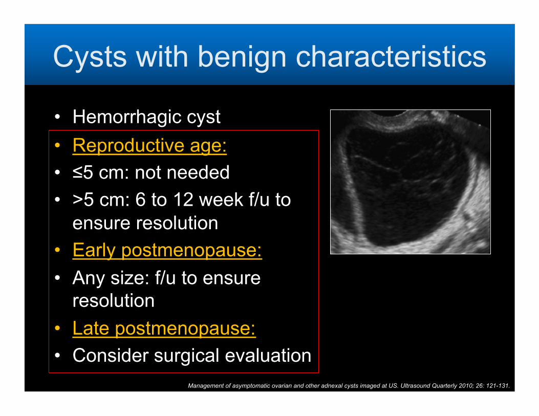
Cysts with benign characteristics
Hemorrhagic cyst Reproductive age: ≤5 cm: not needed >5 cm: 6 to 12 week f/u to
ensure resolution Early postmenopause: Any size: f/u to ensure
resolution Late postmenopause: Consider surgical evaluation
Management of asymptomatic ovarian and other adnexal cysts imaged at US. Ultrasound Quarterly 2010; 26: 121-131.

Cysts with benign characteristics
Endometrioma
Any age: Initial f/u 6 to 12
weeks, then if not surgically removed, f/u yearly
Management of asymptomatic ovarian and other adnexal cysts imaged at US. Ultrasound Quarterly 2010; 26: 121-131.
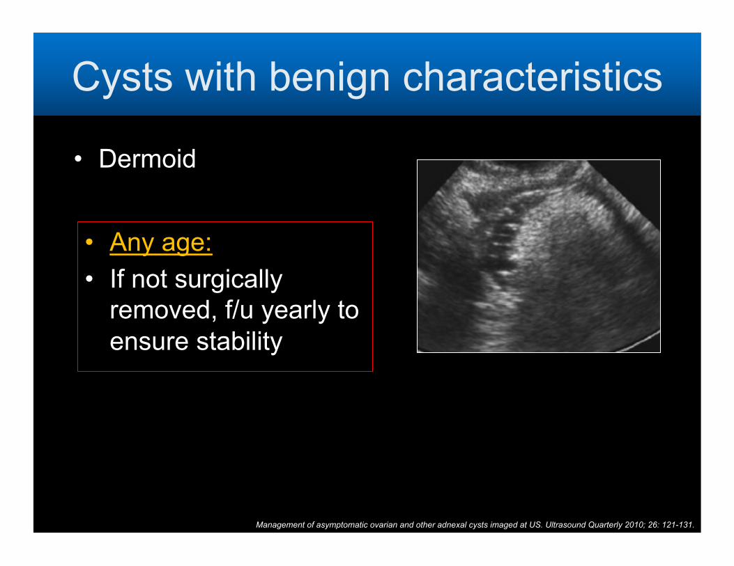
Cysts with benign characteristics
Dermoid
Any age: If not surgically
removed, f/u yearly to ensure stability
Management of asymptomatic ovarian and other adnexal cysts imaged at US. Ultrasound Quarterly 2010; 26: 121-131.

Cysts with benign characteristics
Hydrosalpinx Peritoneal inclusion
cyst
Any age: As clinically indicated
Management of asymptomatic ovarian and other adnexal cysts imaged at US. Ultrasound Quarterly 2010; 26: 121-131.
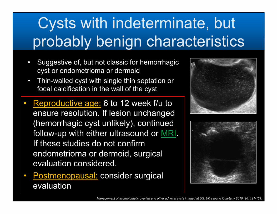
Cysts with indeterminate, but probably benign characteristics Suggestive of, but not classic for hemorrhagic
cyst or endometrioma or dermoid Thin-walled cyst with single thin septation or
focal calcification in the wall of the cyst
Reproductive age: 6 to 12 week f/u to ensure resolution. If lesion unchanged (hemorrhagic cyst unlikely), continued follow-up with either ultrasound or MRI. If these studies do not confirm endometrioma or dermoid, surgical evaluation considered.
Postmenopausal: consider surgical evaluation
Management of asymptomatic ovarian and other adnexal cysts imaged at US. Ultrasound Quarterly 2010; 26: 121-131.

Nodule (non-hyperechoic) without flow
Multiple thin septations ( <3 mm)
Consider surgical evaluation
Surgical evaluation or MRI
Cysts with indeterminate, but probably benign characteristics
Management of asymptomatic ovarian and other adnexal cysts imaged at US. Ultrasound Quarterly 2010; 26: 121-131.

Cysts with characteristics worrisome for malignancy
Thick ( >3 mm) irregular septations Nodule with blood flow
Consider surgical evaluation
Management of asymptomatic ovarian and other adnexal cysts imaged at US. Ultrasound Quarterly 2010; 26: 121-131.

Asymptomatic adnexal cysts
Management of asymptomatic ovarian and other adnexal cysts imaged at US. Ultrasound Quarterly 2010; 26: 121-131.
Simple cyst Hemorrhagic cyst
Endometrioma Dermoid
Reproductive age
Ignore ≤5cm Follow 5 to
7cm Surgical
evaluation >7cm
Ignore ≤5cm
Follow to resolution >5cm
Intial follow-up ultrasound 6 to 12weeks
MRI to confirm if needed
Annual ultrasound if not removed
MRI to confirm if needed
Annual ultrasound if not removed Early post-
menopause Follow 1 to
7cm Surgical
evaluation >7cm
Follow to resolution any size
Late post-menopause
Follow 1 to 7cm
Surgical evaluation 7cm
Consider surgical evaluation any size

Asymptomatic adnexal cysts
Management of asymptomatic ovarian and other adnexal cysts imaged at US. Ultrasound Quarterly 2010; 26: 121-131.
Multiple thin septations
Nodule with no flow
Thick (>3mm) septations
Nodule with blood flow
Reproductive age
Consider surgical evaluation
Likely benign neoplasm
Consider surgical evaluation or MRI
Likely benign neoplasm
Consider surgical evaluation
Likely neoplasm
Consider surgical evaluation
Likely neoplasm
Early post-menopause Late post-menopause
Take home points – Asymptomatic adnexal cysts
Postmenopausal simple cysts ≤1 cm are likely benign, are almost always of no clinical importance in asymptomatic women and can be safely ignored
Simple cysts of any size are unlikely to be malignant lesions, reasonable to f/u with ultrasound when >5 cm in premenopausal women and >1 cm in postmenopausal women
Classic-appearing hemorrhagic cysts in premenopausal women <5 cm do not require follow-up
Management of asymptomatic ovarian and other adnexal cysts imaged at US. Ultrasound Quarterly 2010; 26: 121-131.

Incidental adnexal findings on CT or MRI

Incidental adnexal findings on CT or MRI
Management of incidental CT or MRI findings in nonpregnant postmenarchal patients in whom no adnexal disorder is clinically known or suspected.
Managing Incidental Findings on Abdominal and Pelvic CT and MRI, Part 1: White Paper of the ACR Incidental Findings Committee II on Adnexal Findings. J Am Coll Radiol 2013;10: 675-681.


Exclusions: – A) Normal findings, including hypodense
ovary, corpus luteum – B) unimportant findings, including
calcifications without associated noncalcified mass
– C) previous characterization with ultrasound or MRI
– D) documented stability in size and appearance >2 yrs
Not applicable cysts
Managing Incidental Findings on Abdominal and Pelvic CT and MRI, Part 1: White Paper of the ACR Incidental Findings Committee II on Adnexal Findings. J Am Coll Radiol 2013;10: 675-681.

Benign-appearing cysts Size Action
Pre-menopausal
≤5 cm Benign, no follow-up
>5cm US 6 to 12weeks
Early post-menopause
≤3cm Benign, no follow-up
>3cm, ≤5cm
US 6 to 12 months
>5cm Ultrasound (prompt)
Late post-menopause
≤3cm Benign, no follow-up
>3cm Ultrasound (prompt)
Should have all of the following features: Oval or round Unilocular, with uniform fluid attentuation
or signal (layer hemorrhage acceptable if premenopausal)
Regular or imperceptible wall No solid area, no mural nodule <10 cm maximum diameter
A benign-appearing cyst ≤5cm with suspected internal hemorrhage in a patient aged ≤55 yrs, or within 5 years of menopause, should be followed in 6 to 12 weeks because hemorrhagic cysts in early postmenopause are possible, although rare.
Cysts greater >5 cm on CT should be first characterized promptly with ultrasound, rather than follow-up.
Managing Incidental Findings on Abdominal and Pelvic CT and MRI, Part 1: White Paper of the ACR Incidental Findings Committee II on Adnexal Findings. J Am Coll Radiol 2013;10: 675-681.

Probably benign cysts Size Action
Pre-menopausal
≤3cm Benign, no follow-up
>3cm, ≤5cm US 6 to 12weeks
>5cm Ultrasound (prompt)
Early post-menopause
≤3cm Benign, no follow-up
>3cm Ultrasound (prompt)
Late post-menopause
≤1cm Benign, no follow-up
>1cm Ultrasound (prompt)
May have: a) Angulated
margins b) Not round or oval c) Portion of the cyst
is poorly imaged d) Reduced SNR on
MRI
Managing Incidental Findings on Abdominal and Pelvic CT and MRI, Part 1: White Paper of the ACR Incidental Findings Committee II on Adnexal Findings. J Am Coll Radiol 2013;10: 675-681.

Other imaging features
Examples Action
Probable diagnostic features *adnexal CT and MR findings diagnosed with a high degree of certainty
Paraovarian cyst Hydrosalpinx Peritoneal inclusion cyst Cystic teratoma Endometrioma Leiomyoma Ovarian fibroma Malignancy
Manage as appropriate for diagnosis
Features not specific Solid component Mural nodule Septations Higher than fluid
attenuation Layering hemorrhage
post-menopausal
Ultrasound (prompt)
Managing Incidental Findings on Abdominal and Pelvic CT and MRI, Part 1: White Paper of the ACR Incidental Findings Committee II on Adnexal Findings. J Am Coll Radiol 2013;10: 675-681.

Take home points – CT or MRI adnexal incidentalomas
Premenopausal woman: benign cyst or probably benign cyst ≤3 cm is
normal Early postmenopausal woman:
Benign-appearing cyst >5 cm, prompt ultrasound Probably benign cyst >3 cm, prompt ultrasound
Late postmenopausal woman: Prompt ultrasound follow-up in a probably benign
cyst >1 cm
Managing Incidental Findings on Abdominal and Pelvic CT and MRI, Part 1: White Paper of the ACR Incidental Findings Committee II on Adnexal Findings. J Am Coll Radiol 2013;10: 675-681.

Summary
Transabdominal and transvaginal ultrasound are the first line imaging tests for the pelvis SIS is useful to characterize an endometrial abnormality seen on TVS (especially endometrial polyp), mapping of fibroids CT plays no (little) role in the characterization of pelvic organs **

Summary
Consider MRI: Simple cyst >7cm Adnexal cyst that is probably a benign endometrioma, dermoid (or hemorrhagic cyst), AFTER a 6 to 12 week follow-up to see if the lesion has resolved Cyst with a nodule with no flow, any age

References
Management of asymptomatic ovarian and other adnexal cysts imaged at US. Ultrasound Quarterly 2010; 26: 121-131.
AIUM Practice Guideline for the Performance of Ultrasound of the Female Pelvis. J Ultrasound Med 2014;33: 1122-1130.
Asymptomatic Endometrial Thickening. J Obstet Gynaecol Can 2010;32: 990-999. Abnormal Uterine Bleeding in Premenopausal Women. J Obstet Gynaecol Can 2013;35: S1-S28. Clinical practice guidelines on menorrhagia: management of abnormal uterine bleeding before
menopause. European Journal of Obstetrics & Gynecology and Reproductive Biology 2010;152: 133-137.
Managing Incidental Findings on Abdominal and Pelvic CT and MRI, Part 1: White Paper of the ACR Incidental Findings Committee II on Adnexal Findings. J Am Coll Radiol 2013;10: 675-681.
Management of Abnormal Uterine Bleeding Associated with Ovulatory Dysfunction. Obstetrics & Gynecology 2013;122: 176-185.
FIGO classification system (PALM-COEIN) for causes of abnormal uterine bleeding in nongravid women of reproductive age. International Journal of Gynecology and Obstetrics 2011;113: 3-13.
Change in Endometrial Thickness in Postmenopausal Women Undergoing Hormone Replacement Therapy. Radiology 1995;197: 603-608.
Sonohysterography versus transvaginal sonography for screening of patients with abnormal uterine bleeding. International Journal of Gynecology and Obstetrics 2007;96: 20-23.
AIUM practice guideline for the performance of sonohysterography. J Ultrasound Med 2012;31: 165-172.