Embed Size (px)
Citation preview
Program and Abstract BookProgram and Abstract BookProgram and Abstract Book of the of the of the
Nebraska Physiological SocietyNebraska Physiological SocietyNebraska Physiological Society 11 11 11ththth Annual Meeting Annual Meeting Annual Meeting
Chapter of the American Physiological SocietyChapter of the American Physiological SocietyChapter of the American Physiological Society
Saturday, September 6, 2008Saturday, September 6, 2008Saturday, September 6, 2008
PROGRAM SPONSORS
The Nebraska Physiological Society would like to take this opportunity to gratefully acknowledge the following contributors for their support of the 2008 Meeting of the Nebraska Physiological Society.
The American Physiological Society
The American Physiological Society is proud to be the major sponsor of the 2008 Nebraska Physiological Society Meeting. APS is a nonprofit devoted to fostering education, scientific research, and dissemination of information in the physiological sciences. The Society was founded in 1887 with 28 members. APS now has over 10,500 members. Most members have doctoral degrees in physiology and/or medicine (or other health professions).
APS is governed by an elected Council consisting of a President, President-Elect, Past President, and nine Councilors. The National headquarters of the Society is based in Bethesda, Maryland, on the campus of the Federation of American Societies for Experimental Biology (FASEB).
The following companies contributed support to the Nebraska Physiological Society:
AD Instruments
Bruker BioSpin Corporation - EPR Division
Data Sciences International
The Nebraska Medical Center
North Central Instruments
University of Nebraska College of Medicine
University of Nebraska Medical Center – Department of Cellular and Integrative Physiology
Thank you for your support!
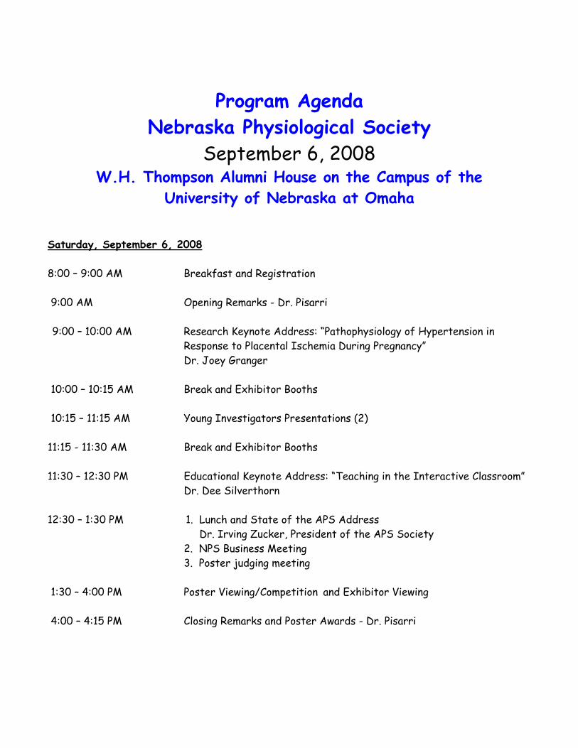
Program Agenda Nebraska Physiological Society
September 6, 2008 W.H. Thompson Alumni House on the Campus of the
University of Nebraska at Omaha Saturday, September 6, 2008 8:00 – 9:00 AM Breakfast and Registration 9:00 AM Opening Remarks - Dr. Pisarri 9:00 – 10:00 AM Research Keynote Address: “Pathophysiology of Hypertension in Response to Placental Ischemia During Pregnancy”
Dr. Joey Granger
10:00 – 10:15 AM Break and Exhibitor Booths 10:15 – 11:15 AM Young Investigators Presentations (2) 11:15 - 11:30 AM Break and Exhibitor Booths 11:30 – 12:30 PM Educational Keynote Address: “Teaching in the Interactive Classroom” Dr. Dee Silverthorn 12:30 – 1:30 PM 1. Lunch and State of the APS Address Dr. Irving Zucker, President of the APS Society 2. NPS Business Meeting 3. Poster judging meeting 1:30 – 4:00 PM Poster Viewing/Competition and Exhibitor Viewing 4:00 – 4:15 PM Closing Remarks and Poster Awards - Dr. Pisarri

Keynote
Speakers

Research Keynote Speaker
Joey P. Granger, Ph.D.
University of Mississippi Medical Center Councilor of the American Physiological Society
Dr. Granger is the Billy S. Guyton Distinguished Professor and Professor of Physiology and Medicine and Associate Director of the Center for Excellence in Cardiovascular-Renal Research and Dean of the School of Graduate Studies in the Health Sciences at the University of Mississippi Medical Center in Jackson, MS. He earned his doctorate from the University of Mississippi School of Medicine in 1983. He received his postdoctoral training in physiology at the Mayo Clinic from 1983–1985. He was appointed Assistant Professor of Physiology at Mayo Medical School in 1985. In 1986, he joined the faculty of the Department of Physiology at Eastern Virginia Medical School. In 1990, he moved back to the University of Mississippi Medical Center.
Dr. Granger is currently an Associate Editor for Hypertension and for the American Journal of Physiology. He has also served as the Editor of the Council for High Blood Pressure Newsletter and a member of Editorial Boards of American Journal of Hypertension, American Journal of Physiology: Renal, Journal of CardioMetabolic Syndrome and News in Physiological Sciences. He has served on numerous scientific committees of the Council for High Blood Pressure Research Inter-American Society of Hypertension, and the American Physiological Society. Within the past year he has been elected to serve on the Leadership committees of the American Physiological Society and the Council for High Blood Pressure Research of the AHA. He has served on scientific study sections for the American Heart Association, National Institutes of Health, NASA, and the Veterans Administration. He has received several awards including the American Physiological Society 2008E.H. Starling Distinguished Lecture Award, American Physiological Society 2008 Bodil M. Schmidt-Nielsen Distinguished Mentor and Scientist Award, Dahl Memorial Lecture of the AHA, American Society of Hypertension Young Scholar Award, the International Society of Hypertension Demuth Research Award, Inter-American Society of Hypertension Young Investigator Award, the Regulatory and Integrative Physiology Young Investigator Award of the American Physiological Society Water and Electrolyte Section, the Harold Lamport Award of the Cardiovascular Section of the American Physiological Society, the Bowditch Lecture of the American Physiological Society, and the Established Investigator Award of the American Heart Association. Granger’s research has been continuously funded by the National Institutes of Health since 1984. Dr. Granger’s research has focused on the role of the kidneys in the pathogenesis of hypertension. His current research focuses on the role of endothelial and neurohormonal factors in mediating hypertension in animal models of preeclampsia. His laboratory is also investigating the role of the renal endothelin system in salt-sensitive hypertension.

Educational Keynote Speaker
Dee Silverthorn, Ph.D.
University of University of Texas-Austin Councilor of the American Physiological Society
Dr. Silverthorn is a comparative physiologist by training, with a B.S.-Honors in Biology from Tulane University and a Ph.D. in marine science from the University of South Carolina. She began her career in the Physiology Department at the Medical University of South Carolina, then followed her husband to Texas. After two years at the University of Texas Medical Branch, Galveston, she ended up at the University of Texas-Austin. Dee’s research interest is epithelial transport, and recent work in her laboratory has focused on transport properties of the chick allantoic membrane. She teaches several undergraduate physiology courses and a graduate course
on developing teaching skills. Dee has received numerous teaching awards and honors, including the American Physiological Society's Claude Bernard Distinguished Lecturer and Arthur C. Guyton Physiology Educator of the Year, the UT-Austin Burnt Orange Apple Award, and Texas Excellence Teaching Award. The first edition of her Human Physiology textbook won the 1998 Robert W. Hamilton Author Award for best textbook published by a UT faculty member. Dee recently completed a six-year term as editor-in-chief of Advances in Physiology Education and she is currently on the governing Council of the American Physiological Society.

State of the American Physiological Address
Irving H. Zucker, Ph.D.
President of the American Physiological Society University of Nebraska Medical Center
Irving H. Zucker, Ph.D. is the Theodore F. Hubbard Professor of Cardiovascular Research and Chairman of the Department of Cellular and Integrative Physiology at the University of Nebraska Medical Center in Omaha, Nebraska. He has been Chairman since 1989. Dr. Zucker received his Ph.D. from New York Medical College in 1972. He continued his post doctoral training at the University of Nebraska Medical Center where he became a faculty member in 1973. Dr. Zucker has been involved in studies related to the neural regulation of cardiovascular function over the past 35 years. His studies have revolved around cardiovascular reflex control of sympathetic nerve activity in animal models of chronic heart failure. These investigations focus on the role of central mediators of sympathetic nerve activity such as angiotensin II and nitric oxide. Dr. Zucker has published over
170 papers in this field and this work has been continuously funded by the National Institutes of Health and the American Heart Association. He serves on the editorial boards of 10 journals. In addition to his research, Dr. Zucker is active in administrative activities for the American Physiological Society and the American Heart Association. He is a member of the National Research Committee of the American Heart Association. He is the Past-President of the Association of Chairs of Departments of Physiology and is the President-of the American Physiological Society.

Spotlight on
Nebraska
Young Investigators

Carol Fassbinder-Orth, Ph.D. Young Investigator
Carol Fassbiner-Orth received her BS from Iowa State University in Genetics in 2003. Carol attended graduate school at the University of Wisconsin-Madison from 2004-2008 where she did research on avian immunology. A central question in Carol’s graduate research was “What aspects of a birds’ immune system make some birds more susceptible to West Nile virus (WNV) infection than others? To investigate this, Carol collaborated with researchers at the USGS National Wildlife Health Center in Madison, WI, and performed the majority of her research in their Biosafety Level 3 facilities. Dr. Fassbinder-Orth received her PhD in Zoology in August, 2008, and was recently hired as a new animal physiology faculty member in the Biology Department at Creighton University. At Creighton this fall, Carol will be teaching an Animal Physiology course, and a new course entitled Ecology of Zoonotic Diseases. Dr. Fassbinder-Orth plans to establish an avian immunology research program involving undergraduate researchers at Creighton, and is currently involved in a research project on the Attwater Prairie Chicken (an endangered bird of the Southern United States). In this project Carol is investigating the immune responses of hatchlings in an attempt to uncover the cause of the massive chick mortality that occurs following introduction of captive birds into the wild. Carol and her husband, Brian have two children, Amara and Quill, and live in Council Bluffs, Iowa.

Young Investigator Abstract
AGE-RELATED SUSCEPTIBILITY OF PIGEONS (COLUMBA LIVIA) TO WEST NILE VIRUS Carol A. Fassbinder-Ortha, William H. Karasovb, and Erik K. Hofmeister c aBiology Department, Creighton University, Omaha, NE 68178 bDepartment of Forest and Wildlife Ecology, University of Wisconsin-Madison, Madison 53706 cUSGS National Wildlife Health Center, Madison, WI 53711
Although birds are considered excellent models for studying immunity and aging, we still have little understanding of comparative avian immunology, or the effects of aging on a bird's susceptibility to infectious diseases. We evaluated the susceptibility of 5-6 week old juvenile pigeons and 12 month old adult pigeons to West Nile virus (WNV). Birds were subjected to a subcutaneous injection of one of two dose levels of WNV (Low dose= 103 plaque forming units (PFU) WNV, High dose=105 PFU WNV). Adults (high dose) had significantly higher viremias compared to adults (low dose) and all juvenile groups. Increased juvenile resistance to WNV infection may be partially explained by the more rapid production of WNV-specific immunoglobulins observed in this group. Juveniles (high dose) produced significantly higher levels of WNV envelope protein-specific immunoglobulin G (IgG) 5 days post infection, and produced WNV nonstructural protein 1-specific IgG sooner than all other groups. The results of this study indicate a possible age-related decline in immunocompetence in pigeons when WNV is used as the challenge agent.

Jennifer Wood, Ph.D. Young Investigator
Jennifer Wood is a native of Indiana and received her BA in microbiology from Indiana University in 1992. Jennifer worked for two years in the Medical Genetics Laboratory at the Indiana University Medical School before beginning her graduate work at the University of Illinois at Urbana-Champaign. Under the direction Dr. Ann Nardulli, Jennifer received her MS in 1996 and her PhD in 2000 from the Department of Molecular and Integrative Physiology. The focus of Jennifer’s thesis work was how differences in estrogen response element sequence alters the conformation of the estrogen receptor and thereby enhances or represses interactions of the receptor with transcriptional cofactors. This thesis work was supported by a NIH reproductive biology training grant and an American Heart Association Illinois Affiliate Student Award. After receiving her PhD, Jennifer pursued post-doctoral training with Dr. Jerome Strauss, III at the University of Pennsylvania. During this time, Jennifer used microarray technology and real-time PCR analysis in order to gain a better understanding of the molecular differences between normal and polycystic ovary syndrome (PCOS) ovarian cells. This post-doctoral work was supported by an Andrew W. Mellon Foundation Junior Investigator Award. Jennifer’s body of work during her post-doc also earned her a Young Investigator’s Travel Grant from the Endocrine Society, a President’s Presenter Award from the Society of Gynecological Investigation, and the Bayard T. Storey Research Award. Jennifer joined the faculty in the Animal Science Department at the University of Nebraska-Lincoln in June 2006. Since that time, her lab has focused its efforts on understanding how metabolic hormones contribute to the growth, development, and maturation of oocytes. The lab is specifically interested in how changes in hormone levels that occur due to changes in body condition including obesity are detrimental to oocyte quality. Collaborative work with colleagues at UNL and the Meat Animal Research Center at Clay Center is also examining how persistent follicles in cattle negatively impact oocyte quality and contributes to embryonic loss. In addition to her research, Jennifer coordinates and is the primary instructor of the Animal Physiology course for the newly formed Veterinary Medicine program at the University of Nebraska-Lincoln. The research carried out by Jennifer has been reported in 20 peer-reviewed manuscripts that appear in journals including the Journal of Biological Chemistry, Physiological Genomics, and Molecular and Cellular Biology. She is a member of the Endocrine Society, American Society of Animal Science, and Society for the Study of Reproduction.

Young Investigator Abstract
EXPRESSION PROFILE OF MTOC-ASSOCIATED GENES IN THE OVARY Kathryn A Cockerill, Lindsey A. Hofman, Jacqueline E. Smith, Jill G. Kerl, and Jennifer R. Wood Department of Animal Science, University of Nebraska-Lincoln, Lincoln, NE
Embryonic loss is relatively common in the human population and particularly in women with metabolic syndromes which are characterized by high circulating insulin levels. Given that successful embryonic development is dependent, in part, on the accumulation of mRNAs within the oocyte, our laboratory has investigated how insulin modifies oocyte gene expression. Previous microarray analysis of MII-arrested oocytes identified increased mRNA abundance of genes associated with microtubule organizing centers (MTOCs) in oocytes exposed to an elevated intrafollicular androgen and insulin environment. Currently, there is little information regarding the normal expression profile or functional significance of MTOC-associated gene expression on developmental competence of the oocyte. Likewise, the insulin-dependent regulation of MTOC-associated gene expression has not been described. To begin to address these questions, we collected ovaries from pubertal mice 2 (n=5), 12 (n=5), 24 (n=5), or 48 (n=5) hours after IP administration of 5 IU equine chorionic gonadotropin (eCG) or 0.5 (n=5), 4 (n=5), 8 (n=5), or 24 (n=5) hours after IP administration of 5 IU eCG followed by 5 IU hCG. RNA collected from each ovary was subjected to quantitative RT-PCR (QPCR) analysis using primers against Nek2, Nek4, and Tacc1. Nek2, Nek4, and Tacc1 mRNA levels were significantly increased (P < 0.01) at 24h post-eCG treatment compared to the other time points. Similar expression profiles were detected for Gata4 and Fkhr which have been shown to be regulated by estrogen in the ovary. In contrast, the mRNA profiles of Nek2, Nek4, and Tacc1 were distinctly different compared to that for the progesterone-regulated Areg and Ereg. Interestingly, NEK2 and NEK4 protein levels were low at 24 – 48 hours post-eCG and were high at 4 and 8 hours post-hCG. When granulosa-oocyte complexes were cultured in vitro in the presence of low (0.1 KM), intermediate (1 KM) or high (10 KM) insulin concentrations, Nek2, Nek4, and Tacc1 mRNA levels were increased in a concentration-dependent manner. Collectively, these data demonstrate that Nek2, Nek4, and Tacc1 are coordinately regulated in the ovary during the estrous cycle and that insulin alters their expression profiles. Furthermore, this study suggests that Nek2 and Nek4 transcription and translation are uncoupled and points to a possible role for these two proteins during oocyte maturation.

Nebraska
Physiological Society
Poster Session
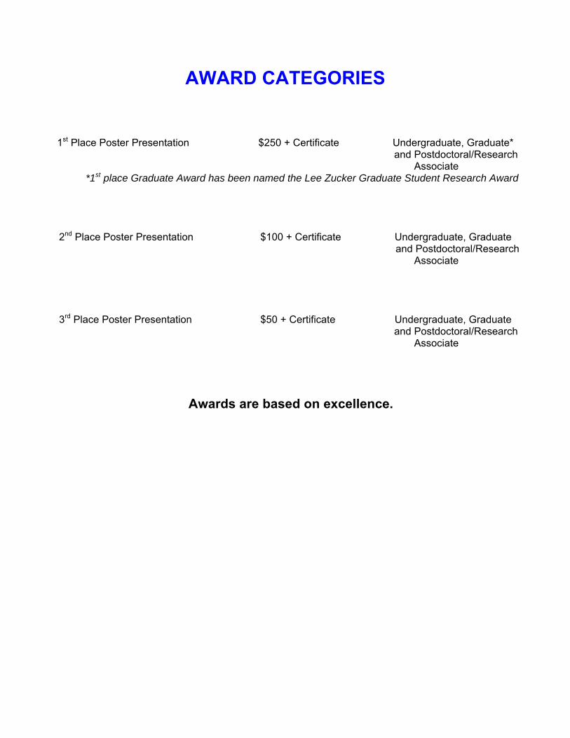
AWARD CATEGORIES
1st Place Poster Presentation $250 + Certificate Undergraduate, Graduate*
and Postdoctoral/Research Associate
*1st place Graduate Award has been named the Lee Zucker Graduate Student Research Award
2nd Place Poster Presentation $100 + Certificate Undergraduate, Graduate and Postdoctoral/Research
Associate
3rd Place Poster Presentation $50 + Certificate Undergraduate, Graduate and Postdoctoral/Research
Associate
Awards are based on excellence.

Undergraduate Posters
Poster U-1 through U-3 to be
considered for the poster award.
POSTER U-1 EXPRESSION OF NEUROPILIN-1 AND -2 DURING MAY INDICATE A ROLE IN SEX-SPECIFIC VASCULAR DEVELOPMENT AND GERM CELL VIABILITY IN THE RAT GONADS Tiffany L. Bohlender, Ningxi Lu, Racheal Slattery, Deb T. Clopton and Andrea S. Cupp University of Nebraska-Lincoln, Lincoln, NE 68583-0908
Neuropilin-1 and 2 act as a co-receptor to Fms-like tyrosine-kinase 1 (FLT1) and Kinase Domain Region (KDR) receptor which is regulated by Vascular Endothelial Growth Factor A (VEGF). Neuropilin-1 serves to stabilize the interaction with VEGF receptors and bind specifically to VEGF isoforms which are critical in establishment of vasculature in most organs in the body. The function of Neuropilin-2 is not yet established. Our laboratory has determined that pro-angiogenic and anti-angiogenic VEGF isoforms are differentially expressed during gonadal development and may be critical to sex-specific vascular development. Therefore, the objective of this experiment was to characterize the expression of Neuropilin-1 and Neuropilin-2 during development in the rat testis and ovary. Gonadal tissue was collected from testes and ovaries at embryonic (E) and postnatal (P) days: E13, E13.5, E14, E16, E18, P0, P3 and P5. RNA from these tissues was extracted and reverse transcribed to produce cDNA pools (n = 6-10 gonads/age/pool). PCR primers for Neuropilin-1 and -2 were optimized for RT-PCR and each developmental age was examined for the Neuropilins. Neuropilin-1 was expressed at E13, E13.5 and E14 in the male at the time of endothelial cell migration during testis development. Furthermore, Neuropilin-1 was expressed after birth at P3 and P5 in the male with some expression in the ovary at P5. During sex specific vascular development endothelial cell migration occurs from the adjacent mesonephros into the testis while no migration occurs in the ovary. Thus, presence of Neuropilin-1 at this time in the male (and not the female) may aid KDR and VEGF interactions to elicit male-specific endothelial cell migration and vascular development. Neuropilin-2 was expressed in both the male and female gonads around gonadal differentiation, E13, E13.5 and E14 and only in the male at E16, E18 and P3 and P5. It is difficult to speculate what role Neuropilin-2 has during early male and female gonadal development. Our laboratory has demonstrated that VEGF is involved in both follicle progression and male germ cell survival, thus, after birth both Neuropilins may be involved in follicle progression in the female and germ cell viability in the male.

POSTER U-2 EFFECTS OF CENTRAL ACE2 OVER EXPRESSION ON CARDIOVASCULAR FUNCTION AND SYMPATHETIC NERVE ACTIVITY IN MICE WITH HEART FAILURE Rachael Farrar, Wei Wang, Lie Gao, Irving H. Zucker Department of Cellular and Integrative Physiology University of Nebraska Medical Center, Omaha, NE 68198-5850
The purpose of the study was to determine the blood pressure, heart rate, urine volume, urine osmolality, and renal sympathetic nerve activity in Wild Type and Angiotensin converting enzyme 2 (ACE2) transgenic mice that were subjected to coronary artery ligation (CHF) or sham surgery. Mice that express ACE2 selectively in neurons (driven by the synapsin promoter) and their Wild Type (WT) counterparts were obtained from Dr. Eric Lazartigues at LSU Health Science Center in New Orleans. Radio telemetry blood pressure transmitters were inserted. Urine volume and urine osmolality were measured in metabolic cages. At the conclusion of the experiments the mice were sacrificed and brains removed and rapidly frozen. Western Blots were performed on brainstem tissue. Renal sympathetic nerve activity (RSNA) was recorded in WT and ACE2 transgenic mice that were subjected to coronary artery ligation or sham surgery. The ACE2 transgenic mice expressed more ACE2 than the WT. The heart failure animals expressed more ACE than the sham animals. The AT1 protein expression appeared to be greater in the ACE2 transgenic animals than the WT animals. The AT2 protein expression appeared to be higher in the ACE2 transgenic sham and CHF animals. The ACE2 transgenic CHF mouse had the highest urine volume. The ACE2 transgenic sham and CHF mice had higher baseline urine volumes than the WT mice. The ACE2 transgenic CHF mouse had the lowest osmolality. The osmolalities of the WT mice and the ACE2 transgenic sham were similar. The transgenic mice had lower RSNA than the WT mice in both the sham and CHF animals. The transgenic mice had lower sympathetic nerve activity than the WT mice in both the sham and CHF animals. These data suggest that over expression of ACE2 and the generation of Ang (1-7) may be sympatho-inhibitory and point to a new mechanism for modulation of sympathetic outflow in CHF.

POSTER U-3 EXERCISE TRAINING NORMALIZESACE AND ACE2 IN THE BRAIN OF RABBITS WITH PACING INDUCED CHRONIC HEART FAILURE Sumit Kar, Lie Gao, Irving H. Zucker Department of Cellular and Integrative Physiology, University of Nebraska Medical Center, Omaha, NE 68198-5850
Over-activation of the renin-angiotensin system (RAS) and elevated Angiotensin II (Ang II) in the brain play a critical role in sympatho-excitation of chronic heart failure (CHF). Exercise training (EX) normalizes sympathetic outflow and plasma Ang II. The central mechanisms by which EX reduces the sympathoexcitatory state are unclear, but EX may alter components of the brain RAS. Angiotensin-converting enzyme (ACE), which synthesizes Ang II, may mediate an increase in sympathetic nerve activity (SNA). ACE2, a homologue of ACE, metabolizes Ang II to Ang 1-7 which has antagonistic effects to Ang II. Little is known about the regulation of ACE and ACE2 in the brain and the effect of EX on the enzymes. This study aimed to investigate the regulation of ACE and ACE2 in various areas of the brain with a pacing induced CHF-EX model. We hypothesized that ACE & ACE2 play a significant role in regulating SNA by mediating the balance of Ang II and Ang 1-7 and will be normalized by EX. Experiments were performed on four groups of New Zealand White rabbits: normal, normal+EX, CHF, and CHF+EX (n=4-5/group). The cortex, cerebellum, medulla, hypothalamus, and punches of the paraventricular nucleus (PVN), nucleus tractus solitarii (NTS), and rostral ventrolateral medulla (RVLM) were analyzed. Western blotting, polymerase chain reaction, and double immunofluorescence were performed to measure and localize expression of ACE and ACE2. ACE protein and mRNA expression in the cerebellum, medulla, hypothalamus, PVN, NTS, and RVLM were significantly upregulated in CHF rabbits (0.3±0.03 to 0.8±0.1 [ratio of ACE to GAPDH] in the RVLM,P<0.05). EX normalized this upregulation compared to CHF(0.4±0.1 to 0.8±0.1 in the RVLM).ACE2 protein and mRNA expression significantly decreased in CHF. EX normalizedACE2 in all areas measured (0.8±0.1 to 0.1±0.01 [ratio of ACE2 to GAPDH] in the RVLM). Immunofluorescence indicated thatACE and ACE2 are present in neurons and vascular endothelial cells. The results suggest that activation of the central RAS system involves an imbalance ofACE and ACE2 in regions of the brain that regulate autonomic function and suggest EX as a possible therapeutic modality to normalize expression of ACE and ACE2 and normalize sympathetic outflow in CHF.

Graduate
Posters
Poster G-1 through G-12 to be
considered for the poster award.

POSTER G-1 ROLE OF ENDOTHELIN IN IMPAIRED RESPONSES OF CEREBRAL ARTERIOLES DURING TYPE 1 DIABETES MELLITUS William G. Mayhan, Glenda M. Sharpe, Denise M. Arrick, and Hong Sun Department of Cellular and Integrative Physiology, University of Nebraska Medical Center, Omaha, NE 68198-5850
Previous studies have suggested that endothelin-1 may contribute to vascular abnormalities during a variety of disease states, including Type 1 diabetes mellitus. Endothelin-1 may contribute to structural and functional abnormalities of the blood vessels, and may also regulate the expression of other growth factors and cytokines to influence vascular function. However, there is a lack of information regarding the precise role of endothelin-1 in altered NOS-dependent responses of cerebral arterioles during Type 1 diabetes. Thus, our goal was to determine whether acute inhibition of endothelin-1 receptors (BQ-123) could influence NOS-dependent responses of cerebral arterioles in diabetic rats. We measured diameter of pial arterioles in nondiabetic and diabetic (STZ; 50 mg/kg) rats in response to eNOS- and nNOS-dependent (ADP and NMDA) and -independent (nitroglycerin) agonists before and during treatment with BQ-123. In addition, we measured superoxide production by cerebral cortex tissue obtained from nondiabetic and diabetic rats. We found that eNOS- and nNOS-dependent dilation of pial arterioles was impaired in diabetic compared to nondiabetic rats. In addition, treatment with BQ-123 restored impaired responses of cerebral arterioles in diabetic rats towards that observed in nondiabetic rats. Further the production of superoxide anion by cortex tissue was increased in diabetic rats when compared to nondiabetic rats. We suggest that endothelin-1 may contribute to impaired responses of cerebral arterioles during Type 1 diabetes. We speculate that endothelin receptor antagonism may be a potential therapeutic tool for the treatment of cerebrovascular dysfunction observed in diabetic subjects.
POSTER G-2 TRANSCRIPTIONAL REGULATION OF THE PORCINE GONADOTROPIN RELEASING HORMONE RECEPTOR II GENE V.M. Brauer, J.R. Wiarda, R.A. Cederberg and B.R. White Department of Animal Science, University of Nebraska-Lincoln
Gonadotropin releasing hormone (GnRH) II has been implicated in female energy balance and reproductive function, in addition to having an anti-proliferative role in cancer cell lines. According to the cDNA sequence, a functional GnRH receptor (GnRHR) II might be present in the pig, unlike many other mammalian species including the mouse. Thus, pigs are an ideal candidate for studying regulation of the GnRHR II gene. Our laboratory isolated 3029 bp of 5´ flanking sequence for the porcine GnRHR II gene. Transient transfections with vectors containing the 5´ untranslated region fused to the cDNA encoding luciferase indicated that this promoter is active in cell lines derived from both reproductive and non-reproductive tissues. Transfection of vectors containing progressive 5´ deletions of the GnRHR II promoter indicated that the -707/-402 and -357/-186 bp regions likely contain elements responsible for promoter activity in a swine testis-derived cell line (ST). Additionally, the region between -1969 and -788 bp of proximal promoter may contain repressive binding sites. To identify the elements contributing to ST cell-specific expression of the porcine GnRHR II gene, oligonucleotides spanning the -707 to -488 bp region of 5´ flanking sequence were radiolabeled and tested in electrophoretic mobility shift assays with nuclear extracts from ST cells. An oligonucleotide spanning -606 to -580 bp produced multiple specific binding complexes. The addition of an unlabeled competitor containing consensus binding sites for Sp1 competed two of the specific complexes. Furthermore, addition of antibodies directed against the Sp1 and Sp3, but not Sp2 or Sp4 transcription factors, resulted in a supershift of two DNA:protein complexes. To determine the specific contribution of the Sp1 and Sp3 transcription factors to GnRHR II promoter activity in ST cells, over-expression vectors containing either Sp1, Sp3 or a control, heterologous protein, were co-transfected with the 3029pGL3 reporter vector. Over-expression of Sp1 increased promoter activity (1.9-fold; P < 0.05) whereas elevated levels of Sp3 did not significantly alter luciferase activity compared to addition of a control expression vector. Thus, the Sp1 and Sp3 transcription factors bind to the -606 to -580 bp region of the porcine GnRHR II gene promoter; although Sp1 may contribute more significantly to activity in ST cells.
POSTER G-3 EXERCISE TRAINING AND RENAL DENERVATION ATTENUATE THE EXPRESSION OF ANGIOTENSIN II TYPE 1 AND 2 RECEPTORS IN RABBITS WITH CHRONIC HEART FAILURE Sarah C. Clayton, Pamela L. Curry, Yu Li, Irving H. Zucker Department of Cellular and Integrative Physiology, University of Nebraska Medical Center, Omaha, NE 68198-5850
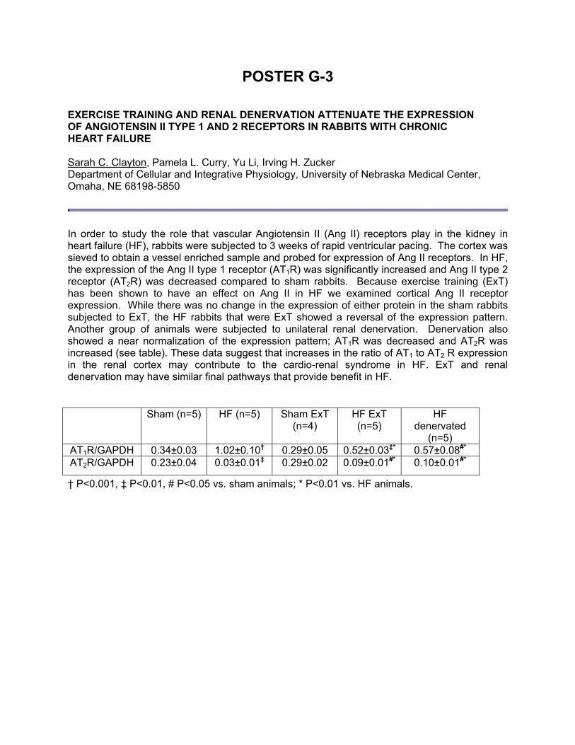
In order to study the role that vascular Angiotensin II (Ang II) receptors play in the kidney in heart failure (HF), rabbits were subjected to 3 weeks of rapid ventricular pacing. The cortex was sieved to obtain a vessel enriched sample and probed for expression of Ang II receptors. In HF, the expression of the Ang II type 1 receptor (AT1R) was significantly increased and Ang II type 2 receptor (AT2R) was decreased compared to sham rabbits. Because exercise training (ExT) has been shown to have an effect on Ang II in HF we examined cortical Ang II receptor expression. While there was no change in the expression of either protein in the sham rabbits subjected to ExT, the HF rabbits that were ExT showed a reversal of the expression pattern. Another group of animals were subjected to unilateral renal denervation. Denervation also showed a near normalization of the expression pattern; AT1R was decreased and AT2R was increased (see table). These data suggest that increases in the ratio of AT1 to AT2 R expression in the renal cortex may contribute to the cardio-renal syndrome in HF. ExT and renal denervation may have similar final pathways that provide benefit in HF.
† P<0.001, ‡ P<0.01, # P<0.05 vs. sham animals; * P<0.01 vs. HF animals.
Sham (n=5) HF (n=5) Sham ExT (n=4)
HF ExT (n=5)
HF denervated
(n=5) AT1R/GAPDH 0.34±0.03 1.02±0.10† 0.29±0.05 0.52±0.03‡* 0.57±0.08#* AT2R/GAPDH 0.23±0.04 0.03±0.01‡ 0.29±0.02 0.09±0.01#* 0.10±0.01#*

POSTER G-4 PARP-1 DEFICIENCY EXACERBATES DIET-INDUCED OBESITY AND INSULIN RESISTANCE Kishor Devalaraja-Narashimha1, and Babu J. Padanilam1,2, 1Department of Cellular and Integrative Physiology, 2Department of Internal Medicine, Section of Nephrology, University of Nebraska Medical Center, Omaha, NE 68198
Poly(ADP-ribose) polymerase-1 (PARP-1) is a major enzyme that has been implicated in regulation of protein functions via poly (ADP-ribosyl)ation and as a transcriptional cofactor in the regulation of gene expression. Here, we report that PARP-1-deficient mice are highly susceptible to diet-induced obesity, accumulate fat tissue and develop augmented insulin resistance and glucose intolerance when compared to their wild-type (WT) counterparts. Male PARP-1-deficient mice are more susceptible and developed weight gain as early as 3 weeks post high fat feeding. Obesity in PARP-1-deficient mice is accompanied by elevated levels of leptin, insulin and higher % of body weight compared to their WT counterparts. Increased abdominal and subcutaneous fat and liver weight accounted for the majority of the weight gain in PARP-1-deficient mice. The increased weight gain was due to increased food intake, decreased basal metabolic rate and total energy expenditure. The motor activity and the consumption of fat energy were paradoxically higher in PARP-1-deficient mice. Absorption of fatty acids was not altered and no significant change in the fecal fatty acid between the groups after high fat diet was observed. These findings demonstrate that PARP-1 mediated signaling pathways play an important role in energy metabolism and malfunction of its signaling could exacerbate diet-induced obesity and insulin resistance.
POSTER G-5 THE INTERACTION OF VASODILATOR-STIMULATED PHOSPHOPROTEIN (VASP) WITH IP3R AND TRPC CHANNELS IN MADIN-DARBY CANINE KIDNEY (MDCK) CELLS P. Richard Grimm, Deann C. Settles, Andrew C. Huss, and Steven C. Sansom University of Nebraska Medical Center, Omaha, NE 69189-5850
Polycystic kidney disease (PKD) is a cellular dedifferentiating disease of the kidneys that accounts for 20% of the patients requiring dialysis or transplant therapy. PKD is partly due to a mutant Ca 2+ channel of the transient receptor family (TRP), that results in aberrantly elevated [Ca2+]i leading to cellular proliferation and dedifferentiation. Therefore it is important to study the mechanism of regulating TRP-mediated cell Ca entry. We have previously found that the cGMP-activated protein kinase (PKG) inhibits TRPC4-mediated Ca2+ entry in mesangial cells. Instead of directly phosphorylating TRPC4, we found that PKG phosphorylated VASP, an actin associated focal adhesion protein, at Ser239, causing its association with TRPC4. This was interesting because Homer, a scaffold protein, interacts with IP3R and TRP channels at the Ena/VASP Homology I (EVHI) domain. Because both VASP and Homer contain the EVH1 domains, we hypothesized that VASP forms a similar interaction with TRPC and IP 3R in MDCK cells, which is a model renal epithelial cell line for studying distal tubule function and polycystic kidney disease. We identified with RT-PCR and immunocytochemistry the presence of TRPC1 and TRPC4 in MDCK cells. We also demonstrated interaction between VASP and TRPC1 with immunocytochemistry and an interaction between VASP with IP3R using immunocytochemistry and Co-IP. Moreover, when VASP was PKG phosphorylated at Ser239, it no longer interacted with TRPC1, but instead interacted with TRPC4 on the cell membrane. These results suggest that PKG controls Ca 2+ entry in MDCK cells by phosphorylating VASP at Ser 239, which causes the linkage of the IP3R to an inhibitory site on TRPC4. This may be an important control mechanism that maintains distal tubule cells in a differentiated state.

POSTER G-6 STRETCH-INDUCED TRANSCRIPTIONAL REGULATION OF THE BK-β4 SUBUNIT IN HUMAN MESANGIAL CELLS Debra L. Irsik, P. Richard Grimm, Steven C. Sansom Department of Cellular and Integrative Physiology, University of Nebraska Medical Center, Omaha, NE 68198-5850
A major complication of Type II diabetes is renal nephropathy. Previously our lab has shown that hyperfiltration and mesangial expansion are characteristics of hyperinsulinemic mice, although the mechanism is not understood. Hyperfiltration results from increased glomerular afferent blood flow, which transmits stretch to podocytes and mesangial cells (MC), two cell types that provide structural support to the vessels of the glomerulus. In MC, stretch increases intracellular Ca2+ levels, possibly by opening mechanosensitive channels, such as those of the Transient Receptor Potential (TRP) family. Increased cytosolic Ca levels can activate the calcineurin/ Nuclear Factor of activated T cells (NFAT) pathway. Membrane potential and contractile tone of MC is partly regulated by large conductance calcium-activated potassium channels (BK). In physiological conditions, the pore-forming BK-α subunits interact with the BK-β1 accessory subunit to hyperpolarize the membrane potential as a feedback response to elevated intracellular Ca. Previous immunohistochemical experiments in this laboratory have shown that glomeruli contain both BK-β1 and BK-β4 subunits. When mice were made insulin resistant with a high fat diet (13 weeks), only the glomerular BK-β4 subunits appeared up-regulated (unpublished observations). For this current study we wished to determine whether MC was one of the glomerular cell types that contained the BK-β4 subunit and investigate the effects of stretch on its transcriptional regulation. Through nested RT PCR, we found the presence of BK-β4 mRNA in MC. Using the Flexercell system with 10% cyclical stretch for 24 hrs, we found a stretch-induced increase in BK-β4 expression six times greater than control (p< 0.05) with a concurrent 50% reduction in BK-β1. Prior studies have shown that the GATA family of transcription factors is important in smooth muscle cell gene expression. Other laboratories have shown an association of NFAT with GATA members in transcriptional complexes. Because MC are considered a modified smooth muscle we proposed that GATA may play a role in the regulation of the BK-β4 subunit. Using the Transcription Element Search System of the University of Pennsylvania, (TESS) we identified 6 GATA4 and 12 NFATc3 binding sites on the β4 promoter. Through chromatin immunoprecipitation, we confirmed that NFATc3 and GATA4 bind to the promoter of BK-β4 1760 bases upstream of the transcription start site. This yielded a 175 bp fragment confirmed as BK-β4 through gel extraction and sequencing. We conclude: 1.Mesangial cells in culture contain the BK-β4 subunit. 2. Stretching MC results in a transcriptional up-regulation of the BK-β4 and down-regulation of the BK- β1. 3. Two candidate transcription factors, NFATc3 and GATA4, bind the BK-β4 at a consensus promoter binding site.

POSTER G-7 WINTER GRAZING SYTEM AND SUPPLEMENTATION DURING LATE GESTATION IMPACT PERFORMANCE OF BEEF COWS AND PROGENY R. N. Funston, J. L. Martin, D. C. Adams, and D.M. Larson University of Nebraska West Central Research and Extension Center, North Platte, NE
A 2x2 factorial study evaluated effects of cow winter system and last trimester supplementation on steer progeny. Composite cows (yr 1 n = 109; yr 2 n = 114; yr 3 n = 116) grazed either range (WR) or corn residue (CR) during winter and within grazing treatment received 0.45 kg/d (DM) 28% CP cubes (PS) or no supplement (NS). Steer calves (yr 1 n = 51; yr 2 n = 58, yr 3 n = 63) entered the feedlot 14 d post-weaning and were harvested 222 d later. Pre-calving BW was greater ( P = 0.02) for PS than NS cows that grazed WR, whereas BCS was greater ( P = 0.03) for cows that grazed CR compared to WR. Calf birth BW was greater ( P = 0.01) for CR than WR and tended to be greater (P = 0.10) for PS than NS cows. Pre-breeding BW and BCS were greater ( P ≤ 0.001) for CR than WR cows and PS than NS ( P = 0.06) cows. At weaning, CR cows were heavier (P < 0.001) than WR cows but had similar BCS ( P = 0.83). Cow weaning BW and BCS were not affected (P > 0.80) by PS. Calf weaning BW was lower (P = 0.01) for calves from NS cows that grazed WR compared to all other treatments. Pregnancy rate was unaffected by treatment (P > 0.46). Steer ADG, 12 th rib fat, yield grade and LM area (P > 0.10) were similar. However, final BW and HCW ( P = 0.06) and marbling score (P < 0.001) were greater for steers from PS cows. Steers from PS cows also graded a higher proportion ( P = 0.05) USDA Choice or greater. Dam PS increased net return of steers at weaning if the dam grazed CR and PS increased net return at harvest regardless of dam grazing system. Heifer calf DMI was similar ( P > 0.11) between all treatments, but PS of cows reduced G:F and increased RFI of heifer progeny (P = 0.02). Heifers born to NS cows that grazed WR were lighter ( P = 0.01) at beginning of breeding than heifers from PS cows. Dam treatment did not affect ( P > 0.10) heifer cyclicity or pregnancy rate. Heifers born to PS cows were younger ( P = 0.03) at puberty and tended to be heavier ( P = 0.07) at pregnancy diagnosis than progeny of NS cows grazing WR. Cows grazing CR with NS produced the most valuable heifer calf at weaning, however, heifers from cows that grazed WR with NS cost the least to develop per pregnant bred heifer. These data support a late gestation dam nutrition effect on calf production via fetal programming that persists through at least 15 months of age. Keywords: carcass quality, fetal programming, maternal nutrition, protein supplement, heifer reproduction

POSTER G-8 GLUCOCORTICOID RESPONSIVENESS OF THE PORCINE GNRH RECEPTOR (GnRHR) GENE IS CONFERRED BY AN ELEMENT(S) LOCATED BETWEEN -290/-270 BP OF PROXIMAL PROMOTER Chanho Lee, Rebecca A. Cederberg and Brett R. White Department of Animal Science, University of Nebraska-Lincoln, Lincoln, NE
The binding of GnRH to its receptor results in the synthesis and secretion of the gonadotropins, as well as stimulation of the gene encoding its own receptor. Thus, the interaction between GnRH and GnRHR represents a central point for regulation of reproductive function. Glucocorticoids can alter reproduction by reducing GnRH responsiveness of gonadotropes within the anterior pituitary gland, potentially via transcriptional regulation of the GnRHR gene. In addition, transcription of the murine GnRHR gene is stimulated by glucocorticoids. To determine the effect of glucocorticoids on porcine GnRHR gene expression, we isolated 5118 bp of 5´ flanking sequence for the porcine GnRHR gene and produced reporter constructs containing the GnRHR promoter fused to the cDNA encoding luciferase (-5118LUC). The gonadotrope-derived &α alpha;T3-1 cell line was transiently transfected with -5118LUC for 12 h and treated with increasing concentrations of the glucocorticoid agonist, dexamethasone (1, 10, 100 and 1,000 nM) for an additional 12 h prior to harvest. A dose-dependent increase in luciferase activity was observed with maximal induction noted at 100 nM dexamethasone (2-fold over vehicle; P < 0.05). The dexamethasone induction of the -5118 promoter was blocked by the glucocorticoid antagonist, mifepristone (100 pM). To determine the location of the glucocorticoid response element(s) within the GnRHR promoter, we performed transient transfection assays with luciferase reporter constructs containing progressively less 5´ flanking region for the porcine GnRHR gene. Dexamethasone-stimulated luciferase activity was maintained following reduction of the full length GnRHR promoter to 323 bp upstream of the translational start site. However, further deletion to 274 bp of proximal promoter eliminated glucocorticoid responsiveness, suggesting the presence of a glucocorticoid response element(s) within this region. Electrophoretic mobility shift assays using 32P-labeled oligomers spanning this region revealed increased protein binding to the -290/-270 bp oligonucleotide in nuclear extracts from & alpha;T3-1 cells treated with 100 nM dexamethasone compared to vehicle treated cells. Sequence analysis of this region has revealed a number of putative elements including binding sites for progesterone receptor (PR), estrogen receptor (ER), glucocorticoid receptor (GR), GATA-1, -3, and -4, as well as retinoid X receptor (RXR) & alpha;, β, and γ. In summary, glucocorticoid responsiveness of the porcine GnRHR gene is conferred by an element(s) located between 270 and 290 bp of proximal promoter.
POSTER G-9 IDENTIFICATION OF PROTEINS INVOLVED IN ESTROGEN-REGULATED PRIMORDIAL FOLLICLE FORMATION IN HAMSTER OVARIES Anindit Mukherjee1 and S. K. Roy1,2, Departments of Cellular and Integrative Physiology1 and OB/GYN2, University of Nebraska Medical Center, Omaha, NE 69198-5850
During early ovarian development oocyte nests are broken down by surrounding undifferentiated somatic cells, which invade the oocyte clusters and align themselves around the oocytes. These cells then differentiate into pre-granulosa cells resulting in the formation of primordial follicles. This event constitutes the critical first step of folliculogenesis and directly determines the eventual number of oocytes available to a mammalian female during her entire reproductive life. Estrogen has been shown to play an important role in mediating primordial follicle formation though the mechanism(s) of its action remains undefined. The objective of this study was to identify protein mediators of estrogen action in primordial follicle formation. We hypothesize that estrogen affects the expression and functions of specific unique proteins in perinatal ovaries to induce primordial follicle formation. The hypothesis was tested using proteomics evaluation of ovaries obtained from 15-day old fetal (E15, complete absence of primordial follicles), 8-day old postnatal (P8, primordial follicles appear for the first time), and estrogen treated P8 golden hamsters. Following staining, gels were compared and analyzed for proteins, which were either upregulated, down regulated or showed unique expression in association with primordial follicle formation. Identified proteins were isolated and analyzed by ion-trap mass spectrometry, and the information was used for database searching to identify the nature and physiological/pathological relevance of the proteins. Several proteins presented a unique expression pattern before and after the formation of primordial follicles, and those proteins were involved in signal transduction, metabolism, post-transcriptional protein modification, gene transcription or translation. The unique protein expression pattern indicates that the differentiation of somatic cells into pre-granulosa cells and their assembly with the oocytes involves proteins that regulate critical molecular events in somatic cells as well as in the oocytes leading to the formation of primordial follicles. The results also indicate some of the possible mechanisms of estrogen-regulated primordial follicle formation.

POSTER G-10 MUTANT SUPEROXIDE DISMUTASE 1 (SOD1) EXPRESSION IN GLIAL CELLS INDUCES NEURONAL TOXICITY IN A CELL CULTURE MODEL OF AMYOTROPHIC LATERAL SCLEROSIS (ALS) Shervin Razavian1, Jocelyn A. Jones1, Matthew C. Zimmerman1,2 1Department of Cellular and Integrative Physiology, University of Nebraska Medical Center, Omaha, NE; 2Redox Biology Center, University of Nebraska-Lincoln, Lincoln, NE
ALS, the most common adult motor neuron disease, is a rapidly progressive and fatal condition characterized by the degeneration of upper and lower motor neurons. Mutations in the cellular antioxidant SOD1 have been identified in 15-20% of familial ALS cases. Interestingly, most of the SOD1 mutants retain SOD1 activity, and thus mutant SOD1-induced neuronal toxicity is believed to be due to an adverse gain of function. Previous studies suggest mutant SOD1 expression in microglia increases levels of damaging reactive oxygen species, which may mediate, at least in part, neuronal toxicity. In this study, we developed a neuronal and glial cell co-culture model of ALS to test the hypothesis that glial cells expressing mutant SOD1 induce toxicity of mutant SOD1-expressing neurons. To express mutant SOD1 in cultured cells, SH-SY5Y neurons and MO59J glial cells plated both individually and as a co-culture were infected with adenoviral vectors (50 MOI) encoding one of three SOD1 mutants (AdG37R, AdG93C, or AdG85R). SOD1 protein and activity levels were measured using Western blot analysis and a native in-gel SOD activity assay. Neuronal toxicity was measured using a WST-8 formazan absorbance-based assay. An increase in mutant SOD1 protein and activity was observed between 3 and 9 days after adenoviral infection; however, this failed to induce toxicity in neurons cultured alone. In contrast, 52-77% neuronal toxicity was observed when neurons were co-cultured with glial cells expressing mutant SOD1. Importantly, these data were confirmed by a decrease in cell number. Overall, this study has established a neuronal and glial cell co-culture model of ALS and indicates that glial cells expressing mutant SOD1 contribute to ALS-associated neuronal toxicity.

POSTER G-11 EFFECTS OF VASCULAR ENDOTHELIAL GROWTH FACTOR (VEGF) ISOFORMS ON RAT TESTIS COMPOSITION AND GERM CELL NUMBERS Racheal G. Slattery, Shantille G. Kruse, Debra T. Clopton and Andrea S. Cupp Department of Animal Science, University of Nebraska-Lincoln, Lincoln NE 68583-0908
Vascular Endothelial Growth Factor (VEGF) is a paracrine growth factor responsible for blood vessel development (neovascularization) as well as endothelial cell migration in many organs including the developing gonad. Multiple isoforms of VEGF are generated from alternative splicing and two of these isoforms are: 1) VEGF164 and 2) VEGF164b. VEGF164b is an anti-angiogenic isoform, which inhibits VEGF164 mediated angiogenesis. In the testis, our laboratory has demonstrated that angiogenic isoforms create a chemoattractive gradient to entice endothelial cell migration from the adjacent mesonephros to allow for sex-specific blood vessel formation and development of seminiferous cords which enclose developing germ cells. We also determined that the receptor for VEGF, Kinase Domain Region (KDR) receptor was expressed in developing germ cells. The objectives of the current experiment were to determine the effect of VEGF isoforms on: 1) testis composition (seminiferous cord area versus interstitial area); and 2) Germ cell numbers. Male rat pups were injected on postnatal day 0 (P0), P1, and P2 with one of five treatments: VEGF 164 (0.5 mg, n= 6), VEGF 164b (0.5 mg; n=6), Anti-VEGFxxxb (antibody to all b isoforms; 1 mg; n=6) IgG Control (1 mg; n=5), or PBS Control (0.5 mg; n=3). Pups were euthanized at day P8 and testis tissue was collected, fixed in bouins, embedded, and sectioned. Hemotyoxylin and eosin sections from each group were utilized to determine seminiferous cord area and interstitial area with Scion image. The males treated with VEGF164b had significantly less seminiferous cord area (1309477 + 26870 vs 1375279 pixels/area), and more interstitial area (332123 + 26870 vs 266321 pixels/area) compared to PBS control (P < 0.05). Furthermore, number of germ cells per area were greater in testes treated with VEGF164 (P< 0.05) than PBS. The antibody to the b isoforms, Anti-VEGFxxxb, increased number of germ cells per seminiferous cord when compared to IgG controls (P <0.05). Therefore, VEGF164 may promote germ cell survival or maturation while VEGF165b or other VEGF b isoforms may reduce the viability of germ cells in vivo.
POSTER G-12 ENHANCED HEAT LOSS DESPITE BLUNTED RENAL SYMPATHOEXCITATION IN DIABETIC RATS DURING HEAT STRESS Laura H. R. Leite,1, 2 Hong Zheng,1 Cândido C. Coimbra,2 and Kaushik P. Patel 1 1Department of Cellular and Integrative Physiology, University of Nebraska Medical Center, Omaha, Nebraska and 2Department of Physiology and Biophysics, Institute of Biological Sciences, Federal University of Minas Gerais, Belo Horizonte, Brazil
Hyperthermia stimulates the sympathetic nervous system to elicit heat dissipation responses in order to maintain body temperature within homeostatic range. Among these responses are vasoconstriction of the visceral vasculature and vasodilation of the skin vasculature to redistribute blood flow to the periphery. Since diabetes is characterized by autonomic nervous system dysfunction, it is possible that regulation of sympathetic activity to heat stress may be altered. In the present study renal sympathetic nerve activity (RSNA), mean arterial pressure (MAP), heart rate (HR), body and tail temperatures were recorded in alpha-chloralose- and urethane-anesthetized (70 mg/Kg and 0.75 g/Kg ip, respectively) control and streptozotocin (STZ)-induced diabetic rats (for 4 weeks; n=6/group) during heat stress. Heat stress was induced by a heating pad with a graded increase in temperature from 37°C to 43°C during 30 minutes. This heat stimulus resulted in blunted RSNA, MAP and HR responses in diabetic rats compared with controls. The highest values were attained at the end of heat stress (delta RSNA: 38.4 ± 9.9 % vs. 159.5 ± 0.1 %; MAP: 91 ± 5 mmHg vs. 114 ± 5 mmHg; HR: 395 ± 11 bpm vs. 460 ± 11 bpm; Diabetic vs Control, p< 0.05), suggesting a decreased renal vasoconstriction. Diabetic animals also showed lower body temperature during the first 13 minutes of heat stress and decreased heat storage rate (HSR: 15.9 ± 1.6 cal min -1 Diabetic vs. 22.0 ± 0.9 cal min -1 Control; p< 0.01). These results may be due to the improvement of heat dissipation shown by diabetic rats as indicated by the decreased change in body temperature (0.3 ± 0.1 °C Diabetic vs. 0.7 ± 0.1 °C Control; p< 0.04) before tail skin vasodilation. In conclusion, although diabetic rats have a decreased RSNA response (renal vasoconstriction), the tail vasodilation was sufficient to decrease body temperature and heat storage rate to heat stress. Supported by: The National Council for Scientific and Technological Development (CNPq, Brazil), National Institute of Health (HL-62222).

Post-doctoral
Posters
Poster P-1 through P-13 to be
considered for the poster award.

POSTER P-1 HUMAN EMBRYONIC KIDNEY CELLS STABLY EXPRESSING BK CHANNELS (HEK- SLO) EXHIBIT ALDOSTERONE (ALDO)-INDUCED TRAFFICKING OF BK CHANNELS TO THE PLASMA MEMBRANE Muhammad R. Bari , Deann C. Settles, J. David Holtzclaw, Liping Liu, Debra L. Irsik and Steven C. Sansom Department of Cellular and Integrative Physiology, University of Nebraska Medical Center, Omaha, NE-68198-5850
Large conductance Ca2+-sensitive K+ channels (BK) reside in the distal nephron where they secrete K+ in response to elevated rates of distal flow. A recent study showed that aldo stimulated BK- mediated K + secretion in an isolated mouse distal colon preparation. However, it is difficult to determine whether aldo affects BK channels in kidney cells because there are no established distal tubule cells in culture that secrete K. Previous investigations have dismissed HEK293 cells as models for studying mineralocorticoid regulated transport activity. However, we examined the potential of HEK- slo cells, which stably express BK, as models to study the mechanism of regulating BK by aldo. We found by immunocytochemistry, Western blotting and RT-PCR that HEK- slo endogenously express mineralocorticoid receptors (MR) and serum and glucocorticoid kinase (SGK), enzymes established as necessary for aldo effects on ion transport. Immunocytochemical staining revealed an aldo (10 nM for 30 min)-evoked increase of MR in the nucleus. Using single channel patch clamp analysis, we determined the 30 min. application of aldo (10 nM) on the NPo (total open time of all channels in a patch; -Vp= -40 mV, cell-attached), from 0.7±0.1 to 1.8±0.4 (p=<0.05). Addition of aldo plus spironolactone, a specific aldo inhibitor, yielded an NPo of 0.6±0.1, a value not different from control. Using Western blot analysis, we found that aldo increased the quantity of plasmalemmal BK-a protein (normalized to caveolin-1) by 120%. The quantity of protein was reduced back to control levels when spironolactone was added with aldo. Similarly, LY294002, an inhibitor of PI3-K, and brefeldin-A, an inhibitor of golgi to surface vesicular trafficking, prevented an aldo-induced increase in plasmalemmal BK protein as determined by Western blot. We conclude: (1) HEK- slo cells contain functional MR receptors and are useful for studying the cellular mechanism of action of aldo on BK channel activity and (2) aldo increases trafficking of BK channels to the plasma membrane of HEK-slo cells within 30 min. via an MR and PI3-K signaling mechanism. Grant support: NIH RO1-DK49561

POSTER P-2 THE ROLE OF THE BETA4 SUBUNIT OF THE LARGE, CALCIUM-ACTIVATED POTASSIUM CHANNEL (BK) IN GLOMERULAR MESANGIAL EXPANSION ASSOCIATEDWITH EARLY STAGE TYPE 2 DIABETES MELLITUS Liping Liu , Deann C. Settles, Steven C. Sansom Department of Cellular and Integrative Physiology, University of Nebraska Medical Center, Omaha, NE 68198-5850
The early stage of type 2 diabetes mellitus (DM) is often associated with high insulin and glomerulopathy that includes expansion of glomerular mesangial cells (MC). Although the pathological mechanisms causing mesangial expansion have been investigated extensively, the role of K channels, which have roles in proliferation in a variety of cells, has not been investigated. Human MC contain large, Ca-activated K channels (BK) comprised of pore-forming a subunits and either accessory BK-b1 or BK-b4 subunits. While the BK-a/b1 is responsible for regulating the contractile tone of MC in physiological conditions, the role of the BK-a/b4 channels in MC is not understood. Our laboratory has found that treating C57/Bl6 mice for 13 weeks with a high fat diet causes mesangial expansion and increased expression of the BK-a and BK-b4, but not the BK-b1 subunit. Increased BK-a/b4 could fuel mesangial proliferation by maintaining a hyperpolarizing driving force for Ca entry via transient receptor cation channels. Often preceding the phase of mesangial expansion is a phase of hyperfiltration, characterized by increased glomerular pressure and stretching of glomerular cells. Stretching often increases cellular proliferation by activating focal adhesion kinase (FAK) via integrins. We therefore investigated the roles of mesangial stretching and high insulin on the expression of BK-b4 in cultured MC (90% confluent, 5 mM glucose, 20% FBS) and the potential role of FAK in this process. We found that high insulin concentrations of 10 nM and 100 nM increased BK-b4 expression slightly (34%, but not significantly) and significantly (170%), respectively, as determined by Western blotting. Cyclical stretching (10% for 24 hours) did not affect expression of BK-b4 protein in MC. However, as determined by immunocytochemical analysis and confocal microscopy, stretching of MC resulted in an association of BK-b4 with FAK and relocalization of BK-b4 from a peri-nuclear region to a sub-plasmalemmal region. We conclude that the combination of high insulin and glomerular stretching in the condition of early stage type 2 DM can result in increased expression of BK-b4 and its trafficking from the a peri-nuclear region to a subplasmalemmal region where it associates with FAK.

POSTER P-3 ROLE OF TRANSCRIPTION FACTOR PATHWAYS IN REGULATION OF BRAIN ANGIOTENSIN RECEPTOR EXPRESSION IN HEART FAILURE Amit Mitra, Lie Gao, Irving H. Zucker Department of Cellular and Integrative Physiology, University of Nebraska Medical Center, Omaha, NE, 68198-5850
It has been clearly established that increased circulating Angiotensin II (Ang II) with concurrent upregulation of brain and peripheral Angiotensin 1-receptors (AT1R) are important mediators in the pathophysiology of heart failure. Our laboratory has previously demonstrated the role of transcription factor AP-1 in the upregulation of AT1R. In this study we aim to determine the role of sequential activation of transcription factors NFκB, AP-1 and Elk-1 in upregulation of brain AT1R. We used Cath.a cells as our neuronal cell model which was treated with Ang II (100nM) over a preset time course. Western blotting was done for protein expression. Our results showed that following Ang II activation, there was a temporal increase of the p65 subunit of NFkB which was observed at 30 minutes and peaked at 1 hr and was sustained upto 24 hours. There was a concomitant decrease of IκB and increased IκK expression. We also observed an increase in AT1R expression which followed the temporal increase of NFkB. The activation of NFκB was blocked by using the inhibitor Parthenolide and this led to a decrease in AT1R expression. The expression of Elk-1 was also examined by western blot and was upregulated over a time period following Ang II activation and was also decreased following NFκB inhibition. Gene silencing using p65-siRNA had similar effects as Parthenolide. Therefore, our results suggest a combined role of the transcription factors NFκB, Elk-1 and AP-1 in the upregulation of brain AT-1R in heart failure.

POSTER P-4 EFFECT OF THIOREDOXIN SYSTEM IN IGF-1-MEDIATED DE-MODELING OF CARDIAC K+ CHANNELS: ROLE OF ASK1-JNK SIGNALING Kang Tang, Ming-Qi Zheng, George J. Rozanski Department of Cellular and Integrative Physiology, University of Nebraska Medical Center, Omaha, NE 68198-5850
The c-Jun NH2-terminal kinase (JNK) pathway plays an important role in the response of cells to pathological stressors. We found that JNK was markedly activated in the rat heart 6-8 wks after myocardial infarction (MI) compared with sham controls, as assessed by Western blotting and JNK assay. Whole-cell patch-clamp recordings in isolated ventricular myocytes from post-MI hearts showed that the characteristic down-regulation of the transient outward K+ current (Ito) density was reversed after 4-5 h by the JNK inhibitor SP600125 (10 BM) or a membrane permeable peptide inhibitor JNKI-1 (10 BM), while a control peptide lacking the c-Jun binding domain sequence had no effect. These results suggest that the JNK pathway is a key contributor to I to remodeling post-MI. We also explored upstream effectors regulating JNK by examining apoptosis signal-regulating kinase (ASK1), which is known to activate JNK as well as p38 MAP kinase. ASK1 has been shown to be inhibited by direct binding with reduced thioredoxin (Trx), and thus we used a co-immunoprecipitation assay to indirectly assess ASK1 activation. These assays showed that ASK1-Trx interaction was markedly decreased post-MI, which is consistent with the activation of ASK1-JNK signaling in response to oxidative stress. To further explore the electrophysiological impact of modulating ASK1-JNK signaling and the role of the Trx system, we investigated the effects of insulin like growth factor-1 (IGF-1), which is involved in anti-apoptotic and other cell survival pathways in the myocardium. Voltage-clamp studies revealed that 10 nM IGF-1 increased Ito density in isolated ventricular myocytes from post-MI rat hearts and that this de-remodeling of Ito was blocked by the specific thioredoxin reductase (TrxR) inhibitor auranofin (10 nM). TrxR is one of the major components of the Trx system which is essential for keeping Trx in its reduced form. We then used an ex vivo organ culture approach by perfusing isolated hearts for 4-6 h with IGF-1 in the presence and absence of auranofin. Western blotting and kinase assay demonstrated that IGF-1 attenuated JNK activation in post-MI hearts, and auranofin blocked this effect. IGF-1 also stimulated TrxR expression in post-MI hearts while auranofin not only blocked TrxR activity but also inhibited the increase in its expression. Moreover, IGF-1 attenuated the activation of activator protein-1 (AP-1), a downstream transcription factor of the JNK pathway. Finally, we found that K + channel protein expression was up-regulated by IGF-1 treatment, providing direct evidence that K + channels are regulated by a redox-sensitive mechanism. Taken together, our data suggest that the expression of K+ channels is redox-regulated by the Trx system whose impaired activity contributes to I to remodeling post-MI through activation of ASK1-JNK signaling. We propose that the cardiac Trx system is part of an essential repair network protecting cellular proteins from oxidation.
POSTER P-5 HOG DUST EXTRACT BINDS TO AND ACTIVATES RABBIT SKELETAL MUSCLE RYANODINE RECEPTOR CALCIUM-RELEASE CHANNEL (RyR1) Chengju Tian1, Danielle S. Fenster1, Myron Toews1, Deborah J. Romberger2, and Keshore R. Bidasee1 1Departments of Pharmacology and Experimental Neuroscience, and 2Department of Internal Medicine, Pulmonary and Critical Care Medicine Section, University of Nebraska Medical Center, Omaha, Nebraska, 68198-5800
BACKGROUND: Individuals working in industrial hog farms as well as people in communities surrounding these animal confinement facilities, exhibit increased muscle weakness and fatigue. To date, molecular mechanisms responsible for this remain incompletely understood. Persistent activation of skeletal muscle ryanodine receptor Ca 2+-release channels (RyR1) increases basal cytosolic Ca 2+ levels, ATP utilization and synthesis and this is known to cause muscle fatigue and weakness. In the present study we investigated whether hog dust contains components that are capable of binding to and activating RyR1. METHODS: Hog dust (3.5g) collected 1-2 meters from the ground of a pig confinement facility in Pender, Nebraska was extracted with chloroform (70ml), filtered and rotor evaporated to dryness. The residue was then redissolved in a 20:1 mixture of hexane:chloroform (30ml) and the precipitate herein referred to as HEX-INS was filtered and air-dried. Displacement binding and single channel assays were then used to determine the effects of HEX-INS on the activity of RyR1. RESULTS: When separated on silica gel thin layer chromatographic plates using chloroform:methanol (80:20 with four drops of triethylamine) and sprayed with 1% sulfuric acid followed by heating, three major compounds were detected in HEX-INS (8.0mg). In [ 3H]ryanodine displacement assays, HEX-INS displaced [ 3H]ryanodine from rabbit skeletal muscle RyR1 in a dose-dependent manner, with an IC values of 1μg/ml. HEX-INS minimally 50 displaced [3H]ryanodine from the cardiac isoform of ryanodine receptor (RyR2) prepared from dog heart. HEX-INS [ 3H]ryanodine displacement curve was parallel to that of the prototype extrinsic ligand, ryanodine as well as Ca 2+-dependent, suggesting that HEX-INS and ryanodine are binding to the same site on RyR1. Using purified rabbit skeletal muscle RyR1 reconstituted into lipid bilayers, HEX-INS increased the open probability of RyR1 in a dose-dependent manner (3.5 to 14.0μg/ml). At a concentration of 17.5μg/ml, HEX-INS induced a state of reduced conductance (55.2% of maximum) of RyR1 with an open probability of 1. This subconductance state was more likely to occur and persist at positive holding potentials. Increasing the concentration of HEX-INS further to 21.0μg/ml resulted in channel closure. CONCLUSIONS: These data are the first to demonstrate that hog dust contains components that selectively bind to and activate RyR1, providing a mechanism for muscle weakness and fatigue reported by indoor hog confinement facility workers and individuals in surrounding communities. Supported in part by grants from NIH (R01-HL085061 KRB, R01-OH008539 DJR).
POSTER P-6 DIFFERENTIAL EXPRESSION OF N- AND E-CADHERIN IN THE HAMSTER OVARY DURING PERINATAL DEVELOPMENT: POTENTIAL REGULATION BY FSH Cheng Wang1, and S. K. Roy1,2, Departments of Obstetrics and Gynecology1 and Cellular and Integrative Physiology2, University of Nebraska Medical Center, Omaha, NE 68198
Factors controlling the size of the initial primordial follicle pool and the development of primordial follicles are largely unknown. Gestational neutralization of FSH attenuates the formation of primordial follicles, but the mechanisms are still unclear. Cadherins mediate homophilic, calcium-dependent cell adhesion; however, whether they are involved in FSH-regulation of primordial follicle formation remains unclear. The objective of the present study was to determine the expression and hormonal regulation of N- and E-cadherins in developing hamster ovaries with special reference to primordial follicle formation. Hamster N- and E-cadherin cDNA and amino acid sequences were highly similar to those of the mouse, rat and human. Both N- and E-cadherins were located mainly in the oocytes during early neonatal life. With the formation of primordial follicles on postnatal day 8 (P8), N-cadherin expression shifted to the pregranulosa cells juxtaposed to the oocytes; however, E-cadherin expression in the oocytes decreased significantly. Subsequently, intense N-cadherin expression was restricted to granulose cells of growing follicles, whereas E-cadherin signal in the oocytes almost disappeared. Levels of N-cadherin mRNA decreased from embryonic day 13 (E13) to P6, but increased markedly on P7, the day before the onset of primordial follicles in the hamster ovary, followed by a decrease on P10. E-cadherin mRNA decreased from E13 through P3 and then remained low. N- and E-cadherin protein levels were consistent with mRNA levels. Exposure of E12 fetuses to an FSH-antiserum in utero resulted in a significant decrease in N-cadherin mRNA levels on P8 (1.7±0.1 ng/mg of total RNA vs. 0.8±0.09 ng/mg of total RNA), which coincided with a block in primordial follicle formation, but the decrease was prevented by a single injection of eCG on P1. A completely opposite result was obtained for E-cadherin mRNA. These results provide evidence for a differential spatio-temporal expression of N- and E-cadherins in perinatal hamster ovaries. Further, the expression is differentially regulated by FSH. The decrease in N-cadherin expression coinciding with the block in primordial follicle formation in FSH antiserum-treated animals and its reversal by eCG suggest that N-cadherin may facilitate the oocyte and somatic cell interaction during the formation of primordial follicles.

POSTER P-7 OXIDATIVE STRESS IN SKELETAL MUSCLE SENSITIZES MECHANORECEPTORS IN HEART FAILURE Han-Jun Wang, Wei-Zhong Wang, Lie Gao, Irving H. Zucker, Wei Wang Department of Cellular and Integrative Physiology, University of Nebraska Medical Center, Omaha, NE 68198-5850
The enhanced skeletal muscle mechanoreflex (mediated via group III fibers) contributes to the exaggerated exercise pressor reflex in chronic heart failure (CHF). However, the mechanism responsible for sensitization of mechanoreceptors is not clear. Here, we proposed that oxidative stress sensitizes mechanoreceptors in skeletal muscle of animals with CHF. We recorded discharge from group III fibers in response to passive stretch (500 grams, 60 s) before and after hindlimb arterial infusion of the superoxide dismutase (SOD) inhibitor diethylthiocarbamate (DETC) or the SOD mimetic tempol in decerebrated rats. The data showed that the expression of SOD protein in triceps surae muscle was significantly decreased in CHF rats compared with sham rats. Pretreatment with DETC significantly increased group III fiber discharge in response to stretch in sham rats (6.09 ± 0.80 vs. 3.03 ± 0.41 Hz, P<0.05, n=6), but not in CHF rats (5.30 ± 1.04 vs. 5.03 ± 1.36 Hz, P>0.05, n=6). Tempol attenuated the response of group III fibers to stretch in both sham (2.15 ± 0.35 vs. 2.98 ± 0.37 Hz, P<0.05, n=6) and CHF rats (3.04 ± 0.49 vs. 5.35 ± 0.83 Hz, P<0.05, n=6). The data indicate that oxidative stress sensitizes mechanoreceptors in CHF. Supported by NIH PO1 HL 62222.

POSTER P-8
TONIC GLUTAMATERGIC INPUT IN THE ROSTRAL VENTROLATERAL MEDULLA IS INCREASED IN RATS WITH CHRONIC HEART FAILURE Wei-Zhong Wang, Lie Gao, Han-Jun Wang, Irving H. Zucker, and Wei Wang Department of Cellular and Integrative Physiology, University of Nebraska Medical Center, Omaha, NE 68198-5850
Chronic heart failure (CHF) is characterized by increased sympathetic tone. The glutamatergic input in the rostral ventrolateral medulla (RVLM), which is a key region involved in sympathetic regulation, seems not to be involved in the generation of sympathetic tone in normal state. The aim of this study was to determine the protein levels of glutamate receptors in the RVLM, and further investigate the effects of RVLM glutamate receptor blockade on sympathetic tone and activity of the RVLM presympathetic neurons in CHF. CHF was produced by coronary artery ligation. The data from western blot analysis showed that the protein expressions of the N-methyl-D-aspartate (NMDA) and non-NMDA glutamate receptor subunits and in the RVLM were significantly higher (2.4-fold and 2.1-fold, respectively) in CHF than in sham rats. Bilateral microinjection of glutamate receptor antagonist kynurenic acid (KYN), NMDA receptor antagonist D-AP5, or the non-NMDA receptor antagonist CNQX into the RVLM dose-dependently reduced the baseline blood pressure and renal sympathetic nerve activity in CHF but not in sham rats. Picoejection of KYN (100 pmol in 5 nl) significantly decreased the discharge of 25 RVLM presympathetic neurons (baseline: 13.2 ± 0.7 spikes/s) by 47% in CHF rats. However, KYN had no effect on the discharge of 22 RVLM presympathetic neurons (baseline: 10.4± 0.6 spikes/s) in sham rats. These data demonstrate that upregulated glutamate receptors including both NMDA and non-NMDA receptors in the RVLM are involved in the tonic control of excitatory sympathetic outflow in CHF. It is suggested that increased glutamatergic input in the RVLM contributes to sympathetic overactivity in CHF.

POSTER P-9 CARDIAC METABOLIC REMODELING IN MICE WITH IMPAIRED THIOREDOXIN Bin Xie, Shumin Li, and George J. Rozanski Department of Cellular and Integrative Physiology, University of Nebraska Medical Center, Omaha, NE 68198-5850
The thioredoxin (Trx) system is a major oxidoreductase network that controls many cellular properties but its functional role in the heart is unclear. Thus, we established a colony of transgenic mice that exhibit cardiac-specific overexpression of a dominant negative mutant of Trx-1 (Tg-DN-Trx1) and characterized their phenotype relative to wild-type (WT) control mice. Spectrophotometric assays of the cytosolic fraction of tissue extracts confirmed that the specific activity of endogenous Trx in hearts of 12 wk Tg-DN-Trx1 mice was 20% less than time-matched WT mice (p<0.05). In vivo echocardiographic measurements under ketamine anesthesia showed markedly greater values for left ventricular (LV) mass (53%), diastolic septal wall thickness (59%), diastolic posterior wall thickness (41%), systolic septal wall thickness (33%), and systolic posterior wall thickness (32%) in Tg-DN-Trx1 compared with WT mice (p<0.05). However, LV ejection fraction and fractional shortening were not significantly different between groups, indicating Tg-DN-Trx1 mice exhibit cardiac hypertrophy with maintained left ventricular function. Additional biochemical assays of other redox markers in Tg-DN-Trx1 mouse hearts showed significant decreases in GSH/GSSG ratio (54%) and protein free SH groups (28%; p<0.05), suggesting that the redox status of cardiac myocytes in Tg-DN-Trx1 mice is shifted to a more oxidized state. However, markers of lipid peroxidation (MDA + 4-HNE) were 17% less in Tg-DN-Trx1 mice (p<0.05). This unexpected finding led us examine whether metabolic remodeling occurs in the myocytes of Tg-DN-Trx1 mice. Thus, we measured NADP/NADPH ratio and the activities of glucose-6-phosphate dehydrogenase (G6PD) and thioredoxin reductase (TrxR), which regulates Trx. NADP/NADPH ratios were 2.1 fold higher in Tg-DN-Trx1 mice than WT (p<0.05), although NADPH levels were not different. These data paralleled a 42% increase in G6PD activity while TrxR activity was 18% lower in Tg-DN-Trx1 mice (p<0.05). Our data suggest that impaired activity of Trx in the heart elicits an oxidative shift in cellular redox state that is associated with LV hypertrophy and compensatory metabolic remodeling. The latter is characterized by increased G6PD activity which regulates NADPH levels required for antioxidant reactions and the detoxication of lipid peroxidation products.

POSTER P-10 CYTOPLASMIC AND MITOCHONDRIAL-PRODUCED SUPEROXIDE MEDIATES ANGIOTENSIN II (ANGII)-INDUCED INHIBITION OF K+ CURRENT IN CATH.A NEURONS Jing-Xiang Yin , Yu-Long Li 1,2, Liang Xiao1, Harold D. Schultz1,3, Matthew C. Zimmerman1,3 1Department of Cellular and Integrative Physiology, 2Department of Emergency Medicine, University of Nebraska Medical Center, Omaha, NE 68198-5850; 3Redox Biology Center, University of Nebraska, Lincoln, NE
Reactive oxygen species, such as superoxide (O·-) and 2 hydrogen peroxide, have been identified as key signaling intermediates in AngII-induced neuronal activation and sympathoexcitation associated with heart failure and hypertension. Here, we hypothesized that AngII-induced inhibition of the delayed rectifier K+ current (I ) is Kv mediated specifically by intracellular O·-. Differentiated 2 CATH.a neurons were infected with adenoviral vectors (50 MOI) encoding the primarily cytoplasmic-localized O·- 2 dismutase (CuZnSOD), or the mitochondrial-targeted isoform (MnSOD). Four days later, I was recorded using Kv the whole cell configuration of the patch-clamp technique. In non-infected and control vector (AdEmpty)-infected neurons, AngII (100 nM) decreased the density of I by 45 ± 4% and 37 ± 6%, respectively (I Kv Kv elicited by 400 ms pulse from -80 to +80 mV, P<0.05 vs vehicle). This AngII effect was significantly blunted in neurons overexpressing active MnSOD or CuZnSOD as AngII inhibited I by merely 18 ± 8% and 11 ± 3%, Kvrespectively (P<0.05 vs AdEmpty-treated and non-infected cells). In contrast, extracellular SOD protein (400 U/ml)-treated cells exhibited the characteristic AngII-induced inhibition of I (34 ± 7%). These data Kv suggest that intracellular O ·- produced from both 2 cytoplasmic and mitochondrial sources attenuates AngII-induced inhibition of IKv-- in neurons. NIH P20RR017675; P01HL062222

POSTER P-11 INCREASED CEREBRAL ISCHEMIC DAMAGE DURING CHRONIC ALCOHOL CONSUMPTION: ROLE OF NAD(P)H OXIDASE Honggang Zhao, William G. Mayhan, Hong Sun Department of Cellular and Integrative Physiology, University of Nebraska Medical Center, Omaha, NE 68198-5850
We hypothesized that NAD(P)H oxidase may contribute to increased cerebral ischemic damage during chronic alcohol consumption. Sprague-Dawley rats were randomly divided into four groups: nonalcohol-fed, alcohol-fed, nonalcohol-fed plus apocynin-treated, and alcohol-fed plus apocynin-treated. Alcohol (8 g/kg/day) was mixed into a liquid diet and given to rats for 8 weeks. Apocynin (7.5 mg/kg/day) was orally administrated for 4 weeks prior to the experiment. Rats were subjected to 2 hours of right middle cerebral artery occlusion (MCAO). Regional cerebral blood flow was monitored through a Laser-Doppler flow probe attached to the lateral aspect of the skull. Infarct volume and neurological score were assessed 24 hours after reperfusion. In addition, superoxide production from ischemic border area was measured by lucigenin-enhanced chemiluminescence. Compared with nonalcohol-fed rats, alcohol-fed rats had a larger infarct volume, worse neurological score and higher superoxide production. Apocynin did not alter infarct size, neurological deficit, and superoxide production in nonalcohol-fed rats, but significantly reduced infarct size, improved neurological outcome and attenuated superoxide production in alcohol-fed rats. Our findings suggest that NAD(P)H oxidase may play an important role in increased cerebral ischemic damage during chronic alcohol consumption.
POSTER P-12 ROLE OF GAMMA-GLUTAMYL TRANSPEPTIDASE IN REDOX REGUALTION OF K + CHANNEL REMODELING IN HEART FAILURE Ming-Qi Zheng, Kang Tang, Bin Xie, Matthew C. Zimmerman, George J. Rozanski Department of Cellular and Integrative Physiology, University of Nebraska Medical Center, Omaha, NE 68198-5850
Gamma-glutamyl transpeptidase (GGT) is a key enzyme in GSH metabolism that regulates intracellular GSH levels in response to extracellular GSH (GSHo). The objective of this study was to identify the role of GGT in reversing pathogenic K+ channel remodeling in the diseased heart. Chronic ventricular dysfunction was induced in rats by myocardial infarction (MI) and studies were done after 6-8wks. Biochemical assays of tissue extracts from post-MI hearts revealed significant increases in GGT activity in left ventricle (47%) and septum (28%) compared with sham hearts, which paralleled increases in protein abundance as determined by Western-blotting. Voltage-clamp studies of isolated left ventricular myocytes from post-MI hearts showed that down-regulation of transient outward K+ current (Ito) was reversed after 4-5 hr by 10 mM GSHo or N-acetylcysteine (NACo), and that the effect of GSHo but not NACo was blocked by the GGT inhibitors, acivicin (10 µM) or S-hexyl-GSH (100 µM). Neither GSHo nor NACo affected Ito density in myocytes from sham hearts. Inhibition of γ-glutamylcysteine synthetase by buthionine sulfoximine (100 μM) did not prevent up-regulation of Ito by GSHo, suggesting that intracellular synthesis of GSH was not involved. However, pre-treatment of post-MI myocytes with an SOD mimetic (MnTPyP, 100 μM) and catalase (400 U/ml) completely blocked recovery of Ito by GSHo. Confocal microscopy using the fluorogenic dye 2’,7’-dichlorodihydrofluorescein diacetate (10 µM) confirmed that GSHo increased reactive oxygen species (ROS) generation by post-MI myocytes and to a lesser extent in myocytes from sham hearts. Furthermore, GSHo-mediated up-regulation of Ito was blocked by inhibitors of tyrosine kinase (genestein, 1 μM; lavendustin A, 10 μM) and thioredoxin reductase (auranofin, 10 nM; 13-cis-retinoic acid, 1 μM). These data suggest that GSHo elicits GGT- and ROS-dependent transactivation of tyrosine kinase signaling that regulates K+ channel activity or expression. The signaling events stimulated by GGT catalysis of GSHo may be a therapeutic target to reverse pathogenic electrical remodeling of the failing heart.

POSTER P-13
GENE TRANSFER OF CU-ZN SUPEROXIDE DISMUTASE TO THE CAROTID BODY REVERSES ENHANCED CHEMORECEPTOR FUNCTION IN HEART FAILURE RABBITS Yanfeng Ding1, Yu-Long Li2, Matthew C Zimmerman1, Robin L Davisson3, Harold D Schultz11Department of Cellular and Integrative Physiology, 2Department of Emergency Medicine, University of Nebraska Medical Center, Omaha, NE 68198-5850. 3Biomedical Sciences,College of Veterinary Medicine and Cell and Developmental Biology, Weill Cornell Medical College, Ithaca, NY 14853-6401
Peripheral chemoreflex sensitivity is potentiated in both clinical and experimental chronic heart failure (CHF). NADPH oxidase-derived superoxide mediates angiotensin II-enhanced carotid body (CB) chemoreceptor sensitivity in CHF rabbits, and tempol, the superoxide dismutase (SOD) mimetic, inhibits this Ang II- and CHF-enhanced superoxide anion effect (Cardiovasc Res. 2007, 75:546-54). Here we investigated the effect of adenoviral Cu/Zn SOD gene transfer in the CB on chemoreceptor activity and function in CHF rabbits. Ad Cu/Zn SOD (1x10⁸ pfu/ml) gene transfer to the CBs significantly reduced the baseline renal sympathetic nerve activity (RSNA) and the response of RSNA to hypoxia in the CHF rabbits. Baseline single-fiber discharge from CB chemoreceptors during normoxia and the response to hypoxia were enhanced in CHF vs. sham rabbits. Ad Cu/Zn SOD decreased the baseline discharge (7.6 ± 1.3 vs. 12.6 ± 1.7 imp/s at 104 ± 2.4 mm Hg PO2) and the response to hypoxia (22.4 ± 1.6 vs. 32.3 ± 1.2 imp/s at 41 ± 2.6 mm Hg PO2, p<0.05) in CHF rabbits. In addition, Ad Cu/Zn SOD normalized the blunted IK in CB glomus cells from CHF rabbits (1271 ± 35 vs. 1809 ± 87 pA at +70 mV, p<0.05 ). Ad SOD did not affect CB afferent discharge and IK of CB glomus cells in sham rabbits. These results suggest that Cu/Zn SOD activity in the CB may be involved in the enhanced activity of the CB chemoreceptors and peripheral chemoreflex function in CHF rabbits.

Young
Investigator
Posters

Poster Y-1
AGE-RELATED SUSCEPTIBILITY OF PIGEONS (COLUMBA LIVIA) TO WEST NILE VIRUS Carol A. Fassbinder-Ortha, William H. Karasovb, and Erik K. Hofmeister c aBiology Department, Creighton University, Omaha, NE 68178 bDepartment of Forest and Wildlife Ecology, University of Wisconsin-Madison, Madison 53706 cUSGS National Wildlife Health Center, Madison, WI 53711
Although birds are considered excellent models for studying immunity and aging, we still have little understanding of comparative avian immunology, or the effects of aging on a bird's susceptibility to infectious diseases. We evaluated the susceptibility of 5-6 week old juvenile pigeons and 12 month old adult pigeons to West Nile virus (WNV). Birds were subjected to a subcutaneous injection of one of two dose levels of WNV (Low dose= 103 plaque forming units (PFU) WNV, High dose=105 PFU WNV). Adults (high dose) had significantly higher viremias compared to adults (low dose) and all juvenile groups. Increased juvenile resistance to WNV infection may be partially explained by the more rapid production of WNV-specific immunoglobulins observed in this group. Juveniles (high dose) produced significantly higher levels of WNV envelope protein-specific immunoglobulin G (IgG) 5 days post infection, and produced WNV nonstructural protein 1-specific IgG sooner than all other groups. The results of this study indicate a possible age-related decline in immunocompetence in pigeons when WNV is used as the challenge agent.
Poster Y-2
EXPRESSION PROFILE OF MTOC-ASSOCIATED GENES IN THE OVARY Kathryn A Cockerill, Lindsey A. Hofman, Jacqueline E. Smith, Jill G. Kerl, and Jennifer R. Wood Department of Animal Science, University of Nebraska-Lincoln, Lincoln, NE
Embryonic loss is relatively common in the human population and particularly in women with metabolic syndromes which are characterized by high circulating insulin levels. Given that successful embryonic development is dependent, in part, on the accumulation of mRNAs within the oocyte, our laboratory has investigated how insulin modifies oocyte gene expression. Previous microarray analysis of MII-arrested oocytes identified increased mRNA abundance of genes associated with microtubule organizing centers (MTOCs) in oocytes exposed to an elevated intrafollicular androgen and insulin environment. Currently, there is little information regarding the normal expression profile or functional significance of MTOC-associated gene expression on developmental competence of the oocyte. Likewise, the insulin-dependent regulation of MTOC-associated gene expression has not been described. To begin to address these questions, we collected ovaries from pubertal mice 2 (n=5), 12 (n=5), 24 (n=5), or 48 (n=5) hours after IP administration of 5 IU equine chorionic gonadotropin (eCG) or 0.5 (n=5), 4 (n=5), 8 (n=5), or 24 (n=5) hours after IP administration of 5 IU eCG followed by 5 IU hCG. RNA collected from each ovary was subjected to quantitative RT-PCR (QPCR) analysis using primers against Nek2, Nek4, and Tacc1. Nek2, Nek4, and Tacc1 mRNA levels were significantly increased (P < 0.01) at 24h post-eCG treatment compared to the other time points. Similar expression profiles were detected for Gata4 and Fkhr which have been shown to be regulated by estrogen in the ovary. In contrast, the mRNA profiles of Nek2, Nek4, and Tacc1 were distinctly different compared to that for the progesterone-regulated Areg and Ereg. Interestingly, NEK2 and NEK4 protein levels were low at 24 – 48 hours post-eCG and were high at 4 and 8 hours post-hCG. When granulosa-oocyte complexes were cultured in vitro in the presence of low (0.1 KM), intermediate (1 KM) or high (10 KM) insulin concentrations, Nek2, Nek4, and Tacc1 mRNA levels were increased in a concentration-dependent manner. Collectively, these data demonstrate that Nek2, Nek4, and Tacc1 are coordinately regulated in the ovary during the estrous cycle and that insulin alters their expression profiles. Furthermore, this study suggests that Nek2 and Nek4 transcription and translation are uncoupled and points to a possible role for these two proteins during oocyte maturation.

Faculty
Posters

POSTER F-1 EXERCISE TRAINING INITIATED AFTER THE ONSET OF DIABETES PRESERVES MYOCARDIAL FUNCTION: EFFECTS ON EXPRESSION OF BETA-ADRENOCEPTORS Keshore R. Bidasee1, Hong Zheng2, Chun-Hong Shao1, Sheeva K. Parbhu1, George J. Rozanski2, and Kaushik P. Patel2 1Departments of Pharmacology and Experimental Neuroscience and 2Department of Cellular and Integrative Physiology, University of Nebraska Medical Center, Omaha, NE 68198
The present study was undertaken to assess cardiac function and characterize beta-adrenoceptor subtypes in hearts of diabetic rats that underwent exercise training (ExT) after the onset of diabetes. Type 1 diabetes was induced in male Sprague-Dawley rats using streptozotocin. Four weeks after induction, rats were randomly divided into two groups. One group was exercised trained for three weeks while the other group remained sedentary. At the end of the protocol, cardiac parameters were assessed using M-Mode echocardiography. A Millar catheter was also used to assess left ventricular hemodynamics with and without isoproterenol stimulation. Beta-adrenoceptors were assessed using Western blots and [ 3H]dihydroalprenolol binding. After seven weeks of diabetes, heart rate decreased by 21%, fractional shortening by 20%, ejection fraction by 9% and basal and isoproterenol induced dP/dt by 35%. beta1- and beta2-adrenoceptor proteins were reduced by 60% and 40%, while beta3-adrenoceptor protein increased by 125%. Ventricular homogenates from diabetic rats bound 52% less [3H]dihydroalprenolol, consistent with reductions in beta 1- and beta 2-adrenoceptors. Three weeks of ExT initiated four weeks after the onset of diabetes minimized cardiac function loss. ExT also blunted loss of beta 1-adrenoceptor expression. Interestingly, ExT did not prevent diabetes-induced reduction in beta 2 -adrenoceptor or the increased of beta 3-adrenoceptor expression. ExT also increased [3 H]dihydroalprenolol binding, consistent with increased beta 1-adrenoceptor expression. These findings demonstrate for the first time that ExT initiated after the onset of diabetes blunts primarily beta1-adrenoceptor expression loss, providing mechanistic insights for exercise-induced improvements in cardiac function.

POSTER F-2 IMBALANCE OF ANGIOTENSIN RECEPTOR EXPRESSION AND FUNCTION IN THE RVLM: POTENTIAL MECHANISM FOR SYMPATHETIC OVERACTIVITY IN HEART FAILURE Lie Gao, Wei-Zhong Wang, Wei Wang, Irving H. Zucker Department of Cellular and Integrative Physiology, University of Nebraska Medical Center, Omaha, NE 68198-5850
Up-regulation of Angiotensin type 1 receptors (AT1R) in the rostral ventrolateral medulla (RVLM) contributes to the sympathetic over activity in the chronic heart failure (CHF) state. However, the role of the central AT2R is not clear. In the current study, we measured both AT1R and AT2R protein expression in the RVLM and determined the effects of activating RVLM these receptors on renal sympathetic nerve activity (RSNA), blood pressure (BP), and heart rate (HR) in anaesthetized sham and CHF rats. We found that: (1) while AT1R protein expression in the RVLM was up regulated (CHF: 0.83 ± 0.07 vs sham: 0.32 ± 0.05, P < 0.05), the AT2R was significantly down regulated (CHF: 0.06 ± 0.02 vs sham: 0.15 ± 0.02, P < 0.05) increasing the ratio of AT1R to AT2R in the RVLM of CHF rats (AT1R/AT2R: 13.8 ± 0.7 in CHF vs 2.1 ± 0.4 in sham, P < 0.05); (2) simultaneously stimulating RVLM AT1R and AT2R by Ang II evoked a sympatho-excitation, hypertension, and tachycardia in both sham and CHF rats, with greater responses in CHF rats (RSNA: 235.3 ± 11.6 % of baseline in CHF vs 146.7 ± 9.4 % of baseline in normal, P < 0.05); (3) stimulating RVLM AT1R with Ang II plus the specific AT2R antagonist PD123319 induced a larger sympatho-excitatory response than simultaneously stimulating AT1R and AT2R in sham rats (RSNA: 193.2 ± 6.8 % of baseline in stimulating AT1R vs 146.7 ± 9.4 % of baseline when stimulating both AT1R and AT2R, p < 0.05), but not in CHF rats; (4) activating RVLM AT2R with CGP42112 induced a sympatho-inhibition, hypotension, and bradycardia only in sham rats (RSNA: 36.4 ± 5.1 % of baseline vs 102 ± 3.9 % of baseline in aCSF, P < 0.05); (5) pretreatment with ETYA, a general inhibitor of AA metabolism, into the RVLM partially abolished the CGP42112 induced sympatho-inhibition. These results suggest that, in contrast to AT1R, AT2R in the RVLM exhibits an inhibitory effect on sympathetic outflow, which is partially mediated by the PLA2/AA/12-LO pathway. These data implicate a down regulation in the AT2R as a contributory factor in the sympatho-excitation in CHF.
POSTER F-3 FLOW-INDUCED SHEAR STRESS REDUCES K CONTENT OF MDCK CELLS BY A CA2+ AND ATP-DEPENDENT K EFFLUX MECHANISM J. David Holtzclaw, Jakeb D. Riggle, and Steven C. Sansom Department of Cellular and Integrative Physiology, University of Nebraska Medical Center; Omaha, NE 68198-5850
High tubular flow induces K+ secretion by large conductance, Ca2+-activated K+ channels (BKCa) in mammalian connecting tubules (CNTs) and cortical collecting ducts (CCDs). Electrophysiological studies have revealed that BKCa channels are expressed in both intercalated cells (IC) and principal cells (PC) of the K+ secreting segments of the nephron. However, IC, which protrude into the lumen, express substantially more BK Ca channels than PC. These observations are puzzling since IC lack sufficient pump activity to support K+ secretion, thus making it unlikely that IC play a role in regulating K+ balance. So, why are the majority of BK Ca channels expressed in IC? We hypothesis that high flow activates BKCa in ICs to reduce IC size, thereby increasing tubule internal diameter and decreasing resistance to flow. To test this hypothesis, PC and IC subclones of MDCK cells, a canine epithelial cell line originating from the distal nephron, were exposed to either static (control) or 10 dynes/cm 2 shear stress in a parallel plate flow chamber (PPFC) at 37ºC for 5 hours. Perfusate was similar to mammalian CNT luminal fluid. After incubation, K+ content was measured by flame photometry and normalized by DNA. With high flow, intracellular K+ decreased by 32% compared to static conditions (5.0 vs. 7.4 Fg K+/Fg DNA, P=0.03, N=6) in IC, but not PC. This decrease was attenuated by removal of luminal Ca 2+ with EGTA (11.0 vs. 9.1 FgK+/Fg DNA, N=4) or addition of 5 mM TEA (12.9 vs. 14.5 FgK +/Fg DNA, N=5), a blocker of BK Ca. Previous studies have indentified nucleotide release as an important factor in flow-induced cation regulation in the distal nephron. Therefore, we examined the role of ATP in IC cell volume regulation. In IC, the addition of 10 mM ATP to static conditions replicated the flow-induced intracellular K+ decreased (3.8 static vs 2.1 Fg K+/Fg DNA static + ATP, P=0.08, N=4), while 2U/ml apyrase, an ATP scavenger, inhibited the flow-induced intracellular K+ decreased (5.9 vs 5.5, Fg K+/Fg DNA, N=2). Futhermore, the addition of 10 FM ATP increased intracellular fura 2 ratio (1.56 vs. 6.85 with ATP, P=0.01, N=4). The BKCa molecular components may explain the large increase in fura 2 ratio in IC cells. By western blot analysis, we show that IC, but not PC cells, contains the BK-ß4 subunit, which dampens the apparent voltage and Ca 2+ sensitivity of the BKCa channel. These data show that an IC subclone of MDCK cells contain the BK Ca molecular components and suggest that the elevated [Ca 2+] is due to shear stress may result in activation of BKCa, which reduces cell K+ content and volume.

POSTER F-4 EXERCISE TRAINING IMPROVES PERIPHERAL CHEMORECEPTOR SENSITIVITY IN HEART FAILURE: INVOLVEMENT OF ANGIOTENSIN II AND NITRIC OXIDE Yu-Long Li1,2, Yanfeng Ding2, Chad Agnew2, Harold D. Schultz2 1Department of Emergency Medicine, 2Department of Cellular & Integrative Physiology, University of Nebraska Medical Center, Omaha, NE 68198-5850
An enhancement of peripheral chemoreflex sensitivity contributes to sympathetic hyperactivity in chronic heart failure (CHF) rabbits. The enhanced chemoreflex function in CHF involves augmented carotid body (CB) chemoreceptor activity via upregulation of the angiotensin (Ang) II-AT1 receptor pathway and downregulation of the neuronal nitric oxide synthase (nNOS)-NO pathway in the CB. Here we investigated whether exercise training (EXT) normalizes the enhanced peripheral chemoreflex function in CHF rabbits and possible mechanism mediating this effect. EXT partially but not fully normalized the exaggerated baseline renal sympathetic nerve activity (RSNA) and the response of RSNA to hypoxia in CHF rabbits. EXT also decreased the baseline carotid body nerve single-fiber discharge (4.9 ± 0.4 vs. 7.7 ± 0.4 imp/s at PO =103 ± 2.3 mm Hg) and the 2 response to hypoxia (20.6 ± 1.1 vs. 36.3 ± 1.3 imp/s at PO =41 ± 2.2 mm Hg) from CB 2 chemoreceptors in CHF rabbits. Our results also showed that NO concentration and protein expression of nNOS were increased in the CBs from EXT + CHF rabbits, compared with that in CHF rabbits. On the other hand, elevated Ang II concentration and AT receptor overexpression of the CBs in CHF state were blunted by EXT. These 1 results indicate that EXT normalizes the CB chemoreflex in CHF by preventing an increase in afferent CB chemoreceptor activity. EXT reverses the alterations in the nNOS-NO and Ang II-AT receptor pathways in the CB responsible for chemoreceptor 1 sensitization in CHF.

POSTER F-5 CASTRATION REDUCES VENOCONSTRICTOR RESPONSES TO PVN STIMULATION Doug Martin, Basic Biomedical Sciences, Sanford School of Medicine, University of South Dakota 414 East Clark Street, Vermillion SD, 57069
Our previous work showed that venous tone is elevated during hypertension development in the spontaneously hypertensive rat (SHR) and that the hypothalamic paraventricular nucleus (PVN) contributes to the control of venous tone. Androgens exacerbate hypertension development in the SHR. This study tested the hypothesis that androgens modulate PVN control of venous tone in SHR. Five week old male SHR underwent sham operation or castration. At approximately 12 weeks of age they were anesthetized and fitted with arterial and venous catheters. A latex balloon was placed in the right atrium for assessment of mean circulatory filling pressure (an index of venous tone). The rats were placed in a stereotaxic frame and an injector lowered into the PVN. Mean arterial pressure, heart rate (HR) and MCFP were recorded at baseline and during graded PVN stimulation with bicuculline (BIC) (50-200 pmol). Injection of BIC into the PVN caused dose dependent increases in MAP, HR and MCFP. The MAP and MCFP responses obtained in SHR were greater than those in SHR subjected to castration (Sham; 32±1 vs Castrated 19±4 mm Hg) and MCFP (Sham; 2±0.6 mm Hg vs Castrated; 1±0.3 mm Hg). Pretreatment with an injection of losartan (angiotensin AT1 antagonist) into the PVN markedly reduced the MAP, HR and MCFP responses to BIC and largely reduced the differences between Sham operated and castrated SHR. These data suggest that PVN-induced venoconstrictor responses are modulated by endogenous male sex steroids. Supported by NIH #63053, and AHA #0515443Z.

POSTER F-6 EFFECTS OF ALCOHOL ON SUPEROXIDE PRODUCTION AND NITRIC OXIDE RELEASE IN HUMAN BRAIN MICROVASCULAR ENDOTHELIAL CELLS Hong Sun, Hong Zheng, Honggang Zhao, Glenda M. Sharpe, Denise M. Arrick, and William G. Mayhan Department of Cellular and Integrative Physiology, University of Nebraska Medical Center, Omaha, NE 68198-5850
Oxidative stress may play an important role in impaired endothelial nitric oxide (NO) synthase (eNOS)-dependent dilation of cerebral arterioles during chronic alcohol consumption. The purpose of this study was to examine the effects of acute (3 hours) and chronic (7 days) alcohol on superoxide production and NO release in human brain microvascular endothelial cells (HBMECs). Primary HBMECs were acutely and chronically exposed to 5, 20, 50, and 100 mM alcohol. In the absence and presence of NAD(P)H oxidase inhibitor, apocynin, basal superoxide production and NO release to L-arginine and acetylcholine was measured by lucigenin-enhanced chemiluminescence and a NO microsensor, respectively. eNOS expression was determined by Western blot analysis and immunofluorescence staining. Acute and chronic exposure of low (5 mM) to moderate dose (20 mM) alcohol did not alter superoxide production and NO release. Chronic exposure of high dose (50 and 100 mM) alcohol significantly enhanced superoxide production. Acute exposure of high dose alcohol increased NO production to L-arginine, but not to acetylcholine. In contrast, chronic exposure of high dose alcohol significantly reduced NO release to L-arginine and acetylcholine. Apocynin (1 mM, 30 min) restored enhanced superoxide production and impaired NO release towards normal. In addition, acute exposure of high dose and chronic exposure of moderate dose alcohol up-regulated eNOS expression. However, chronic exposure of high dose alcohol down-regulated eNOS expression. Our findings suggest that impaired NO release during chronic exposure of high dose alcohol may be related to reduced eNOS expression and enhanced superoxide production via NAD(P)H oxidase in HBMECs.

POSTER F-7 HOG BARN DUST ACTIVATION AND REGULATION OF EPIDERMAL GROWTH FACTOR RECEPTORS ON AIRWAY EPITHELIAL CELLS Myron L. Toews1, Nancy A. Schulte1, Puttappa R. Dodmane1, Anna C. Jacobs1, Art J. Heires1,2 and Debra J. Romberger1,2
1University of Nebraska Medical Center and 2 Omaha Veterans' Administration Medical Center, Omaha, NE
Rationale. Inhalation of hog confinement facility dust can lead to chronic inflammatory lung disease in susceptible workers. An aqueous extract of this dust (HDE) induces inflammation in animal models and promotes IL-6 and IL-8 release from airway epithelial cells in culture. Lysophosphatidic acid (LPA) has been shown to stimulate IL-8 release from airway epithelial cells via transactivation of epidermal growth factor receptors (EGFRs) and to induce a biphasic decrease in EGFR binding. We hypothesized that similar EGFR activation and regulation would be involved for HDE. Methods. BEAS-2B or H292 cells were incubated for 15 min or 18 hr in the absence or presence of HDE, with and without various inhibitors. Cells were then assayed for 125I-EGF binding to intact cells on ice or for IL-6 and IL-8 release by ELISA. Results. Treatment of BEAS-2B cells with HDE led to a concentration-dependent decrease in EGFR binding within 15 min that was sustained to 18 hr; in H292 cells, HDE decreased EGFR binding at 15 min but not at 18 hr, similar to the differential effects of LPA in these cells. The HDE-induced decrease in EGFR binding in BEAS-2B cells was partially blocked by the EGFR tyrosine kinase inhibitor AG1478, similar to results for LPA. The metalloprotease inhibitor GM6001 partially blocked the decrease in EGFR binding induced by LPA but not by HDE. Finally, AG1478 also caused a partial inhibition of the HDE-induced stimulation of IL-6 and IL-8 release from BEAS-2B cells, but GM6001 did not. Conclusions. HDE induces a rapid decrease in airway epithelial cell EGFR binding that involves EGFR tyrosine kinase activation. EGFR tyrosine kinase activation is also involved in HDE stimulation of IL-6 and IL-8 release. The ability of GM6001 to inhibit these effects for LPA but not for HDE suggests that these agents activate and regulate EGFRs by somewhat different mechanisms. Supported by NIH grant R01-OH008539, D.J. Romberger, PI.

POSTER F-8 CONTRIBUTION OF RENAL EPITHELIAL SODIUM CHANNEL IN SODIUM RETENTION DURING CHRONIC HEART FAILURE Hong Zheng1, Xuefei Liu1, U. S. Rao2, and Kaushik P. Patel1 1Department of Cellular and Integrative Physiology, University of Nebraska Medical Center, Omaha, NE 68198-5850; 2Department of Pharmaceutical Sciences, Texas Tech University Health Sciences Center, Amarillo, TX
The goal of the present study was to examine the contribution of renal epithelial sodium channel (ENaC) in the increased sodium and water retention during chronic heart failure (CHF). We hypothesized that over expression of ENaC could be involved in sodium retention during CHF. The left coronary ligation-induced heart failure model in the rat was used. There was significant increase in diuretic (7-fold of sham) and natriuretic responses (3-fold of sham) to the ENaC inhibitor benzamil in the rats with CHF compared to sham rats. Real-time PCR and Western blotting demonstrated that the mRNA and protein abundance of a, b, and g-subunits of ENaC were significantly increased in the cortex (alpha-ENaC: mRNA 104±24%, protein 32±7% compared to sham rats) and outer medulla (alpha-ENaC: mRNA 52±18%, protein 33±9% compared to sham rats) in CHF compared to sham rats. Immunohistochemical imaging confirmed the increased labeling of alpha-ENaC, beta-ENaC and gamma-ENaC subunits in the collecting duct segments in rats with CHF compared to sham rats. These results suggest that the increased expression of renal ENaC subunits may contribute to the renal sodium and water retention observed during CHF. (Supported by NIH)

POSTER F-9 COMPUTERIZED DOG LAB FOR TEACHING CARDIOVASCULAR PHYSIOLOGY TO FIRST YEAR MEDICAL STUDENTS Kurtis G. Cornish and Irving H. Zucker Department of Integrative and Cellular Physiology, University of Nebraska College of Medicine, Omaha, Nebraska 68198-5850
Purpose This laboratory objective is to provide the students with an experience similar to that provided through animal labs but without using animals. Methods Dogs were chronically instrumented to demonstrate cardiovascular function and reflexes. This included aortic, left atrial and left ventricular catheters (LV), aortic and coronary flow probes, inferior vena cava, coronary artery and aortic occluders and in some instances LV dimension crystals. The instrumentation allowed demonstration of the cardiac cycle and the direct and reflex responses to various interventions in conscious animals. The interventions included increasing and decreasing preload and afterload with drugs or vascular occluders, injecting an alpha or beta agonist, nitroglycerine or veratradine into the left atria. Reactive hyperemia was demonstrated with coronary occlusion. The data were recorded with the Power Lab system (ADInstruments). Results Data were obtained over a period of years, negating the need for animals. Some additional animals were prepared to fill gaps. The data were organized in a html format allowing the playback of avi files or figures showing the response to the intervention. For more advanced analysis the Chart 5 program (ADI) was used.
Conclusion This lab has repeatedly been very highly evaluated by the students. They appreciate observing the integration of the cardiovascular system without the necessity of using animals for their Aeducational use.@ Such a demonstration can replace the experience previously obtained with live animal labs. However, it is the opinion of the authors that such educational tools will never completely duplicate the educational experience obtained in animal labs.

Nebraska Physiological Society Business Meeting Agenda
September 6, 2008
Item 1: State of American Physiological Society – Dr. Zucker Item 2: Introduction of Slate of Candidates – Dr. Pisarri Item 3: Update on PHUN Week - Dr. Holtzclaw Item 4: Update on Chapter Advisory Committee – Dr. Schultz Item 5: Treasurer Report – Cindy Norton Item 6: Poster judging meeting – Dr. Pisarri Item 7: Good to the order

Find out more about the
Nebraska Physiological Society
Slate of Candidates

Janet Steele, Ph.D. Nominated for President-Elect
Janet E. Steele is a Professor of Biology at the University of Nebraska at Kearney, where she has served since her appointment as Assistant Professor in 1993. Born in Charleston, Illinois, she attended Texas A&M University in College Station, Texas, and earned the B.S. degree in Bioenvironmental Sciences in 1984. She began graduate study at Eastern Illinois University in Charleston, Illinois, and completed the M.S. degree in Environmental Biology in 1987. She continued her graduate education at Miami University in Oxford, Ohio, under the direction of Ronald L. Wiley, earning the Ph.D. in 1991. Her dissertation research focused on the influence of exercise training on autonomic control of arterial blood pressure in borderline hypertensive rats. In the fall of that year she began post-doctoral training under the direction of Steven L. Britton and Patricia J. Metting at the Medical College of Ohio in Toledo, Ohio. There she worked on the influence of high and low salt diets on the expression of spontaneous pressure diuresis in rats. Her current research interests focus on the influence of exercise training on enzyme repair mechanisms and gene expression in diabetic rats. She was a part of the Nebraska BRIN/INBRE team and Courtesy Adjunct Associate Professor in the Department of Cell Biology and Anatomy at the University of Nebraska Medical Center from 2002-2007. She has served the Nebraska Physiological Society as a Councilor (1999-2001; 2005-2008) and Secretary (2001-2003), and she heads the Nebraska Local Outreach Team, a branch of the American Physiological Society’s Frontiers in Physiology program.

George J. Rozanski, Ph.D. Nominated for President-Elect
George J. Rozanski is Professor of Cellular and Integrative Physiology at the University of Nebraska Medical Center in Omaha, Nebraska, where he has served since his appointment as Assistant Professor in 1985. Born in Chicago, Illinois, he attended Luther College in Decorah, Iowa, earning the BA degree in 1974. He began his graduate study in the Department of Physiology at the University of Minnesota in 1975, and later transferred to Loyola University of Chicago, where he received the Ph.D. degree in 1981 under the direction of Walter C. Randall and Stephen L. Lipsius. His dissertation research focused on the electrophysiological properties of ectopic pacemaker cells in the right atrium. In the fall of 1981, he began his post-doctoral training under the direction of Gordon K. Moe and Jose Jalife at the Masonic Medical Research Laboratory in Utica, New York. There he worked on mechanisms of reentrant ventricular arrhythmias due to discrete regions of cell-to-cell uncoupling. He was appointed to Staff Researcher at MMRL in 1983, a position he held until moving to UNMC in 1985. His current research interests focus on the role of cell redox state in ion channel remodeling of the diseased ventricle and its relationship to the genesis of arrhythmias.
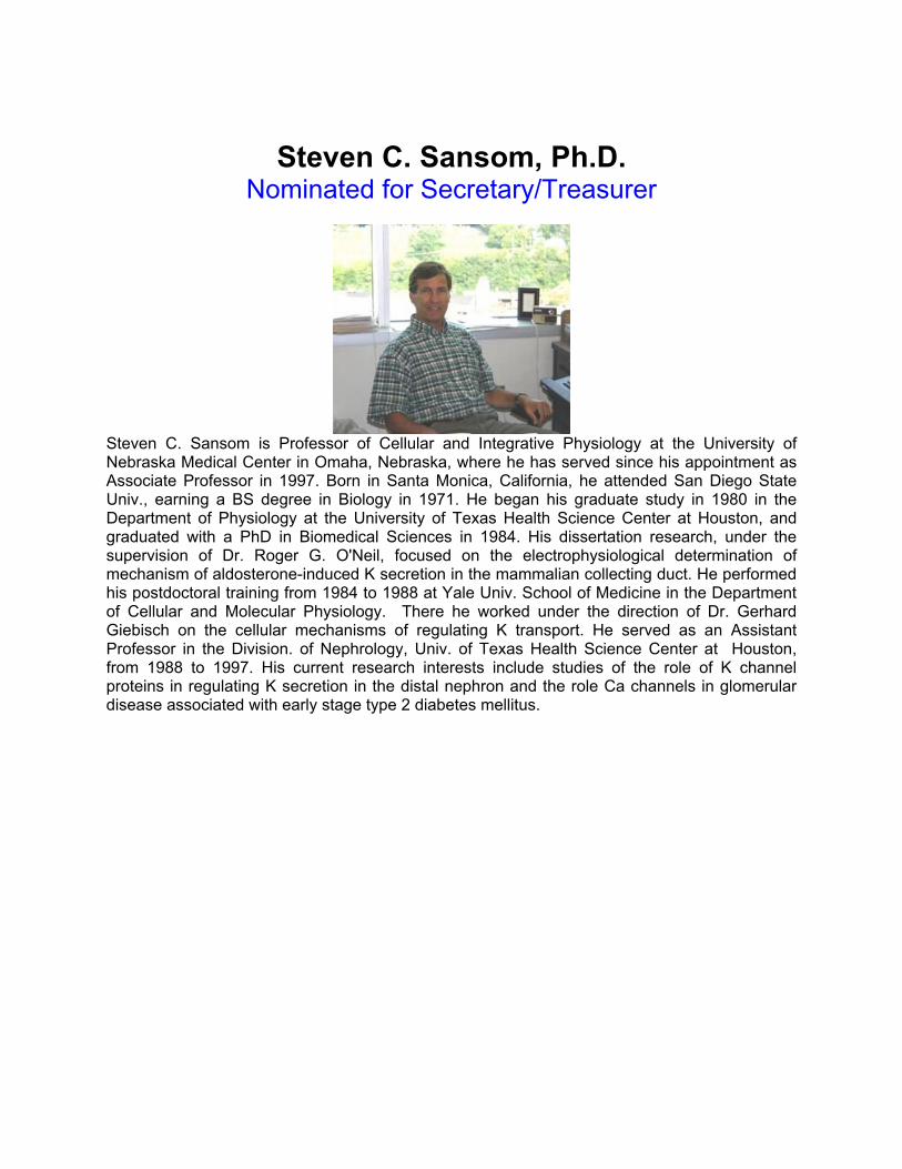
Steven C. Sansom, Ph.D. Nominated for Secretary/Treasurer
Steven C. Sansom is Professor of Cellular and Integrative Physiology at the University of Nebraska Medical Center in Omaha, Nebraska, where he has served since his appointment as Associate Professor in 1997. Born in Santa Monica, California, he attended San Diego State Univ., earning a BS degree in Biology in 1971. He began his graduate study in 1980 in the Department of Physiology at the University of Texas Health Science Center at Houston, and graduated with a PhD in Biomedical Sciences in 1984. His dissertation research, under the supervision of Dr. Roger G. O'Neil, focused on the electrophysiological determination of mechanism of aldosterone-induced K secretion in the mammalian collecting duct. He performed his postdoctoral training from 1984 to 1988 at Yale Univ. School of Medicine in the Department of Cellular and Molecular Physiology. There he worked under the direction of Dr. Gerhard Giebisch on the cellular mechanisms of regulating K transport. He served as an Assistant Professor in the Division. of Nephrology, Univ. of Texas Health Science Center at Houston, from 1988 to 1997. His current research interests include studies of the role of K channel proteins in regulating K secretion in the distal nephron and the role Ca channels in glomerular disease associated with early stage type 2 diabetes mellitus.

Jessica R. Meendering, Ph.D. Nominated for Secretary/Treasurer
Jessica R. Meendering is an Assistant Professor in the School of Health, Physical Education, and Recreation at the University of Nebraska at Omaha. Originally from Brookings, SD, Meendering attended South Dakota State University, earned her BS degree with a double major in athletic training and health promotion in 2002. She then began graduate study under the direction of Dr. Christopher T. Minson at the University of Oregon, in Eugene. She earned her MS in 2004, and her Ph.D. in 2007, in exercise science and human physiology, both from the University of Oregon. Her research is in the area of women’s health, with a focus on how natural and synthetic female sex hormones affect blood pressure regulation and endothelial function. Her dissertation examined the effects of various progestins on markers of cardiovascular risk in young women, with a specific focus on endothelial function. She started at the University of Nebraska Omaha in January 2007, and is excited to expand this line of women’s health research in her new laboratory. She is an advocate for women’s health education programs and hopes to develop a social support program to educate women about cardiovascular disease basics, prevention, and the benefits of exercise in Omaha. Throughout her career she has been deeply involved with the American Physiological Society (APS); as she has published numerous articles in APS Journals, serves as a reviewer for APS Journals, and frequently attends the Experimental Biology annual meeting. She is currently a NPS council member. Now that she calls Omaha home, Jessica looks forwards to becoming more involved in the Nebraska Physiological Society.

G. Patrick Lambert, Ph.D. Nominated for Councilor
G. Patrick Lambert is Assistant Professor of Exercise Science and Athletic Training at Creighton University. He also holds a secondary appointment in Biomedical Sciences at Creighton. Born in Michigan, he received his B.S. degree in Exercise and Health Science from Alma College (1988), his M.S. degree in Exercise Physiology from Ball State University (1990), and his Ph.D. degree in Exercise Physiology from the University of Iowa (2001). At Ball State, he worked under Dr. David Costill. At the University of Iowa he was mentored by the late Dr. Carl Gisolfi and by Dr. Kevin Kregel. His dissertation examined the effects of heat stress on intestinal barrier function. Following his Ph.D. work, he conducted postdoctoral research in the Pulmonary and Critical Care Division of Internal Medicine at the University of Nebraska Medical Center. While at UNMC he performed studies on the inflammatory response to endotoxin under the direction of Drs. Todd Wyatt, Debra Romberger, and Susanna Von Essen. His current research interests are in gastrointestinal barrier dysfunction with exercise-heat stress.

Barbara Engebretsen, Ph.D. Nominated for Councilor
Barbara Engebretsen is Associate Professor of Health, Human Performance and Sport at Wayne State College, where she has held a faculty appointment since 1995. She received her BA from the University of California at Riverside, and her MEd in Exercise Physiology and PhD in Physiology from Colorado State University. Her PhD was completed under the direction of the late Alan Tucker. Her research examined acute hypoxic signaling in the pulmonary circulation. The goal was to understand the molecular mechanisms of individual variability in hypoxic sensitivity and its contribution to hypoxic pulmonary hypertension and high altitude pulmonary edema (High Altitude Medicine and Biology. 2007. 8(4):312-321). Barbara is a teaching physiologist. Since November 2006, she has collaborated with colleagues in the departments of HHPS and Life Sciences to sponsor the annual “Physiology Understanding (PhUn) Open House” each fall (The Physiologist, (2007), 50:2). Current research interests include animal models for hypoxic signaling and the effects of body composition on glucose and insulin responses to exercise in women. She has been a member of the American Physiological Society since 1999 and attended Nebraska Physiological Society meetings since 2003. Barbara is honored to be nominated for NPS Councilor and welcomes the opportunity to contribute to the continued excellence of physiology education and research in Nebraska.

Babu J. Padanilam, Ph.D. Nominated for Councilor
Babu J. Padanilam is an Associate Professor of Cellular and Integrative Physiology at the University of Nebraska Medical Center in Omaha, Nebraska, where he has served since his appointment as Associate Professor in 2000. Born in Kerala, India, he attended Kerala University, earning a B.Sc and M.Sc degree in Chemistry. He began his graduate study in 1981 in the Department of Cellular and Molecular Biology at the Medical College of Georgia, Augusta and graduated with a PhD in Biochemistry in 1985. His dissertation research, under the supervision of Dr. Titus H. Huisman, focused on characterization genetic mutations in beta-thalassemia. He performed his postdoctoral training from 1986 to 1988 in the Department of Cell Biology at the Baylor College of Medicine, Houston, Texas. There, he worked on the role of apolipoproteins in development of atherosclerosis under the mentorship of Dr. Lawrence Chan. In 1988 he joined the Department of Biology at the University of Iowa as an Assistant Research Scientist and worked on the role of homeobox genes and growth factors in craniofacial development. In 1994, he joined Washington University in St. Louis as Research Assistant Professor in the Division of Nephrology. His current research interests include studies on the mechanism of cellular injury and repair in ischemia induced kidneys and characterization of genes involved in obesity and type 2 diabetes mellitus.

Sarah C. Clayton Nominated for Student Council
After graduating from University of Nebraska at Omaha with a bachelor’s degree, Ms. Clayton began graduate school in the summer of 2005 in the Biomedical Research Training Program, a common entry program for graduate students. She completed four rotations and started in Dr. Irving H. Zucker’s laboratory in May 2006. Her thesis project is focused on the cardiorenal syndrome in heart failure with particular interest on the regulation of renal blood flow by Angiotensin II receptors. She uses chronically instrumented rabbits for conscious experiments and harvests tissue for biochemical assays. During her tenure at UNMC, she has been an active member of the Graduate Student Association. She has participated in 4 national meetings and 4 regional meetings, receiving a first place in a poster presentation contest at one of the regional meetings. She has presented 2 departmental seminars. She was also awarded an American Heart Association pre-doctoral fellowship for her research in 2007.

Racheal Slattery Nominated for Student Council
Racheal Slattery is currently beginning the second year of her Master’s degree in Reproductive Physiology/Animal Science at the University of Nebraska- Lincoln with Dr. Andrea Cupp. She completed her B.S. in Animal Science at the University of Wisconsin-River Falls in May 2007 and is a native of Rudolph, Wisconsin. Racheal has two main research projects. Her first project investigates the effect of VEGF angiogenic and inhibitory isoforms on ovarian follicle formation and progression in postnatal rat pups. Racheal’s second project involves measuring VEGF angiogenic and inhibitory isoforms in persistent and “normal” follicles in the bovine ovary. The hypotheses for both experiments is that VEGF isoforms are involved in ovarian follicle progression and arrest and may be potential regulators of oocyte quality. Racheal has served as teaching assistant for Animal Science Physiology and Anatomy in the Animal Science Department and is a member of Society for the Study of Reproduction and the American Society of Animal Science.

NEBRASKA PHYSIOLOGICAL SOCIETY BALLOT
PRESIDENT-ELECT - (Vote for One)
� Janet Steele, Ph.D. University of Nebraska at Kearney
� George J. Rozanski, Ph.D. University of Nebraska Medical Center
� _______________________________ Write-in (must agree to serve)
SECRETARY/TREASURER – (Vote for One) � Jessica R. Meendering, Ph.D. University of Nebraska at Omaha
� Steven C. Sansom, Ph.D. University of Nebraska Medical Center
� _______________________________ Write-in (must agree to serve)

COUNCILOR - (Vote for One) � Barbara Engebretsen, Ph.D. Wayne State College
� G. Patrick Lambert, Ph.D. Creighton University
� Babu J. Padanilam, Ph.D. University of Nebraska Medical Center
� ________________________________ Write in (must agree to serve)
STUDENT COUNCILOR - (Vote for One) � Sarah Clayton University of Nebraska Medical Center
� Racheal Slattery University of Nebraska - Lincoln
� ________________________________ Write in (must agree to serve)
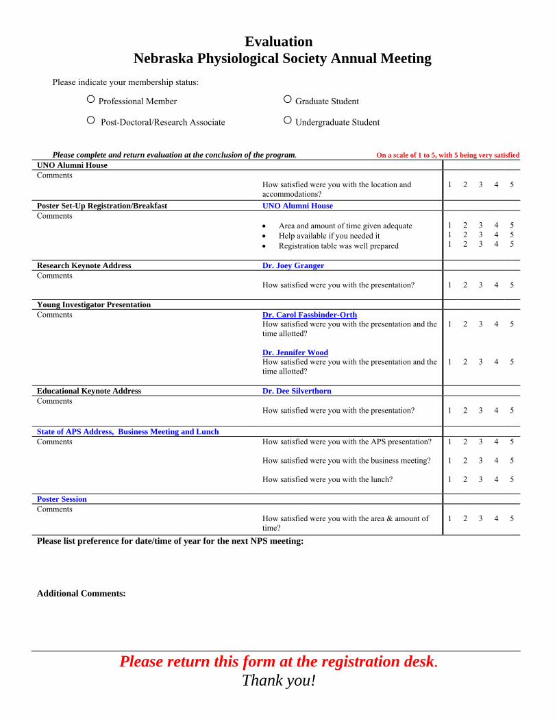
Evaluation Nebraska Physiological Society Annual Meeting
Please indicate your membership status:
○ Professional Member ○ Graduate Student
○ Post-Doctoral/Research Associate ○ Undergraduate Student
Please complete and return evaluation at the conclusion of the program. On a scale of 1 to 5, with 5 being very satisfied UNO Alumni House Comments
How satisfied were you with the location and accommodations?
1
2
3
4
5
Poster Set-Up Registration/Breakfast UNO Alumni House Comments
• Area and amount of time given adequate • Help available if you needed it • Registration table was well prepared
1 11
2 2 2
3 3 3
4 44
5 5 5
Research Keynote Address Dr. Joey Granger Comments
How satisfied were you with the presentation?
1
2
3
4
5
Young Investigator Presentation Comments Dr. Carol Fassbinder-Orth
How satisfied were you with the presentation and the time allotted? Dr. Jennifer Wood How satisfied were you with the presentation and the time allotted?
1 1
2 2
3 3
4 4
5 5
Educational Keynote Address Dr. Dee Silverthorn Comments
How satisfied were you with the presentation? 1
2
3
4
5
State of APS Address, Business Meeting and Lunch Comments
How satisfied were you with the APS presentation? How satisfied were you with the business meeting? How satisfied were you with the lunch?
1 1 1
2 2 2
3 3 3
4 4 4
5 5 5
Poster Session Comments
How satisfied were you with the area & amount of time?
1
2
3
4
5
Please list preference for date/time of year for the next NPS meeting: Additional Comments:
Please return this form at the registration desk. Thank you!

Acknowledgments
We would like to acknowledge and thank our sponsors who have provided support to the
Nebraska Physiological Society Annual Meeting.
EPR Division
We would especially like to thank Pearl Sorensen, Linda Tegeder, Janine Wilson and Cindy
Norton from the Department of Cellular and Integrative Physiology at the University of Nebraska Medical Center for their help in organizing the meeting.
For additional information on the NPS sponsors, please visit www.unmc.edu/physiology/nps/sponsors.