Embed Size (px)
Citation preview

MOLECULAR AND CELLULAR BIOLOGY, June 2005, p. 4826–4840 Vol. 25, No. 120270-7306/05/$08.00�0 doi:10.1128/MCB.25.12.4826–4840.2005Copyright © 2005, American Society for Microbiology. All Rights Reserved.
Progestins Induce Transcriptional Activation of Signal Transducer andActivator of Transcription 3 (Stat3) via a Jak- and Src-Dependent
Mechanism in Breast Cancer CellsCecilia Proietti,1 Mariana Salatino,1 Cinthia Rosemblit,1 Romina Carnevale,1 Adalı́ Pecci,2
Alberto R. Kornblihtt,2 Alfredo A. Molinolo,3 Isabel Frahm,4 Eduardo H. Charreau,1Roxana Schillaci,1 and Patricia V. Elizalde1*
Instituto de Biologı́a y Medicina Experimental (IBYME), CONICET, Obligado 2490,1 Facultad de Ciencias Exactas y Naturales,Universidad de Buenos Aires,2 and Servicio de Patologı́a, Sanatorio Mater Dei,4 Buenos Aires, Argentina, and
Oral and Pharyngeal Cancer Branch, National Institute of Dental and Craniofacial Research,National Cancer Institute, National Institutes of Health, Bethesda, Maryland3
Received 5 November 2004/Returned for modification 10 December 2004/Accepted 29 March 2005
Interactions between steroid hormone receptors and signal transducer and activator of transcription (Stat)-mediated signaling pathways have already been described. In the present study, we explored the capacity ofprogestins to modulate Stat3 transcriptional activation in an experimental model of hormonal carcinogenesisin which the synthetic progestin medroxyprogesterone acetate (MPA) induced mammary adenocarcinomas inBALB/c mice and in the human breast cancer cell line T47D. We found that C4HD epithelial cells, from theMPA-induced mammary tumor model, expressed Stat3 and that MPA treatment of C4HD cells up-regulatedStat3 protein expression. In addition, MPA induced rapid, nongenomic Stat3, Jak1, and Jak2 tyrosine phos-phorylation in C4HD and T47D cells. MPA treatment of C4HD cells also resulted in rapid c-Src tyrosinephosphorylation. These effects were completely abolished by the progestin antagonist RU486. Abrogation ofJak1 and Jak2 activity by transient transfection of C4HD cells with dominant negative (DN) Jak1 or DN Jak2vectors, or inhibition of Src activity by preincubation of cells with the Src family kinase inhibitor PP2, blockedthe capacity of MPA to induce Stat3 phosphorylation. Treatment of C4HD cells with MPA induced Stat3binding to DNA. In addition, MPA promoted strong Stat3 transcriptional activation in C4HD and T47D cellsthat was inhibited by RU486 and by blockage of Jak1, Jak2, and Src activities. To investigate the correlationbetween MPA-induced Stat3 activation and cell growth, C4HD cells were transiently transfected with a DNStat3 expression vector, Stat3Y705-F, or with a constitutively activated Stat3 mutant, Stat3-C. While expres-sion of Stat3Y705-F mutant had an inhibitory effect on MPA-induced growth of C4HD cells, transfection withthe constitutively activated Stat3-C vector resulted in MPA-independent proliferation. Finally, we addressedthe effect of targeting Stat3 in in vivo growth of C4HD breast tumors. Blockage of Stat3 activation bytransfection of C4HD cells with the DN Stat3Y705-F expression vector significantly inhibited these cells’ abilityto form tumors in syngeneic mice. Our results have for the first time demonstrated that progestins are able toinduce Stat3 transcriptional activation, which is in turn an obligatory requirement for progestin stimulationof both in vitro and in vivo breast cancer growth.
Progesterone regulates diverse biological effects in a broadrange of tissues, mostly by interaction with the classical pro-gesterone receptor (PR), a member of the nuclear receptorsuperfamily of ligand-dependent transcription factors. Partic-ularly in the mammary gland, progesterone plays a key role inthe control of cell proliferation and differentiation (31 andreferences within). Accumulated evidence also indicates thatprogestins are involved in controlling mammary gland tumor-igenesis, both in women and in animal models (6, 8, 15, 21, 24,26, 31, 32, 48). Although the mechanisms by which progestinsstimulate growth of breast cancer cells have not been com-pletely deciphered, several lines of evidence (6, 26, 33), includ-ing our own work (3, 4, 25, 44), have shown that convergencebetween progestins and growth factor (GF) signaling pathways
mediates proliferative effects of progestins in mammary tumorcells.
In addition to their direct transcriptional effects, rapid ornongenomic biological effects of progestins have been de-scribed in several species, including fish, amphibian, and mam-malian (27). Whether the recently cloned membrane PR fromhumans and other vertebrates (59) is involved in mediatingnongenomic progestin effects in mammalians remains to beelucidated. Nongenomic effects of progestins in breast cancercells have been unraveled by startling reports from Auricchioand coworkers (5, 13, 33), who demonstrated that progestintreatment of human breast cancer T47D cells activates thesignal-transducing c-Src/p21ras/MAPK pathway, which resultsin cell proliferation (5, 13, 33). Progestin activation of thec-Src/p21ras/MAPK pathway has also been described by Ed-wards and coworkers (6), who further explored mechanismsinvolved in progestin modulation of c-Src activity.
Over the last years, a unique family of proteins, the signaltransducers and activators of transcription (Stats), was foundto be involved in cross talks with both steroid hormones and
* Corresponding author. Mailing address: Laboratory of MolecularMechanisms of Carcinogenesis, Instituto de Biologı́a y Medicina Ex-perimental (IBYME), Obligado 2490, Buenos Aires 1428, Argentina.Phone: 5411-4783-2869. Fax: 5411-4786-2564. E-mail: [email protected].
4826
Dow
nloa
ded
from
http
s://j
ourn
als.
asm
.org
/jour
nal/m
cb o
n 13
Jan
uary
202
2 by
62.
12.6
4.15
1.

GF signaling pathways. There are seven Stat genes in mam-mals, Stat1, Stat2, Stat3, Stat4, Stat5a, Stat5b, and Stat6 (9).They have dual functions: as signaling molecules in the cyto-plasm and as transcription factors following nuclear transloca-tion (9, 55). Cytoplasmic Stats were found to be recruited toand activated either by tyrosine kinase receptors (RTKs), afterbinding of GFs (38, 42, 50), or by soluble tyrosine kinases ofthe Janus (JAK) and Src kinase families, after binding of cy-tokines to their receptors (16, 52). Tyrosine-phosphorylatedStats then dimerize and translocate to the nucleus, where theybind DNA and regulate gene transcription.
Interaction between steroid hormone receptors and Stat-mediated pathways has a bidirectional nature, where steroidhormone receptors regulate Stat-dependent transcription andwhere, conversely, Stats are able to modulate steroid hormone-mediated transcription (18, 51, 58). Functional interactionsbetween progestins and Stats were demonstrated by Lange etal. (26) and Richer et al. (43), who found that treatment ofT47D human breast cancer cells with the synthetic progestinR5020 resulted in up-regulation of Stat3 and Stat5 proteinlevels. In addition, these authors described constitutive associ-ation between Stat5 and PRB in HeLa cells transfected withthe B isoform of PR (43). Recently, progesterone was found toinduce Stat5a expression in MDA-MB-231 breast cancer cellstransfected with PR (29). Progesterone also enhanced Stat3cytoplasmic stores in the decidualized mesometrium duringpregnancy in rats (30), an effect which was inhibited by theprogestin antagonist RU486, showing that Stat3 is a progest-erone-dependent protein. On the other hand, involvement ofdifferent RTKs in the activation of Stat proteins has beenreported. The best elucidated are the roles of the epidermalgrowth factor receptor (EGF-R/ErbB-1) (38, 42) and the plate-let-derived growth factor receptor (50). Interestingly, constitu-tive activation of Stats, especially of Stat3 and Stat5, has beenfound in transformed cells and in a variety of human tumortypes, supporting the concept that Stats play a role in malig-nant transformation (55).
We have already exhaustively characterized mechanisms ofprogestin-induced growth in an experimental model of hor-monal carcinogenesis in which the synthetic progestin me-droxyprogesterone acetate (MPA) induced mammary adeno-carcinomas in female BALB/c mice (3, 4, 25, 44). In thepresent study, we addressed the question of whether Statsmight play a role in the scenario of progestin-induced growthof breast cancer. We focused our present study on Stat3 notonly because accumulated evidence indicates that its activationis associated with cell transformation (9, 11, 55) but also be-cause tumor-derived cell lines or samples from human breastcancer frequently contain activated forms of Stat3 (12, 20, 28,53). Our findings have for the first time demonstrated thatprogestins are able to induce Stat3 transcriptional activation inboth mouse and human mammary tumor cells by a mechanismrequiring Jak and c-Src activities. We also found that the pres-ence of activated Stat3 is a requisite for progestin stimulationof in vitro and in vivo breast cancer growth. Moreover, in thecourse of our studies of the molecular mechanisms involved inprogestin-induced Stat3 activation, we unfolded several rapid,nongenomic progestin effects. Thus, we have here for the firsttime described the ability of progestin to induce rapid Jak1,Jak2, and Stat3 tyrosine phosphorylation.
MATERIALS AND METHODS
Animals and tumors. Experiments were carried out with virgin female BALB/cmice raised at the Institute of Biology and Experimental Medicine of BuenosAires. All animal studies were conducted in accordance with the highest stan-dards of animal care as outlined in the National Institutes of Health Guide forthe Care and Use of Laboratory Animals and were approved by the Institute ofBiology and Experimental Medicine Animal Research Committee. Hormone-dependent ductal tumor line C4HD originated in mice treated with 40 mg MPAevery 3 months for 1 year and has been maintained by serial transplantation inanimals treated with a 40-mg subcutaneous (s.c.) MPA depot in the flank oppo-site the tumor inoculum (3, 4, 25, 44). The C4HD tumor line is of ductal origin,expresses PR and estrogen receptor (3, 4, 25, 44), and lacks glucocorticoidreceptor and EGF-R/ErbB-1 expression (4, 25).
Cell culture and treatments. Primary cultures of epithelial cells from C4HDtumors growing in MPA-treated mice were performed as previously described (3,4, 25, 44). Cells were incubated in Dulbecco modified Eagle medium (DMEM)-F12 (without phenol red, with 100 U/ml penicillin, and with 100 �/ml strepto-mycin) with 0.1% charcoal-stripped fetal calf serum (ChFCS) in the presence orabsence of 10 nM MPA and MPA–10 nM RU486. To block c-Src activation,4-amino-5-(4-chlorophenyl)-7-(t-butyl)pyrazolo[3,4-d]pyrimidine (PP2) (10 �M,dissolved in 1:2,000 dimethyl sulfoxide) was added to cells 90 min before incu-bation with MPA. Controls were performed in order to verify that dimethylsulfoxide (1:2,000) did not modify MPA-modulated c-Src tyrosine phosphoryla-tion. Proliferation of C4HD cells was evaluated by propidium iodide (PI) stainingand flow cytometry analysis, as described below. T47D cells were obtained fromthe American Type Culture Collection. T47D-Y cells were a generous gift fromK. Horwitz (University of Colorado Health Sciences Center, Denver). LM3 cellswere cultured as previously described (40). In experiments assessing the effect ofMPA on Stat3 activity, T47D, T47D-Y, and LM3 cells were cultured in DMEM(without phenol red) supplemented with 0.1% ChFCS and subjected to thetreatments described above for C4HD cells.
Transient transfections. In experiments assessing the roles of Jak1 and Jak2 inMPA-induced effects on Stat3, C4HD and T47D cells were transiently trans-fected with 2 �g of a dominant negative (DN) Jak1 vector or with 2 �g of a DNJak2 vector (38, 39), kindly provided by O. Silvennoinen (Tampere UniversityHospital, Finland) via N. Hynes (Friedrich Miescher Institute, Basel, Switzer-land). C4HD cells were transfected for 48 h in DMEM-F12 supplemented with10 nM MPA and 2.5% ChFCS, and T47D cells were transfected for 48 h inDMEM with 10% ChFCS without antibiotics. The Fugene 6 transfection reagenttechnique (Roche Biochemicals) was used in accordance with the manufacturer’sinstructions. After transfection, cells were washed and cultured for 24 h in 0.1%ChFCS before treatment with MPA for the indicated times. Total cell lysateswere then prepared as described below for use in Western blot assays. To studythe role of Stat3 in proliferation, C4HD cells were transiently transfected, asdescribed above, for 48 h with 2 �g of a DN Stat3 expression vector, Stat3Y705-F(10, 23, 28), which carries a tyrosine-to-phenylalanine substitution at codon 705that reduces phosphorylation on tyrosine of the wild-type Stat3 protein, there-fore inhibiting both dimerization and DNA binding of Stat3. C4HD cells werealso transfected with 2 �g of a constitutively activated Stat3 mutant, Stat3-C,which dimerizes spontaneously, binds to DNA, and activates transcription (11).Both were kindly provided by J. Darnell (New York, NY). As a control, cellswere transfected with 2 �g of the empty pRc/CMV vector. After transfection,cells were washed and cultured for 24 h in 0.1% ChFCS before treatment withMPA for 48 h. Cells were then harvested for flow cytometry. Transfectionefficiency in C4HD cells was evaluated using the pEGFP-N1 vector (BD Bio-sciences Clontech, Palo Alto, CA) and determined by the percentage of cells thatexhibited green fluorescence 24 h after transfection. Green fluorescent proteinwas visualized by direct fluorescence imaging using a Nikon Eclipse E800 con-focal laser microscopy system (Nikon Instruments, Inc., Melville, NY). In C4HDcells, fluorescence intensities were also measured by fluorescence-activated cellsorter. To investigate the capacity of MPA to induce the transcriptional activa-tion of Stat3, C4HD and T47D cells were transiently transfected with 2 �g of aluciferase reporter plasmid containing four copies of the m67 high-affinity bind-ing site (11, 56) and 1 �g of a �-galactosidase expression plasmid, CMV-�gal(Clontech, Palo Alto, CA), used to correct variations in transfection efficiency.As a control, cells were also transfected with a pTATA-Luc reporter lacking them67 insertion (11, 56). In experiments assessing the role of Jaks in Stat3 tran-scriptional activation, cells were cotransfected with 2 �g DN Jak1 or 2 �g DNJak2 expression vector. The total amount of transfected DNA was standardizedby adding the pTATA-Luc reporter lacking the m67 insertion. Cells were thentreated with MPA or MPA-RU486 or preincubated with the c-Src inhibitor PP2for 90 min and then treated with MPA for 48 h or were left untreated growing
VOL. 25, 2005 Stat3 ACTIVATION BY PROGESTINS 4827
Dow
nloa
ded
from
http
s://j
ourn
als.
asm
.org
/jour
nal/m
cb o
n 13
Jan
uary
202
2 by
62.
12.6
4.15
1.
in ChFCS. Cells were washed once with phosphate-buffered saline without cal-cium and magnesium and then harvested and lysed in 1� reporter buffer (Pro-mega, Madison, Wis.) using one freeze-thaw cycle. Protein concentrations weredetermined using a Bio-Rad kit (Bio-Rad, Hercules, CA). Luciferase resultswere calculated as the ratio of luciferase activity per unit of �-galactosidaseactivity. Duplicate samples were analyzed for each datum point.
Cell cycle analysis. Primary cultures of C4HD cells subjected to the differenttreatments described in Results were harvested for flow cytometric analysis andfixed in 70% ethanol for 24 h at 4°C. They were washed twice with phosphate-buffered saline, followed by RNA digestion (RNase A at 50 U/ml) and PI (20�g/ml) staining for 30 min at room temperature in the dark. Cell cycle analysiswas performed using a FACScalibur flow cytometer (Becton-Dickinson, Moun-tain View, CA) and Modfit LT software.
Apoptosis assays. C4HD cells were transiently transfected with the DN Stat3expression vector Stat3Y705-F or with the empty pRc/CMV vector as describedabove. Apoptosis was investigated through cell surface binding of fluorescentAnnexin V by using the fluorescein isothiocyanate-annexin V binding assay (Im-munotech).
Western blot analysis and immunoprecipitation assays. Lysates were pre-pared from C4HD or T47D cells subjected to the different treatments describedin each experiment. Cells were lysed in buffer containing 50 mM Tris (pH 7.4),150 mM NaCl, 1 mM EDTA, 1 mM EGTA, 10% glycerol, 0.5% Nonidet P-40,1 mM Mg2Cl, 1 mM phenylmethylsulfonyl fluoride (PMSF), 10 �g/ml leupeptin,5 �g/ml pepstatin, 5 �g/ml aprotinin, 1 mM sodium orthovanadate, 5 mM NaF,20 mM sodium molybdate, and 5 mM sodium pyrophosphate. Lysates werecentrifuged at 40,000 � g for 40 min at 4°C, and protein content in the super-natant was determined using a Bio-Rad kit (Bio-Rad, Richmond, CA). Proteinswere solubilized in sample buffer (60 mM Tris-HCl [pH 6.8], 2% sodium dodecylsulfate [SDS], 10% glycerol, 0.01% bromophenol blue) and subjected to SDS-polyacrylamide gel electrophoresis (PAGE). Proteins were electroblotted ontonitrocellulose. Membranes were immunoblotted with the following antibodies:phosphotyrosine Stat3 (B-7), total Stat3 (C-20), phosphotyrosine Stat1 (A-2),total Stat1 (E-23), c-Src (N-16), phosphotyrosine Jak1 (Tyr1022/1023), total Jak1(HR-785), the p85 regulatory subunit of phosphatidylinositol 3-kinase (PI-3K)(Z-8), and Bcl-xL (S-18), all from Santa Cruz Biotechnology (Santa Cruz, CA),phosphotyrosine Src (Tyr416) and phosphotyrosine Jak2 (Tyr1007/1008) fromCell Signaling (Beverly, MA), hPR Ab-7 (clone 7), and actin (clone ACTN05)from Neomarkers (Freemont, CA), and FLAG-M2 monoclonal antibody fromSigma (St. Louis, MO). Association between PR and Stat3 was studied byperforming coimmunoprecipitation experiments. Total cell lysates (500 �gprotein) from C4HD cells, treated and untreated with MPA, were immunopre-cipitated using the PR mouse monoclonal antibody hPR Ab-7 (clone 7; Neo-markers). Immunoprecipitations were rocked for 2 h at 4°C, and the immuno-complexes were then captured by adding protein A-agarose and rocked for anadditional 2 h. Beads were washed three times with lysis buffer and then boiledfor 10 min in sample buffer and subjected to SDS-PAGE. Proteins were elec-troblotted onto nitrocellulose and probed with an anti-Stat3 antibody (C-20;Santa Cruz). When the immunoprecipitation-immunoblotting procedure wasreversed, lysates were immunoprecipitated with the anti-Stat3 antibody, andWestern blotting was performed with the anti-PR antibody (Ab-7; Neomarkers).As a negative control, preimmune rabbit serum was used.
Preparation of nuclear and cytosolic extracts. C4HD and T47D cells werelysed by scraping at 4°C in buffer A, which contains 10 mM Tris (pH 7.4), 1.5 mMEDTA, 10% glycerol, 0.5 mM dithiothreitol, 1 mM PMSF, 10 �g/ml leupeptin,5 �g/ml pepstatin, 5 �g/ml aprotinin, and 5 mM NaF. The homogenate wascentrifuged for 10 min at 1,000 � g, and the supernatant fraction (cytosol) wasthen collected. The crude nuclear pellet was washed once with buffer A contain-ing 0.01% NP-40 and centrifuged again at 1,000 � g, and the supernatant wasadded to the cytosol, which was then centrifuged at 105,000 � g for 30 min. Thesupernatant yielded the cytoplasmic fraction. The washed crude nuclear pelletwas resuspended in buffer A containing 0.4 M KCl and was later incubated for30 min at 4°C with resuspension every 10 min. The suspension was centrifuged at105,000 � g (30 min) and diluted with the same volume of buffer A to reduce thesalt concentration.
EMSA. The DNA-binding activity of Stat3 in nuclear cell extracts of C4HDcells was analyzed by electrophoretic mobility shift assay (EMSA). C4HD cellswere treated for 15 min at 37°C with MPA or MPA-RU486 or were left un-treated growing in ChFCS. Double-stranded DNA containing the high-affinitymutant of the sis-inducible element (SIE) from the human c-fos promoter (5�GTGCATTTCCCGTAAATCTTGTCTACA3�) (m67) was used as probe. M67oligonucleotide was end labeled with [�-32P]ATP by T4 polynucleotide kinase toa specific activity of approximately 30,000 to 50,000 cpm/0.1 ng. Twenty micro-grams of protein from nuclear extracts was incubated for 20 min at room tem-
perature in a total volume of 30 �l with 1 ng of the [32P]m67. The DNA-bindingreaction mixture also consisted of 2.5 �g poly(dI-dC), as nonspecific competitorDNA, in a binding buffer containing 10 mM Tris-HCl (pH 7.4), 50 mM NaCl,10% glycerol, 1 mM dithiothreitol, 5 mM MgCl2, 5 �g/�l gelatin, 1 mM PMSF,10 �g/ml leupeptin, 5 �g/ml pepstatin, and 5 �g/ml aprotinin. The specificity ofStat3-DNA complexes was studied by competition with a 25- and 100-fold massexcess of unlabeled m67 oligonucleotide and by the lack of competition with a100-fold mass excess of a mutant m67 with a TTC-to-CCA substitution in theDNA-binding region (5�GTGCATCCACCGTAAATCTTGTCTACA3�) (SantaCruz). An anti-Stat3� antibody (clone C-20) or an anti-Stat1 antibody (cloneE-23), each from Santa Cruz, was used in the supershift assays at a concentrationof 4 �g/assay. Preimmune rabbit serum was used as a control. Samples weresubjected to electrophoresis on nondenaturing 5.5% acrylamide gels (30:1) inlow-ionic-strength TBE buffer (10 mM Tris-borate [pH 7.5], 0.025 mM EDTA).Gels were preelectrophoresed at 10 mA for 60 min, and samples were run at 20mA/gel for 120 min at room temperature. To prevent warming of the gels, waterwas recirculated through the gel apparatus. Gels were dried without fixationunder vacuum and were autoradiographed by exposure to Kodak X-OMAT filmat �70°C.
In vivo blockage of Stat3 expression. C4HD cells growing in 10 nM MPA weretransiently transfected as already described above with the DN Stat3Y705-Fexpression vector and with the empty pRc/CMV vector or remained untreated.After 48 h of transfection, 106 cells from each experimental group were inocu-lated s.c. into animals treated with a 40-mg MPA depot in the flank opposite thecell inoculum. Tumor growth was measured three times a week with a verniercaliper. Tumor volume (mm3) was calculated as (L � W2)/2, where L length(mm) and W width (mm). Tumor growth rates were determined as the slopesof growth curves. Percentage of tumor growth inhibition was calculated bydividing the mean tumor volume of the animal group injected with C4HD cellstransiently transfected with the DN Stat3Y705-F expression vector, by the meantumor volume of control groups, subtracting the resulting value from 1, andmultiplying it by 100. Tumor growth delay was evaluated as T � C, where T andC are the median times for treated and control tumors, respectively, to reach thesame volume. At day 21, animals from each group were killed and tumors wereremoved. Tissues for molecular studies were stored at �80°C, and tissues forhistopathological analysis were fixed in 10% buffered formalin. Samples of liver,lung, heart, and pancreas were also fixed for histological examination. Compar-ison of tumor volumes between the different groups for specific times was doneby analysis of variance followed by Tukey’s t test among groups. Linear regres-sion analysis was performed on tumor growth curves, and the slopes were com-pared using analysis of variance followed by a parallelism test to evaluate thestatistical significance of differences.
RESULTS
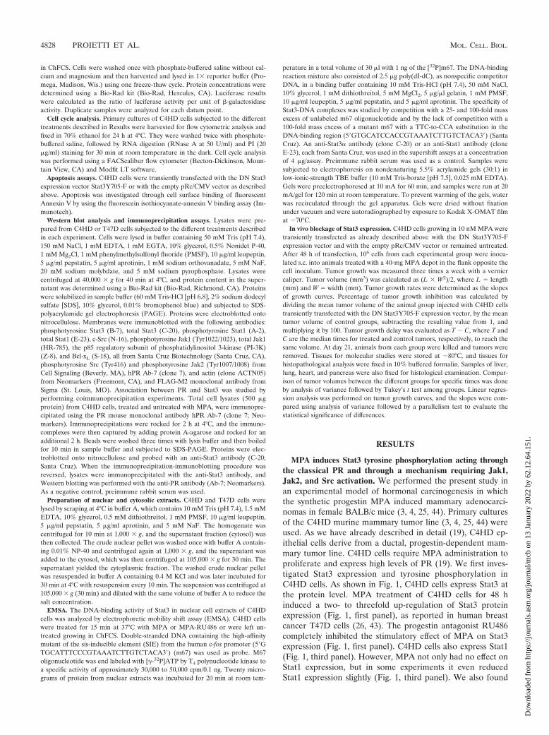
MPA induces Stat3 tyrosine phosphorylation acting throughthe classical PR and through a mechanism requiring Jak1,Jak2, and Src activation. We performed the present study inan experimental model of hormonal carcinogenesis in whichthe synthetic progestin MPA induced mammary adenocarci-nomas in female BALB/c mice (3, 4, 25, 44). Primary culturesof the C4HD murine mammary tumor line (3, 4, 25, 44) wereused. As we have already described in detail (19), C4HD ep-ithelial cells derive from a ductal, progestin-dependent mam-mary tumor line. C4HD cells require MPA administration toproliferate and express high levels of PR (19). We first inves-tigated Stat3 expression and tyrosine phosphorylation inC4HD cells. As shown in Fig. 1, C4HD cells express Stat3 atthe protein level. MPA treatment of C4HD cells for 48 hinduced a two- to threefold up-regulation of Stat3 proteinexpression (Fig. 1, first panel), as reported in human breastcancer T47D cells (26, 43). The progestin antagonist RU486completely inhibited the stimulatory effect of MPA on Stat3expression (Fig. 1, first panel). C4HD cells also express Stat1(Fig. 1, third panel). However, MPA not only had no effect onStat1 expression, but in some experiments it even reducedStat1 expression slightly (Fig. 1, third panel). We also found
4828 PROIETTI ET AL. MOL. CELL. BIOL.
Dow
nloa
ded
from
http
s://j
ourn
als.
asm
.org
/jour
nal/m
cb o
n 13
Jan
uary
202
2 by
62.
12.6
4.15
1.
that MPA treatment of C4HD cells for 5 to 10 min inducedphosphorylation of Stat3 on tyrosine residue 705 by performingWestern blot assays with the specific antiphosphotyrosine Stat3antibody (Fig. 2A, first part). This effect was completely abol-ished by RU486 (Fig. 2A, first part). On the other hand, Stat1showed basal levels of phosphorylation on tyrosine 701, whichremained unaffected by MPA treatment (Fig. 2A, third part).The involvement of members of the Jak tyrosine kinase familyin cytokine-mediated Stat phosphorylation has been well ac-knowledged (1). However, the role of Jaks in progestin-induced Stat3 activation has never been explored. Here, wefound that both Jak1 and Jak2 were expressed in C4HD cells(Fig. 2A, sixth and eighth parts). MPA treatment resulted instrong tyrosine phosphorylation of Jak1 and Jak2, observedas early as 5 min after treatment, which was inhibited by anti-progestin RU486 (Fig. 2A, fifth and seventh parts, respec-tively). Similar experiments were conducted with T47D cells.As shown in Fig. 2B, MPA was able to induce Stat3 tyrosinephosphorylation in T47D cells, and this effect was abolished byRU486. MPA treatment of T47D cells also resulted in Jak1and Jak2 tyrosine phosphorylation, which was blocked byRU486 (Fig. 2B). Interestingly, although Stat3 tyrosine phos-phorylation was very transient, Jak activation persisted for atleast 15 min and returned to basal levels at 30 min of MPAtreatment in both the C4HD (Fig. 2A) and T47D (Fig. 2B) celllines. To further demonstrate that conventional PR is mediat-ing nongenomic MPA-induced Stat3 tyrosine phosphorylation,we used our well-characterized mouse metastatic mammary
tumor line LM3, which lacks PR expression (40). MPA was notable to induce Stat3 phosphorylation in wild-type LM3 cells(Fig. 2C). However, as we previously demonstrated (40), treat-ment of LM3 cells with the type I RTK ligand heregulinstrongly induced Stat3 tyrosine phosphorylation (Fig. 2C). Onthe other hand, when we transiently transfected LM3 cells witha human PRB expression vector, MPA treatment resulted instrong induction of Stat3 tyrosine phosphorylation, which wasabolished by RU486 (Fig. 2C). We used a similar strategy withhuman PR null T47D-Y cells (26). As shown in Fig. 2D, MPAtreatment of T47D-Y cells did not induce Stat3 tyrosine phos-phorylation. However, when these cells were transfected withPRB, strong Stat3 tyrosine phosphorylation in response toMPA was detected (Fig. 2D). RU486 inhibited MPA-inducedStat3 phosphorylation (Fig. 2D).
We then investigated the participation of Jaks in MPA-induced Stat3 phosphorylation. For this purpose, C4HD cellswere transiently transfected with 2 �g DN Jak1 or DN Jak2vector (38, 39) and then treated with MPA. Transfection effi-ciency in C4HD cells, evaluated using the pEGFP-N1 vector,varied from 65 to 75%, as determined by the percentage ofcells that exhibited green fluorescence 24 h after transfection.Green fluorescent protein was visualized by direct fluorescenceimaging using confocal laser microscopy. High efficiency oftransfection in our cell cultures induced high levels of DN Jakexpression, shown by an increase in total Jak1 and Jak2 ex-pression in cells transfected with DN Jak expression vectors,compared with nontransfected cells (Fig. 3A and B, respec-tively). As shown in Fig. 3A and B, when DN Jak1 and Jak2were expressed in C4HD cells, they were able to work domi-nant negatively against MPA-induced tyrosine phosphoryla-tion of wild-type Jaks present in C4HD cells. Experimentsdone transfecting C4HD cells with increasing amounts of DNJak vectors showed that inhibition of wild-type Jak tyrosinephosphorylation was dose dependent (data not shown). Spec-ificity of DN Jak action was demonstrated by lack of effect ofthe DN Jak2 vector on MPA-induced tyrosine phosphorylationof endogenous Jak1 (Fig. 3A) and of the DN Jak1 vector onMPA-induced phosphorylation of endogenous Jak2 (Fig. 3B).Abolishment of Jak1 and Jak2 activity resulted in inhibition ofthe capacity of MPA to induce Stat3 tyrosine phosphorylation(Fig. 3C, upper parts), clearly showing that both kinases areinvolved in the effect of MPA.
The requirement of Src activity for Stat3 activation in breastcancer cells has already been acknowledged (7, 20, 53, 55, 57).In addition, accumulating data have shown the ability of pro-gestin to induce c-Src phosphorylation by a nongenomic mech-anism in mammary tumor cells (5, 6, 13, 33). Therefore, wehere assessed the effect of MPA on c-Src activity in C4HDcells. MPA treatment of C4HD cells for 2 to 10 min inducedstrong c-Src tyrosine phosphorylation, which was significantlyinhibited by the selective Src family kinase inhibitor PP2 (Fig.3D). We then explored the role of c-Src in MPA-induced Stat3tyrosine phosphorylation. As shown in Fig. 3E, inhibition ofSrc activity by preincubation of cells with PP2 blocked thecapacity of MPA to induce Stat3 phosphorylation. The aboveresults show that both Jaks and Src are able to activate Stat3.To gain further insight into the molecular mechanism throughwhich these kinases participate in Stat3 tyrosine phosphoryla-tion, we examined the tyrosine phosphorylation state of Jak1
FIG. 1. MPA up-regulates Stat3 protein expression. Primary cul-tures of C4HD cells were treated for 48 h in medium with ChFCSsupplemented with 10 nM MPA or MPA–10 nM RU486. Fifty micro-grams of protein from cell lysates was electrophoresed and immuno-blotted for Stat3 and Stat1. A Western blot assay using an antiactinantibody was carried out using identical protein lysates as a control forthe specificity of the effect of MPA. This is a representative experimentof a total of four in which the standard error of the mean was within10%. W, Western blot assay.
VOL. 25, 2005 Stat3 ACTIVATION BY PROGESTINS 4829
Dow
nloa
ded
from
http
s://j
ourn
als.
asm
.org
/jour
nal/m
cb o
n 13
Jan
uary
202
2 by
62.
12.6
4.15
1.

and Jak2 in C4HD cells pretreated with the Src inhibitor PP2.We found that Jak1 and Jak2 tyrosine phosphorylation waseffectively inhibited by PP2 (Fig. 3E). These findings indicatethat Jaks are phosphorylated by c-Src in C4HD cells.
MPA induces association between Stat3 and PR. Stats havebeen found to be physically associated with several members ofthe steroid receptor superfamily, including PR (30, 43). In thiswork, we investigated the capacity of MPA to induce associa-tion between Stat3 and PR by performing coimmunoprecipi-tation experiments. Protein extracts from C4HD cells wereimmunoprecipitated with an anti-PR antibody and immuno-blotted with an anti-Stat3 antibody. As shown in Fig. 4A, as-sociation between PR and Stat3 was detected in C4HD cellsgrowing in 0.1% ChFCS and was dramatically increased byMPA treatment. Coimmunoprecipitation of Stat3 and PR wasalso detected when the immunoprecipitation-immunoblottingprocedure was reversed. Thus, when cell extracts were immu-noprecipitated with an anti-Stat3 antibody and Western blot-ting was performed with an anti-PR a clear MPA-inducedassociation between Stat3 and PR was found (Fig. 4B). Sincewe found that Src activity is an absolute requirement for MPA-induced Jak and Stat3 tyrosine phosphorylation, we exploredSrc involvement in the association between PR and Stat3. Asshown in Fig. 4A and B, abolishment of Src activity resulted inblockage of the capacity of MPA to induce a physical associ-ation between PR and Stat3.
MPA induces Stat3 nuclear translocation and bindingto DNA. Upon tyrosine phosphorylation, Stats dimerize andtranslocate to the nucleus, where they bind DNA and regulategene transcription. In order to investigate the effects of MPAon these events, nuclear and cytoplasmic extracts from C4HDcells were prepared and Western blot assay was performedwith an anti-Stat3 antibody. While Stat3 was only detected inthe cytoplasmic fraction in untreated cells growing in 0.1%ChFCS, MPA treatment induced an extensive nuclear translo-cation of Stat3 (Fig. 5, upper panel). In addition, we found thatinhibition of Src activity by PP2 inhibited MPA-induced Stat3nuclear translocation (Fig. 5, upper panel). Cellular fraction-ation was controlled by immunoblotting with either an anti-PI-3K (Fig. 5, middle panel) or an anti-retinoblastoma (Rb)(Fig. 5, lower panel) antibody.
We then investigated the capacity of MPA to induce Stat3binding to DNA, using as a labeled probe a high-affinity mu-tant of the SIE from the human c-fos promoter (m67), whichbinds both Stat3 and Stat1 (11, 56). EMSAs using nuclearextracts from C4HD cells treated with MPA for 15 min dem-onstrated that MPA was able to promote formation of DNA-binding complexes (Fig. 6, left panel). As has been acknowl-edged in numerous studies (11, 56), three major complexeswere found in C4HD cells corresponding to Stat3 homodimers,
FIG. 2. MPA induces tyrosine phosphorylation by Stat3, Jak1, andJak2. Cultures of C4HD (A) and T47D (B) cells were treated with 10nM MPA or MPA–10 nM RU486 for the indicated times. Fifty mi-crograms of protein from cell lysates was electrophoresed, and West-ern blot assays were performed with antiphosphotyrosine 705 Stat3,antiphosphotyrosine 701 Stat1, antiphosphotyrosine 1022/1023 Jak1,and antiphosphotyrosine 1007/1008 Jak2 antibodies. Membranes werethen stripped and hybridized with anti-Stat3, -Stat1, -Jak1, and -Jak2antibodies. This experiment was repeated six times for C4HD cells andthree times for T47D cells with similar results. LM3 (C) or T47D-Y(D) cells were transfected with PRB or with the empty pSG5 plasmid
or remained untreated. Cells were then stimulated for 5 min with MPAor pretreated with RU486 before MPA stimulation. LM3 cells werealso treated with heregulin for 10 min. Fifty micrograms of proteinfrom cell lysates was electrophoresed, and Western blot assays wereperformed with antiphosphotyrosine 705 Stat3 (upper parts of panelsC and D). Membranes were then stripped and hybridized with anti-Stat3 (middle panes of parts C and D) and anti-PR (lower parts ofpanels C and D) antibodies. This experiment was repeated three timeswith similar results. W, Western blot assay.
4830 PROIETTI ET AL. MOL. CELL. BIOL.
Dow
nloa
ded
from
http
s://j
ourn
als.
asm
.org
/jour
nal/m
cb o
n 13
Jan
uary
202
2 by
62.
12.6
4.15
1.

Stat3-Stat1 heterodimers, and Stat1 homodimers (Fig. 6, leftpanel). Specificity of the Stats-DNA complexes was demon-strated by competition with excess unlabeled m67 oligonucle-otide and by lack of competition with a mutated m67 probe(Fig. 6, left panel). Stat1 binding to DNA was observed in cellsgrowing in 0.1% ChFCS, while no increase in the levels ofStat1-DNA complexes was observed after MPA treatment(Fig. 6, left panel), in accordance with our finding of basalStat1 tyrosine phosphorylation, which remained unaffected byMPA treatment (Fig. 2). On the contrary, MPA strongly in-duced the formation of Stat3-DNA complexes, which was com-pletely abolished by preincubation with RU486 (Fig. 6, leftpanel). To confirm the identity of the DNA-binding complexes,either anti-Stat3 or anti-Stat1 antibodies were included in theEMSA reaction mixture (Fig. 6, right panel). The use of ananti-Stat3� antibody (clone C-20) resulted in complete abol-ishment of the MPA-induced Stat3 homodimer-DNA complexand dramatically decreased the Stat3-Stat1 heterodimer-DNAcomplex abundance (Fig. 6, right panel). This antibody alsoinduced the formation of a complex with decreased mobility(Fig. 6, right panel). The Stat1 antibody (clone E-23) was raisedagainst a Stat1 epitope corresponding to an amino acid se-quence within the carboxy-terminal SH2 domain sequencepresent in both Stat1� and Stat1�. The inclusion of this anti-body in the EMSA reaction mixture eliminated Stat1 homo-dimer-DNA complexes (Fig. 6, right panel) and induced ahighly significant decrease in Stat3-Stat1-DNA heterodimercomplex abundance (Fig. 6, right panel). As previously re-ported for this antibody (Fig. 6, right panel) (46, 47), no super-shift was detected. An equivalent amount of the preimmunerabbit serum used as a control had no effect (Fig. 6, right panel,NRS). These data clearly show the presence of Stat3 in MPA-induced DNA-protein complexes. We found similar results inthe EMSAs using the �2-macroglobulin probe, which containsthe GAS element in the �2-macroglobulin enhancer (49, 56),as has been previously reported in HEPG2 cell types (49).
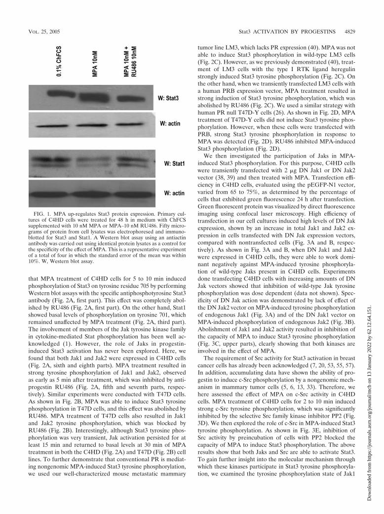
MPA induces Stat3 transcriptional activation through aJak1-, Jak2-, and Src-dependent pathway. To investigate thecapacity of MPA to induce the transcriptional activation ofStat3, C4HD cells were transiently transfected with a luciferasereporter plasmid containing four copies of the m67 high-affin-ity binding site (11, 56) and a �-galactosidase expression vector
FIG. 3. Jak1 and Jak2 are involved in MPA-induced Stat3 phos-phorylation. C4HD cells were transiently transfected with 2 �g of DNJak1 or DN Jak2 vector and then treated with MPA for 5 min or leftuntreated. Fifty micrograms of protein from cell lysates was electro-phoresed, and Western blot assays were performed with antiphospho-tyrosine Jak1 (A, upper part) or antiphosphotyrosine Jak2 (B, upperpart) antibodies. Membranes were then stripped and hybridized withanti-Jak1 (A, lower part) and anti-Jak2 (B, lower part) antibodies.(C) Fifty micrograms of protein from cells transfected with the DNJak1 (left part) or the DN Jak2 (right part) vector and subsequently
treated with MPA for 5 min or left untreated was electrophoresed, andWestern blot assays were performed with antiphosphotyrosine Stat3(upper parts). Membranes were then stripped and hybridized withanti-Stat3 (lower parts) antibodies. (D) C4HD cells were treated withMPA for the indicated times or preincubated with the selective Srcfamily kinase inhibitor PP2 or RU486 for 90 min and then treated withMPA. Fifty micrograms of protein from cell lysates was electropho-resed and immunoblotted with an antiphosphotyrosine c-Src antibody(upper part). Membrane was then stripped and hybridized with anti-c-Src antibody (lower part). (E) C4HD cells were preincubated withthe selective Src family kinase inhibitor PP2 for 90 min and thentreated with MPA for 5 min. Fifty micrograms of protein from cell ly-sates was electrophoresed and immunoblotted with antiphosphoty-rosine Stat3, antiphosphotyrosine Jak1, and antiphosphotyrosine Jak2antibodies. Membranes were then stripped and hybridized with anti-Stat3, anti-Jak1, and anti-Jak2 antibodies, respectively. These experi-ments were repeated three times with similar results. W, Western blotassay.
VOL. 25, 2005 Stat3 ACTIVATION BY PROGESTINS 4831
Dow
nloa
ded
from
http
s://j
ourn
als.
asm
.org
/jour
nal/m
cb o
n 13
Jan
uary
202
2 by
62.
12.6
4.15
1.

as an internal control. Treatment of C4HD cells with MPAinduced Stat3 transcriptional activation that was completelyinhibited by preincubation with RU486 (Fig. 7A). Blockage ofJak1 and Jak2 activities by the use of a DN Jak1 or Jak2expression vector and of Src activity with PP2 also inhibited thecapacity of MPA to activate the m67-Luc reporter plasmid(Fig. 7A). Comparable results were obtained with T47D cells,in which MPA also induced Stat3 transcriptional activation.RU486 pretreatment, transfection with a DN Jak1 or Jak2expression vector, and PP2 preincubation all abolished thecapacity of MPA to activate the m67-Luc reporter in T47Dcells (Fig. 7B). Similar results with both C4HD and T47D cells
were obtained by using a luciferase reporter plasmid contain-ing three copies of the Ly6E Stat1 and Stat3 binding site (10)in the transient-transfection assays (data not shown).
Stat3 activity is a requisite in MPA-induced proliferation ofC4HD cells. To investigate the correlation between MPA-
FIG. 4. MPA induces association of Stat3 with PR. (A) C4HD cellswere treated with 10 nM MPA for the indicated times or preincubatedwith PP2 before MPA treatment for 5 min, and PR was immunopre-cipitated from 500 �g of protein extracts. As a control, lysates werealso immunoprecipitated with normal mouse serum (NMS). Immuno-complexes were subjected to SDS-PAGE and analyzed by Westernblotting with an anti-Stat3 antibody (upper part). Twenty microgramsof protein from cell extracts was directly immunoblotted with the Stat3antibody (last lane, upper part). Identical aliquots of each immuno-precipitate were subjected to immunoblot analysis with anti-PR anti-body to verify that nearly equal amounts of immunoprecipitated pro-teins were loaded (lower part). (B) Protein lysates (500 �g) from cellstreated as indicated in panel A were immunoprecipitated with ananti-Stat3 antibody or with normal rabbit serum (NRS). Immunocom-plexes were subjected to SDS-PAGE and analyzed by Western blottingwith an anti-PR antibody (upper part). Twenty micrograms of proteinfrom cell extracts was directly immunoblotted with the PR antibody(last lane, upper part). Identical aliquots of each immunoprecipitatewere subjected to immunoblot analysis with anti-Stat3 antibody toverify that nearly equal amounts of immunoprecipitated proteins wereloaded (lower part). This is a representative experiment out of a totalof three. W, Western blot assay; IP, immunoprecipitation.
FIG. 5. MPA induces Stat3 nuclear translocation. C4HD cells weretreated with 10 nM MPA for the time indicated or were pretreatedwith PP2 before MPA stimulation. Nuclear (nuc) and cytosolic (cyt)fractions were prepared, and 30 �g of protein from cell extracts wasanalyzed by Western blot assay for Stat3 expression level. Membraneswere then stripped and hybridized with an anti-p85 PI-3K subunit anti-body (middle) or an anti-retinoblastoma (Rb) antibody (bottom) inorder to control cellular fractionation efficiency. W, Western blot assay.
FIG. 6. MPA induces Stat3 binding to the high-affinity mutant ofthe SIE from the human c-fos promoter. C4HD cells were treated for15 min at 37°C with MPA or MPA-RU486 or were left untreated grow-ing in ChFCS. Twenty micrograms of protein from nuclear extracts wasincubated for 20 min at room temperature with 1 ng of 32P-labeleddouble-stranded DNA containing the high-affinity mutant of the SIEfrom the human c-fos promoter (5�GTGCATTTCCCGTAAATCTTGTCTACA3�) (m67) used as a probe and analyzed by EMSA. Thespecificity of the Stat3-DNA complexes is shown by competition with25- and 100-fold mass excesses unlabeled m67 oligonucleotide and bythe lack of competition with a 100-fold mass excess of mutant m67(100xm67 mut). The right panel shows a supershift analysis that wasperformed by including either anti-Stat3 or anti-Stat1 antibodies. Anequivalent amount of preimmune rabbit serum was used as a control inthe EMSA reaction mixture (NRS [normal rabbit serum]). This exper-iment was repeated six times with similar results. wt, wild type.
4832 PROIETTI ET AL. MOL. CELL. BIOL.
Dow
nloa
ded
from
http
s://j
ourn
als.
asm
.org
/jour
nal/m
cb o
n 13
Jan
uary
202
2 by
62.
12.6
4.15
1.
induced Stat3 activation and cell growth, C4HD cells weretransiently transfected with a DN Stat3 expression vector,Stat3Y705-F (10, 23, 28), which carries a tyrosine-to-pheny-lalanine substitution at codon 705 that reduces phosphoryla-tion on tyrosine of the wild-type Stat3 protein, therefore in-hibiting both dimerization and DNA binding of Stat3. C4HDcells were also transfected with a constitutively activated Stat3mutant, Stat3-C, which dimerizes spontaneously, binds toDNA, and activates transcription (11). Proliferation of trans-fected C4HD cells was evaluated by PI staining and flow cy-
tometry analysis. As shown in Fig. 8A (middle parts, bottom),expression of the Stat3Y705-F mutant had an inhibitory effecton MPA-induced growth of C4HD cells, compared with MPA-stimulated C4HD cells transfected with the empty vector(middle parts, top). On the other hand, transfection with theconstitutively activated Stat3-C vector (Fig. 8A, right part)resulted in MPA-independent proliferation with a percentageof cells in the S and G2/M phases comparable to the one inMPA-treated cells, transfected with the empty vector (Fig. 8A,middle parts, top). Interestingly, profiles of PI staining showeda significant subdiploid peak in C4HD cells transfected withthe Stat3Y705-F mutant vector (Fig. 8A, middle parts, bot-tom), compared with C4HD cells transfected with the emptyvector (Fig. 8A, middle parts top), indicating a higher percent-age of apoptotic cells. To further explore this finding, apoptosiswas investigated through cell surface binding of fluorescentAnnexin V, as an early indicator of programmed cell death. Asshown in Fig. 8B, the level of apoptosis in Stat3Y705-F-trans-fected cells was found to increase significantly (right parts,bottom), compared with empty-vector-transfected cells (rightparts, top). As a whole, our results indicate that Stat3Y705-F-mediated growth inhibition of C4HD cells involves both cellcycle arrest and apoptosis.
Previous reports (11, 14) have identified members of theBcl-2 family of antiapoptotic regulatory proteins as being en-coded by Stat3-regulated genes. Here, we investigated Bcl-xL
gene expression in C4HD transfected with either Stat3Y705-For Stat3-C plasmids. MPA treatment of C4HD cells resulted inup-regulation of Bcl-xL protein expression (Fig. 8C), in accor-dance with previous results demonstrating progesterone induc-tion of Bcl-xL expression in breast cancer cells (34). As shownin Fig. 8C, transfection with Stat3Y705-F resulted in significantinhibition of MPA-induced Bcl-xL expression. Inhibition of Srcactivity by preincubation of cells with PP2 also induced a de-crease in MPA-induced Bcl-xL protein (Fig. 8C). On the otherhand, the constitutively activated Stat3-C mutant induced anincrease in Bcl-xL protein levels, comparable to the effect ex-erted by MPA (Fig. 8C).
Expression and function of Stat3Y705-F and Stat3-C plas-mids under the conditions described above, in which they mod-ulate C4HD cell growth, were then assessed. Expression ofeither Stat3Y705-F or Stat3-C in transfected C4HD cells wasstudied by Western blot assay with an anti-FLAG M2 antibodysince both Stat3Y705-F and Stat3-C are tagged at the carboxyl-terminal site with the FLAG epitope (Fig. 8D, upper part).Compared with the nontransfected cells, the levels of Stat3were markedly higher after transfection with both plasmids(Fig. 8D, lower part). As a control of Stat3Y705-F function,Fig. 8E shows that when expressed in C4HD cells, Stat3Y705-Fwas able to work dominant negatively against MPA-inducedtyrosine phosphorylation of Stat3. Specificity of Stat3Y705-Faction was demonstrated by its lack of effect on Stat1 consti-tutive phosphorylation on tyrosine 701 (Fig. 8F). In addition,the capacity of MPA to induce transcriptional activation ofendogenous Stat3 was completely inhibited by Stat3Y705-Fexpression, as shown in the transient-transfection assay withthe m67-Luc reporter plasmid (Fig. 8G). In contrast, Stat3-Cexpression resulted in activated transcription of the reportergene in cells untreated with MPA (Fig. 8G). As happened withDN Jak transfection, experiments done by transfecting C4HD
FIG. 7. MPA induces Stat3 transcriptional activation. C4HD (A)and T47D (B) cells were transiently transfected with 2 �g/well of a luc-iferase reporter plasmid containing four copies of the m67 high-affinitybinding site and with 1 �g/well of a CMV-�gal expression vector as aninternal control. In the indicated lanes, C4HD cells were cotransfectedwith the DN Jak1 and DN Jak2 expression vectors or pretreated withPP2. Cells were also transfected with a pTATA-Luc reporter lackingthe m67 insertion. The total amount of transfected DNA was standard-ized by adding the empty vector. After transfection, cells were treatedwith MPA and MPA-RU486 at 37°C for 48 h or left untreated growingin ChFCS. C4HD cells were then harvested and lysed. Luciferase and�-galactosidase activities were measured as described in Materials andMethods. Results are presented as n-fold induction of luciferase ac-tivity with respect to cells growing in ChFCS. The data shown repre-sent the mean of six independent experiments the standard error ofthe mean. For b versus a and c versus b, P � 0.001.
VOL. 25, 2005 Stat3 ACTIVATION BY PROGESTINS 4833
Dow
nloa
ded
from
http
s://j
ourn
als.
asm
.org
/jour
nal/m
cb o
n 13
Jan
uary
202
2 by
62.
12.6
4.15
1.

cells with increasing amounts of Stat3Y705-F showed that in-hibition of MPA-induced tyrosine phosphorylation of Stat3and of the capacity of MPA to induce transcriptional activationof endogenous Stat3 was dose dependent (data not shown).
In vivo blockage of Stat3 expression results in C4HD tumorgrowth inhibition. We have already demonstrated that prolif-eration in our model system is driven by a complex cross talkbetween progestins and growth factors pathways (3, 4, 25, 44).In the present work, we found that Stat3 is yet one more playerin this scenario, since MPA-induced Stat3 transcriptional ac-tivation (Fig. 7) is a requisite for MPA stimulation of C4HDcell growth (Fig. 8). Therefore, we here addressed the effect oftargeting Stat3 in in vivo growth of C4HD breast tumors. Forthis purpose, C4HD cells growing in 10 nM MPA were tran-siently transfected with the DN Stat3Y705-F expression vectoror with the empty pRc/CMV vector or were left untreated.After 48 h of transfection, 106 cells from each experimentalgroup were inoculated s.c. into animals treated with a 40-mgMPA depot in the flank opposite the cell inoculum, and tumor
FIG. 8. Stat3 is involved in MPA-induced proliferation of C4HDcells. (A) C4HD cells were transiently transfected with 2 �g DN Stat3expression vector, Stat3Y705-F, with 2 �g constitutively activated Stat3mutant, Stat3-C, or with 2 �g empty pRc/CMV vector, as a control, for48 h. Cells were treated with MPA for another 48 h or remained un-treated and were then stained with PI and analyzed for cell cycledistribution by flow cytometry. The percentages of total cells in the cellcycle phases are indicated. (B) C4HD cells were transiently transfectedwith 2 �g DN Stat3 expression vector or with 2 �g empty pRc/CMVvector, as a control, for 48 h. Cells were treated as described for panelA, and cell surface Annexin V binding was measured by flow cytom-etry. (C) Fifty micrograms of protein from lysates of cells transfectedwith Stat3Y705-F, Stat3-C, and empty pRc/CMV plasmids and fromnontransfected cells, treated with MPA for 48 h or left untreated, waselectrophoresed, and Western blot assays were performed with an antiBcl-xL antibody (upper part). Membrane was then stripped and hy-bridized with an antiactin antibody (lower part). (D) Fifty microgramsof protein from lysates of cells treated as described for panel C andstimulated or not with MPA for 5 min was electrophoresed, and West-ern blot assays were performed with an anti-FLAG M2 antibody (up-per part). Membrane was then stripped and hybridized with an anti-Stat3 antibody (lower part). (E) Fifty micrograms of protein fromC4HD cells transfected with 2 �g Stat3Y705-F vector or with emptypRc/CMV plasmid and subsequently left untreated treated or withMPA for 5 min was electrophoresed, and Western blot assays wereperformed with antiphosphotyrosine Stat3 antibody (upper part).Membranes were then stripped and hybridized with anti-Stat3 anti-bodies (lower part). (F) Fifty micrograms of protein from C4HD cellstransfected with 2 �g Stat3Y705-F vector and then treated with MPAfor 5 min or left untreated was electrophoresed, and Western blot as-says were performed with antiphosphotyrosine 701 Stat1 antibody (up-per part). Membrane was then stripped and hybridized with anti-Stat1antibody (lower part). Experiments described in panels A to F wererepeated three times with similar results. W, Western blot assay. (G)C4HD cells were transiently transfected with 2 �g/well of the m67-Lucreporter plasmid and with 1 �g/well of a CMV-�gal expression vectoras an internal control. In the indicated lanes, C4HD cells were co-transfected with either Stat3Y705-F or Stat3-C plasmid. The totalamount of transfected DNA was standardized by adding the emptyvector. After transfection, cells were treated when indicated with MPAfor 48 h and were then harvested and lysed. Luciferase and �-galac-tosidase activities were measured as described in Materials and Meth-ods. Results are presented as n-fold induction of luciferase activity withrespect to cells growing in ChFCS. Data shown represent the mean oftwo independent experiments the standard error of the mean. For bversus a and c versus b, P � 0.001. FITC, fluorescein isothiocyanate.
4834 PROIETTI ET AL. MOL. CELL. BIOL.
Dow
nloa
ded
from
http
s://j
ourn
als.
asm
.org
/jour
nal/m
cb o
n 13
Jan
uary
202
2 by
62.
12.6
4.15
1.

width and length were measured three times a week in order tocalculate volume. All mice (n 9) injected with C4HD cellstransfected with the pRc/CMV vector or injected with wild-type C4HD cells (n 9) developed tumors which becamepalpable after 8 days of inoculation. On the contrary, only fourout of nine mice injected with C4HD cells transfected with theDN Stat3Y705-F expression vector developed tumors, with adelay of 3 days in tumor latency compared with tumors fromcontrol groups. The mean volume of tumors developed fromDN Stat3Y705-F-transfected C4HD cells was significantlylower than that of tumors from both control groups (Fig. 9).Tumor growth rates, determined as the slopes of growthcurves, were significantly lower in tumors developed from DNStat3Y705-F-transfected C4HD cells than those of controlgroups (Table 1). At day 21, animals in each group were killedand tumors were excised. Results are summarized in Table 1.Both tumor growth and growth rates were found to decreasesignificantly in tumors developed from DN Stat3Y705-F-trans-fected C4HD cells (Table 1). At day 21, a delay of 7 daysin tumor growth was observed in mice injected with DNStat3Y705-F-treated cells with respect to tumors developed inmice injected with wild-type C4HD cells and of 6 days withrespect to tumors growing in mice injected with pRc/CMVvector-transfected cells. No statistically significant differenceswere found either in tumor growth, in growth rates, or ingrowth delay between tumors growing in mice injected withwild-type C4HD cells and tumors growing in mice injected withpRc/CMV vector-transfected cells (Table 1). Histopathologi-cal analysis was performed by hematoxylin-and-eosin stainingof histological sections obtained from tumors excised at day
FIG. 9. In vivo blockage of Stat3 expression. (A) C4HD cells grow-ing in 10 nM MPA were transiently transfected with the DN Stat3Y705-F expression vector (�) or the empty pRc/CMV vector (■ ) or remaineduntreated (Œ), as described in the legend to Fig. 8. After 48 h oftransfection, 106 cells from each experimental group were inoculateds.c. into animals treated with a 40-mg MPA depot in the flank oppositethe cell inoculum, and tumor width and length were measured threetimes a week in order to calculate volume as described in Materials andMethods. Each point represents the mean volume the standarderror of nine independent tumors for each control group and of fourtumors that developed in mice injected with Stat3Y705-F-transfectedcells. (B) One hundred micrograms of protein from tumor lysates waselectrophoresed and immunoblotted with an anti-phospho-Stat3 anti-body (upper part). Shown are two representative samples of mice in-jected with C4HD wild-type cells (lanes 1 and 2), C4HD cells trans-fected with the empty pRc/CMV vector (lanes 3 and 4), and C4HDcells transfected with the DN Stat3Y705-F expression vector (lanes 5and 6). Membrane was stripped and hybridized with an anti-Stat3 an-tibody (lower part). This is a representative experiment out of a totalof three. Densitometric analysis of Stat3 phosphorylated bands fromthe four tumors that developed in mice injected with C4HD cellstransfected with the DN Stat3Y705-F vector and from multiple C4HDtumors that developed in mice injected with either wild-type C4HDcells or with C4HD cells transfected with the empty pRc/CMV vectorshowed a significant decrease in Stat3 tyrosine phosphorylation in tu-mors from mice injected with cells transfected with the DN Stat3Y705-Fvector, with respect to tumors growing in control animals (P � 0.001).W, Western blot assay. (C) One hundred micrograms of protein fromtumor lysates was electrophoresed and immunoblotted with an anti-Bcl-xL antibody (upper part). Shown are two representative samples ofmice injected with C4HD wild-type cells (lanes 1 and 2), with C4HDcells transfected with the empty pRc/CMV vector (lanes 3 and 4), and
TABLE 1. Tumor growth rates on day 21a
Treatment Mean tumor vol(mm3) SE
Growth rate(mm3/day)
% Growthinhibition
Stat3Y705-F 101.1 39.09* 7.55 1.15* 57.14,b 63.84c
pRc/CMV 235.9 52.55† 16.52 1.287†Wild-type C4HD 279.6 49.15† 18.92 1.597†
a All experimental protocols were performed with mice treated with a 40-mgs.c. MPA depot in the flank opposite the cell inoculum. Mice were inoculatedwith C4HD cells transfected with either the DN Stat3Y705-F expression vectoror the empty pRc/CMV vector and with wild-type C4HD cells. While controlgroups contained nine mice each, only four of the nine mice injected with cellstransfected with the DN Stat3Y705-F vector developed tumors. Growth rate wascalculated as the slopes of growth curves. At day 21, tumor volume and percent-age of growth inhibition in tumors from mice injected with cells transfected withthe DN Stat3Y705-F expression vector with respect to mice injected with theempty pRc/CMV vector or with wild-type C4HD cells were calculated as de-scribed in Materials and Methods. * versus †, P � 0.001.
b With respect to pRc/CMV treatment.c With respect to wild-type C4HD treatment.
with C4HD cells transfected with the DN Stat3Y705-F expressionvector (lanes 5 and 6). Membrane was then stripped and hybridizedwith an antiactin antibody (lower part), as a control for the specificityof the DN Stat3Y705-F effect. Densitometric analysis of the Bcl-xLband from tumors that developed in mice injected with C4HD cellstransfected with the DN Stat3Y705-F, expressed as a percentage of thecontrol values (i.e., tumors growing in control groups), ranged between25 and 40% for tumors growing in mice injected with DN Stat3Y705-F-transfected cells. There was significant inhibition of Bcl-xL expres-sion in mice injected with DN Stat3Y705-F-transfected cells with re-spect to mice injected with empty pRc/CMV vector-transfected cells orwith wild-type C4HD cells (P � 0.001).
VOL. 25, 2005 Stat3 ACTIVATION BY PROGESTINS 4835
Dow
nloa
ded
from
http
s://j
ourn
als.
asm
.org
/jour
nal/m
cb o
n 13
Jan
uary
202
2 by
62.
12.6
4.15
1.

21. Tumors from mice receiving DN Stat3Y705-F-transfectedC4HD cells showed a significantly lower number of mitoses (0to 5 mitoses/10 high-power fields) compared to tumors fromanimals receiving wild-type C4HD cells or pRc/CMV vector-transfected C4HD cells, both of which showed over 10 mito-ses/10 high-power fields. To gain further insight into the mo-lecular mechanisms involved in inhibition of tumor growth byStat3Y705-F transfection, we explored levels of Stat3 tyrosinephosphorylation and of antiapoptotic Bcl-xL gene expression intumor samples. As shown in Fig. 9B, significantly lower levelsof Stat3 tyrosine phosphorylation were found in tumors devel-oped in mice injected with DN Stat3Y705-F-transfected cellsthan in tumors of mice injected with either pRc/CMV vector-transfected C4HD cells or wild-type C4HD cells. Furthermore,Bcl-xL expression was dramatically lower in tumors from DNStat3Y705-F-transfected cells than that of both control groups(Fig. 9C).
There were no signs of overt toxicity in mice injected withDN Stat3Y705-F-transfected C4HD cells. In addition, we ob-served no weight loss in mice receiving DN Stat3Y705-F-trans-fected cells, which shows good tolerance by animals. Histolog-ical examination of the liver, lung, heart, and pancreas did notreveal any pathological changes (data not shown).
DISCUSSION
In the present study, we have demonstrated that MPA up-regulates Stat3 protein expression in C4HD mammary tumorcells, our experimental model of progestin-induced mammarycarcinogenesis. This effect was completely inhibited by theprogestin antagonist RU486, indicating involvement of classi-cal PR. The ability of progestin to modulate Stat expression inbreast cancer cells has been previously found by Lange et al.(26) and by Richer et al. (43), who demonstrated that treat-ment of T47D cells with the synthetic progestin R5020 resultedin up-regulation of Stat3, Stat5a, and Stat5b protein levels.These authors also found a constitutive association betweenStat5 and PRB in HeLa cells transfected with the B isoform ofPR (43). Recently, progesterone was found to induce Stat5aexpression in MDA-MB-231 breast cancer cells transfectedwith PR (29). Functional interaction between progestins andStat3 was also found in decidual cells (30). Thus, progesteroneenhanced cytoplasmic Stat3 stores in the decidualized meso-metrium during pregnancy in rats (30), and this effect wasinhibited by RU486, showing that Stat3 is a progesterone-dependent protein. Association between PR and Stat3 in de-cidualized mesometrium has also been found (30). RegardingStat1, our results showing that MPA not only had no effect onStat1 expression but even reduced it slightly are in accordancewith those previously reported for T47D cells (43). Since MPAexerts a strong proliferative stimulus in C4HD cells, our pres-ent findings add further support to accumulating evidence fa-voring a growth inhibitory role of Stat1 (7, 9, 55).
Here, we have for the first time demonstrated that proges-tins induce rapid Stat3, Jak1, and Jak2 tyrosine phosphoryla-tion. Moreover, MPA stimulates association between PR andStat3, Stat3 nuclear translocation, binding to DNA, and tran-scriptional activation. All these effects were abrogated byRU486, indicating involvement of the classical intracellularPR. Our studies with PR-null LM3 and T47D-Y cells, in which
MPA was not able to induce Stat3 tyrosine phosphorylation,and the finding that transfection of these cells with PRB re-stored the effects of MPA further showed the involvement ofconventional PR in progestin-induced Stat3 tyrosine phos-phorylation We found these results particularly interesting be-cause of two reasons. First, because they provide support toaccumulating evidence showing Stat3 activation in breast can-cer cells. In fact, constitutive Stat3 tyrosine phosphorylationand DNA-binding activity have been found in breast cancercell lines and in breast tumor samples (12, 20, 28, 41, 53).Interestingly, a high frequency of constitutive Stat3 activationhas been reported in human breast cancer cell lines possessingelevated levels of EGF-R (45). Second, because we have forthe first time uncovered several rapid or nongenomic effects ofprogestin, i.e., the capacity of MPA to induce Stat3, Jak1, andJak2 tyrosine phosphorylation in both mouse and humanbreast cancer cells. Evidence of the capacity of progestins topromote Jak2 tyrosine phosphorylation was provided in pivotalstudies by Lange et al. (26) and Richer et al. (43). However, inthese studies, short-term treatment of T47Dco cells failed toinduce Jak2 tyrosine phosphorylation, which instead becamereadily detectable after 48 h of R5020 stimulation. Differencesbetween the T47D cell clones used could account for the dis-crepancy. Nevertheless, it is worth pointing out that eitherthrough transcriptional effects (26, 43) or through the rapid,nongenomic mechanism we have described here, progestinsare able to modulate Jak2 phosphorylation in breast cancercells. From our studies with DN Jak1 and Jak2 expressionvectors, it is clear that progestins induce Stat3 tyrosine phos-phorylation by a Jak-dependent pathway. It should be notedthat while MPA-induced Stat3 tyrosine phosphorylation is verytransient, Jak activation persists for at least 15 min and returnsto basal levels after 30 min of MPA treatment in both C4HDand T47D cells. Prolonged Jak activation may reflect involve-ment of Jaks in other signaling pathways, activated by MPA bya nongenomic mechanism, besides induction of Stat3 tyrosinephosphorylation.
In the present work, we have also assessed the role of c-Srcin progestin-induced Stat3 activation in C4HD cells. We wereinterested in c-Src because a series of previous reports hadshown the requirement of Src activity for Stat3 activation inbreast cancer cells (7, 20, 53, 55, 57). In addition, accumulatingdata showed the ability of progestin to induce Src phosphory-lation by a nongenomic mechanism in mammary tumor cells(5, 6, 13, 33). Interestingly, we here found that 2 min of MPAtreatment of C4HD cells induced strong c-Src tyrosine phos-phorylation in C4HD cells. Blockage of Src activity inhibitedboth the capacity of MPA to induce Stat3 phosphorylation andtranscriptional activation in C4HD and T47D cells. Notably,we also found that abolishment of Src activity results in inhi-bition of MPA-induced Jak1 and Jak2 tyrosine phosphoryla-tion. Therefore, our findings indicate that progestins induceStat3 tyrosine phosphorylation via Jak- and Src-dependentpathways. The molecular mechanism through which Jaks andSrc participate in Stat3 phosphorylation remains to be eluci-dated. Jaks and Src can be hypothesized to cooperate in theactivation of Stat3 in C4HD cells, as described in other breastcancer cells (20). Our findings led us to hypothesize a mecha-nism in which Src, activated by progestin binding to the clas-sical nuclear PR, acts as the upstream kinase for phosphory-
4836 PROIETTI ET AL. MOL. CELL. BIOL.
Dow
nloa
ded
from
http
s://j
ourn
als.
asm
.org
/jour
nal/m
cb o
n 13
Jan
uary
202
2 by
62.
12.6
4.15
1.

lation of Jak1 and Jak2. We hereby propose two possiblemolecular mechanisms downstream to this event, consistentwith results in our model system. First, both Src and Jaks mightact as kinases for Stat3. Second, activated Jaks might serve torecruit Stat3 to Src, which in turn could directly phosphorylateStat3. A mechanism like the latter has already been proposedin v-Src-transformed NIH 3T3 cells (57). Interestingly, we alsounraveled several events in the molecular mechanism of MPA-induced Stat3 activation. Thus, we found that MPA-mediatedSrc activation, which in turn induces Stat3 tyrosine phosphor-ylation, is a requirement for Stat3 nuclear translocation andassociation with PR.
Here have here provided the first demonstration that pro-gestins are able to induce transcriptional activation of Stat3 inboth mouse and human breast cancer cells. As expected fromresults with Stat3 tyrosine phosphorylation, assessment of themolecular mechanism involved in MPA-stimulated Stat3 tran-scriptional activity in C4HD and T47D cells showed that it ismediated by the classical nuclear PR and that it requires Jak1,Jak2, and Src activities. Similarly, Src and the Jak family oftyrosine kinases have been found to cooperate in mediatingconstitutive Stat3 activation in several human breast cancercell lines (20).
We have already demonstrated that proliferation in ourmodel system is driven by a bidirectional interaction betweenprogestins and growth factors pathways (3, 4, 25, 44). In thepresent work, we found that Stat3 is yet another player in thisscenario, since MPA-induced Stat3 transcriptional activation(Fig. 7) is a requisite for MPA stimulation of C4HD cellgrowth (Fig. 8). Thus, transfection of C4HD cells with the DNStat3 expression vector Stat3Y705-F (10, 23, 28) strongly in-hibited MPA-induced proliferation by inducing cell cycle arrestand apoptosis. These findings, providing the first evidence thatStat3 participates in progestin-induced breast cancer growth,are in line with accumulating evidence showing involvement ofStat3 in the proliferation of breast cancer cells. Thus, in severalbreast cancer cell lines possessing activated Stat3, pharmaco-logical inhibitors of Src and Jaks, whose activation is a require-ment for Stat3 phosphorylation and DNA-binding capacity,resulted in abrogation of cell growth (12, 20), with a significantincrease in the percentage of cells undergoing apoptosis (12,20). In addition, induction of apoptosis and inhibition of cellgrowth were observed by transfection of breast tumor cells,displaying constitutive Stat3 activity, with a naturally occurringDN Stat3 form, Stat3�, or with DN Stat3Y705-F and DNStat33E/V expression vectors (12, 20, 28). Very recently, adramatic increase in Stat3 tyrosine phosphorylation with con-fluence was found in normal mammary cells, as well as inbreast cancer cells (53). Confluence-dependent Stat3 activa-tion was dependent on cell-to-cell contact and on Jak activitybut did not require functional Src (53). On the other hand, wehave demonstrated that transfection with a constitutively acti-vated Stat3 form, Stat3-C (11), induced progestin-independentproliferation of breast cancer cells. This is the first evidence ofa direct link between hormone-independent growth of breasttumors and Stat3 activity. Our results agree with previous stud-ies with the constitutively activated Stat3-C mutant that provedthat Stat3 has an intrinsic capacity to induce cell transforma-tion. Thus, rodent fibroblasts expressing Stat3-C form coloniesin soft agar and induce tumors in nude mice (11). Moreover, it
has recently been shown that Stat3-C transforms immortalizedhuman mammary epithelial cells which then acquire the ca-pacity to develop tumors in irradiated nonobese diabetic-se-vere combined immunodeficient mice (17).
Members of the Bcl-2 family of antiapoptotic regulatoryproteins have been identified as being encoded by Stat3-regu-lated genes, under conditions in which Stat3 modulates cellgrowth and survival (11, 14). Thus, constitutive Stat3 activationresults in Bcl-xL expression in a range of tumor cells (55) thatdirectly correlates with induction of proliferative responses byStat3 (reviewed in references 41 and 55). Conversely, blockageof Stat3 activation or transfection with DN Stat3 variants re-sulted in induction of apoptosis, inhibition of tumor growth,and down-regulation of Bcl-xL (2). In our present work, wefound that MPA-induced up-regulation of Bcl-xL protein ex-pression in C4HD cells was inhibited by transfection withStat3Y705-F. In addition, blockage of Src activity resulted alsoin significant inhibition of MPA-induced Bcl-xL expression,further demonstrating the involvement of Stat3 in progestin-induced Bcl-xL expression. On the other hand, transfection ofC4HD cells with constitutively activated Sta3t-C mutant in-duced an increase in Bcl-xL protein levels, comparable to theeffect exerted by MPA. These findings are consistent witha mechanism of malignant transformation in which persis-tent MPA-induced Stat3 activation might contribute to tumorgrowth by preventing apoptosis of C4HD cells. Blockage ofStat3 activity by transfection of DN Stat3Y705-F would there-fore result in inhibition of MPA-induced growth by sensitizingtumor cells to apoptosis.
Finally, our results demonstrated that blockage of Stat3 ac-tivation by the DN Stat3Y705-F expression vector resulted ininhibition of in vivo breast tumor growth in our model ofimmunocompetent mice. We have used an experimental strat-egy that relied on modification of cells ex vivo before miceinoculation. Thus, C4HD cells were transiently transfectedwith the DNStat3Y705-F expression vector and were theninoculated s.c. into syngeneic mice. No tumors developedin a significantly high percentage of mice injected withDNStat3Y705-F-transfected C4HD cells. In addition, the lowpercentage of tumors that grew in animals injected withDNStat3Y705-F-transfected C4HD cells showed a greater de-lay in tumor formation and significantly lower tumor growthand growth rates. A likely explanation for the lack of tumorgrowth in five out of nine mice injected with DNStat3Y705-F-transfected C4HD cells is that DNStat3Y705-F induced mas-sive apoptosis of C4HD cells (Fig. 8B), which absolutely pre-vented them from growing at the site of injection. On the otherhand, assessment of the mechanisms involved in reduced pro-liferation of the low percentage of tumors that grew in miceinjected with DNStat3Y705-F-transfected C4HD cells re-vealed a significantly lower Stat3 tyrosine phosphorylationcompared to control groups. Lack of Stat3 activation was ac-companied by a dramatic reduction in Bcl-xL protein expres-sion. To our knowledge, there is only one more study assessingthe effect of inhibition of Stat3 activity in breast tumor growth,in which G-quartet oligodeoxynucleotides (GQ-ODNs) weredeveloped to inhibit Stat3 activation by abolishment of thecapacity of Stat3 to bind DNA (22). Systemic administration ofthese GQ-ODNs blocked the growth of breast and prostatetumor xenografts in nude mice (22). Inhibitory mechanisms of
VOL. 25, 2005 Stat3 ACTIVATION BY PROGESTINS 4837
Dow
nloa
ded
from
http
s://j
ourn
als.
asm
.org
/jour
nal/m
cb o
n 13
Jan
uary
202
2 by
62.
12.6
4.15
1.

tumor growth by GQ-ODNs were strikingly similar to those wehave reported in our experimental strategy. Thus, levels ofStat3 activation and of Bcl-2 and Bcl-xL protein expressionwere found to be markedly inhibited and apoptosis was foundto be much higher in tumors from mice receiving GQ-ODNscompared with tumors from placebo-treated cells (22). Wecould not define significantly higher levels of apoptosis in tu-mors from DNStat3Y705-F-transfected C4HD cells than thosein control groups, using the terminal deoxynucleotidyltrans-ferase-mediated dUTP-biotin nick end labeling method (datanot shown). Although to some extent this was an unexpectedresult, a probable explanation is that the rapidity of the deathprocess makes it difficult to visualize and to quantify apoptosis.Our samples were tumors that developed in mice for 21 days.It could be hypothesized that the mechanism of tumor growthinhibition in our in vivo experimental approach relies on block-age of MPA-induced Stat3 activation that results in the abro-gation of expression of antiapoptotic proteins, such as Bcl-xL,which in turn triggers apoptosis of breast cancer cells.
In addition to direct regulation of gene expression by trans-fection with DN variants of Stat3, which might account for thegrowth inhibitory properties of DN Stat3, a strong bystandereffect mediated by DN Stat3 expression has been reported.Thus, a series of previous studies demonstrated that gene ther-apy by electroinjection of the DN Stat3� expression vector intopreexisting murine melanoma B16 tumors resulted in inhibi-tion of tumor growth and tumor regression (35, 36). Interest-ingly, although only 10 to 15% of the tumor cells were trans-fected in vivo in these studies, the antitumor effect of Stat3�was associated with massive apoptosis of B16 cells, suggestinga bystander effect (36). Stat3�-transfected B16 cells releasedsoluble factors capable of inducing apoptosis and cell cyclearrest of nontransfected B16 cells, which suggests that thebystander effect could be mediated in part by soluble factors(36). It was very recently demonstrated that blockage of Stat3in tumor cells increased expression of proinflammatory cyto-kines and chemokines that activate innate immune cells toproduce immunological danger signals such as nitric oxide andtumor necrosis factor alpha (54). Inflammatory mediators syn-thesized as a consequence of the abrogation of Stat3 functionin tumor cells also activate dendritic cells, resulting in tumor-specific T-cell responses (54). Therefore, antitumor immuneresponses might be involved in the bystander effect. Finally,evidence has been provided demonstrating that inhibition ofangiogenic signals could also mediate the bystander effect ofabrogation of Stat3 signaling (37). Remarkably, all of themechanisms mentioned above as mediators of the bystandereffect of abrogation of Stat3 signaling might be able to inhibitDNStat3Y705-F-transfected C4HD cells in vivo growth. Par-ticularly interesting is the concept of involvement of a hostimmune response in the antitumor effect, since we have workedwith a model of immunocompetent mice, in contrast to theaforementioned study in which systemic administration ofthese GQ-ODNs inhibited the growth of breast tumor xeno-grafts in nude mice (22). An intriguing question is the mech-anism mediating the inhibition of Stat3 tyrosine phosphoryla-tion in tumors arising from DNStat3Y705-F-transfected C4HDcells. Our in vitro studies showed both a significant increase inStat3 expression after transfection of C4HD cells with theDNStat3Y705-F vector (Fig. 8D) and a direct correlation be-
tween the increase in Stat3 expression after DNStat3Y705-Ftransfection and the decrease in the levels of Stat3 tyrosinephosphorylation (Fig. 8E). It remains to be elucidated whetherthe levels of expression of the DNStat3Y705-F vector could behigh enough to account for decreased levels of Stat3 phosphor-ylation after 21 days of in vivo tumor growth. Another inter-esting possibility to consider is that soluble factors released bycells expressing DNStat3Y705-F might result in abrogation ofMPA-induced Stat3 tyrosine phosphorylation.
Here, we have not only unraveled a novel mechanism ofprogestin-Stat3 interaction but have also demonstrated novelnongenomic progestin effects in breast cancer cells directlyassociated with proliferation. Our findings have for the firsttime demonstrated that progestins are able to induce Stat3transcriptional activation, which is in turn an obligatory re-quirement for progestin stimulation of both in vitro and in vivobreast cancer growth. Accumulated evidence, including ourown results, has shown that breast cancer proliferation isdriven by bidirectional cross talk between steroid hormonesand growth factors. In the present study, we have demon-strated that Stat3 is yet another player in this scenario. Takinginto account the role of Stat3 as a transcription factor, it couldinteract on chromatin with other transcription factors, such asPR, to regulate gene transcription both in normal proliferationand in the aberrant growth of malignant cells. On the otherhand, proliferative effects of GFs are mediated by signalingpathways that ultimately result in activation of transcriptionfactors. In this context, what makes Stats so attractive is thatthey could constitute a convergence point between steroid hor-mones and GF signaling pathways so that targeting of Stat3might in turn be an alternative therapy for effective treatmentof breast cancer.
ACKNOWLEDGMENTS
This work was supported by grants IDB 201/OC-AR and PICT 200205-11055 from the National Agency of Scientific Promotion of Argen-tina and Oncomed-Reno CONICET 1819/03 from the Henry MooreInstitute of Argentina.
We thank E. Arzt and A Carbia-Nagashima for expert assistancewith the Stat3 transient-transfection assays, N. Lope for assistance withanimal care, G. Rabinovich for assistance with the Annexin V bindingassay, C. Lanari for providing the MPA-induced mammary tumormodel, and the Gador Laboratory of Argentina for kindly providingthe MPA.
REFERENCES
1. Aaronson, D. S., and C. M. Horvath. 2002. A road map for those who don’tknow JAK-STAT. Science 296:1653–1655.
2. Amin, H. M., T. J. McDonnell, Y. Ma, Q. Lin, Y. Fujio, K. Kunisada, V.Leventaki, P. Das, G. Z. Rassidakis, C. Cutler, L. J. Medeiros, and R. Lai.2004. Selective inhibition of STAT3 induces apoptosis and G1 cell cyclearrest in ALK-positive anaplastic large cell lymphoma. Oncogene 23:5426–5434.
3. Balana, M. E., L. Labriola, M. Salatino, F. Movsichoff, G. Peters, E. H.Charreau, and P. V. Elizalde. 2001. Activation of ErbB-2 via a hierarchicalinteraction between ErbB-2 and type I insulin-like growth factor receptor inmammary tumor cells. Oncogene 20:34–47.
4. Balana, M. E., R. Lupu, L. Labriola, E. H. Charreau, and P. V. Elizalde.1999. Interactions between progestins and heregulin (HRG) signaling path-ways: HRG acts as mediator of progestins proliferative effects in mousemammary adenocarcinomas. Oncogene 18:6370–6379.
5. Ballare, C., M. Uhrig, T. Bechtold, E. Sancho, M. Di Domenico, A. Migli-accio, F. Auricchio, and M. Beato. 2003. Two domains of the progesteronereceptor interact with the estrogen receptor and are required for progester-one activation of the c-Src/Erk pathway in mammalian cells. Mol. Cell. Biol.23:1994–2008.
6. Boonyaratanakornkit, V., M. P. Scott, V. Ribon, L. Sherman, S. M. Ander-
4838 PROIETTI ET AL. MOL. CELL. BIOL.
Dow
nloa
ded
from
http
s://j
ourn
als.
asm
.org
/jour
nal/m
cb o
n 13
Jan
uary
202
2 by
62.
12.6
4.15
1.

son, J. L. Maller, W. T. Miller, and D. P. Edwards. 2001. Progesteronereceptor contains a proline-rich motif that directly interacts with SH3 do-mains and activates c-Src family tyrosine kinases. Mol. Cell 8:269–280.
7. Bowman, T., R. Garcia, J. Turkson, and R. Jove. 2000. STATs in oncogen-esis. Oncogene 19:2474–2488.
8. Braunsberg, H., N. G. Coldham, R. E. Leake, S. K. Cowan, and W. Wong.1987. Actions of a progestogen on human breast cancer cells: mechanisms ofgrowth stimulation and inhibition. Eur. J. Cancer Clin. Oncol. 23:563–571.
9. Bromberg, J., and J. E. Darnell, Jr. 2000. The role of STATs in transcrip-tional control and their impact on cellular function. Oncogene 19:2468–2473.
10. Bromberg, J. F., C. M. Horvath, D. Besser, W. W. Lathem, and J. E. Darnell,Jr. 1998. Stat3 activation is required for cellular transformation by v-src.Mol. Cell. Biol. 18:2553–2558.
11. Bromberg, J. F., M. H. Wrzeszczynska, G. Devgan, Y. Zhao, R. G. Pestell, C.Albanese, and J. E. Darnell, Jr. 1999. Stat3 as an oncogene. Cell 98:295–303.
12. Burke, W. M., X. Jin, H. J. Lin, M. Huang, R. Liu, R. K. Reynolds, and J.Lin. 2001. Inhibition of constitutively active Stat3 suppresses growth ofhuman ovarian and breast cancer cells. Oncogene 20:7925–7934.
13. Castoria, G., M. V. Barone, M. Di Domenico, A. Bilancio, D. Ametrano, A.Migliaccio, and F. Auricchio. 1999. Non-transcriptional action of oestradioland progestin triggers DNA synthesis. EMBO J. 18:2500–2510.
14. Catlett-Falcone, R., T. H. Landowski, M. M. Oshiro, J. Turkson, A. Levitzki,R. Savino, G. Ciliberto, L. Moscinski, J. L. Fernandez-Luna, G. Nunez, W. S.Dalton, and R. Jove. 1999. Constitutive activation of Stat3 signaling confersresistance to apoptosis in human U266 myeloma cells. Immunity 10:105–115.
15. Clarke, C. L., and R. L. Sutherland. 1990. Progestin regulation of cellularproliferation. Endocr. Rev. 11:266–301.
16. Darnell, J. E., Jr., I. M. Kerr, and G. R. Stark. 1994. Jak-STAT pathways andtranscriptional activation in response to IFNs and other extracellular signal-ing proteins. Science 264:1415–1421.
17. Dechow, T. N., L. Pedranzini, A. Leitch, K. Leslie, W. L. Gerald, I. Linkov,and J. F. Bromberg. 2004. Requirement of matrix metalloproteinase-9 forthe transformation of human mammary epithelial cells by Stat3-C. Proc.Natl. Acad. Sci. USA 101:10602–10607.
18. De Miguel, F., S. O. Lee, S. A. Onate, and A. C. Gao. 2003. Stat3 enhancestransactivation of steroid hormone receptors. Nucl. Recept. 1:3.
19. Dran, G., I. A. Luthy, A. A. Molinolo, F. Montecchia, E. H. Charreau, C. D.Pasqualini, and C. Lanari. 1995. Effect of medroxyprogesterone acetate(MPA) and serum factors on cell proliferation in primary cultures of anMPA-induced mammary adenocarcinoma. Breast Cancer Res. Treat. 35:173–186.
20. Garcia, R., T. L. Bowman, G. Niu, H. Yu, S. Minton, C. A. Muro-Cacho, C. E.Cox, R. Falcone, R. Fairclough, S. Parsons, A. Laudano, A. Gazit, A. Lev-itzki, A. Kraker, and R. Jove. 2001. Constitutive activation of Stat3 by the Srcand JAK tyrosine kinases participates in growth regulation of human breastcarcinoma cells. Oncogene 20:2499–2513.
21. Groshong, S. D., G. I. Owen, B. Grimison, I. E. Schauer, M. C. Todd, T. A.Langan, R. A. Sclafani, C. A. Lange, and K. B. Horwitz. 1997. Biphasicregulation of breast cancer cell growth by progesterone: role of the cyclin-dependent kinase inhibitors, p21 and p27(Kip1). Mol. Endocrinol. 11:1593–1607.
22. Jing, N., Y. Li, W. Xiong, W. Sha, L. Jing, and D. J. Tweardy. 2004. G-quartetoligonucleotides: a new class of signal transducer and activator of transcrip-tion 3 inhibitors that suppresses growth of prostate and breast tumorsthrough induction of apoptosis. Cancer Res. 64:6603–6609.
23. Kaptein, A., V. Paillard, and M. Saunders. 1996. Dominant negative stat3mutant inhibits interleukin-6-induced Jak-STAT signal transduction. J. Biol.Chem. 271:5961–5964.
24. Kiss, R., R. J. Paridaens, J. C. Heuson, and A. J. Danguy. 1986. Effect ofprogesterone on cell proliferation in the MXT mouse hormone-sensitivemammary neoplasm. J. Natl. Cancer Inst. 77:173–178.
25. Labriola, L., M. Salatino, C. J. Proietti, A. Pecci, O. A. Coso, A. R. Korn-blihtt, E. H. Charreau, and P. V. Elizalde. 2003. Heregulin induces tran-scriptional activation of the progesterone receptor by a mechanism thatrequires functional ErbB-2 and mitogen-activated protein kinase activationin breast cancer cells. Mol. Cell. Biol. 23:1095–1111.
26. Lange, C. A., J. K. Richer, T. Shen, and K. B. Horwitz. 1998. Convergenceof progesterone and epidermal growth factor signaling in breast cancer.Potentiation of mitogen-activated protein kinase pathways. J. Biol. Chem.273:31308–31316.
27. Leonhardt, S. A., V. Boonyaratanakornkit, and D. P. Edwards. 2003. Pro-gesterone receptor transcription and non-transcription signaling mecha-nisms. Steroids 68:761–770.
28. Li, L., and P. E. Shaw. 2002. Autocrine-mediated activation of STAT3correlates with cell proliferation in breast carcinoma lines. J. Biol. Chem.277:17397–17405.
29. Lin, V. C., R. Jin, P. H. Tan, S. E. Aw, C. T. Woon, and B. H. Bay. 2003.Progesterone induces cellular differentiation in MDA-MB-231 breast cancercells transfected with progesterone receptor complementary DNA. Am. J.Pathol. 162:1781–1787.
30. Liu, T., and T. F. Ogle. 2002. Signal transducer and activator of transcription3 is expressed in the decidualized mesometrium of pregnancy and associates
with the progesterone receptor through protein-protein interactions. Biol.Reprod. 67:114–118.
31. Lydon, J. P., L. Sivaraman, and O. M. Conneely. 2000. A reappraisal ofprogesterone action in the mammary gland. J. Mammary Gland Biol. Neo-plasia 5:325–338.
32. Manni, A., B. Badger, C. Wright, S. R. Ahmed, and L. M. Demers. 1987.Effects of progestins on growth of experimental breast cancer in culture:interaction with estradiol and prolactin and involvement of the polyaminepathway. Cancer Res. 47:3066–3071.
33. Migliaccio, A., D. Piccolo, G. Castoria, M. Di Domenico, A. Bilancio, M.Lombardi, W. Gong, M. Beato, and F. Auricchio. 1998. Activation of theSrc/p21ras/Erk pathway by progesterone receptor via cross-talk with estro-gen receptor. EMBO J. 17:2008–2018.
34. Moore, M. R., J. L. Conover, and K. M. Franks. 2000. Progestin effects onlong-term growth, death, and Bcl-xL in breast cancer cells. Biochem. Bio-phys. Res. Commun. 277:650–654.
35. Niu, G., R. Heller, R. Catlett-Falcone, D. Coppola, M. Jaroszeski, W. Dalton,R. Jove, and H. Yu. 1999. Gene therapy with dominant-negative Stat3 sup-presses growth of the murine melanoma B16 tumor in vivo. Cancer Res.59:5059–5063.
36. Niu, G., K. H. Shain, M. Huang, R. Ravi, A. Bedi, W. S. Dalton, R. Jove, andH. Yu. 2001. Overexpression of a dominant-negative signal transducer andactivator of transcription 3 variant in tumor cells leads to production ofsoluble factors that induce apoptosis and cell cycle arrest. Cancer Res.61:3276–3280.
37. Niu, G., K. L. Wright, M. Huang, L. Song, E. Haura, J. Turkson, S. Zhang,T. Wang, D. Sinibaldi, D. Coppola, R. Heller, L. M. Ellis, J. Karras, J.Bromberg, D. Pardoll, R. Jove, and H. Yu. 2002. Constitutive Stat3 activityup-regulates VEGF expression and tumor angiogenesis. Oncogene 21:2000–2008.
38. Olayioye, M. A., I. Beuvink, K. Horsch, J. M. Daly, and N. E. Hynes. 1999.ErbB receptor-induced activation of stat transcription factors is mediated bySrc tyrosine kinases. J. Biol. Chem. 274:17209–17218.
39. Paukku, K., S. Valgeirsdottir, P. Saharinen, M. Bergman, C. H. Heldin, andO. Silvennoinen. 2000. Platelet-derived growth factor (PDGF)-induced ac-tivation of signal transducer and activator of transcription (Stat) 5 is medi-ated by PDGF beta-receptor and is not dependent on c-src, fyn, jak1 or jak2kinases. Biochem. J. 345(Pt. 3):759–766.
40. Puricelli, L., C. J. Proiettii, L. Labriola, M. Salatino, M. E. Balana, G. J.Aguirre, R. Lupu, O. P. Pignataro, E. H. Charreau, J. E. Bal de Kier, andP. V. Elizalde. 2002. Heregulin inhibits proliferation via ERKs and phos-phatidyl-inositol 3-kinase activation but regulates urokinase plasminogenactivator independently of these pathways in metastatic mammary tumorcells. Int. J. Cancer 100:642–653.
41. Real, P. J., A. Sierra, A. De Juan, J. C. Segovia, J. M. Lopez-Vega, and J. L.Fernandez-Luna. 2002. Resistance to chemotherapy via Stat3-dependentoverexpression of Bcl-2 in metastatic breast cancer cells. Oncogene 21:7611–7618.
42. Ren, Z., and T. S. Schaefer. 2002. ErbB-2 activates Stat3 alpha in a Src- andJAK2-dependent manner. J. Biol. Chem. 277:38486–38493.
43. Richer, J. K., C. A. Lange, N. G. Manning, G. Owen, R. Powell, and K. B.Horwitz. 1998. Convergence of progesterone with growth factor and cytokinesignaling in breast cancer. Progesterone receptors regulate signal transduc-ers and activators of transcription expression and activity. J. Biol. Chem.273:31317–31326.
44. Salatino, M., R. Schillaci, C. J. Proietti, R. Carnevale, I. Frahm, A. A.Molinolo, A. Iribarren, E. H. Charreau, and P. V. Elizalde. 2004. Inhibitionof in vivo breast cancer growth by antisense oligodeoxynucleotides to type Iinsulin-like growth factor receptor mRNA involves inactivation of ErbBs,PI-3K/Akt and p42/p44 MAPK signaling pathways but not modulation ofprogesterone receptor activity. Oncogene 23:5161–5174.
45. Sartor, C. I., M. L. Dziubinski, C. L. Yu, R. Jove, and S. P. Ethier. 1997. Roleof epidermal growth factor receptor and STAT-3 activation in autonomousproliferation of SUM-102PT human breast cancer cells. Cancer Res. 57:978–987.
46. Schaefer, L. K., S. Wang, and T. S. Schaefer. 2000. Oncostatin M activatesstat DNA binding and transcriptional activity in primary human fetal astro-cytes: low- and high-passage cells have distinct patterns of stat activation.Cytokine 12:1647–1655.
47. Schulze, H., M. Ballmaier, K. Welte, and M. Germeshausen. 2000. Throm-bopoietin induces the generation of distinct Stat1, Stat3, Stat5a and Stat5bhomo- and heterodimeric complexes with different kinetics in human plate-lets. Exp. Hematol. 28:294–304.
48. Shen, T., K. B. Horwitz, and C. A. Lange. 2001. Transcriptional hyperactivityof human progesterone receptors is coupled to their ligand-dependent down-regulation by mitogen-activated protein kinase-dependent phosphorylationof serine 294. Mol. Cell. Biol. 21:6122–6131.
49. Sherman, C. T., and A. R. Brasier. 2001. Role of signal transducers andactivators of transcription 1 and 3 in inducible regulation of the humanangiotensinogen gene by interleukin-6. Mol. Endocrinol. 15:441–457.
50. Silvennoinen, O., C. Schindler, J. Schlessinger, and D. E. Levy. 1993. Ras-
VOL. 25, 2005 Stat3 ACTIVATION BY PROGESTINS 4839
Dow
nloa
ded
from
http
s://j
ourn
als.
asm
.org
/jour
nal/m
cb o
n 13
Jan
uary
202
2 by
62.
12.6
4.15
1.

independent growth factor signaling by transcription factor tyrosine phos-phorylation. Science 261:1736–1739.
51. Stoecklin, E., M. Wissler, D. Schaetzle, E. Pfitzner, and B. Groner. 1999.Interactions in the transcriptional regulation exerted by Stat5 and by mem-bers of the steroid hormone receptor family. J. Steroid Biochem. Mol. Biol.69:195–204.
52. Taga, T., and T. Kishimoto. 1997. Gp130 and the interleukin-6 family ofcytokines. Annu. Rev. Immunol. 15:797–819.
53. Vultur, A., J. Cao, R. Arulanandam, J. Turkson, R. Jove, P. Greer, A. Craig,B. Elliott, and L. Raptis. 2004. Cell-to-cell adhesion modulates Stat3 activityin normal and breast carcinoma cells. Oncogene 23:2600–2616.
54. Wang, T., G. Niu, M. Kortylewski, L. Burdelya, K. Shain, S. Zhang, R.Bhattacharya, D. Gabrilovich, R. Heller, D. Coppola, W. Dalton, R. Jove, D.Pardoll, and H. Yu. 2004. Regulation of the innate and adaptive immuneresponses by Stat-3 signaling in tumor cells. Nat. Med. 10:48–54.
55. Yu, H., and R. Jove. 2004. The STATs of cancer—new molecular targetscome of age. Nat. Rev. Cancer 4:97–105.
56. Zhang, X., M. H. Wrzeszczynska, C. M. Horvath, and J. E. Darnell, Jr. 1999.Interacting regions in Stat3 and c-Jun that participate in cooperative tran-scriptional activation. Mol. Cell. Biol. 19:7138–7146.
57. Zhang, Y., J. Turkson, C. Carter-Su, T. Smithgall, A. Levitzki, A. Kraker,J. J. Krolewski, P. Medveczky, and R. Jove. 2000. Activation of Stat3 inv-Src-transformed fibroblasts requires cooperation of Jak1 kinase activity.J. Biol. Chem. 275:24935–24944.
58. Zhang, Z., S. Jones, J. S. Hagood, N. L. Fuentes, and G. M. Fuller. 1997.STAT3 acts as a co-activator of glucocorticoid receptor signaling. J. Biol.Chem. 272:30607–30610.
59. Zhu, Y., J. Bond, and P. Thomas. 2003. Identification, classification, andpartial characterization of genes in humans and other vertebrates homolo-gous to a fish membrane progestin receptor. Proc. Natl. Acad. Sci. USA100:2237–2242.
4840 PROIETTI ET AL. MOL. CELL. BIOL.
Dow
nloa
ded
from
http
s://j
ourn
als.
asm
.org
/jour
nal/m
cb o
n 13
Jan
uary
202
2 by
62.
12.6
4.15
1.


![[VI]. Post-Transcriptional Processing and Post-Transcriptional Control of Gene Expression](https://img.dokumen.tips/doc/110x75/56815a87550346895dc7f921/vi-post-transcriptional-processing-and-post-transcriptional-control-of-gene.jpg)