Embed Size (px)
Citation preview
1
Title: Lipolytic Actions of Secretin in Mouse Adipocytes
Author names: Revathi Sekara, Billy K.C. Chow
a
Affiliations: aSchool of Biological Sciences; The University of Hong Kong, Pokfulam, Hong
Kong
Corresponding author: Prof. Billy K. C. Chow, School of Biological Sciences, The University
of Hong Kong, Pokfulam Road, Hong Kong. E-mail: [email protected] Phone: 852-22990850. Fax:
852-25599114
Disclosure summary: The authors have nothing to disclose.
by guest, on May 24, 2018
ww
w.jlr.org
Dow
nloaded from
2
Abstract
Secretin (Sct), a classical gut hormone, is now known to play pleiotropic functions in the body
including osmoregulation, digestion, and feeding control. As Sct has long been implicated to
regulate metabolism, in this report, we have investigated a potential lipolytic action of Sct. In our
preliminary studies, both Sct levels in circulation and Sct receptor (SctR) transcripts in adipose
tissue were upregulated during fasting, suggesting a potential physiological relevance of Sct in
regulating lipolysis. Using secretin receptor knockout (SctR-/-
) and secretin knockout (Sct-/-
) mice
as controls, we show that Sct is able to stimulate lipolysis in vitro in isolated adipocytes dose-
and time-dependently, as well as acute lipolysis in vivo. H-89, a protein kinase A (PKA)
inhibitor, was found to attenuate lipolytic effects of 1 µM Sct in vitro, while a significant
increase in PKA activity upon Sct injection was observed in the adipose tissue in vivo. Sct was
also found to stimulate phosphorylation at 660ser
of Hormone Sensitive Lipase (HSL) and to
bring about the translocation of HSL from cytosol to the lipid droplet. In summary, our data
demonstrate for the first time the in vivo and in vitro lipolytic effects of Sct, and that this
function is mediated by PKA and HSL.
Keywords: Lipolysis; secretin (Sct); secretin receptor (SctR); hormone sensitive lipase (HSL);
protein kinase A (PKA)
by guest, on May 24, 2018
ww
w.jlr.org
Dow
nloaded from

3
Introduction
Lipolysis is a tightly regulated process involving enzymatic hydrolysis of stored triacylglycerol
in adipocytes in response to physiological demands for maintaining body energy homeostasis.
During times of energy depletion, lipolysis provides the needed free fatty acids (FFAs) as fuel
for ATP production (1). For example, the energy for the continuous contractile activity of the
heart muscle is met by the beta-oxidation of long-chain fatty acids (2). Fatty acids that are
released from lipolysis are also involved in heat production through beta-oxidation and
mitochondrial uncoupling, leading to adaptive thermogenesis in brown adipose tissue (3).
Dysregulation of lipolysis, such as impaired responsiveness to stimulated lipolysis and elevated
circulating FFA levels could lead to lipotoxicity, which is associated with conditions such as
obesity and insulin resistance (4, 5).
Among the hormones involved in the regulation of lipolysis, catecholamines and insulin are two
well-recognized factors. Catecholamines via circulation or sympathetic innervations either
stimulate lipolysis through the beta-adrenergic receptor or exert anti-lipolytic activity by binding
to the Gαi-coupled alpha-2 adrenergic receptor (α2AR). On the other hand, insulin remains the
most potent anti-lipolytic hormone by activation of phosphodiesterase 3B (PDE3B) through the
PI3K/Akt pathway (6). Other hormones such as glucocorticoids, glucagon, thyroid hormone,
growth hormone, natriuretic peptide and α-melanocyte stimulating hormone have also been
shown to stimulate lipolysis. For instance, glucocorticoids activate lipolysis by down-regulating
PDE3B and perilipin, and up-regulating adipose triglyceride lipase (ATGL) (7). TNF-α, a
cytokine, downregulates perilipin thereby bringing about lipolysis. In addition to PKA-dependent
by guest, on May 24, 2018
ww
w.jlr.org
Dow
nloaded from
4
lipolysis, other kinases such as ERK1/2 through the PKC/MAPK pathway, AMP-activated
protein kinase (AMPK) as well as cGMP-dependent kinase are also involved in regulating
lipolysis (6, 8, 9). With new discoveries on the molecular mechanisms of lipolysis, some of the
proteins involved in its metabolic pathway have been proposed as drug targets for metabolic
disorders (10).
Secretin (Sct) is best known for its action in stimulating bicarbonate release from pancreatic
ductal epithelial cells and has been thoroughly studied for its gastro-intestinal functions (11, 12).
More recently, the role of Sct as a neuroactive peptide has been substantiated (11, 13-16) and it
has been found to regulate, at multiple levels, in osmoregulation (17-20). Although Sct has
recently been shown to be an anorectic peptide (21, 22), its metabolic role in regulating lipolysis
remains a controversial issue. There are evidences supporting (23-26) and some negating (27-29)
the lipolytic effects of Sct. Besides, there was no information as yet on the underlying cellular
mechanism, secondary messenger pathway and mode of action on the actions of Sct on
adipocytes. In this report, using SctR-/-
and Sct-/-
mice as controls in comparison with Wt
animals, we sought to study the function of Sct in lipolysis and investigate the molecular
mechanisms involved in this process in mouse adipocytes.
Materials and methods
Reagents
by guest, on May 24, 2018
ww
w.jlr.org
Dow
nloaded from

5
Antibodies for HSL, HSL-660ser
, HSL-563ser
, HSL-565ser
, perilipin, ATGL and GAPDH were
purchased from Cell Signaling Technology (Beverly, MA, USA); antibody for ABDH5 was from
Abcam (Cambridge, MA, USA), G0S2 from LifeSpan BioSciences (Seattle, WA, USA) and
perilipin-522ser
from Vala Sciences (San Diego, CA, USA). The β3-specific adrenergic receptor
(β3-AR) agonist, CL-316,243 (CL), was from Sigma (St. Louis, MO, USA). HSL inhibitor
CAY10499 was from Cayman Chemical (Ann Arbor, MI, USA). EIA kit for Sct was purchased
from Phoenix Pharmaceuticals, Inc. (Burlingame, CA, USA). Glycerol and Free fatty acid kits
were from BioAssay Systems (Hayward, CA, USA). Other reagents were from Sigma (St. Louis,
MO, USA).
Animals
Procedures of animal care and handling were in accordance with the protocols approved by the
Committee on the Use of Live Animals in Teaching and Research of the University of Hong
Kong. All experiments were carried out using adult mice (23–26 g) of at least N5 generation,
which were kept in a temperature-controlled room with a 12-h light-dark cycle. Mice were fed ad
libitum with standard rodent chow (no. 5010; Test Diet, Richmond, IN, USA) and water unless
otherwise stated.
Fasting experiments
by guest, on May 24, 2018
ww
w.jlr.org
Dow
nloaded from

6
Age and weight matched mice (8-9 weeks old) were used for all the experiments. Food was
removed from a cohort of mice at the beginning of the dark cycle and epididymal fat pads were
collected from fasted and control mice at 0, 12, 18 and 24 hr, and were immediately snap frozen
with liquid nitrogen for real-time quantitative measurements of SctR transcripts. Blood was
collected from mice at the same time points and plasma Sct concentrations were measured by
enzyme immunoassays (Phoenix Pharmaceuticals Inc., Burlingame, CA, USA).
Adipocyte isolation and in vitro lipolysis assay
Adipocyte isolation and in vitro lipolysis assay were done as described (30, 31), with minor
modifications. Briefly, epididymal fat pads from Wt, SctR-/-
and Sct-/-
mice were placed in
Krebs-Ringer bicarbonate (KRB) buffer with 30 mM HEPES (KRBH buffer) supplemented with
3% fatty acid free-BSA, 500 nmol/l adenosine, and 1 mg/ml collagenase type I (Worthington
Biochemical, Lakewood, NJ, USA) and agitated for 60 min at 37°C. The cells were washed three
times and then resuspended in KRBH buffer with 3% fatty acid–free BSA. Suspended adipocytes
(105 cells/ml) were used for a lipolysis assay by incubating them with or without Sct (1 pM to 1
µM) in the presence or absence of a non-selective β-adrenergic agonist isoproterenol (Iso) (1
µM) or β3-AR-specific agonist CL-316,243 (5 µM). After 60 mins, glycerol release was
measured by a kit from BioAssay Systems (Hayward, CA, USA). For pathway analysis, cells
were pretreated with 10 µM H-89 (PKA inhibitor) or 1 µM R0-31-8220 (PKC inhibitor) or 10
µM SP 600125 (JNK inhibitor) or 10 µM or 1 µM CAY10499 (HSL inhibitor) in the presence or
absence of 1 µM Sct. For time-dependent effects, cells were treated with or without 1 µM Sct,
and glycerol release was measured 0, 10, 20, 30, 40, 50, 60 and 90 mins after stimulation.
by guest, on May 24, 2018
ww
w.jlr.org
Dow
nloaded from

7
Serum analysis
For in vivo lipolysis assay, baseline serum samples were collected from 18 hr-fasted Wt, SctR-/-
and Sct-/-
mice by blood drawn from tail vein. These mice were then treated with an intra-
peritoneal (i.p.) injection of either Sct (0.5 mg/kg) or CL-316,243 (0.1 mg/kg), and serum
samples were collected after 15 min. For time point analysis, serum was collected at 5, 10, 15,
20, 30 and 45 mins after i.p. injection of Sct (0.5 mg/kg). Free fatty acid (FFA) levels were
determined by using a kit from BioAssay Systems (Hayward, CA, USA).
PKA assay
A PepTag PKA activity assay kit (Promega) was used. Mice were fasted for 18 hr and i.p.
injected with either PBS or Sct (0.5 mg/kg). Fifteen minutes after i.p. injection, epididymal fat
pads were removed and processed as per manufacturer’s instruction.
Quantitative Real time-PCR
Total-RNA from epididymal fat tissue was extracted using Trizol reagent (Invitrogen). Total-
RNA (1 µg) was used for the synthesis of first strand cDNA. The cDNA was diluted three-fold
and Real time-PCR was performed either with specific TaqMan probes (GAPDH: 4352339E and
SctR: Mm01290790_m1) or with a SYBR PCR master mix kit (Applied Biosystems) and
primers (sequences listed in Table 1). The specificity of the SYBR Green PCR signal measured
by guest, on May 24, 2018
ww
w.jlr.org
Dow
nloaded from
8
by the 7300 Real-Time PCR System (Applied Biosystems) was confirmed by melting curve
analysis and agarose gel electrophoresis. The threshold cycle (Ct) value was used for calculating
the ratio change in the target gene relative to the GAPDH control gene which was determined by
the 2−ΔΔ
Ct method (32).
Western blot analysis
Mice were treated with an i.p. injection of either PBS or Sct (0.5mg/kg) and 15 mins later
epididymal fat depots (100 mg) was subsequently homogenized in a buffer composed of 25 mM
Tris·HCl (pH 7.4), 25 mM NaCl, 1 mM MgCl2, 2.7 mM KCl, and protease and phosphatase
inhibitors (0.5 mM Na3VO4, 1mM NaF, 1 µM leupeptin, 1 µM pepstatin, 1 µM okadaic acid, and
0.1 mM PMSF). For isolated adipocytes, cells were stimulated with or without Sct (1 µM) or Iso
(1 µM) for 30 min before lysed with buffer as described above. Western blot analysis of the
lysates was performed as described (33) with dilutions of primary antibodies as suggested by
their respective companies.
Continuous Sct infusion
Adult male mice (8-9 weeks old) were treated with PBS or Sct (2.5 nmol/kg/day, a dose similar
to that published previously (34) ) by i.p. implantation of Alzet® osmotic minipumps (Alzet, CA,
USA) as described (17). Blood samples were collected from tail vein at 0, 18 and 24 hr for
measurements of free fatty acid (BioAssay Systems, Hayward, CA, USA). Epididymal fat pads
were removed after 24 hr, snap frozen in liquid nitrogen for subsequent real-time PCR analysis.
by guest, on May 24, 2018
ww
w.jlr.org
Dow
nloaded from

9
Immunofluorescence Imaging and Immunohistochemical Staining
Immunocytochemical staining was performed as described (35) with minor modifications.
Isolated adipocytes were treated with or without 1 µM Sct or 1 µM Iso for 20 mins, fixed and
then blocked for one hour in 5% goat serum at room temperature. Cells were then incubated
overnight at 4°C with the primary antibody, anti-HSL (Cell Signaling, Beverly, MA, USA) (1:50
dilution). This was followed by a wash and one hour incubation at room temperature with Alexa
Fluor 488 goat anti-rabbit secondary antibody (1:300 dilution; Molecular Probes). After washing,
the cells were then placed in SlowFade Gold antifade reagent (Invitrogen), confocal images were
obtained using a Carl Zeiss LSM 710-NLO (Carl Zeiss, Oberkochen, Germany). For
immunohistochemical staining, epididymal adipose tissue were fixed in 3.7% formalin,
embedded in paraffin, and sectioned (7 μm). Immunostaining was performed with Leica Bond-
Max automatic immunostainer (Leica, Bannockburn, IL, USA) according to the recommended
procedure using a rabbit anti-mouse secretin receptor antibody (1:300; raised in our laboratory
using a synthetic peptide [R-A-E-C-L-R-E-L-S-E-E-K-K] that is present in the mouse secretin
receptor) (18, 36).
Statistical analysis
All data are shown as means ± SE. The deviations between groups were analyzed using Prism 3.0
software (GraphPad Software Inc., San Diego, CA, USA). Unpaired t-test was performed when 2
by guest, on May 24, 2018
ww
w.jlr.org
Dow
nloaded from
10
groups were under consideration, whereas data from >2 groups were analyzed by 1-way
ANOVA, followed by Dunnett's test.
Results
Sct level in circulation and SctR expression in epididymal adipose tissue are increased
during starvation
To determine whether Sct plays a physiological role in lipid mobilization, we first studied the
effects of fasting on Sct level in the circulation and SctR expression in the epididymal adipose
tissue of mice. Interestingly, we found that, Sct level in the circulation (Fig. 1A) and SctR (Fig.
1B) transcript in the adipose tissue increased in a time-dependent manner, and the most
significantly increases were observed after 18 hr fasting (3.2±1.1-fold and 9.4±1.6-fold increase
in Sct peptide in blood and SctR transcript in adipose tissue, respectively). Expression of SctR
protein in adipocyte cell membrane was confirmed by immunohistochemical staining (Fig. 1C)
using the same tissue from SctR-/-
mice as a negative control. In summary, increase of both the
ligand in the circulation and receptor in the fat tissue suggests potential activity of Sct in fat
metabolism in response to starvation.
Sct stimulates lipolysis in isolated adipocytes from mice via SctR and PKA
by guest, on May 24, 2018
ww
w.jlr.org
Dow
nloaded from
11
To determine whether Sct stimulates lipolysis, we investigate the in vitro rate of lipolysis in the
presence of graded concentrations of Sct in isolated adipocytes in Wt mice, using SctR-/-
and Sct-
/- as negative and positive controls, respectively. In this study, rate of glycerol release from
isolated adipocytes into the culture medium was determined to reflect its lipolytic activity. Sct is
able to stimulate a dose-dependent release of glycerol in Wt adipocytes (Fig. 2A) with an EC50
of 37.5 nM. This lipolytic response of Sct was found to be specific to its receptor as this effect
was completely abolished in SctR-/-
adipocytes (Fig 2A), but could be reproduced in Sct-/-
adipocytes (Fig. 2A). Sct also stimulates lipolysis in Wt adipocytes in a time-dependent manner
(Fig. 2B). Isolated adipocytes from Wt, Sct-/-
and SctR-/-
mice had similar basal lipolytic activity
(Glycerol release in µmol/105
cells/hr) as well as there were no significant differences in their
lipolytic responses to 1 µM Iso (a non-selective beta-adrenergic agonist) or 5 µM CL-316243
(β3-AR-specific agonist) (Fig. 2C), indicating that SctR-/-
adipocytes are able to respond
normally to other lipolytic agents.
After showing the lipolytic actions of Sct, we next seek to investigate its secondary messenger
pathway. In this study, specific inhibitor of PKA (10 µM H-89), PKC (1 µM Ro-31-8220) or
JNK (10 µM SP 600125) was pre-incubated with adipocytes before stimulation by 1 µM Sct, and
it was found that only H-89, but not R0-31-8220 or SP 600125, could completely abolish
lipolytic actions of Sct in Wt and Sct-/-
adipocytes (Fig. 3A). The rates of glycerol release in
SctR-/-
adipocytes are similar to basal levels in all treatments groups due to the lack of SctR in
these cells. Taken together, our data show that the lipolytic effect of Sct is mediated by the PKA
pathway. Additionally, incubation with HSL specific inhibitor 10 µM or 1 µM CAY10499
(CAY) completely abolished the glycerol release stimulated by 1 µM Sct (Fig. 3A). Furthermore,
FFA release by 1 µM Sct was attenuated to around 33-35% on co-incubation with 10 µM or 1
by guest, on May 24, 2018
ww
w.jlr.org
Dow
nloaded from

12
µM CAY10499 (CAY) (Fig. 3B). Such a response is similar to that of catecholamine in HSL
null mice adipocytes wherein catecholamine-stimulation completely abolished glycerol release
and severely reduced FFA release (37, 38). This study suggests that HSL is the primary
downstream enzyme involved while delineating the role for other enzymes to be minimal or
none.
Sct activates phosphorylation of 660ser
and 563ser
in HSL and translocation of HSL from
cytosol to lipid droplet in adipocytes
To confirm the involvement of HSL as a downstream regulator, we measure changes in the
phosphorylation of the serine residues at positions 660, 563 and 565 of HSL after Sct
stimulation. While the protein levels of GAPDH and total HSL of the Wt adipocytes were similar
with or without Sct stimulation (Fig. 3C), Sct incubation led to 4.26±0.75-fold (P<0.05) and to
1.99±0.32-fold (P<0.05) increase in phosphorylation at positions 660 and 563, respectively, but
not Ser565
(Fig. 3C). Perilipin phosphorylation is essential for HSL stimulated lipolysis and
consistently, Sct stimulated phosphorylation of perilipin (Fig. 3C).Additionally, stimulation with
Sct did not have any effect on the triglyceride lipase ATGL and its positive and negative
regulators ABDH5 and G0S2 respectively (Fig. 3C), thus confirming that HSL is the primary
enzyme involved in the lipolytic effect of Sct. In summary, our findings suggest that Sct-
activated lipolysis in adipocytes could be mediated by the phosphorylation of HSL at 660ser
and
563ser
.
by guest, on May 24, 2018
ww
w.jlr.org
Dow
nloaded from

13
To understand further the cellular mechanism of Sct on lipolysis, translocation of HSL from the
cytosol to the lipid droplet in isolated adipocytes was studied. Through immunofluorescent
imaging, in the positive control, treatment of isolated adipocytes with Iso clearly resulted in the
translocation of HSL from the cytosol to the phospholipid monolayer surface of the lipid droplet
in Wt and SctR adipocytes (Fig. 4). This translocation of HSL was observed also in Sct-treated
Wt cells, but neither in Sct-treated SctR-/-
adipocytes nor in un-stimulated Wt and SctR-/-
cells
(Fig. 4). Our findings suggest the lipolytic action of Sct is mediated via the translocation of HSL
from the cytosol to the lipid droplet.
Sct induces an acute lipolytic response through SctR and PKA in vivo
After showing the in vitro lipolytic actions of Sct through PKA and HSL in isolated adipocytes,
our next objective is to investigate if Sct can induce lipolysis in vivo. After 5, 10 and 15 mins i.p.
injection of Sct (0.5 mg/kg), we found significant elevation of circulating FFA in the blood (Fig.
5A). This lipolytic effect of Sct is specific to SctR since the rise in FFA level was not observed
in SctR-/-
while it could be reproduced in Sct-/-
mice (Fig. 5B). In addition, both the Wt and SctR-
/- mice show normal lipolytic response to i.p. CL-316243 (0.1 mg/kg) as controls (Fig 5C),
indicating the normal functioning of these animals in response to β3-AR-specific agonist.
To test in vivo involvement of PKA and HSL in carrying out lipolytic effects of Sct, mice were
i.p.-injected with Sct (0.5 mg/kg) or control PBS, and epididymal adipose tissue was removed 15
mins after for PKA measurements. It was found that PKA activity in epididymal adipose tissue
was significantly up-regulated in Sct-injected mice when compared to control (Fig. 6A). Similar
by guest, on May 24, 2018
ww
w.jlr.org
Dow
nloaded from

14
to the in vitro studies, Sct injection resulted in increased phosphorylation of 660ser
in HSL (Fig.
6B), while there were no significant changes in phosphorylation status of 563ser
and 565ser
as
well as protein levels of HSL. In summary, our in vivo data confirm that Sct activates lipolysis
through PKA to stimulate phosphorylation of HSL-660ser
.
Continuous Sct infusion does not increase circulating FFA levels
Our in vivo and in vitro data consistently show an acute lipolytic effect of Sct, we seek to
investigate its long-term effect on lipid metabolism by continuous infusion of Sct (2.5
nmol/kg/day) with a mini-osmotic pump. Sct infusion for 18 to 24 hr was found unable to
significantly change levels of circulating FFA (Fig. 7A). When we measure expression changes
of lipogenic and lipolytic genes, interestingly, 24 hr Sct infusion was found to elevate both
lipolytic HSL as well as lipogenic gene cluster of differentiation 36 (CD 36) transcript levels
(Fig. 7B). These data suggest a dual lipolytic and lipogenic role of long-term infusion of Sct that
has to be explored and clarified in future studies.
Discussion
Being a simple biochemical reaction to catabolize triacylglycerols stored in lipid droplets, the
process however is highly regulated and is required for energy homeostasis (4-6). Catecholamine
is known so far to be the key hormone involved in fasting-induced lipolysis while
by guest, on May 24, 2018
ww
w.jlr.org
Dow
nloaded from

15
glucocorticoids has also been shown to play a role (6). Among hormones that stimulate lipolysis
such as catecholamines, insulin, glucagon, growth hormone, thyroid hormone (5, 6), the lipolytic
effect of Sct remains elusive (24-29). Being a recently recognized anorectic hormone (21) with
elevated plasma concentrations during fasting as shown in our study and other studies (39-41), as
well as an increased SctR expression in the epididymal adipose tissue (Fig. 1B), we have
therefore hypothesized the potential function of Sct in lipid metabolism. Consistent with this
hypothesis, using Sct-/-
and SctR-/-
mouse model, we show here that Sct is able to stimulate dose-
and time-dependent lipolysis in both in vitro adipocytes and in in vivo animals. This lipolytic
effect is mediated by SctR expressed on the fat cells, which stimulate the cAMP-dependent
protein kinase PKA, leading to phosphorylation of HSL, the rate-limiting enzyme for lipolysis
(42), to bring about translocation of HSL to fat droplet for the activation lipolysis (Fig 8). PKA is
known to phosphorylate HSL at residues 565ser
, 563ser
, 660ser
and 659ser
where 565ser
is
considered the basal phosphorylation site, while the other three serines are regulatory sites.
Among them, phosphorylation at 660ser
was shown to be important for lipolysis since mutation
of this residue resulted in a loss of enzymatic activity (43, 44). Consistent with this, we show that
Sct is able to stimulate phosphorylation of HSL-660ser
in both in vitro and in vivo studies. Similar
to catecholamines (43) and glucagon (45) that act via the PKA/HSL pathway to promote
translocation of HSL from the cytosol to the phospholipid monolayer of the lipid droplet, we
were also able to observe an accumulation of HSL surrounding the lipid droplet after Sct
stimulation in Wt but not in SctR-/-
adipocytes . It seems that catecholamines, glucagon and Sct
use the same intracellular pathway to stimulate lipolysis in the fat cells. A working model
summarizing the molecular mechanism in the lipolytic effect of Sct is shown in Fig. 8.
by guest, on May 24, 2018
ww
w.jlr.org
Dow
nloaded from
16
While catecholamines are mainly responsible for acute lipolysis, other stimulants such as TNF-α
and glucocorticoids have more chronic lipolytic effects (46). As shown in this study, Sct is able
to stimulate an acute lipolytic response in vivo, evidenced by an increase in circulating FFA
levels upon Sct injection (Fig. 5A). A 24-hour continuous infusion of Sct through a mini-osmotic
pump had no significant on levels of circulating FFA, but could stimulate expressions of lipolytic
HSL and lipogenic CD 36 genes, suggesting that effects of long-term Sct on lipid metabolism is
different from its acute response. In fact, it has been reported that chronic elevated levels of Sct
could increase FA uptake and triglyceride storage in fat cells, as well as upregulation of FA
uptake genes including CD 36 in adipose cells in vitro (26). In addition, the expression of SctR
has been found to be increased in the omental adipose tissue of obese individuals (47) and
positively correlated with the body mass index (26). These evidences along with our findings
suggest a potential lipogenic role of long-term Sct, although this remains to be confirmed and
molecular mechanisms to be elucidated in future.
The balance between triacylglycerol hydrolysis and FFA esterification controls lipid homeostasis
in our body. A slight alteration in these processes could therefore lead to metabolic disorders
such as type-2 diabetes (48), hepatic steatosis (49), cardiovascular diseases (50), lipotoxicity
(51), dyslipidemia (52) and certain types of cancer (53). In the case of obesity, higher levels of
FFA in circulation due to changes in lipolytic rates result in the development of insulin resistance
(54). In familial hyperlipidemia, decreased expression or function of HSL causes an impaired
lipolytic function of catecholamines (55). It is therefore not surprising that proteins involved in
lipid metabolism are targeted as pharmacological strategies against disorders such as obesity and
metabolic syndrome (10, 56). This reinforces the relevance and importance of the need for more
by guest, on May 24, 2018
ww
w.jlr.org
Dow
nloaded from
17
research on Sct and lipid metabolism. Our group and others have recently highlighted the
importance of re-evaluating the metabolic effects of Sct (21, 57). In this report, we have provided
evidences to conclude that Sct has an acute lipolytic effect in intro and in vivo. The relationship
between lipid metabolism and Sct should warrant further studies in the future to provide an
alternative therapeutic means to tackle various metabolic diseases.
Acknowledgements
This work was supported by the Hong Kong Government RGC grants 7648/12, CRF grants
HKU6/CRF/11G and HKU CRCG grants 201111159046 to Billy K.C. Chow.
by guest, on May 24, 2018
ww
w.jlr.org
Dow
nloaded from
18
References
1. Raclot, T., and R. Groscolas. 1995. Selective mobilization of adipose tissue fatty acids during energy depletion in the rat. Journal of Lipid Research 36: 2164-2173. 2. Lopaschuk, G. D., J. R. Ussher, C. D. L. Folmes, J. S. Jaswal, and W. C. Stanley. 2010. Myocardial Fatty Acid Metabolism in Health and Disease. Physiological Reviews 90: 207-258. 3. CANNON, B., and J. NEDERGAARD. 2004. Brown Adipose Tissue: Function and Physiological Significance. Physiological Reviews 84: 277-359. 4. Lass, A., R. Zimmermann, M. Oberer, and R. Zechner. 2011. Lipolysis – A highly regulated multi-enzyme complex mediates the catabolism of cellular fat stores. Progress in Lipid Research 50: 14-27. 5. Duncan, R. E., M. Ahmadian, K. Jaworski, E. Sarkadi-Nagy, and H. S. Sul. 2007. Regulation of lipolysis in adipocytes. Annual review of nutrition 27: 79-101. 6. Jaworski, K., E. Sarkadi-Nagy, R. E. Duncan, M. Ahmadian, and H. S. Sul. 2007. Regulation of Triglyceride Metabolism.IV. Hormonal regulation of lipolysis in adipose tissue. American Journal of Physiology - Gastrointestinal and Liver Physiology 293: G1-G4. 7. Serr, J., Y. Suh, S.-A. Oh, S. Shin, M. Kim, J. D. Latshaw, and K. Lee. 2011. Acute Up-Regulation of Adipose Triglyceride Lipase and Release of Non-Esterified Fatty Acids by Dexamethasone in Chicken Adipose Tissue. Lipids 46: 813-820. 8. Carmen, G.-Y., and S.-M. Víctor. 2006. Signalling mechanisms regulating lipolysis. Cellular signalling 18: 401-408. 9. Greenberg, A. S., W.-J. Shen, K. Muliro, S. Patel, S. C. Souza, R. A. Roth, and F. B. Kraemer. 2001. Stimulation of Lipolysis and Hormone-sensitive Lipase via the Extracellular Signal-regulated Kinase Pathway. Journal of Biological Chemistry 276: 45456-45461. 10. Langin, D. 2006. Adipose tissue lipolysis as a metabolic pathway to define pharmacological strategies against obesity and the metabolic syndrome. Pharmacological Research 53: 482-491. 11. Lam, I. P. Y., F. K. Y. Siu, J. Y. S. Chu, and B. K. C. Chow. 2008. Multiple Actions of Secretin in the Human Body. In International Review of Cytology. W. J. Kwang, editor. Academic Press. 159-190. 12. Chey, W. Y., and T.-M. Chang. 2003. Secretin, 100 years later. Journal of Gastroenterology 38: 1025-1035. 13. Chu, J. Y., W. H. Yung, and B. K. Chow. 2006. Endogenous release of secretin from the hypothalamus. Ann N Y Acad Sci 1070: 196-200. 14. Yung, W.-H., P.-S. Leung, S. S. M. Ng, J. Zhang, S. C. Y. Chan, and B. K. C. Chow. 2001. Secretin Facilitates GABA Transmission in the Cerebellum. The Journal of Neuroscience 21: 7063-7068. 15. Ng, S. S., W. H. Yung, and B. K. Chow. 2002. Secretin as a neuropeptide. Molecular neurobiology 26: 97-107. 16. Chu, J. Y. S., W. H. Yung, and B. K. C. Chow. 2006. Endogenous Release of Secretin From the Hypothalamus. Annals of the New York Academy of Sciences 1070: 196-200. 17. Lee, V. H. Y., L. T. O. Lee, J. Y. S. Chu, I. P. Y. Lam, F. K. Y. Siu, H. Vaudry, and B. K. C. Chow. 2010. An indispensable role of secretin in mediating the osmoregulatory functions of angiotensin II. The FASEB Journal 24: 5024-5032. 18. Chu, J. Y. S., S. C. K. Chung, A. K. M. Lam, S. Tam, S. K. Chung, and B. K. C. Chow. 2007. Phenotypes Developed in Secretin Receptor-Null Mice Indicated a Role for Secretin in Regulating Renal Water Reabsorption. Molecular and Cellular Biology 27: 2499-2511. 19. Chu, J. Y. S., L. T. O. Lee, C. H. Lai, H. Vaudry, Y. S. Chan, W. H. Yung, and B. K. C. Chow. 2009. Secretin as a neurohypophysial factor regulating body water homeostasis. Proceedings of the National Academy of Sciences 106: 15961-15966.
by guest, on May 24, 2018
ww
w.jlr.org
Dow
nloaded from
19
20. Chu, J. Y. S., C. Y. Y. Cheng, V. H. Y. Lee, Y. S. Chan, and B. K. C. Chow. 2011. Secretin and body fluid homeostasis. Kidney Int 79: 280-287. 21. Sekar, R., and B. K. Chow. 2013. Metabolic effects of secretin. Gen Comp Endocrinol 181: 18-24. 22. Cheng, C. Y. Y., J. Y. S. Chu, and B. K. C. Chow. 2011. Central and Peripheral Administration of Secretin Inhibits Food Intake in Mice through the Activation of the Melanocortin System. Neuropsychopharmacology 36: 459-471. 23. Rudman, D., and A. E. Del Rio. 1969. Lipolytic activity of a peptide fragment of porcine secretin. Endocrinology 85: 610-611. 24. Butcher, R. W., and L. A. Carlson. 1970. Effects of secretin on fat mobilizing lipolysis and cyclic AMP levels in rat adipose tissue. Acta physiologica Scandinavica 79: 559-563. 25. Rodbell, M., L. Birnbaumer, and S. L. Pohl. 1970. Adenyl cyclase in fat cells. 3. Stimulation by secretin and the effects of trypsin on the receptors for lipolytic hormones. The Journal of biological chemistry 245: 718-722. 26. Miegueu, P., K. Cianflone, D. Richard, and D. H. St-Pierre. 2012. Effect of secretin on preadipocyte, differentiating and mature adipocyte functions. Int J Obes. 27. Dehaye, J., J. Winand, and J. Christophe. 1977. Lipolysis and cyclic AMP levels in epididymal adipose tissue of obese-hyperglycaemic mice. Diabetologia 13: 553-561. 28. Beringer, T. R., R. W. Henry, and K. D. Buchanan. 1984. Physiological circulating levels of secretin-like immunoreactivity in the human do not stimulate free fatty acid production. Regulatory peptides 9: 69-75. 29. Richter, W. O., and P. Schwandt. 1985. Glycerol release from incubated human adipocytes is not affected by gastrointestinal peptides. Int J Obes 9: 25-27. 30. Carpéné, C. 2001. Assays of Adrenergic Receptors. In Adipose Tissue Protocols. G. Ailhaud, editor. Springer New York. 129-140. 31. Tansey, J. T., C. Sztalryd, J. Gruia-Gray, D. L. Roush, J. V. Zee, O. Gavrilova, M. L. Reitman, C.-X. Deng, C. Li, A. R. Kimmel, and C. Londos. 2001. Perilipin ablation results in a lean mouse with aberrant adipocyte lipolysis, enhanced leptin production, and resistance to diet-induced obesity. Proceedings of the National Academy of Sciences 98: 6494-6499. 32. Livak, K. J., and T. D. Schmittgen. 2001. Analysis of Relative Gene Expression Data Using Real-Time Quantitative PCR and the 2−ΔΔCT Method. Methods 25: 402-408. 33. Gaidhu, M. P., N. M. Anthony, P. Patel, T. J. Hawke, and R. B. Ceddia. 2010. Dysregulation of lipolysis and lipid metabolism in visceral and subcutaneous adipocytes by high-fat diet: role of ATGL, HSL, and AMPK. American Journal of Physiology - Cell Physiology 298: C961-C971. 34. Glaser, S., I. P. Lam, A. Franchitto, E. Gaudio, P. Onori, B. K. Chow, C. Wise, S. Kopriva, J. Venter, M. White, Y. Ueno, D. Dostal, G. Carpino, R. Mancinelli, W. Butler, V. Chiasson, S. DeMorrow, H. Francis, and G. Alpini. 2010. Knockout of secretin receptor reduces large cholangiocyte hyperplasia in mice with extrahepatic cholestasis induced by bile duct ligation. Hepatology 52: 204-214. 35. Krawczyk, S. A., J. F. Haller, T. Ferrante, R. A. Zoeller, and B. E. Corkey. 2012. Reactive oxygen species facilitate translocation of hormone sensitive lipase to the lipid droplet during lipolysis in human differentiated adipocytes. PloS one 7: e34904. 36. Chow, B. K. C., K. H. Cheung, E. M. W. Tsang, M. C. T. Leung, S. M. Y. Lee, and P. Y. D. Wong. 2004. Secretin Controls Anion Secretion in the Rat Epididymisin an Autocrine/Paracrine Fashion. Biology of Reproduction 70: 1594-1599. 37. Osuga, J.-i., S. Ishibashi, T. Oka, H. Yagyu, R. Tozawa, A. Fujimoto, F. Shionoiri, N. Yahagi, F. B. Kraemer, and O. Tsutsumi. 2000. Targeted disruption of hormone-sensitive lipase results in male sterility and adipocyte hypertrophy, but not in obesity. Proceedings of the National Academy of Sciences 97: 787-792.
by guest, on May 24, 2018
ww
w.jlr.org
Dow
nloaded from

20
38. Haemmerle, G., R. Zimmermann, M. Hayn, C. Theussl, G. Waeg, E. Wagner, W. Sattler, T. M. Magin, E. F. Wagner, and R. Zechner. 2002. Hormone-sensitive lipase deficiency in mice causes diglyceride accumulation in adipose tissue, muscle, and testis. Journal of Biological Chemistry 277: 4806-4815. 39. Stout, R. W., R. W. Henry, and K. D. Buchanan. 1976. Triglyceride metabolism in acute starvation: the role of secretin and glucagon. European journal of clinical investigation 6: 179-185. 40. Mason, J. C., R. F. Murphy, R. W. Henry, and K. D. Buchanan. 1979. Starvation-induced changes in secretin-like immunoreactivity of human plasma. Biochimica et biophysica acta 582: 322-331. 41. Bell, P. M., R. W. Henry, K. D. Buchanan, and K. G. Alberti. 1984. The effect of starvation on the gastro-entero-pancreatic hormonal and metabolic responses to exercise. (GEP hormones in starvation and exercise). Diabete & metabolisme 10: 194-198. 42. Londos, C., D. L. Brasaemle, C. J. Schultz, D. C. Adler-Wailes, D. M. Levin, A. R. Kimmel, and C. M. Rondinone. 1999. On the Control of Lipolysis in Adipocytes. Annals of the New York Academy of Sciences 892: 155-168. 43. Holm, C. 2003. Molecular mechanisms regulating hormone-sensitive lipase and lipolysis. Biochemical Society transactions 31: 1120-1124. 44. Kraemer, F. B., and W.-J. Shen. 2002. Hormone-sensitive lipase: control of intracellular tri-(di-)acylglycerol and cholesteryl ester hydrolysis. Journal of Lipid Research 43: 1585-1594. 45. Slavin, B. G., J. M. Ong, and P. A. Kern. 1994. Hormonal regulation of hormone-sensitive lipase activity and mRNA levels in isolated rat adipocytes. Journal of Lipid Research 35: 1535-1541. 46. Campbell, J. E., A. J. Peckett, A. M. D'souza, T. J. Hawke, and M. C. Riddell. 2011. Adipogenic and lipolytic effects of chronic glucocorticoid exposure. American Journal of Physiology - Cell Physiology 300: C198-C209. 47. Gómez-Ambrosi, J., V. Catalán, A. Diez-Caballero, L. A. Martínez-Cruz, M. J. Gil, J. García-Foncillas, J. A. Cienfuegos, J. Salvador, J. M. Mato, and G. Frühbeck. 2003. Gene expression profile of omental adipose tissue in human obesity. The FASEB Journal. 48. Stumvoll, M., B. J. Goldstein, and T. W. van Haeften. 2005. Type 2 diabetes: principles of pathogenesis and therapy. Lancet 365: 1333-1346. 49. Marchesini, G., M. Brizi, G. Bianchi, S. Tomassetti, E. Bugianesi, M. Lenzi, A. J. McCullough, S. Natale, G. Forlani, and N. Melchionda. 2001. Nonalcoholic Fatty Liver Disease: A Feature of the Metabolic Syndrome. Diabetes 50: 1844-1850. 50. Björntorp, P. 1990. "Portal" adipose tissue as a generator of risk factors for cardiovascular disease and diabetes. Arteriosclerosis, Thrombosis, and Vascular Biology 10: 493-496. 51. Schaffer, J. E. 2003. Lipotoxicity: when tissues overeat. Current opinion in lipidology 14: 281-287. 52. Mooradian, A. D. 2009. Dyslipidemia in type 2 diabetes mellitus. Nature clinical practice. Endocrinology & metabolism 5: 150-159. 53. Strickler, H. D., J. Wylie-Rosett, T. Rohan, D. R. Hoover, S. Smoller, R. D. Burk, and H. Yu. 2001. The relation of type 2 diabetes and cancer. Diabetes technology & therapeutics 3: 263-274. 54. Guilherme, A., J. V. Virbasius, V. Puri, and M. P. Czech. 2008. Adipocyte dysfunctions linking obesity to insulin resistance and type 2 diabetes. Nature reviews. Molecular cell biology 9: 367-377. 55. Arner, P. 2005. Human fat cell lipolysis: Biochemistry, regulation and clinical role. Best Practice & Research Clinical Endocrinology & Metabolism 19: 471-482. 56. Pike, N. B., and A. Wise. 2004. Identification of a nicotinic acid receptor: is this the molecular target for the oldest lipid-lowering drug? Current opinion in investigational drugs (London, England : 2000) 5: 271-275. 57. St-Pierre, D. H., and F. Broglio. 2010. Secretin: Should we revisit its metabolic outcomes? Journal of endocrinological investigation 33: 266-275.
by guest, on May 24, 2018
ww
w.jlr.org
Dow
nloaded from
21
Figure legends
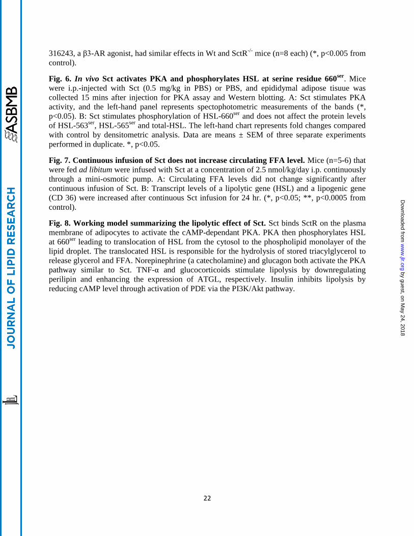
Fig. 1. Increases of both Sct peptide level in circulation and SctR transcript level in
epididymal fat tissue during starvation. Mice were starved and blood samples or fat tissues at
specified hours were obtained for measurements of Sct peptide and SctR transcript levels,
respectively. A: Sct peptide concentrations in circulation measured by an EIA kit were found
significantly increased after starvation. B: SctR transcript levels measured by real time-PCR.
were found significantly increased after starvation. For A and B, n=9-11 (*, p<0.05; **,
p<0.0005; versus 0 hr): C: SctR protein was localized on the adipose cell membrane of Wt mice
by immunohistochemical staining using SctR-/-
as negative controls.
Fig. 2. Sct via SctR stimulates lipolysis in dose- and time-dependant manners. A: Glycerol
release is increased in the presence of graded concentrations of Sct treatment for 1 hr in Wt and
Sct-/-
adipocytes but not in SctR-/-
adipocytes, indicating specificities of the lipolytic actions of
Sct. B: Sct (1 µM) stimulates glycerol release time-dependently in Wt adipocytes. C:
Adipocytes from Wt, Sct-/-
and SctR-/-
mice were found to exhibit similar basal lipolytic activities
and responded similarly to 1 µM isoproterenol or 5 µM CL-316243 treatment for 1 hr. Data are
means ± SEM of three separate experiments performed in triplicate. *, p<0.0001.
Fig. 3. Lipolytic actions of Sct is mediated by PKA and by phosphorylation of 660ser
and
563ser
of HSL in vitro. A: Isolated adipocytes were treated with 10 µM H-89 (PKA inhibitor), 1
µM Ro-31-8220 (PKC inhibitor) or 10 µM SP 600125 (JNK inhibitor) or 10 µM or 1 µM
CAY10499/CAY (HSL inhibitor) before stimulation with 1 µM Sct for 1 hr. Lipolysis by Sct in
Wt and Sct-/-
adipocytes was completely abrogated by H-89, but no effects observed for Ro-31-
8220 and SP 600125. SctR-/-
serves as the negative control. Data are means ± SEM of three
separate experiments performed in triplicate (*, p<0.0001 from basal). B: Small amounts of FFA
release was still observed in adipose cells incubated with 10 µM or 1 µM CAY10499/CAY
(HSL inhibitor) along with 1 µM Sct for 1 hr (**, p<0.0001; *, p<0.001 from basal). C: Isolated
adipocytes incubated with 1 µM Sct for 1 hr were used for Western analysis. Sct phosphorylates
HSL at 660ser
and 563ser
, but not 565ser
and phosphorylates perilipin at 522ser
while total HSL,
ATGL, G0S2, ABDH5, perilipin and GAPDH proteins remain unaltered by Sct. The left-hand
chart represents fold changes compared with control by densitometric analysis. Data are means ±
SEM of three separate experiments performed in duplicate. *, p<0.05 from control.
Fig. 4. Sct stimulates translocation of HSL from cytosol to lipid droplet. Isolated adipocytes
from Wt or SctR-/-
were stimulated with or without 1 µM Sct or 1 µM Isoproterenol (Iso; positive
control). Cells were fixed and translocation of HSL from the cytosol to lipid droplet was
visualized by confocal microscopy using an anti-HSL Ab (1:50 dilution) and subsequent
incubation with Alexa-488 secondary Ab (1:300 dilution). Red arrows in the figure denote the
translocation process. In the figure, BF: bright field, Iso: isoproterenol, Sct: secretin.
Fig. 5. Sct stimulates an acute FFA release in vivo through SctR. Stimulation of lipolysis by
Sct was detected by measuring changes in the levels of FFA in circulation after i.p. injection of
Sct. Blood was collected from the tail vein for measurement of FFA. A: Sct (0.5mg/kg, i.p.)
induced an acute lipolytic response in Wt mice (n=9) up to 15 mins after injection (*, p<0.05). B:
The same dose of i.p.-Sct significantly augments FFA concentrations in Wt (n=8) and Sct-/-
(n=9)
but not in SctR-/-
(n=7) mice (*, p<0.0005; **, p<0.0001 from control). C: 0.1 mg/kg CL-
by guest, on May 24, 2018
ww
w.jlr.org
Dow
nloaded from
22
316243, a β3-AR agonist, had similar effects in Wt and SctR-/-
mice (n=8 each) (*, p<0.005 from
control).
Fig. 6. In vivo Sct activates PKA and phosphorylates HSL at serine residue 660ser
. Mice
were i.p.-injected with Sct (0.5 mg/kg in PBS) or PBS, and epididymal adipose tisuue was
collected 15 mins after injection for PKA assay and Western blotting. A: Sct stimulates PKA
activity, and the left-hand panel represents spectophotometric measurements of the bands (*,
p<0.05). B: Sct stimulates phosphorylation of HSL-660ser
and does not affect the protein levels
of HSL-563ser
, HSL-565ser
and total-HSL. The left-hand chart represents fold changes compared
with control by densitometric analysis. Data are means ± SEM of three separate experiments
performed in duplicate. *, p<0.05.
Fig. 7. Continuous infusion of Sct does not increase circulating FFA level. Mice (n=5-6) that
were fed ad libitum were infused with Sct at a concentration of 2.5 nmol/kg/day i.p. continuously
through a mini-osmotic pump. A: Circulating FFA levels did not change significantly after
continuous infusion of Sct. B: Transcript levels of a lipolytic gene (HSL) and a lipogenic gene
(CD 36) were increased after continuous Sct infusion for 24 hr. (*, p<0.05; **, p<0.0005 from
control).
Fig. 8. Working model summarizing the lipolytic effect of Sct. Sct binds SctR on the plasma
membrane of adipocytes to activate the cAMP-dependant PKA. PKA then phosphorylates HSL
at 660ser
leading to translocation of HSL from the cytosol to the phospholipid monolayer of the
lipid droplet. The translocated HSL is responsible for the hydrolysis of stored triacylglycerol to
release glycerol and FFA. Norepinephrine (a catecholamine) and glucagon both activate the PKA
pathway similar to Sct. TNF-α and glucocorticoids stimulate lipolysis by downregulating
perilipin and enhancing the expression of ATGL, respectively. Insulin inhibits lipolysis by
reducing cAMP level through activation of PDE via the PI3K/Akt pathway.
by guest, on May 24, 2018
ww
w.jlr.org
Dow
nloaded from

23
Tables
Table 1
Sequence of the oligonucleotides used for real time
Primer Sequence 5’ 3’
HSL CAACATGGCATCAACCACTG
GCCTGGGATCAGAGGTGATG
ATGL TGTGGCCTCATTCCTCCTAC
TCGTGGATGTTGGTGGAGCT
β3-AR GAGCCAGTGGTGGCGTGTAGG
ACAGCAGCGATTGGAGT
GAPDH TGTGTCCGTCGTGGATCTGA
CCTGCTTCACCACCTTCTTGAT
CD 36 GCCAAGCTATTGCGACATGA
ATCTCAATGTCCGAGACTTTTCAAC
LPL GGGAGTTTGGCTCCAGAGTTT
TGTGTCTTCAGGGGTCCTTAG
AP2 TGGAAGCTTGTCTCCAGTGA
AATCCCCATTTACGCTGATG
ADIPONECTIN CAGGCATCCCAGGACATCC
CCAAGAAGACCTGCATCTCCTTT
by guest, on May 24, 2018
ww
w.jlr.org
Dow
nloaded from
Figure 1
C
A
B
0
2
4
6
8
10
12
0 hr 12 hr 18 hr 24 hr
Exp
ress
ion
of
SctR
mR
NA
/GA
PD
H m
RN
A
Fold
re
lati
ve t
o 0
hr
con
tro
l
Time
control
starved
**
** *
0
1
2
3
4
5
6
0 hr 12 hr 18 hr 24 hr
Pla
sma
Sct
leve
l (n
g/m
l)
Time
*
by guest, on May 24, 2018
ww
w.jlr.org
Dow
nloaded from

Figure 2
0
2
4
6
8
10
12
basal 1pM 100pM 10nM 100nM 1µM
Gly
cero
l re
leas
e(µ
mo
l/1
05
cells
/hr)
Concentration of Sct
Sct-/-
Wt
SctR-/-*
*
*
0
2
4
6
8
10
12
14
0 10 20 30 40 50 60 70 80 90
Gly
cero
l re
leas
e
(µm
ol/
10
5 c
ells
/1µ
M S
ct)
Time in min
Basal
Sct-stimulated
* *
*
*
*
0
5
10
15
20
25
30
basal CL-316243 Isoproterenol
Gly
cero
l re
leas
e(µ
mo
l/1
05
cells
/hr)
Wt
SctR-/-
Sct-/-
A
B
C
by guest, on May 24, 2018
ww
w.jlr.org
Dow
nloaded from

0
2
4
6
8
10
12
14
Wt SctR-/- Sct-/-
Gly
cero
l re
leas
e(µ
mo
l/1
05
cells
/hr)
basal
1µM Sct
H-89
R0-31-8220
SP 600125
10µM CAY
1µM CAY
*
**
*
* *
Figure 3
A
1µM Sct - + + + + + + - + + + + + + - + + + + + +
H-89 - - + - - - - - - + - - - - - - + - - - -
R0-31-8220 - - - + - - - - - - + - - - - - - + - - -
SP 600125 - - - - + - - - - - - + - - - - - - + - -
10µM CAY - - - - - + - - - - - - + - - - - - - + -
1µM CAY - - - - - - + - - - - - - + - - - - - - +
1µM Sct - + + + - + + + - + + +
10µM CAY - - + - - - + - - - + -
1µM CAY - - - + - - - + - - - +
B
0
5
10
15
20
Wt SctR-/- Sct-/-
FFA
re
leas
e(m
M/1
05
cells
/hr) basal
1µM SCT
10µM CAY
1µM CAY
****
* * * *
by guest, on May 24, 2018
ww
w.jlr.org
Dow
nloaded from

Control Sct
HSL-660ser
HSL-563ser
HSL-565ser
HSL
Perilipin- 522ser
Perilipin
ABDH5
G0S2
ATGL
GAPDH
C
0 1 2 3 4 5
Fold increase over control (protein/GAPDH)
*
*
*
Figure 3
by guest, on May 24, 2018
ww
w.jlr.org
Dow
nloaded from

Wt -control
Wt -Sct
Wt -Iso
SctR-/- -control
SctR-/- -Sct
SctR-/- -Iso
HSL BF Merge HSL BF Merge
Figure 4
lipiddroplet
cytoplasm
translocation
Cytoplasm with nucleus
translocation
cytosol
cytosol
by guest, on May 24, 2018
ww
w.jlr.org
Dow
nloaded from

Figure 5
0
0.2
0.4
0.6
0.8
1
1.2
Wt SctR-/- Sct-/-
FFA
(m
M)
control
i.p. Sct
***
A
B
C
0
0.2
0.4
0.6
0.8
1
1.2
1.4
1.6
0 5 10 15 20 30 45
FFA
(m
M)
Time in min
* **
0
0.2
0.4
0.6
0.8
1
1.2
1.4
1.6
1.8
Wt SctR-/-
FFA
(m
M)
control
i.p. CL-316243
* *
by guest, on May 24, 2018
ww
w.jlr.org
Dow
nloaded from
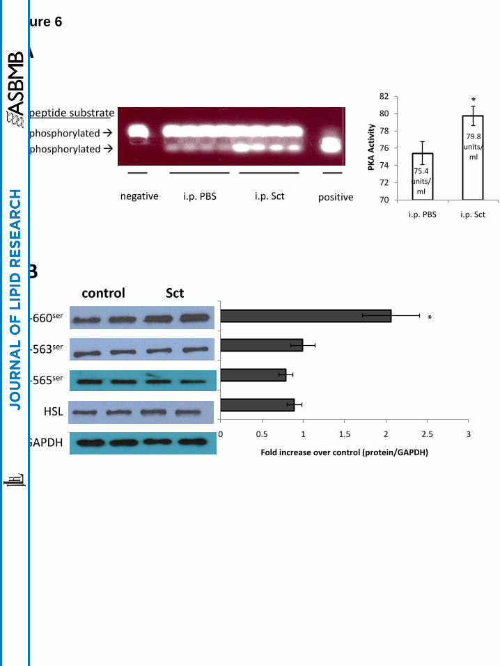
Figure 6
i.p. PBS i.p. Sctnegative positive
75.4 units/
ml
79.8units/
ml
70
72
74
76
78
80
82
i.p. PBS i.p. Sct
PK
A A
ctiv
ity
*
A
PKA peptide substrate
Non-phosphorylated
phosphorylated
control Sct
HSL-660ser
HSL-563ser
HSL-565ser
HSL
GAPDH
B
0 0.5 1 1.5 2 2.5 3
Fold increase over control (protein/GAPDH)
*
by guest, on May 24, 2018
ww
w.jlr.org
Dow
nloaded from

Figure 7
Time of infusion
Treatment 0 h 18 h 24 h
PBS (FFA in mM) 0.52±0.06 0.42±0.07 0.42±0.07
Sct (FFA in mM) 0.54±0.08 0.53±0.07 0.56±0.09
Table 2 : Circulating FFA levels in response to continuous infusion of Sct
0
0.5
1
1.5
2
2.5
Exp
ress
ion
of
targ
et
gen
e/G
AP
DH
mR
NA
(fo
ld r
ela
tive
to
PB
S)
i.p. PBS
i.p. Sct
*
**
A
B
by guest, on May 24, 2018
ww
w.jlr.org
Dow
nloaded from