Embed Size (px)
Citation preview

www.elsevier.com/locate/matbio
Matrix Biology 24 (
Pro-a3(V) collagen chain is expressed in bone and its basic
N-terminal peptide adheres to osteosarcoma cells
Kenji Yamaguchia, Noritaka Matsuoa, Hideaki Sumiyoshia, Noritaka Fujimotoa,
Ken-ich Iyamac, Shigetaka Yanagisawab, Hidekatsu Yoshiokaa,*
aDepartment of Anatomy, Biology and Medicine, Oita University, Faculty of Medicine, 1-1 Hasama-machi, Oita 879-5593, JapanbDepartment of Oncological Science, Faculty of Medicine, Oita University, 1-1 Hasama-machi, Oita 879-5593, Japan
cDepartment of Surgical Pathology, Kumamoto University School of Medicine, Kumamoto 860-8556, Japan
Received 11 August 2004; received in revised form 9 March 2005; accepted 9 March 2005
Abstract
The third a-chain of type V collagen, a3(V) chain, was initially identified in the placenta more than 20 years ago, but was poorly
characterized with regard to its expression and function. We generated a specific monoclonal antibody against the N-terminal domain of the
pro-a3(V) chain and examined gene expression using immunohistochemical methods combined with in situ hybridization. The pro-a3(V)
chain was seen in funis and amnion, but not chorionic villi and deciduas of mouse placenta. In mouse embryo, the transcripts of the pro-
a3(V) gene were seen in tissues that were related to bone formation as well as developing muscle and nascent ligament previously reported
(J. Biol. Chem. 275, 8749–8759, 2000). However, immunohistochemistry showed that pro-a3(V) protein accumulated rather in the
developing bone of mouse embryo. On the other hand, the N-terminal globular domain of the pro-a3(V) chain has a unique structure that
contains a highly basic segment of 23 amino acids. The peptide derived from the basic segment showed a specific adhesive feature to
osteosarcoma cells but not to chondrosarcoma cells. The four heparin binding sites in the basic segment equally contribute toward adhesion to
the osteosarcoma cells. Our data suggested that N-terminal globular domain of the pro-a3(V) chain influence bone formation of osteoblasts
through proteoglycan on the cell surface during development or regeneration.
D 2005 Elsevier B.V./International Society of Matrix Biology. All rights reserved.
Keywords: Pro-a3(V) collagen; Bone formation; Heparin binding; Gene expression
1. Introduction
The collagens are major constituents of the extracel-
lular matrix. Among them, fibrillar collagen, which
include five different molecular types, I, II, III, V and
XI, participate in the formation of fibrils (Vuorio and de
Crombrugghe, 1990; van der Rest and Garrone, 1991;
Brown and Timpl, 1995). Collagen V is a minor
0945-053X/$ - see front matter D 2005 Elsevier B.V./International Society of Ma
doi:10.1016/j.matbio.2005.03.006
Abbreviations: PBS, Phosphate-buffered saline; RT-PCR, Reverse
transcription-polymerase chain reaction; IPTG, Isopropyl-h-d-thiogalacto-pyranoside; GST, Glutathione S-transferase; ELISA, Enzyme-linked
immunosorbent assay; DMEM, Dulbecco’s modified Eagle’s medium.
* Corresponding author. Tel.: +81 97 586 5670; fax: +81 97 549 6302.
E-mail address: [email protected] (H. Yoshioka).
component of connective tissues but it plays an important
role in matrix organization. Mutations in the collagen V
gene produce abnormal fibril aggregates in the tissue
(Andrikopoulos et al., 1995; Toriello et al., 1996; De
Paepe et al., 1997; Michalickova et al., 1998). Type V
collagen co-polymerizes with the major collagen, type I,
and regulates the diameter of collagen fibers in non-
cartilage tissues. Collagen V is widely distributed in
vertebrate tissues as an [a1(V)]2a2(V) heterotrimer
(Fessler and Fessler, 1987; Fichard et al., 1994). Other
forms of collagen V include an [a1(V)]3 homotrimer
secreted by a line of Chinese hamster cells (Haralson et
al., 1980) that may also exist in normal tissues (Moradi-
Ameli et al., 1994; Kumamoto and Fessler, 1980), and a
poorly characterized a1(V)a2(V)a3(V) heterotrimer that
2005) 283 – 294
trix Biology. All rights reserved.

Fig. 1. RT-PCR analysis of pro-a3(V) collagen gene expression in mouse
embryo. A: RNAs from whole mouse embryos at E12.5–E18.5 were used.
RT-PCRs using specific primers for the mouse pro-a3(V), pro-a1(V)
collagen and GAPDH genes were performed. B: RNAs from different
tissues of the E18.5 mouse embryo were used. GAPDH was used as an
internal control. Expected size of the PCR product is shown on the right of
each panel.
K. Yamaguchi et al. / Matrix Biology 24 (2005) 283–294284
is primarily isolated from placenta (Rhodes and Miller,
1981; Niyibizi et al., 1984) but also reported in uterus,
skin, and synovial membranes (Fessler and Fessler, 1987;
Abedin et al., 1982).
Collagen specifically interacts with other macromole-
cules in the extracellular matrix such as fibronectin, laminin
and proteoglycan (Hynes and Yamada, 1982; Timpl and
Brown, 1994; Scott, 1988) and with cell-surface receptor,
integrins (Kramer and Marks, 1989; Plow et al., 2000;
Ruggiero et al., 1996). The interactions are important in
regulating cell behavior, including proliferation, migration,
and differentiation during development and physiological
and pathological conditions. Heparin is abundant in the
tissues as a form of heparin sulfate proteoglycans. Heparin
binding sites are found in the extracellular matrix proteins
such as collagens, fibronectin, tenascin, and laminin.
Common structural motifs have been proposed from the
analysis of different heparin binding sites (Cardin and
Weintraub, 1989).
Type V collagen, [a1(V)]2a2(V) form, was shown to
possess a site that binds heparin/heparan sulfate under
physiological conditions (Cardin and Weintraub, 1989;
LeBaron et al., 1989). This site is located within the
NH2-terminal half of the a1(V) chain (Yaoi et al., 1990;
Delacoux et al., 1998). Delacoux et al. (2000) narrowed
the region down to a 12 kDa fragment that contains a
cluster of seven basic amino acids. Unlike a1(V) chains,
a2(V) and a3(V) chains do not bind heparin under
physiological or denaturing conditions (Mizuno and
Hayashi, 1996). Triple helical type V collagen trimers
bind to heparin with decreasing affinity in the order
[a1(V)]3> [a1(V)]2a2(V)>a1(V)a2(V)a3(V), indicating
that a1(V) chains mediate heparin binding, but a2(V)
and a3(V) chains do not (Mizuno and Hayashi, 1996).
Mann (1992) presented the first partial human amino acid
sequence of the N-terminus of a3(V) collagen chain.
Recently, Imamura et al. (2000) provided the primary
structure of the human and mouse pro-a3(V) collagen
chains. The structure of the pro-a3(V) chain was shown to
be closely related to that of the pro-a1(V) chain. It has a
unique acidic domain in the N-terminal globular domain,
which is also contained in the pro-a1(V) chain as well as
pro-a1(XI) and pro-a2(XI) chains. They showed expression
of the pro-a3(V) gene in the epimysial sheaths of developing
muscles, within nascent ligaments adjacent to forming bones
and in joints using in situ hybridization. Alternatively,
Chernousov et al. (2000) reported that rat pro-a4(V) chain
must be the counterpart of mouse and human pro-a3(V)
chains based on its high similarity of structure as shown in
the genomic databases (Gopalakrishnan et al., 2004). Their
group also found a new heparan sulfate binding site that
mediates Schwann cell adhesion in the unique N-terminal
domain of rat pro-a4(V) (Erdman et al., 2002). They showed
that the major binding proteins are glypican-1 and perlecan
using pull down assays and immunofluorescent staining
(Rothblum et al., 2004).
In this study, we generated a specific monoclonal
antibody directed against the unique N-terminal domain of
the pro-a3(V) chain. We examined the expression of the
pro-a3(V) chain using immunohistochemical methods and
in situ hybridization. The pro-a3(V) chain was observed in
tissues related to bone formation. Furthermore, the peptide
derived from the basic segment flanking the acidic domain
of the pro-a3(V) chain binds to osteosarcoma cells.
2. Results and discussion
2.1. Transcripts of pro-a3(V) collagen gene
The pro-a3(V) chain was originally isolated from human
placenta but is distributed in many more tissues than
previously expected (Rhodes and Miller, 1981; Abedin et
al., 1982; Imamura et al., 2000). Initially, to examine the
pro-a3(V) gene expression temporally and spatially in
mouse, we performed RT-PCR. As shown in Fig. 1A, pro-
a3(V) transcripts were detectable from E16.5, whereas pro-
a1(V) transcripts were readily detectable in embryos at
E12.5. In different tissues of E18.5, relatively high levels of
pro-a3(V) expression were seen in placenta, kidney,
vertebrae, calvaria and tongue (Fig. 1B). Weak bands were
also detectable in tail and heart, but almost undetectable in
liver, lung, intestine, limb, brain and skin (Table 1). By
contrast, pro-a1(V) gene was expressed in many tissues
tested (data not shown) (Wu et al., 1998).
To characterize the expression pattern of the pro-a3(V)
gene in detail, we applied in situ hybridization. In situ
hybridization was performed on sagittal sections of E16.5
mouse embryos using specific probes for pro-a3(V) basic/
acidic domain, pro-a1(V) acidic domain and type II
collagen (Yoshioka et al., 1995). Although pro-a1(V)

Fig. 3. In situ hybridization of the tissues related to the bone formation of
E16.5 mouse embryos. Photomicrographs are H–E stained with brightfield
(A and B) or hybridization with darkfield (C–H). Panels are shown at high
magnification of Fig. 2 to clearly show the portion of calvaria (A, C, E and G)
and vertebrae (B, D, F and H). The sections were hybridized with a
radioactively labeled pro-a3(V) (C and D), pro-a1(V) (E and F) and a1(II)
(G and H) collagen antisense riboprobe. Specific regions were labeled as
follows: skin (Sk), subcutaneous connective tissue (CT), calvaria (frontal
bone) (Ca), midbrain (Br), first ossification center (OsC), hypertrophic
chondrocyte (Hc), prehypertrophic chondrocyte (Pc), ganglion (Ga). Scale
bar: 100 Am.
Table 1
Expression of a3(V) collagen gene in the placenta and the mouse embryo
RT-PCR In situ
hybridization
Immunohistochemistry
Placenta + / /
Chorionic villi nd � �Funis nd + +
Amnion nd + +
Deciduas nd � �Liver � � �Lung � � �Intestine � � �Kidney + � �
Renal facia nd + +
Tail T � �Vertebrae + + +
Heart T � �Vessel nd � �Limb � � �Calvaria + + +
Tongue + + �Brain � � �Skin � � �Superficial facia
of developing
muscle
nd + +
+, Positive; T, weakly positive; �, negative; nd, not determined.
The E18.5 mouse embryos were used for RT-PCR, and E16.5 for in situ
hybridization and immunohistochemistry.
K. Yamaguchi et al. / Matrix Biology 24 (2005) 283–294 285
expression was widely distributed throughout developing
connective tissues (Fig. 2C), pro-a3(V) expression was
restricted (Fig. 2B). Type II collagen was strongly expressed
in cartilage, especially prehyper/hypertrophic chondrocytes
(Figs. 2D and 3H). Pro-a3(V) mRNA was expressed in
osteoblasts of calvaria (Fig. 3C) and in the associated
periosteal cells surrounding first ossification center of
vertebrae (Fig. 3D). The former represent bones formed
by intramembranous ossification, and the latter by endo-
chondrial ossification. By contrast, pro-a1(V) expression
was seen in calvaria and its surrounding connective tissue
(Fig. 3E), and type II collagen was never seen in calvaria
(Fig. 3G). In vertebrae, pro-a1(V) mRNA was expressed in
perichondrial cell layers surrounding cartilage primordium
(Fig. 3F).
Fig. 2. In situ hybridization of E16.5 mouse embryos. Photomicrographs are hemat
with darkfield (B–D). Sagittal sections from E16.5 embryos were hybridized with a
antisense riboprobe. Scale bar: 1 mm.
2.2. Distribution of pro-a3(V) collagen chain in bone-
related tissues
To detect the protein in tissues by immunohistochemis-
try, we prepared a specific monoclonal antibody directed
against the basic/acidic domain of pro-a3(V). The antibody
oxylin and eosin (H–E) staining with brightfield (A) or in situ hybridization
radioactively labeled pro-a3(V) (B), pro-a1(V) (C) and a1(II) (D) collagen
200K
118K
1 2 3 4
A
B C
a b c d
0.8
1.2
dilution
O.D
.
0.4
0.2
0.0x20 x40 x80 x160 x320 x640 x1280
0.6
1.0α3(V)-GST α3(V)-Hisα1(V)-GST
Fig. 4. Characterization of monoclonal antibody against the basic/acidic domain of pro-a3(V) chain. A: Western blot analysis using recombinant proteins.
Purified recombinant proteins were run on a 15% SDS-PAGE gel and stained with Coomassie brilliant blue (panel a). The proteins blotted on the filters were
hybridized with anti-pro-a3(V) antibody (panel b), anti-pro-a1(V) antibody (panel c) and anti-GST antibody (panel d). The samples in each lane were as
follows—lane 1: pro-a3(V)–GST fusion protein, lane 2: pro-a1(V)–GST fusion protein, lane 3: empty vector (GST), lane 4: pro-a3(V)–His-tag fusion protein,
lane M: marker. The positions of the molecular mass standards are shown on the left. B: Western blot analysis using native collagenous protein. Proteins were run
on a 10% SDS-PAGE gel and stained with Coomassie brilliant blue (lanes 1 and 2). The proteins blotted on the filters were hybridized with anti-pro-a3(V)
antibody (lanes 3 and 4). The samples were digested with collagenase (lanes 2 and 4). The positions of the molecular mass standards are shown on the left. C:
ELISA using anti-pro-a3(V) antibody. The pro-a3(V) monoclonal antibody reacted with the pro-a3(V)–GST (open circles) and pro-a3(V)–His (closed
squares) recombinant protein applied to a plate as a serial dilution, but not with similar dilutions of the pro-a1(V)–GST (closed triangles) fusion proteins.
K. Yamaguchi et al. / Matrix Biology 24 (2005) 283–294286
was purified by protein G affinity column chromatogra-
phy and Western blot and ELISA confirmed its specific-
ity. As shown in Fig. 4A and C, the anti-pro-a3(V)
antibody recognized the GST fusion and His-tagged pro-
a3(V) recombinant polypeptide but not the tagged pro-
a1(V) recombinant polypeptide. The antibody also recog-
nized a collagenous peptide of approximately 220 kDa
from a fraction of neutral salt-extracted osteosarcoma cells
(Fig. 4B). An anti-pro-a1(V) polyclonal antibody directed
against the acidic domain was also generated.
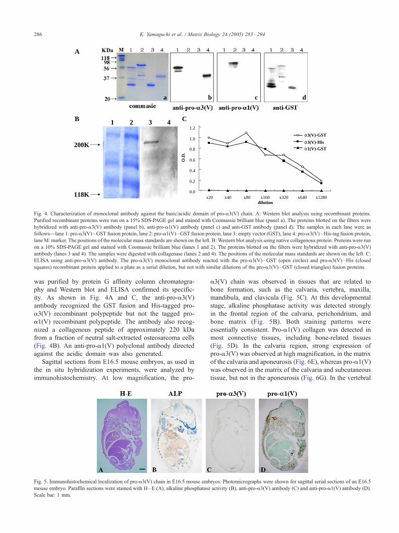
Sagittal sections from E16.5 mouse embryos, as used in
the in situ hybridization experiments, were analyzed by
immunohistochemistry. At low magnification, the pro-
Fig. 5. Immunohistochemical localization of pro-a3(V) chain in E16.5 mouse emb
mouse embryo. Paraffin sections were stained with H–E (A), alkaline phosphatase
Scale bar: 1 mm.
a3(V) chain was observed in tissues that are related to
bone formation, such as the calvaria, vertebra, maxilla,
mandibula, and clavicula (Fig. 5C). At this developmental
stage, alkaline phosphatase activity was detected strongly
in the frontal region of the calvaria, perichondrium, and
bone matrix (Fig. 5B). Both staining patterns were
essentially consistent. Pro-a1(V) collagen was detected in
most connective tissues, including bone-related tissues
(Fig. 5D). In the calvaria region, strong expression of
pro-a3(V) was observed at high magnification, in the matrix
of the calvaria and aponeurosis (Fig. 6E), whereas pro-a1(V)
was observed in the matrix of the calvaria and subcutaneous
tissue, but not in the aponeurosis (Fig. 6G). In the vertebral
ryos. Photomicrographs were shown for sagittal serial sections of an E16.5
activity (B), anti-pro-a3(V) antibody (C) and anti-pro-a1(V) antibody (D).

Fig. 6. Immunohistochemical localization of pro-a3(V) chain in the tissues
that are related to bone formation in E16.5 mouse embryos. Photomicro-
graphs are H–E stained (A and B) or immunohistochemical stained (C–H).
Panels are shown at a higher magnification than Fig. 5 to clearly show the
portion of calvaria (A, C, E and G) and vertebrae (B, D, F and H). The
sections were stained for alkaline phosphatase activity (C and D), anti-pro-
a3(V) (E and F) and anti-pro-a1(V) (G and H) antibodies. Specific regions
were labeled as follows: skin (Sk), subcutaneous connective tissue (CT),
calvaria (frontal bone)(Ca), midbrain (Br), aponeurosis (Ap), first ossifica-
tion center (OsC), hypertrophic chondrocyte (Hc), prehypertrophic chon-
drocyte (Pc), perichondrial (Pe), periosteal (Po). Scale bar: 100 Am.
K. Yamaguchi et al. / Matrix Biology 24 (2005) 283–294 287
region, pro-a3(V) was detected in the periosteum and bone
matrix, where ossification initially occurs (Fig. 6F). This
staining pattern is consistent with that of alkaline phos-
phatase (Fig. 6D). Weak staining of the pro-a3(V) chain
was also seen in ligamentous attachments. On the other
hand, staining for pro-a1(V) was more intense in the
periosteum and ligamentous attachments than in the bone
matrix (Fig. 6H). This pattern differed from that of alkaline
phosphatase.
2.3. Expression in non-bone-related tissues
The a3(V) chain was originally isolated from the
placenta. As shown in Fig. 1B, the transcripts of pro-
a3(V) were detected in the placenta using RT-PCR.
However, Chernousov et al. (2000) did not detect pro-
a3(V) in the placenta of rat. This was one of the reasons for
them to call the collagenous peptide, the a4(V) chain, not
the a3(V) chain. We examined the distribution of the mouse
pro-a3(V) chain in the placenta using in situ hybridization
and immunohistochemical methods. Signal from the tran-
scripts were seen in the soft connective tissue, Wharton’s
jelly, in funis and amnion (Fig. 7B and E). However, they
could never be seen in chorionic villi and decidua. The
immunohistochemical data were consistent with those of in
situ hybridization (Fig. 7C and F).
Imamura et al. (2000) reported that the pro-a3(V) gene
was expressed in the epimysial sheaths of developing
muscles and within nascent ligaments adjacent to forming
bones and joints in the E15.5 mouse embryo. Similarly, we
could detect the signals in those regions in the E16.5 mouse
embryo. The staining of pro-a3(V) could be detected in the
superficial fascia of developing muscle and renal fascia
around the kidney and adrenal gland (Fig. 7I and L).
However, we could not clearly detect a signal in other
regions where expression was positive by RT-PCR or in situ
hybridization (Table 1). This discrepancy may be due to the
fast turnover or disseminated distribution of the protein in
these tissues.
2.4. Cell adhesion of pro-a3(V) chain
To elucidate the biological function of type V collagen in
osteoblast, we used an osteosarcoma cell line to examine
whether the N-terminal domain of pro-a3(V) chain is
involved in cell adhesion. The N-terminal globular domain
of the pro-a3(V) chain can be divided into subdomains
(Fig. 8). Among them, the acidic domain of pro-a3(V)
chain, which is flanked by a highly basic segment of 23
amino acids at the N-terminus, is unique because it is only
found in mouse and human pro-a3(V) and rat pro-a4(V)
chains (Imamura et al., 2000; Chernousov et al., 2000).
We tested the adhesive ability of the basic/acidic domain
of pro-a3(V) chain using recombinant proteins to ROS and
RCS cells. As shown in Fig. 9, ROS cells adhere to the basic/
acidic domain of pro-a3(V) recombinant protein but not to
the acidic domain of pro-a1(V). The adhesion of RCS cells
was hardly recognizable in both acidic domains of pro-a1(V)
and basic/acidic domain of pro-a3(V). This adhesive
function was restricted to the region of 239–269 amino
acids where the basic segment is contained. This segment has
four repeats of the heparin binding consensus sequence
BBXB (B and X are a basic and any amino acids,
respectively) (Fig. 12A) (Cardin and Weintraub, 1989). To
assess the heparin binding activity of the recombinant
protein, heparin affinity chromatography was performed.
As shown in Fig. 10A and B, pro-a3(V) (239–369) and pro-
a3(V) (239–269) fragments, containing the basic segment,
bound to the heparin column and were eluted at a high NaCl
concentration, reflecting high affinity binding to heparin. In
contrast, the pro-a1(V) acidic domain and pro-a3(V)(269–
369) that deletes the basic segment, dramatically abolished
the heparin binding activity. Thus, the basic segment in the
N-globular domain of pro-a3(V) chain is important to bind
heparin.
These results suggest that ROS cells adhesion to pro-
a3(V) peptide might be mediated by cell-surface heparan
sulfate proteoglycans. The contribution of glycosaminogly-

Fig. 7. Expression in non-bone-related tissues. Photomicrographs are H–E stained (A, D, G and J), in situ hybridization with darkfield (B, E, H and K) and
immunohistochemical staining (C, F, I and L). The sections were hybridized with a radioactively labeled pro-a3(V) collagen antisense riboprobe (B, E, H and
K) and stained for anti-pro-a3(V) collagen antibody (C, F, I and L). The tissues are placenta (A–C), funis attached to fetal abdomen (D–F), superficial fascia
of developing muscle (G–H) and kidney and adrenal gland (J–L). Specific regions were labeled as follows: chorionic villi in placenta (Pl), amnion (Am), funis
(Fu), superficial fascia in fetus (Fa), skin (Sk), vertebrae (Ve). Scale bar: 100 Am.
K. Yamaguchi et al. / Matrix Biology 24 (2005) 283–294288
can to ROS adhesion was examined by plating the cells onto
the recombinant proteins of pro-a3(V) basic/acidic domain
in the presence of heparin, chondrotin sulfate and dextran
239
239 269
270
37
37
255
1(V)
3(V)
(37)
(29)
(217) (189) (115) (1
(209) (133) (70) (1011
Npp AD SC/NT C NH2 -SP
Npp BS/AD C O LNH2 - PS
239
239 269
270
37
37
255
α α
α α
(37)
(29)
(217) (189) (115) (1
(209) (133) (70) (1011
Npp AD SC/NT C NH2 -SP
Npp BS/AD C O LNH2 - SC/NTPS
Fig. 8. The constructs of recombinant protein of the mouse pro-a3(V) and pro-a1(
domain of pro-a3(V) and the acidic domain pro-a1(V) chains were generated. Th
peptide (SP), N-terminal propeptide (Npp), basic/acidic (BS/AD) or acidic dom
continuous collagenous domain (COL) and C-terminal propeptide (Cpp). A short b
Vertical arrows and numbers in parenthesis indicate the cleaved sites of pro-a
respectively. The recombinant protein numbers indicate the number of amino aci
sulfate. As shown in Fig. 11, heparin and dextran sulfate,
added at a concentration of 10 Ag/mL, inhibited cell
adhesion by 76%. Chondrotin sulfate inhibited adhesion
1
1
443
3(V) 239/371
3(V) 239/269
3(V) 270/371
014) (266)
) (251)
O L Cpp -COOH
Cpp -COOH
1
1
443α1(V) 255/443
3(V) 239/371
3(V) 239/269
3(V) 270/371
α α
α α
α α
014) (266)
) (251)
O L Cpp -COOH
Cpp -COOH
V) collagen polypeptides. The recombinant protein covered the basic/acidic
e domains of prepro-a3(V) and prepro-a1(V) chain are consist of a signal
ain (AD), short collagenous segment (SC), N-telopeptide (NT), central
ar in the pro-a3(V) chain indicates a basic segment flunked acidic domain.
1(V) chains and number of amino acid residues in individual domains,
d residues from the N-terminal end.

ROSRCS
1
0.9
0.8
0.7
0.6
0.5
0.4
0.3
0.2
0.1
0cell
adhe
sion
(ab
sorb
ance
595
nm)
ROSRCS
GST α 1(V)255/443
GST α 3(v)239/371
GST α 3(V)239/269
GST α 3(V)270/371
GST
Fig. 9. Cell adhesion to the recombinant proteins of pro-a3(V) and pro-a1(V) collagen polypeptides. ROS and RCS cells were plated on dishes coated with the
recombinant proteins. Cell adhesion was quantitated by staining with crystal violet.
K. Yamaguchi et al. / Matrix Biology 24 (2005) 283–294 289
by about 30%. The interaction between ROS cells and basic
segment might be mediated by a cell-surface heparan sulfate
proteoglycan receptor. Erdman et al. (2002) showed the N-
terminal binding activity to Schwann cells. Rothblum et al.
(2004) suggested that this binding is mediated via glypican-
1, that is a lipid-anchored proteoglycan of the plasma
membrane. In osteoblasts, the same or similar proteoglycan
might also contribute to the interaction with the basic
segment.
2.5. Adhesion activity of the four heparin binding sites
To know which heparin binding site is the most
important in cell adhesion among the four heparin binding
0
0.01
0.02
0.03
0.04
0.05
0.06
0.07
0.08
0.2 0.4 0.6 0.8 1.0 1.
NaC
A
B
abso
rban
ce (
280n
m)
GST α α3(V)239/371
GST α α3(V)239/269
GST α α3(V)270/371
GST α α1(V)255/443
F 0.4 0.5 0.6 0.7
Fig. 10. Heparin binding of the recombinant pro-a3(V) and pro-a1(V) collagen
polypeptides were applied to a heparin–sepharose column and eluted by step gradi
shown in the graph. B: Aliquots of column fractions were subjected to Western bl
sites, we replaced each heparin binding site by site-
directed mutagenesis. These amino acids were replaced by
alanine (Fig. 12A). The mutated fusion proteins were
expressed in bacteria and tested by the heparin binding
assay and cell adhesion assay. As shown in Fig. 12B, M1,
M2, M3 and M4 mutated proteins were eluted from the
heparin column at NaCl concentration of 0.8–1.5 M.
However, the binding affinities were decreased compared
with that of wild type pro-a3(V)(247–269), which eluted
at the concentration of 1.0–2.0 M. Similarly, as shown in
Fig. 12C, each mutated protein, M1, M2, M3 and M4,
showed approximately 60–70% of the adhesive activity to
ROS compared with that of wild type pro-a3(V)(247–
269). These results indicate that each heparin binding site
25 1.5 2.0
l (M)
GST α α3(V)239/371
GST α α3(V)239/269
GST α α3(V)270/371
GST α α1(V)255/443
(M)0.8 0.9 1.0 1.25 1.5 2.0
polypeptides. GST fusion proteins of pro-a3(V) and pro-a1(V) collagen
ent at the indicated concentrations from 0 to 2.0 M NaCl. A: Elution data are
ot analysis with anti-GST antibody. F indicates the fraction of flowthrough.

0.0
0.2
0.4
0.6
0.8
1.0
1.2
hepdex
chon
cell
adhe
sion
(abs
orba
nce
595n
m)
( - )
Fig. 11. Inhibition of cell adhesion by glycosaminoglycan. ROS cells were
suspended in serum-free medium containing 10 Ag/mL of heparin (hep),
dextran sulfate (dex), and chondroitin sulfate (chon), and plated onto wells
coated with pro-a3(V)(239–369) recombinant protein. The values shown
are the meanTSD of five independent wells.
PPRRKGKGKKKGRGRKGKGRKKNKETSEL
-----------------------
---AAAA----------------------
--------AAAA-----------
-------------AAAA------
-------------------AAAA---
3(V)247/269WTM1
M2
M3
M4
sequence247 269
PPRRKGKGKKKGRGRKGKGRKKNKETSEL
-----------------------
---AAAA----------------------
--------AAAA-----------
-------------AAAA------
-------------------AAAA---
α α V)247/269WT
M1
M2
M3
M4
247 269
basic segment
2.01.51.00.50.10
0.08
0.06
0.04
0.02
1.00.90.80.7
0.60.50.40.30.20.10.0
WTM1M2M3M4
Abs
orba
nce
(280
nm
)WTM1M2M3M4
NaCl (M)
** *
*
W T M 1 M 2 M 3 M 4
cell
adhe
sion
(ab
sorb
ance
595
nm)
A
B
C
Fig. 12. Assessment of four heparin binding sites in the basic segment of
pro-a3(V) chain for heparin binding affinity and ability of cell adhesion. A:
Amino acid sequence of the constructs with an alanine mutation. Note that
it was difficult to generate a 23 amino acids-size pro-a3(V)M1 and pro-
a3(V)M4 using the PCR procedure because of annealing problems with the
primers. Therefore, pro-a3(V)M1 and pro-a3(V)M4 are six and three
amino acids, respectively, longer than the others. B: Heparin binding assay
using substituted mutant proteins. Bound proteins were eluted by step
gradient of indicated concentrations of 0–2.0 M NaCl. Elution data are
shown in the graph. C: Cell adhesion assay using substituted mutant
proteins. ROS cells were plated on dishes coated with mutated protein. The
results are displayed graphically. The values shown are the meanTSD for
four independent wells. *p <0.01.
K. Yamaguchi et al. / Matrix Biology 24 (2005) 283–294290
in the pro-a3(V) chain equally contributes with regard to
adhesive interactions with ROS cells.
Delacoux et al. (2000) showed that a heparin binding site
is present in the collagenous domain of the a1(V) chains.
The sequence contains a cluster of seven basic amino acids
that are different from the consensus sequence of the heparin
binding site previously reported (Cardin and Weintraub,
1989). However, its binding affinity is significantly lower
than that of the pro-a3(V) chain. The former is eluted at
0.35 M NaCl concentration whereas the latter is at 1.0–2.0
M. This basic segment would play a specific role through a
strong electrostatic interaction with molecules such as
heparan sulfate proteoglycan in vivo. This portion of the
protein is retained in the fibrils that contain the rat a4(V)
chain synthesized by Schwann cells (Chernousov et al.,
1996; Chernousov et al., 2000) and is exposed on the fibril
surface (Linsenmayer et al., 1993). However, Gopalakrish-
nan et al. (2004) have recently reported that this portion of
the recombinant human pro-a3(V) chain was removed by
BMP-1 in a fibroblast culture system. The processing of the
N-terminal domain of the pro-a3(V) chain (or rat a4(V)
chain) may depend on the cell type or the circumstances of
the tissue. The processing of the pro-a3(V) chain in bone is
unknown. Whether the basic segment is retained in the
fibrils or removed, it might still affect the osteoblast in the
developing bone.
2.6. Conclusion
Using immunohistochemistry and in situ hybridization,
this study has demonstrated that the pro-a3(V) chain is
expressed in tissues that are related to bone formation as
well as in the funis and amnion of the placenta. The
peptide derived from the basic segment of the N-terminal
domain of the pro-a3(V) chain showed specific adherence
to osteosarcoma cells but not to chondrosarcoma cells.
The unique segment should play an important role in bone
formation. Further study is required to determine the
precise role of this chain in osteoblasts, and to character-
ize its function in bone formation during development and
regeneration.
3. Materials and methods
3.1. Animals
Mice and rabbits were purchased from commercial
sources (Yoshitomi, Fukuoka, Japan; Kyudo, Saga, Japan).

K. Yamaguchi et al. / Matrix Biology 24 (2005) 283–294 291
The animals were treated in accordance with the Oita
University Guidelines for the Care and Use of Laboratory
Animals based on the National Institutes of Health Guide
for the Care and Use of Laboratory Animals.
3.2. Staging of mouse embryos and preparation of sections
Gestational age was initially determined by the date of
formation of the copulation plug and confirmed by crown-
rump length. For in situ hybridization and immunohisto-
chemistry, mouse embryos were fixed overnight in fresh 4%
paraformaldehyde in phosphate-buffered saline (PBS),
dehydrated, and embedded in paraffin, and 7 Am consecu-
tive sections were prepared.
3.3. Reverse transcription-polymerase chain reaction
(RT-PCR)
RNA samples were prepared from E12.5, 14.5, 16.5, and
18.5 mouse whole embryos, and from different tissues of the
E18.5 mouse embryo, using Isogen (Wako, Osaka, Japan)
according to the manufacturer’s instructions. Total RNAs
were used as templates to synthesize cDNAs using Moloney
murine leukemia virus reverse transcriptase (Invitrogen,
Carlsbad, CA) and random hexamers. The PCR program
was as follows: 1 min at 94 -C for denaturation, 1 min at 55
-C for annealing, and 1 min at 72 -C for extension; repeated
for 30 cycles with a final extension of 7 min 30 s at 72 -C.The gene-specific primers used in the PCR reactions were:
pro-a1(V)255/443: (forward) 5V-CAGGACCCTAAC-CCGGATGA-3V
(reverse) 5V-CTCAAAGATGGTGTCCTGGT-3Vpro-a3(V)239/371: (forward) 5V-GATGAACCAGAAA-CCCCTGC-3V
(reverse) 5V-AGCACCAGGAAAGATCTGGA-3VGAPDH: (forward) 5V-AGAGGTGCTGCCCAGAA-CATCATC-3V
(reverse) 5V-GTGGGGAGACAGAAGGGAACAGA-3V
3.4. In situ hybridization
In situ hybridizations were carried out using [35S]-labeled
riboprobes on tissue sections of mouse embryos as
described previously (Iyama et al., 2001; Sumiyoshi et al.,
2001). The fragments of the acidic domain of the pro-a1(V)
and the basic/acidic domain of pro-a3(V) mouse collagen
cDNAwere generated by the RT-PCR procedure mentioned
above. The amplified fragments were cloned into TA-Easy
vector (Promega, Madison, WI). Type II collagen cDNA
was described elsewhere (Yoshioka et al., 1995). After
linearization at appropriate restriction sites, sense and
antisense probes were generated by in vitro transcription
with T3 and T7 polymerases. In situ hybridization was
performed on deparaffinized and proteinase K-treated
sections. The sections were incubated with [35S]-labeled
antisense and sense cRNA riboprobe at 52 -C for 16 h and
washed several times with increasingly stringent conditions.
The slides were dipped in Kodak NTB-2 (Tokyo, Japan),
dried for 1 h and exposed for 7 days at 4 -C. Sections werecounterstained with hematoxylin and eosin. Photographs
were taken by a camera attached to a microscope (Olympus
BX-50 and Keyence VB-6000/6010).
3.5. Expression of recombinant proteins
cDNAs encoding the different region of the basic/acidic
domain of the pro-a3(V) collagen (Fig. 7) were generated
by RT-PCR procedure using the pro-a3(V)239/371 frag-
ment as a template.
Pro-a3(V)239/269:(forward) It is the same as the forward
primer of pro-a3(V)239/371.
(reverse) 5V-CTTGTTTTTCTTTCTTCCCT-3Vpro-a3(V)270/371: (forward) 5V-GAGACCTCAGAGC-TGAGTCC-3V(reverse) It is the same as the reverse primer of pro-
a3(V)239/371.
Various mutant cDNAs covering the basic segment of the
pro-a3(V) collagen were also generated using the pro-
a3(V)239/269 fragment as a template.
Pro-a3(V)247/269WT: (forward) 5V-CGTCGTCGAAA-GGGCAAAGG-3V
(reverse) 5V-CTTGTTTTTCTTTCTTCCCT-3Vpro-a3(V)247/269M1: (forward) 5V-CCTCGTCGTGCA-GCGGCCGCAGGGAAGAAA-3V
(reverse) 5V-CAGCTCTGAGGTCTCCTTGT-3Vpro-a3(V)247/269M2: (forward) 5V-CGTCGTCGAAA-GGGCAAAGGGAAGGCAGC, AGCGGCGGGT-3V
(reverse) It is the same as the reverse primer of pro-
a3(V)247/269WT.
pro-a3(V)247/269M3: (forward) It is the same as the
forward primer of pro-a3(V)247/269WT.
(reverse) 5V-CTTGTTTTTCTTTCTTCCCGCGGCG-GCTGCACC-3V
pro-a3(V)247/269M4: (forward) It is the same as the
forward primer of pro-a3(V)247/269WT.
(reverse)5V-TGAGGTCTCCGCGGCTGCCGCTCTT-CCCTT-3V
The amplified fragment was subcloned into the TA-Easy
vector (Promega). Following digestion at appropriate
restriction sites, the fragments were subcloned into pGEX-
4T vector (Amersham-Bioscience) to produce glutathione S-
transferase (GST) fusion proteins. And His-tag pro-a3(V)
recombinant protein was also prepared (Novergen, Darm-
stadt, Germany).
Nucleotide sequences of the inserts and junctions linked
to the vector were determined by automated DNA sequenc-
ing (ABI PRISM 310 Genetic Analyzer, Applied Biosys-

K. Yamaguchi et al. / Matrix Biology 24 (2005) 283–294292
tems, Foster City, CA) using the BigDye Terminator Cycle
Sequencing Kit (Applied Biosystems).
Large-scale preparation of bacterial sonicates for the
purification of GST fusion proteins were performed
according to the manufacturer’s protocol (Amersham-
Bioscience). In brief, a single colony of E. coli BL21 cells
containing the recombinant pGEX plasmid was grown
overnight and used to inoculate 2�YT medium containing
100 Ag/mL ampicillin and 2% glucose. The cells were
grown at 37 -C until OD600 of 0.5 was obtained, and protein
expression was induced by incubation for an additional 3
h in 0.1 mM isopropyl-h-d-thiogalactopyranoside (IPTG).
The cells were collected by centrifugation, resuspended in
PBS, sonicated, solubilized in 1�PBS and 1% Triton-X-
100, and bound to glutathione sepharose 4B. The glutathi-
one fusion protein matrix was washed three times in five
bed volumes of 1�PBS and eluted with glutathione elution
buffer (10 mM reduced glutathione, 50 mM Tris–HCl pH
8.0). The purified proteins were collected and concentrated
by using Centricon (Millipore, Bedford, MA).
3.6. Production of anti-pro-a3(V) and anti-pro-a1(V)antiboies
Antibody against the pro-a3(V) basic/acidic domain was
established by the rat lymph node method (Kishiro et al.,
1995). In brief, the purified GST recombinant protein was
mixed with an equal volume of Freund’s complete or
incomplete Adjuvant (Wako). Initial subcutaneous injec-
tions contained 100 Ag of recombinant protein with
complete Freund’s adjuvant. Two booster injections, which
contained the same amount of protein with incomplete
Freund’s adjuvant, were given 1 and 2 weeks after the initial
injection. The rats were bled 3 weeks after the second
booster injection. The inguinal lymph nodes were dissected
out for production of monoclonal antibody. Antigen against
the pro-a1(V) acidic domain was prepared by immunizing
rabbits using purified GST recombinant protein as described
previously (Iyama et al., 2001). Specificity of the antibodies
was confirmed by ELISA (enzyme-linked immunosorbent
assay) as previously described (Iyama et al., 2001) and
Western blot analysis (see below).
3.7. Cell culture
Rat ROS 17/2.8 osteosarcoma cells and rat RCS
chondrosarcoma cells were cultured in Dulbecco’s modified
Eagle’s medium (DMEM) supplemented with 10% FBS,
penicillin (10 IU/mL), streptomycin (10 Ag/mL) and
glutamine (200 Ag/mL). The cell lines were maintained at
37 -C in a humidified environment of 5% CO2.
3.8. Western blot analysis
A fraction containing collagenous protein was prepared
from the cell layer of ROS cells. Cells were cultured in
medium described above plus 50 Ag/mL l-ascorbic acid and
50 Ag/mL h-aminopropionitrile to promote collagen syn-
thesis and prevent cross-linking. Cells were lysed in
extraction buffer containing 50 mM Tris–HCl pH 7.5,
100 mM NaCl, 0.5% NP-40, 2.5 mM EDTA and 0.2 mM
PMSF. An aliquot of the sample was digested with bacterial
collagenase (Form III Wakojunyaku, Tokyo, Japan) before
loading onto the gel. The sample for digestion was first
neutralized with 0.5 M NaOH and then incubated with
bacterial collagenase solution (250 U/mL collagenase, 50
mM Tris–HCl, pH 7.5, 0.2 M NaCl, 5 mM CaCl2)
containing 5.5 mM CaCl2 for 5 h at 37 -C.Native collagenous and recombinant proteins were
electrophoresed on 10% and 15% polyacrylamide gels,
respectively. Proteins were transferred electrophoretically at
60 V for 5 h onto fluoro transmembrane (Japan Genetics,
Tokyo, Japan). The blotted membrane was treated with a
blocking solution (PBS containing 4% nonfat milk) at 4 -Covernight. The membrane was subsequently incubated at
room temperature for 1 h with the primary antibody. After
being washed, the membrane was incubated with anti-rat or
rabbit-IgG-HRP at 1 : 5000 dilution (Wako) at room
temperature for 1 h and finally immunoreactive signals
were detected with ECL Plus Western Blotting Detection
Reagents (Amersham Biosciences).
3.9. Immunohistochemistry
For immunostaining of pro-a3(V) and pro-a1(V) colla-
gen, deparaffinized sections were pretreated for antigen
retrieval by autoclave heating (121 -C, 110 kPa) in 10
mmol/L citrate buffer (pH 4.0) for 5 min (Iyama et al.,
2001). These sections were blocked for endogenous
peroxidase activity with 1% H2O2 methanol for 30 min
and then washed in PBS. Subsequently, sections were
pretreated for removal of glycosaminoglycan by 500 U/mL
hyaluronidase (Sigma) for 20 min at 37 -C. Thereafter,
sections were immersed in 5% normal rabbit serum in PBS
for 30 min, covered with anti-pro-a3(V) monoclonal or anti-
pro-a1(V) polyclonal antibody, and incubated for 18 h at 4
-C. Immunoreactions were performed using a Vectastain
peroxidase ABC kit (Vector Laboratories, Burlingame, CA).
The antigenic sites were demonstrated by reacting the
sections with a mixture of 0.05% 3,3V-diaminobenzidine
tetrahydrochloride (Dojin Chemicals, Tokyo, Japan) in 0.05
mol/L Tris–HCl pH 7.6, containing 0.01% H2O2 for 7 min.
After washing in distilled water, the nuclei were stained with
methylgreen, and then the sections were dehydrated in
ethanol, cleared in xylene, and mounted in Permount (Fisher
Scientific).
3.10. Heparin affinity chromatography
Purified recombinant protein was subjected to heparin
affinity chromatography on prepacked HiTrap-Heparin HP
columns (Amersham Biosciences). The columns were

K. Yamaguchi et al. / Matrix Biology 24 (2005) 283–294 293
equilibrated with 50 mM Tris–HCl pH 7.5 at a flow rate of
0.5 mL/min. The proteins were diluted in equilibration
buffer, and 500 Ag of each protein was applied to the
column. The column was eluted at a flow rate of 0.5 mL/
min with 50 mM Tris–HCl pH 7.5 followed by a linear
gradient of 0–2.0 M NaCl in 50 mM Tris–HCl pH 7.5.
3.11. Cell adhesion assay
The recombinant proteins were used to coat 24-well
plates (2 Ag/cm2). The proteins were diluted in 20 mM
Tris–HCl pH 7.5, 100 mM NaCl to a final concentration of
10 Ag/mL, and incubated in the wells at 37 -C for 18 h. The
solution was removed, and the plates were blocked with a
solution of 1% bovine serum albumin in 20 mM Tris–HCl
pH 7.5, 100 mM NaCl at 37 -C for 1 h. The blocking
solution was removed, and the wells were washed with
PBS. ROS and RCS cells were removed from the dishes by
trypsinization. ROS and RCS cells were harvested by
centrifugation, resuspended in serum-free medium, and
added to the protein-coated wells. The plates were incubated
for 3 h at 37 -C. At the end of the incubation period, the
medium was aspirated, and the wells were washed with
DMEM to remove nonadherent cells. Attached cells were
fixed with 3% paraformaldehyde in PBS and stained with
0.5% crystal violet in 10% ethanol for 20 min. After
extensive washing with water, bound dye was solubilized
with 1% SDS, and the absorbance was read at a wavelength
of 595 nm.
Acknowledgements
We thank the staff of Division of Biomolecular Medicine
and Medical Imaging, and Division of Radioisotope
Research, Institute of Scientific Research, Oita University
where we performed some experiments. This work was
supported by Grants-In-Aid for Scientific Research
(11470312 and 14370468 to H.Y.) from the Ministry of
Education, Culture, Sports, Science and Technology of
Japan, and Grant of Possible Trial (to H.Y.) from The Oita
Prefectural Organization For Industry Creation.
References
Abedin, M.Z., Ayad, S., Weiss, J.B., 1982. Isolation and native character-
ization of cysteine-rich collagens from bovine placental tissues and
uterus and their relationship to types IVand V collagens. Biosci. Rep. 2,
493 – 502.
Andrikopoulos, K., Liu, X., Keene, D.R., Jaenisch, R., Ramirez, F., 1995.
Targeted mutation in the col5a2 gene reveals a regulatory role for type
V collagen during matrix assembly. Nat. Genet. 9, 31 – 39.
Brown, J.C., Timpl, R., 1995. The collagen superfamily. Int. Arch. Allergy
Immunol. 107, 484 – 490.
Cardin, A.D., Weintraub, H.J., 1989. Molecular modeling of protein–
glycosaminoglycan interactions. Arteriosclerosis 9, 21 – 32.
Chernousov, M.A., Stahl, R.C., Carey, D.J., 1996. Schwann cells secrete a
novel collagen-like adhesive protein that bind N-syndecan. J. Biol.
Chem. 271, 13844 – 13853.
Chernousov, M.A., Rothblum, K., Tyler, W.A., Stahl, R.C., Carey, D.J.,
2000. Schwann cells synthesize type V collagen that contains a novel
a4 chain. J. Biol. Chem. 275, 28208 – 28215.
Delacoux, F., Fichard, A., Geourjon, C., Garrone, R., Ruggiero, F., 1998.
Molecular features of the collagen V heparin binding site. J. Biol.
Chem. 273, 15069 – 15076.
Delacoux, F., Fichard, A., Cogne, S., Garrone, R., Ruggiero, F., 2000.
Unraveling the amino acid sequence crucial for heparin binding to
collagen V. J. Biol. Chem. 275, 29377 – 29382.
De Paepe, A., Nuytinck, L., Hausser, I., Anton-Lamprecht, I., Naeyaert,
J.M., 1997. Mutation in the COL5A1 gene are causal in the Ehlers–
Danlos syndromes I and II. Am. J. Hum. Genet. 60, 547 – 554.
Erdman, R., Stahl, R.C., Rothblum, K., Chernousov, M.A., Carey, D.J.,
2002. Schwann cell adhesion to a novel heparin sulfate binding site in
the N-terminal domain of a4 type V collagen is mediated by syndecan-
3. J. Biol. Chem. 277, 7619 – 7625.
Fessler, J.H., Fessler, L.I., 1987. Type V collage. In: Mayne, R., Burgeson,
R.E. (Eds.), Structure and Function of Collagen Types. Academic Press,
Inc., Orlando, FL, pp. 81 – 103.
Fichard, A., Kleman, J.-P., Ruggiero, F., 1994. Another look at collagen V
and XI molecules. Matrix Biol. 14, 515 – 531.
Gopalakrishnan, B., Wang, W.-M., Greenspan, D.S., 2004. Biosynthetic
processing of the pro-a1(V)pro-a2(V)pro-a3(V) procollagen hetero-
trimer. J. Biol. Chem. 279, 30904 – 30912.
Haralson, M.A., Michell, W.M., Rhodes, R.K., Kresina, T.F., Gay, R.,
Miller, E.J., 1980. Chinese hamster lung cells synthesize and confine to
the cellular domain a collagen composed solely of B chains. Proc. Natl.
Acad. Sci. U. S. A. 77, 5206 – 5210.
Hynes, R., Yamada, K.M., 1982. Fibronectins: multifunctional modular
glycoproteins. J. Cell Biol. 95, 369 – 377.
Imamura, Y., Scott, I.C., Greenspan, D.S., 2000. The pro-a3(V) collagen
chain. J. Biol. Chem. 275, 8749 – 8759.
Iyama, K., Sumiyoshi, H., Khaleduzzaman, M., Matsuo, N., Ninomiya, Y.,
Yoshika, H., 2001. Differential expression of two exons of the a1(XI)
collagen gene (Col11a1) in the mouse embryo. Matrix Biol. 20, 53 – 61.
Kishiro, Y., Kagawa, M., Naito, I., Sado, Y., 1995. A novel methods of
preparing rat-monoclonal antibody-producing hybridomas by using rat
medial iliac lymph node cells. Cell Struct. Funct. 20, 151 – 156.
Kramer, R.H., Marks, N., 1989. Identification of integrin collagen receptors
on human melanoma cells. J. Biol. Chem. 264, 4684 – 4688.
Kumamoto, C.A., Fessler, J.H., 1980. Biosynthesis of A, B procollagen.
Proc. Natl. Acad. Sci. U. S. A. 77, 6434 – 6438.
LeBaron, R.G., Hook, A., Esko, J.D., Gay, S., Hook, M., 1989. Binding of
heparin sulfate to type V collage. J. Biol. Chem. 264, 7950 – 7956.
Linsenmayer, T.F., Gibney, E., Igoe, F., Gordon, M.K., Fitch, J.M.,
Fessler, L.I., Birk, D.E., 1993. Type V collagen: molecular structure
and fibrillar organization of the chick a1(V) NH2-terminal domain,
a putative regulator of corneal fibrillogenesis. J. Cell Biol. 121,
1181 – 1189.
Mann, K., 1992. Isolation of the a3-chain of human type V collagen and
characterization by partial sequencing. Biol. Chem. Hoppe-Seyler 373,
69 – 75.
Michalickova, K., Susic, M., Willing, M.C., Wenstrup, R.J., Cole, W.G.,
1998. Mutation of the a2(V) chain of type V collagen impair matrix
assembly and produce Ehlers–Danlos syndrome I. Hum. Mol. Genet. 7,
249 – 255.
Mizuno, K., Hayashi, T., 1996. Separation of the subtypes of type V
collagen molecules [a1(V)]2a2(V) and a1(V)a2(V)a3(V), by chain
composition-dependent affinity for heparin: single a1(V) chain shows
intermediate heparin affinity between those of the type V collagen
subtypes composed [a1(V)]2a2(V) and of a1(V)a2(V)a3(V). J.
Biochem. 120, 934 – 939 (Tokyo).
Moradi-Ameli, M., Rousseau, J.-C., Kleman, J.-P., Champliaud, M.-F.,
Boutillon, M.-M., Bernillon, J., Wallach, J., van der Rest, M., 1994.
K. Yamaguchi et al. / Matrix Biology 24 (2005) 283–294294
Diversity in the processing events at the N-terminus of type V collagen.
Eur. J. Biochem. 221, 987 – 995.
Niyibizi, C., Fietzek, P.P., van der Rest, M., 1984. Human placenta type V
collagens. J. Biol. Chem. 259, 14170 – 14174.
Plow, E.F., Haas, T.A., Zhang, L., Loftus, J., Smith, J.W., 2000. Ligand
binding to integrins. J. Biol. Chem. 275, 21785 – 21788.
Rhodes, R.K., Miller, E.J., 1981. Evidence for the existence of an
a1(V)a2(V)a3(V) collagen molecule in human placental tissue.
Collagen Relat. Res. 1, 337 – 343.
Rothblum, K., Stahl, R.C., Carey, D.J., 2004. Constitutive release of a4
type V collagen N-terminal domain by Schwan cells and binding to cell
surface and extracellular matrix heparin sulfate proteoglycans. J. Biol.
Chem. 279, 51282 – 51288.
Ruggiero, F., Comte, J., Cabanas, C., Garrone, R., 1996. Structural
requirements of a1h1 and a2h1 integrin mediated cell adhesion to
collagen V. J. Cell Sci. 109, 1865 – 1874.
Scott, J.E., 1988. Proteoglycan– fibrillar collagen interactions. Biochem. J.
252, 313 – 323.
Sumiyoshi, H., Laub, F., Yoshioka, H., Ramirez, F., 2001. Embryonic
expression of type XIX collagen is transient and confined to muscles
cells. Dev. Dyn. 220, 155 – 162.
Timpl, R., Brown, J.C., 1994. The laminins. Matrix Biol. 14, 275 – 281.
Toriello, H.V., Glover, T.W., Takahara, K., Byers, P.H., Miller, D.E.,
Higgins, J.V., Greenspan, D.S., 1996. A translocation interrupts the
COL5A1 gene in a patient with Ehlers–Danlos syndrome and
hypomelanosis of Ito. Nat. Genet. 13, 361 – 365.
van der Rest, M., Garrone, R., 1991. Collagen family of proteins. FASEB J.
5, 2814 – 2823.
Vuorio, E., de Crombrugghe, B., 1990. The family of collagen genes. Ann.
Rev. Biochem. 59, 837 – 872.
Wu, Y.-L., Sumiyoshi, H., Khaleduzzaman, M., Ninomiya, Y., Yoshioka,
H., 1998. cDNA sequence and expression of the mouse a1(V) collagen
gene (Col5a1). Biochim. Biophys. Acta 1397, 275 – 284.
Yaoi, Y., Hashimoto, K., Koitabashi, H., Takahara, K., Ito, M., Kato, I.,
1990. Primary structure of the heparin-binding site of type V collagen.
Biochim. Biophys. Acta 1035, 139 – 145.
Yoshioka, H., Iyama, K., Inoguchi, K., Khaleduzzaman, M., Ninomiya, Y.,
Ramirez, F., 1995. Developmental pattern of expression of the mouse
a1(XI) collagen gene (Col11a1). Dev. Dyn. 204, 41 – 47.


![Biochemical and Biophysical Research Communications · Type I collagen is mainly expressed in odontoblast [7]. How-ever, little is known regarding the expression of minor fibrillar](https://img.dokumen.tips/doc/110x75/5e88b90270b5d44b3918b8db/biochemical-and-biophysical-research-type-i-collagen-is-mainly-expressed-in-odontoblast.jpg)