Embed Size (px)
Citation preview

Underlined structures: do not dissect or find, note in the illustration(s), and if bold, know them and their significance
PRIORITIZING ITEMS & VISUAL CLUES
13, #1, 2nd, etcBold Arabic numerals (ex., 1, 3, 5) in
text (not dissection) = Learn!Arabic numerals also used to help vi-
sualize dissection measurements & learn cranial nerves
Bold text, legend or label: most important (read, memorize, remember) Light text = not as important Light legend = least important, LPI (low priority item)
^Light legend: higher level of importance in legend (but still LPI)
Bold legend = important, defining text on another page
c
e
F. Transverse humeral lig.
1*
B. Infraspinous tendon
d
b
e
f
3. Joint capsule
E. Synovial sheath
g
3
2
C. Subscapular tendon
E. Biceps brachii tendon
c
D*
Review, on individual bones and an articulated skeleton, the bones and processes making up the shoulder joint (see box).
A. Shoulder joint (glenohumeral, scapulohumeral, or hu-meral#) (I, III): a ball-and-socket (spheroid/universal) type synovial joint between the glenoid cavity and the humeral head. Functionally, a freely movable joint, it is chiefly used as a hinge joint, allowing flexion and extension.
Return to the proximal end of the right limb and dissect out the joints:
Scrape the deep pectoral and supraspinatus mm. off the lesser tubercle of the humerus to expose the bicipital groove, the large tendon in it, and the ligament holding this tendon down.
Locate the main replacements for the collateral ligaments the shoulder lacks:
B. Infraspinous tendon (cut) (I)C. Subscapular tendon (cut) (II)D. Intertubercular (bicipital) groove (II): the sulcus be-
tween the greater and lesser tubercles holding the biceps brachii tendon.
E. Biceps brachii tendon: arises from the supraglenoid tubercle, it passes through the bicipital groove where it is held down by the transverse humeral ligament.
E. Synovial sheath of the biceps brachii tendon/intertuber-cular sheath (II): an extension of the roomy shoulder joint capsule around the tendon of the biceps brachii m. in the
intertubercular (bicipital) groove of the humerus.F. Transverse humeral ligament (II): attaches to the greater
and lesser tubercles to hold the biceps brachii tendon in the intertubercular (bicipital) groove.
Transect the tendon of the subscapularis m. at the level of the shoulder joint. Observe:
G. Shoulder joint capsule (II): the dense connective tissue enveloping the shoulder joint.
Incise the shoulder joint capsule and manipulate the limb to find the shoulder joint. Twist the humerus to visualize:
H. Hyaline cartilage (I): the shiny, bluish articular cartilage over the head of the humerus and the glenoid cavity.
Optional: Relocate the bursa under the cut tendon of inser-tion of the infraspinatus m.:
I. Infraspinatus bursa (I): represented by the shiny surface on the greater tubercle under the tendon of insertion of the infraspinatus m.
1. ^Glenohumeral ligaments (medial and lateral) (I): thickenings of the joint capsule
2. Coracobrachialis m. (II) 3. Joint capsule
II. Shoulder joint, medial view
I. Shoulder joint, lateral view
CLINICAL:• Osteochondrosis: a failure of
cartilage maturation (grows thicker than synovial fluid nutrition supply. Deep cells die. Trauma may cause cracking and dissecting a piece of cartilage ("joint mouse") into the synovial space (osteo-chondrosis dissecans). The #1 site is the caudal head of the humerus in dogs.
E*
I. Infraspinatus bursa
* Defined in text or legend
aa
JOINT - SHOULDER
1
II Thoracic Limb
* = Defined in legend or text
(I, III)Figure labeled in
( ): Labeled in ALL figures
Bolded instruction: vital direction to find structures that follow. Can't find a structure? Scan back and complete bold instruction = structure identifiable
a. Scapula b. Glenoid cavity (I) c. Supraglenoid tubercle d. Acromion (I)
CLINICAL ANATOMYplaced in round-cornered boxes
No small "l" in legend/ labels as looks like 1
Joint capsuell & sheath connected
Empasized fact, find in text
Legend
Text
Shaded Instruction

Sudz
76258
Phone: (940) 686-9208Fax: (940) 686-0164web site: www.sudzpublishing.come-mail: [email protected] [email protected]
PublishingP.O. Box 1199Pilot Point, TX
© 2014 by Sudz Publishing

Dog Dissection Guide
Edition1
Chris Pasquini, MS, DVMSt. George's UniversitySusan Pasquini, DVMSt. George's University
Contributors:Mike Smith, MS, PhDSt. George's UniversityMrs Hester McAllister MVB DVR MRCVSUniversity of DublinNewell McArthur, DVM, MS, PhDEd Smallwood, DVM, MSUinveristy of North Carolina State
Student Contributors:Jessica Franchini, DVM, MSJeff BradyAndria ZappNat GoolickAbbey PasquiniGina Pasquini
Illustrations & CartoonsChris Pasquini
st
A Regional Clinical Approach
Clinical

To Our Grand children
Anna, Kate, Luke, and OwenWho give us Great delight
Sussex Spaniel
Miniature Bulldog
Flatcoated Retreiver
Shit Tzu
Ibizan Hound
English Toy Spaniel
Polish Sheepdog
Wirehaired VislaSilkyTerrier
Lowchen
Yorkshire terrier
Entlebucher Mountain Dog

Bojrab, JM, Current Techniques in Small Animal Surgery, 4th Ed. Lippincott, Williams & Wilkins, 1998Boyd JS, Color Atlas of Clinical Anatomy of the Dog and Cat, Mosby, 2001Caywood, DD, AJ Lipowitz. Atlas of General Small Animal Surgery. Illus: ME Finch. Mosby, 1989 DeLahunta, A. Veterinary Neuroanatomy and Clinical Neurology, 2nd edition, W. B. Saunders Co., 1983.Dennis R, RM Kirberger, F Barr, RH Wrigley, Handbook of Small Animal radiology and Ultrasound, 2nd, Churchill Livingstone, 2010Done SH, PC Goody, S Evans, NC Strickland, color atlas of veterinary anatomy, vol. 3, Mosby 1996Dyce, KM, WO Sack, CJG Wensing.
Textbook of Veterinary Anatomy. W. B. Saunders Co., 1996.Flo, GL, DL Permattei, C deCamp, Brinker, Paermattei & Flo's Handbook of Small Animal Orthopedics & Fracture Repair, 4th Ed., Illus: FD
Giddings, RM Fritzler. WB Saunders Co., 2006 Fossum, TW. Small Animal Surgery, 3rd Ed. Mosby, 2007Getty, R. Sisson and Grossman's The Anatomy of the Domestic Animals. W.B. Saunders Co., 1975.Goshal, NG, T Koch, P Papesco. The Venous drainage of the Domestic Animals. W.B. Saunders Co., 1981.Habel, RE, A de Lahunta. Applied Veterinary Anatomy, W.B. Saunders Co., Philadelphia, PA, 1986.Harari J, Small Animal Surgery. Williams & Wilkens, 1996Harari J, Small Animal Surgery Techniques, 2nd Ed. Hanley and Belfus, 2003Harvey, CE, CD Newton, A Schwart, Small Animal Surgery, 2nd Ed. JB Lippincott Co., 1990Hubbell, JAE, WW Muir, Handbook of Veterinary Anesthesia, 4th Ed., Mosby, 2006Kealy JK, Hester McAllister Diagnostic Radiology and Ultrasonography of the Dog and Cat, W.B. Saunders Co., 2004 Koch T, Lehrbuch Der Veterinar-Anatomie, Band III. Jena, Veb Gustav Fisher Verlag, 1970.Lane D, B Cooper, L Turner. BSAVA Textbook of Veterinary Nursing, 4th Ed. BSAVA, 2007Lavin LM, Radiography in Veterinary Technology, Saunders, 1994Leonard EP, Fundamentals of Small Animal Surgery. Illustrations M Newsom, WB Saunders Co, 1968McCurnin DM, McCurnin’s Clinical Textbook for Veterinary Technicians, 7th Ed. WB Saunders Co, 2009McCraken TO, RA Kanier, Color Atlas of Small Animal Anatomy. Wiley-Blackwell, 2008McCraken TO, RA Kanier, TL Spurgeon, Spurgeon's Color Atlas of Large Animal Anatomy. Wiley-Blackwell, 2008Nickel R, A Schummer, E Seiferle. Lehrbuch der Anatomie der Haustiere. Band IV. Verlag Paul Perry, Berlin, 1975.Nickel R, A Schummer, E Seiferle. The Locomotor System of the Domestic Mammals. Verlag Paul Perry, 1986.Nickel R, A Schummer, E Seiferle, WO Sack. The Viscera of the Domestic Mammals. Springer Verlag, NY, 1973.Nickel R, Schummer, E S, The Circultory System, the Skin, and the Cutaneous Organs of the Domestic Mammals. Verlag Paul Perry, 1981 Nomina Anatomica Veterinaria. International Committee on Veterinary Anatomical Nomenclature, Vienna, 1983.Oliver Jr JE, ZF Hoerlein, IG Mayhew. Veterinary Neurology. W. B. Saunders Co., 1987.Owens JM, DN Biery, Radiographic Interpretation, William & Wilkens, 1992Pasquini, C, S Pasquini, Dissection Guide to the Dog & Cat. Sudz Publishing, Pilot Point, Tx. 2009Pasquini, C, S Pasquini, R Bruhl-Day, Veterinary Surgical Preparation and Protocol. Sudz Publishing, 2011Pasquini C, S Pasquini, J Hoover. Tschauner's Guide to Small Animal Clinics. Sudz Publishing. 1995Pasquini C, S Pasquini, J Kirpatrick. Guide to Bovine Clinics. Sudz Publishing, 1995Pasquini C, S Pasquini, P Woods. Guide to Equine Clinics, Vol. 1. Sudz Publishing, 1995Pasquini C, H Jann, S Pasquini. Guide to Equine Clinics, Vol. 2 - Lameness. Sudz Publishing, 1995Pasquini C, S Pasquini, R Barr, H Jann, Guide to Equine Clinics, Vol. 3 - Lameness Diagnosis. Sudz Publishing, 1995Popesko P, Atlas of the Topographical Anatomy of the Domestic Animals, W.B. Saunders Co., 1977.Pratt PW, Principles and Practice of Veterinary Technology, Mosby, 1997Sack WO, RE Habel. Rooney's Guide to the Dissection of the Horse. Veterinary Textbooks, Ithaca, NY, 1977.Schaller O. Illustrated Veterinary Anatomical Nomenclature. Ferdinand Enke Verlag, Stuttagart, 1992.Schummer A, H Wilkens, Vollmer, B Haus, KH Habermehl, Anatomy of Domestic Animals. Vol. 3, The Circulatory System, the Skin, and
the Cutaneous Organs of the Domestic Mammals. Springer Verlag , 1981.Seiferle E, Anatomy of Domestic Animals. The Nervous System of the Domestic Mammals. Verlag Paul Perry, 984.
BIBLIOGRAPHY
Shively, MJ. Veterinary Anatomy. Texas A&M University Press, College Station, 1984.Sisson S, JD Grossman. The Anatomy of the Domestic Animals. 4th ed. Philadelphia PA,
W.B. Saunders Co., 1967.Slatter D Textbook of Small Animal Surgery, 3rd Ed. WB Saunders Co. 2002Slatter D Pocket Companion to Textbook of Small Animal Surgery. WB Saunders Co. 1995Solberg V, Laboratory Manual for Animal Technicians. Iowa State University Press, Ames
Iowa, 1985 Tilley L, FWK Smith. The 5 Minute Veterinary Consult - Canine & Feline, 4th Ed. Blackwell,
2008 Tobias, KM, Manuel of Small Animal Soft Tissue Surgery, Wiley-Blackwell, 2009Tracy DL, Small Animal Surgical Nursing: Mosby's Fundamental of Veterinary Technology,
2nd Ed. Mosby, 1994Williams DJ, FH Johnson, AR Allen, Knecht CD, Fundamental Techniques in Veterinary
Surgery. 3rd Ed., WB Saunders, 1987
Bichon FriezeEntlebucher Mountain Dog

Pericardium 131 Heart Circulation 132 Fetal Circulation 133 External Heart 134 Coronary Vessels 136 Veins - Thorax 137 Arteries - Thorax 138 Interior Heart 140 Heart Valves 144 Palpation - Thorax 146 Radiology - Thorax 148 Structures - Thorax 152
Chapter IV Abdomen 155 Quadrants - Regions - Abdomen 156 Exterior Abdomen - Inguinal Region 158 Cutaneous Vessels & Nerves 160 Abdominal - Muscles 162 Somatic Nerves - Abdomen 164 Inguinal Canal 166 Opening Abdomen 167 Abdominal Wall 168 Topography - Abdominal Cavity 169 Peritoneum 170 Abdominal Tour 172 Diaphragm 174 Liver 176 Stomach 178 Intestines 180 Perineum 182 Pancreas, Gall Bladder 184 Spleen 185 Kidneys and Adrenal Glands 186 Sublumbar Muscles 189 ANS - Abdomen 190 Vessels - Abdomen 192 Lymph Nodes of the Abdomen 197 Palpation - Abdomen 198 Radiology - Abdomen 200 Structures of the Abdomen 204
Chapter V Back 207 Spinal Column 208
Fascia - Back 214 Muscles - Back & Neck 216 Superficial Structures - Back 222 Ventral Neck 224 Nerves - Neck 226 Joints, Ligaments - Spinal Column 228
Common Singular & Plural Forms Inside front cover
Prioritizing Items & Visual clues 1st page
Table of Contents vi
Introduction viii
Chapter I Body 1 Descriptive Terms 2 Skeleton 4 Shapes of Bones 6 Bone Processes 8 Joints - Types 10 Joints - Body 12 Synovial joints - Classification 14 Anatomical Palpation - Live Animal 16
Radiology - Introduction 22
Chapter II Thoracic limb 31 Bones - Thoracic limb 32 Mammary Glands 40 Regions 41 Dissection - Begin 42 Regions - Thoracic Limb 47 Dissection 48 Fascia - Cutaneous muscles 44 Muscles of the Thoracic limb 46 Joints - Thoracic limb 72 Superficial Veins - Thoracic limb 76 Nerves - Thoracic limb 78 Cutaneous Innervation - Thoracic limb 90 Arteries - Thoracic limb 92 Palpation - Thoracic limb 96 Radiology - Thoracic limb 98 Structures - Thoracic limb 103
Chapter III Thorax 107 Bones - Thorax 108 Vessels & Nerves- Thorax 112 Muscles - Thorax 114 Opening - Thorax 116 Pleura 118 Lungs 120 Thoracic Cavity - Diaphragm 124 Nerves - Thoracic cavity 126
Table of Contents
vi
WeimaranerPortugesue Water Dog
Wirehaired Pointing Griffin
Irish Water Spaniel
Lakeland Terrier
Field Spaniel

James Thurber's dog
Table of Contents
vii
Peruvian Inca Orchid
Sussex Spaniel
Harrier
Basset Hound
Meninges 232 Myelogram and Epidural 233 Spinal Cord 234 LMN/UMN Lesion Localization 236 Vasculature - Spinal Column 238 Palpation - Back 239 Radiology - Back 240 Structures - Back 242
Chapter VI Pelvic Limb 245 Pelvic Limb Overview 246 Bones of the Pelvic limb 247 Perineum 254 Fascia and Regions 255 Muscles of the Pelvic limb 256 Synovial Sheaths and Bursae 270 Joints of the Pelvic Limb 271 Superficial Veins 276 Nerves 278 Vessels 284 Palpation of the Pelvic Limb 288 Radiology of the Pelvic Limb 290 Structures of the Pelvic Limb 294
Chapter VII Reproductive Organ 297 Bones of the Pelvis 298
Perineum and Inguinal Region 299 Female Genitalia Overview 300 Scrotum 301 Mammary glands 302 Spermatic Cord 304 Inguinal Canal 306 Testicle to Ductus Deferens 308 Peritoneal Pouches 310 Urinary Bladder 311 Accessory Sex Glands 312 Prepuce & Penis 313 Arteries - Reproduction 317 Nerves - Reproduction 321 Broad Ligament 322 Female Reproductive Tract 323 Urinary Bladder 328 Radiology - Reproduction 329 Palpation - Reproduction - Live Animal 330 Structures - Reproduction 331
Chapter VIII Head 335 External Features of the Head 336
Bones of the Head 337 Joints of the Skull 343 Foramina of the Skull 344 Superficial Head 346 Muscle - Head 350 Salivary Glands 352 Oral Cavity 354 Tongue 355 Teeth 356 Nostrils 360 Nasal Cavity 361 Pharynx 362 Larynx 364 Pharyngeal Muscles 369 Trachea 370 Veins - Head 371 Lymph nodes - Head & Neck 373 Superficial Blood Supply 374 Nerves - Retromandibular Region 375 Muscles of Mastication 376 Eye - Adnexa 378 Lacrimal Apparatus 380 Third Eyelid 381 Muscles - Eye 382 Nerves - Orbit 383 Eye 384 Ear 388 Trigeminal Nerve 390 Blood Supply 396 Retromandibular Nerves 379 Meninges 398 Vessels of the Brain 400 Brain - Overview 402 Cranial Nerves 404 Brain 410 Ventricles 418 Nerve Tracts 420 Neurological exam - Lesion Localization 422 Palpation of the Head 424 Radiology of the Head 428 Structures of the Head 432
Medical Terminology 436
Index 441
Table of Contents Inside back coverWeimaraner

R. Radius U. UlnaCr. Radial carpal bone
1. Intercarpal joints (I, II): plane joints between the individual carpal bones of each row.
2. ^Distal radioulnar joint (II): the articulation between the distal radius and ulna, it shares a cavity with the antebrachiocarpal joint.
3. Intermetacarpal joints (I, II): articulations between the bases (proximal ends) of the metacarpal bones.
4-8. Intercarpal, antebrachiocarpal, and carpometacarpal ligaments (V)
CARPUS, "wrist": the composite joint between the ante-brachial, carpal, and metacarpal bones. A modified hinge (ginglymus) type joint, it allows flexion and extension with some lateral movement. The carpus is called the "knee" in the horse and ruminants.
Review the bones of the carpus on an articulated skeleton of the limb (see box).
Cut into the joint capsule under the extensor tendons at the distal end of the radius to open the most proximal joint of the carpus:
A. Antebrachiocarpal joint (I, II, IV): the highly movable hinge articulation between the distal radius and ulna and the proximal row of carpal bones.
Cut into the joint capsule under the extensor tendons, be-
A. Antebrachiocarpal joint*
I. Left carpus, lateral view
tween the proximal and distal rows of the carpus: B. Midcarpal/middle carpal joint (I, II, IV): a hinge articula-tion between the 2 rows of carpal bones. It communicates distally with the carpometacarpal joint between the third and fourth carpal bones. Although less than the antebrachio-carpal joint, it also allows a great deal of movement.
C. Carpometacarpal joint (I, II, IV): the plane articulation between the distal row of carpal bones and the metacarpal bones that has a little sliding or gliding movement. Its cavity communicates with the middle carpal joint.
D. Joint capsule (III): consists of fibrous and membranous parts. Its fibrous part is common to all the joints, attaching to the margins of the radius and metacarpal bones. The
CARPUS -1
G. Interosseous space*
B. Middle carpal joint*
C. Carpometacarpal joint*
R
U
CrCu
Ca
Mc 5
1
3*
* Defined in text or legend
13, #1, 2nd, etcBold Arabic numerals in text (not
dissection) = Learn #Arabic numerals also used to help
visualize dissection instruction & learn cranial nerves
^
Bold names, letters, or numbers in text or legend = important Light text = not as importantLight legend = least important, LPI (low priority item)^Light legend: higher level of importance in legend (still LPI)Bold legend = important, defining text on another page
GOAL of the Dog Dissection Guide is to make the learning of essential and clinical anatomy more accessible, pleasant, and to be placed in long term memory. Anatomy and physiology are the ABC's of veterinary medicine, thus, the student needs not only to learn them, but to master them. Just as we didn't learn our ABC's in one lecture with a requisite test in three weeks, anatomy requires constant repetition and regurgita-tion before it can be utilized in critical thinking. To be placed in long term memory, exposure to information must occur seven to twelve times, thus, anatomy class should be accumulative.
The Dog Dissection Guide is mainly due to modification of the Dog & Cat Dissection Guide by removing the cat information and illustrations. It is designed for a five credit hour Canine Anatomy course with three laboratory dissection periods per week. It is a region-oriented book.
REGIONAL SECTIONS: Body, Thoracic limb, Thorax, Abdomen, Back, Pelvic limb, Pelvis and Reproductive Organs, and Head: each region is organized into distinctive sections. Basic anatomy covered in each section includes surface structures, bones, muscles, joints, nerves, and vessels. A palpation section follows. The final section of each region is radiographic anatomy to initiate the student into reading and recogniz-ing normal radiographic anatomy. The chapter ends with a study list of the anatomical structures (each followed by a fact to jog the memory) for which the students are responsible on lab tests.
"RELEVANT" ANATOMY: when the authors went to veterinary school, the teachers complained about anatomy students asking for only rel-evant facts. We agree with the students then and now; and try to lead the students into prioritizing information. The goal is remembering anatomy when in surgery, medicine, clinics, or practice. Therefore, emphasis is on important structures and clinical reasons for learning them, and de-emphasizing other structures. Although correctly nam-ing structures is important in journal articles, we emphasize being able to communicate in the clinics and to clients, using lay terms and
INTRODUCTION TO THE FIRST EDITION
even contractions; along with proper terminology, as this is the way normal people talk.
FACING PAGES: Each regional section is further divided into compo-nent parts set up on single or facing pages, the illustrations grouped with the supporting text and legends. This allows reading structural information as it is visualized. Capital letters, small letters, numerals are used to group information.
CARTOONS are used to enliven the book and help with memory. CLINICAL ANATOMY is placed in round-cornered boxes to increase
interest in anatomy and emphasize why there is a need for learning the different structures.
PRIORITIZE INFORMATION: Information is prioritized throughout the book by using Bold and light printing to indicate relative importance. Any bold structure is to be learned and remembered whether in the text, labels, or legend. Light structures in the text should be read, and if possible learned (author limits use of them on written tests and lab labeling to 10%, to prevent students from not reading them at all). Light structures in the legends are considered low priority items (LPIs)
by the author (they are not tested on written or lab tests). ^ is added to some light legend structures to prioritize these low priority items (author's choice), as the most important LPIs, they are still LPIs. These prioritizations are influenced by all the authors' prejudices and hang ups. If your professors disagrees, go with them; and realize structures not emphasized in this book will be four times as likely to be tagged on lab tests, especially light text structures (heads up!).
LABELS: Bold written labels are usually defined on the
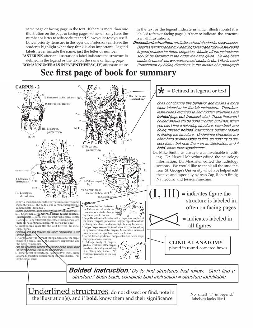
E. Short med. (radial) collateral lig.*
Synovial sacs
CARPUS - 2
III. Lt carpus, palmar view
synovial membranes form three synovial sacs correspond-ing to the joints. The middle and carpometacarpal joints communicate (distal two).Locate in pictures, don't dissect the following ligaments: E, F. Short medial (radial) and lateral (ulnar) collateral ligaments (II, III): only cross the antebrachiocarpal joint to stabilize it. Long collateral ligaments are lacking; therefore, there are no continuous ligaments over all the joints.
G. Interosseous space (II): the void between the meta-carpal bones.
Relocate and cut through the flexor retinaculum, if not already done.
H. Carpal canal (VI): formed by the palmar side of the carpal bones, the medial side of the accessory carpal bone, and the flexor retinaculum.
Pull the structures passing through the carpal canal aside to view the dorsal wall of the carpal canal:
I. Palmar carpal fibrocartilage/ligament (VI): thick, firmly attached connective tissue forming the smooth dorsal wall of the carpal canal.
D. Fibrous joint capsule*
CLINICAL:
V. Rt carpus, palmar view
A
B
C
IV. Lt carpus, dorsal view
R
Ca
Mc I
Mc V
R
B & C joints communicate
U
Ca
Mc I-V
R U
Mc V
Mc I
U4*
5*
6*
7*
F. Short lat. (ulnar) collateral lig.*
8*
VI. Carpus cross section (schematic)
a
b
e
H. Carpal canal*
c
df
h
iCr
Cu
CaI. Palmar carpal lig.*
jk
FE
g
• Communication between the 2 distal carpal joints be-comes important when block-ing the carpus in horses.
• Carpal luxation, subluxation of the carpal joint: rupture of the palmar carpal ligament and the joint capsule results in a plantigrade stance and nonweight bearing lameness.
• Puppy carpal weakness: insufficient exercises resulting in hyperextension of the carpus. Moderately increased exercise results in spontaneously resolution.
• Carpal flexion syndrome: puppies stand on flexed carpi, they spontaneous recover.
• Old age laxity of carpus: gradual weakness of the carpus in old and obese dogs, resulting in a plantigrade stance. No treatment is needed as the dog does fine.
= Defined in legend or text
CLINICAL ANATOMYplaced in round-cornered boxes
*
Underlined structures: do not dissect or find, note in the illustration(s), and if bold, know them and their significance
See first page of book for summary
Bolded instruction: Do to find structures that follow. Can't find a structure? Scan back, complete bold instruction = structure identifable
= indicates labeled in all figures
= indicates figure the structure is labeled in, often on facing pages
(I, III)
( )
same page or facing page in the text. If there is more than one illustration on the page or facing pages; some will only have the number or letter to reduce clutter and allow you to test yourself. Lower priority items are in the legends. Professors can have the students highlight what they think is also important. Legend labels never include the name, just the letter or number.
*ASTERISK after an illustration's label indicates the structure is defined in the legend or the text on the same or facing page.
ROMAN NUMERALS IN PARENTHESES (I, IV) after a structure
in the text or the legend indicate in which illustration(s) it is labeled (often on facing pages). Absence indicates the structure is in all illustrations.
Dissection instructions are italicized and shaded for easy access. Besides learning anatomy, learning to read and follow instructions is good practice for future surgeries. Ideally, all the instructions should be followed in the order they are given. Having been students ourselves, we realize most students don't like to read! Punishment by hiding directions in the middle of a paragraph
does not change this behavior and makes it more labor intensive for the lab instructors. Therefore, instructions required to find hidden structures are bolded (e.g., cut, transect, etc.). Those that aren't bolded should still be done in order, but if not, when you can't find a following structure, scan back and doing missed bolded instructions usually results in finding the structure. Underlined structures are often hard or impossible to find, so don't try to dis-sect them, but note them on an illustration, and if bold, know their significance.
Dr. Mike Smith, as always, was invaluable in edit-ing. Dr. Newell McArthur edited the neurology information. Dr. McAlsiter edited the radiology sections. We would like to thank all the students from St. George's University who have helped edit the text, and especially Adrean Zap, Robert Brady, Nat Goolik, and Jessica Franchini.
No small "l" in legend/ labels as looks like 1
Alaskan Malamute
American Eskimo Dog
Chinese Shar Pei
American Water Spaniel
Neopolitan Matiff Cairn Terrier
Basenji
Parson Russel Terrier
American English Coonhound
Maltese
Staffordshire Terrier
Shar Pei
(English) Mastiff
Itlian Spinone
Your diploma woun't include a new personality
- you're working on that now!