Embed Size (px)
Citation preview

Principles of Tooth Preparation
Rola Shadid, BDS, MSc, Associate Fellow AAID

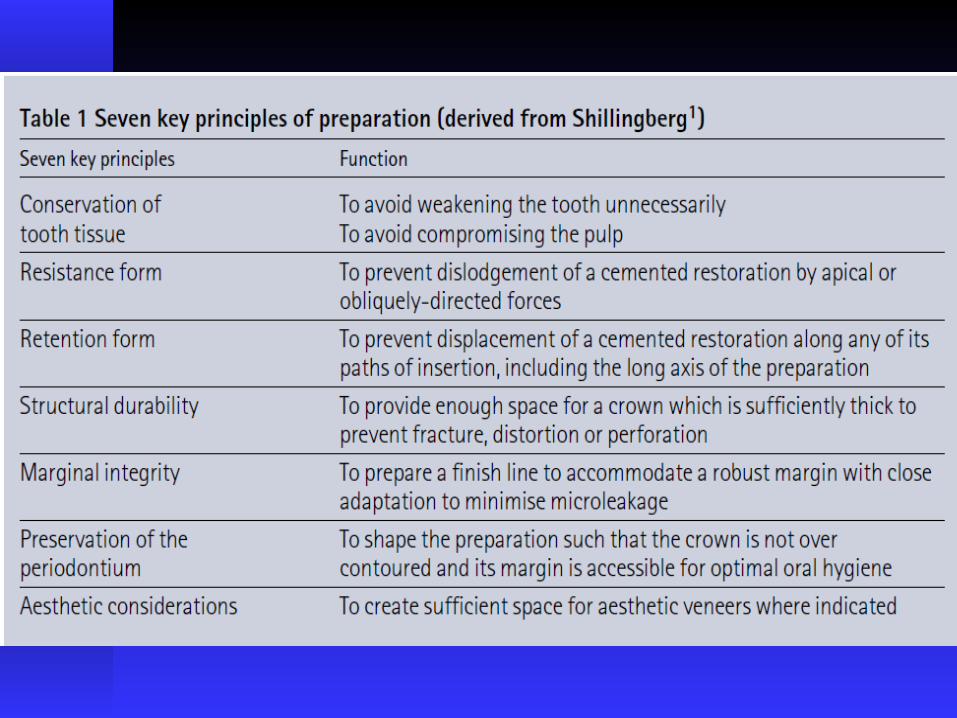

Tooth structure is conserved by using the following guidelines
1. Use of partial coverage rather than complete coverage restoration
2. Preparation of teeth with minimum practical convergence angle (taper) between axial walls
3. Preparation of the occlusal surface so that reduction follows the anatomic planes to give uniform thickness in the restoration
4. Preparation of the axial wall so tooth structure is removed evenly if necessary teeth should be orthodontically positioned
5. selection of conservative margin compatible with other principles of tooth preparation

Preservation of tooth structure
· Histologic studies showed that specimen with dentin thickness greater than 2 mm demonstrated very little or no pulp response , while when cutting was done within 1.5mm of the pulp, 23%of teeth developed an abscess

Reduction of axial walls with maximal thickness of remaining dentin surrounded the pulp

Preparation of tooth with minimal practical convergence angle (Taper) between axial walls
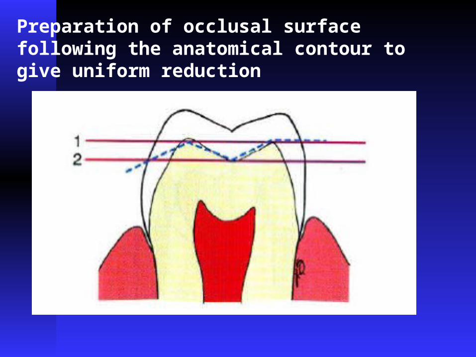
Preparation of occlusal surface following the anatomical contour to give uniform reduction

Selection of marginal geometry which is conservative and compatible with other principles

Avoidance of unnecessary apical extension of the preparation
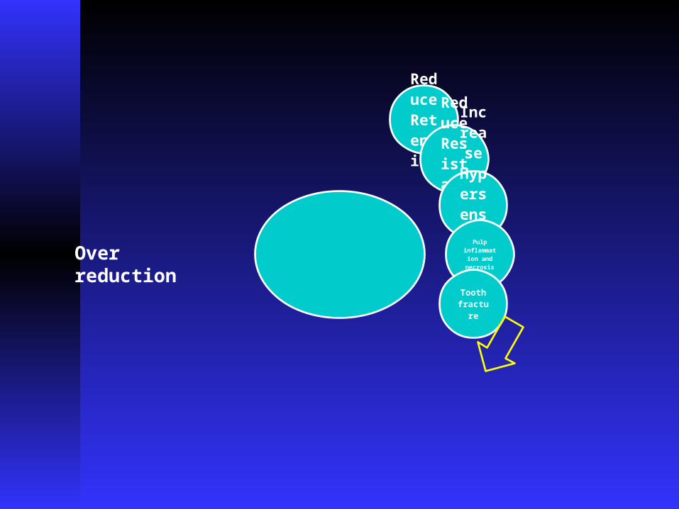
Reduce Retentio
n
Reduce Resistance
Increase HypersensitivityPulp inflammation and necrosis
Tooth fractu
re
Over reduction

Also you should be careful about:1- Adjacent tooth
- by using matrix band of the adjacent tooth
- cutting in the enamel of prepared tooth with
fine tapered stone
2- Soft tissues
- by using mirror or the flange of saliva ejector

Retention & Resistance

RETENTION FORM· The feature of a tooth preparation
that resists dislodgement of a crown in a vertical direction or along the path of placement.

RESISTANCE FORM· The features of a tooth preparation that
enhance the stability of restoration and resist dislodgement along an axis other than the path of placement
· It prevents dislodgement of a restoration by forces directed in an apical, oblique or horizontal direction.
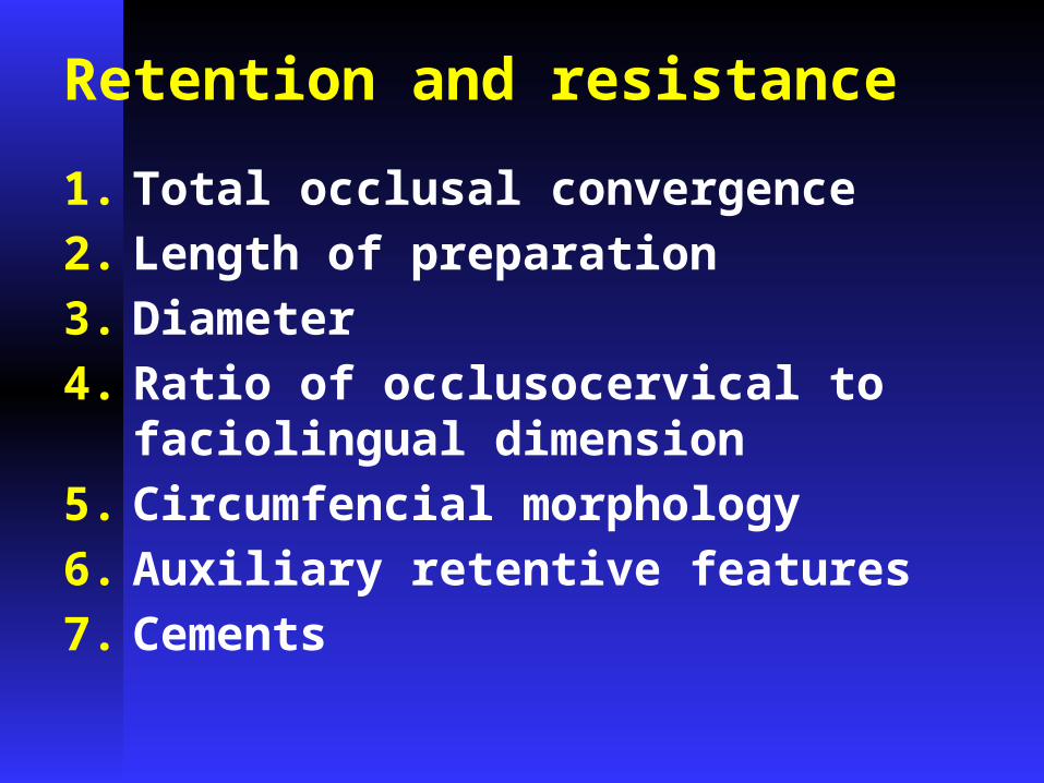
Retention and resistance
1. Total occlusal convergence2. Length of preparation3. Diameter 4. Ratio of occlusocervical to
faciolingual dimension 5. Circumfencial morphology 6. Auxiliary retentive features 7. Cements

Total occlusal convergence

Factors that may create greater total occlusal convergence
1. Posterior teeth are prepared with greater total occlusal convergence than anterior teeth
2. Mandibular teeth were prepared with greater convergence than maxillary teeth
3. Faciolingual surfaces had greater convergence than mesiodistal surfaces.
4. Abutments of fixed partial denture usually prepared with greater total occlusal convergence
5. Monocular vision created greater total occlusal convergence than binocular vision

Parallel walls are impossible to create in the mouth without producing preparation undercuts.
The axial walls of the preparationmust taper slightly to permit the restoration to seat.

· A tapered diamond or bur will impart an inclination of 2 to 3 degrees to any surface it cuts if the shank of the instrument is held parallel to the intended path of insertion of the preparation.
· Ideal convergence angle of 4-10° is seldom achieved
· Mack estimates that a minimum taper of 12 degrees is necessary Just to insure the absence of undercuts.

· A taper or total convergence of 16 degrees has been proposed as being achievable clinically while still affording adequate retention. It can be as low as 10 degrees on preparations anterior teeth and as high as 22 degrees on molars

· Resistance is more sensitive than retention to changes in convergence angle
· As a rule we will go for the minimal tapering without having undercuts in the preparation

SURFACE AREA VS. RETENTION
Provided the restoration has limited path of withdrawal, the greater the surface area of a preparation, the greater is its retention.
Length….. Crowns with long axial walls are more retentive…
Molar crowns are more retentive than premolar crowns of same taper
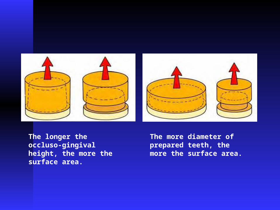
The longer the occluso-gingival height, the more the surface area.
The more diameter of prepared teeth, the more the surface area.

Length of preparation vs. resistance· Because the axial wall occlusal to the finish
line interferes with displacement, the length and inclination of that wall become factors in resistance to tipping forces.
· The shorter the wall, the more important its inclination
· The walls of shorter preparations should have as little taper a possible to increase the resistance.


Width of prep. vs resistance· A narrow tooth can have greater
resistance to tipping.· The preparation on the smaller tooth will
have a short rotational radius for the arc of displacement, and the incisal portion of the axial wall will resist displacement.
· The longer rotational radius on the larger preparation allows for a more gradual arc of displacement, and the axial wall does not resist removal

· Parker et al found that approximately 95% of anterior preparations analyzed had resistance form, while only 46% of those on molars did.


· Resistance to displacement for a short-walled preparation on a large tooth can be improved by placing grooves in the axial walls.
· In effect, this reduces the rotational radius, and that portion of the walls of the grooves near the occlusal surface of the preparation will interfere with displacement

Ratio of occlusocervical to faciolingual dimension
· The longer the faciolingual dimension of prepared molars compared with other teeth and shorter occlusogingival dimension compared with anterior teeth produce a lower ratio and lower resistance to dislodgment of molar crown

· Weed and Baez recommended that the occlusocervical / faciolingual ratio should be 0.4 mm or higher for all teeth

STRESS CONCENTRATION
· If line angle between axial and occlusal surface is sharp, it leads to concentration of stresses around that junction
· Induced stresses exceeds the strength of the cement
· Leads to cohesive failure of cement
TYPE OF RESTORATION VS RETENTION
· Full veneer crown has excellent retention when compared to partial veneer crown because reducing the path of insertion to a narrow range.

TYPE OF RESTORATION VS RESISTANCE
· Partial coverage restoration may have less resistance than a complete crown because it has no buccal resistance area

Surface Roughness
· Adhesion of dental cements depends primarily on projections of the cement into microscopic irregularities.
· Jorgensen found retention of castings cemented with ZnPO4 cement on test dies with a 10° taper to be twice as great on preparations with 40µm scratches than 10µm.
· Retention increases when restoration is roughened or grooved

Materials being cemented vs retention· Retention is affected by both the casting
alloy and the core material. · More reactive the alloy is more adhesion.· Base metal alloys are better retained
than less reactive high gold content metals.
· Type of luting agent: Studies show that adhesive resin cements are more retentive than conventional ZnPO4 and GIC cements

PHYSICAL PROPERTIES OF LUTING AGENT VS RESISTANCE
· Resistance to deformation is affected by physical properties of the luting agent, such as compressive strength and modulus of elasticity
· Adhesive resin >GIC > ZnPO4> Polycarboxylate > ZOE
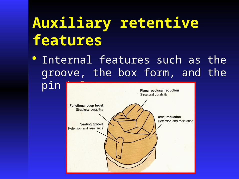
Auxiliary retentive features · Internal features such as the groove, the box
form, and the pin hole
· Secondary retentive features does not significantly affect the retention because the surface area is not increased significantly.
· But where these features limits the path of withdrawal, retention is increased


· Kent et al reported a marked difference between the degree of taper of full crown preparations (18.4 to 22.2 degrees) and that of boxes and grooves in the axial surfaces of those preparations (7.3 degrees).

· Kent et al found that grooves and boxes had less convergence than the convergence of the axial wall to enhance resistance
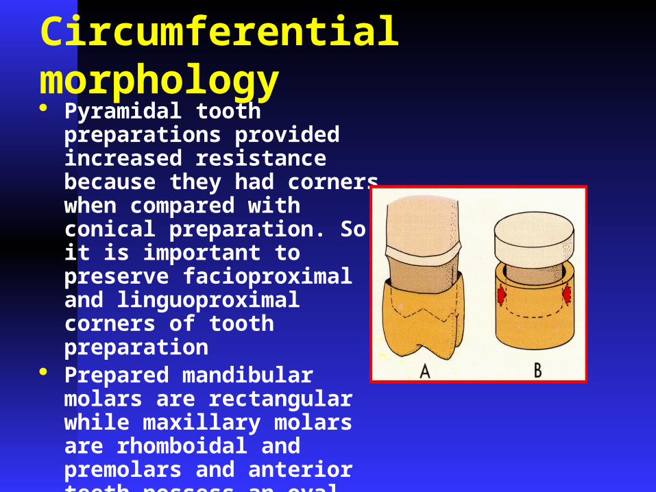
Circumferential morphology · Pyramidal tooth
preparations provided increased resistance because they had corners when compared with conical preparation. So it is important to preserve facioproximal and linguoproximal corners of tooth preparation
· Prepared mandibular molars are rectangular while maxillary molars are rhomboidal and premolars and anterior teeth possess an oval form
Path of Insertion
· It is an imaginary line along which the restoration will be placed onto or removed from the preparation.
· It is of special importance when preparing teeth to be fixed partial denture abutments, since the paths of all the abutment preparations must parallel each other.


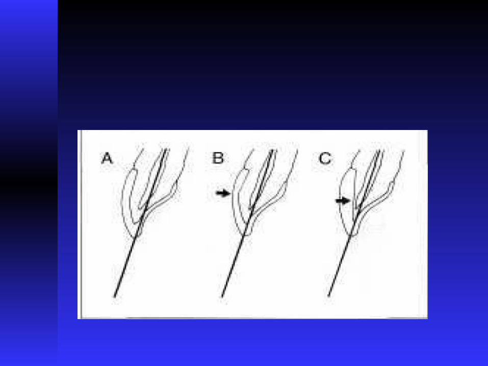
Path of insertion
Parallel to the long axis of the tooth
Parallel to the incisal 2/3 of the tooth

The path of insertion must be considered in two dimensions:· Faciolingually: the faciolingual
orientation of the path can affect the esthetics of metal-ceramic or partial veneer crowns.
· Mesiodistally


Structural durability
· A restoration must have sufficient strength to prevent permanent deformation during function
· “The ability of a restoration to withstand destruction due to external forces “

Structural durability
· Adequate tooth reduction- occlusal reduction - functional cusp bevel - axial reduction
· Alloy selection· Metal-ceramic framework design· Margin design

Occlusal reduction
· Zurcherman has shown that the placement of inclined planes on occlusal surfaces of a crown preparation rather than flat surface increases resistance form

Functional cusp bevel
· This includes wide bevel on the lingual surface inclines of maxillary lingual cusps and buccal inclines of mandibular buccal cusps, this will provide space for adequate bulk of metal in an area of heavy occlusal contact


Axial reduction

Margin Placement

Biologic widthIt is the dimension of space that the healthy gingival tissues occupy above the alveolar bone. It refers to the combined connective tissue-epithelial attachment from the crest of the alveolar bone to the base of the sulcus (2mm;connective tissue-1.07mm and epithelium-0.97mm).
Evaluation of the biological width by radiographs, probing, and sounding of bone

Location of restorative margins
· Supragingival · Equigingival · Subgingival


Indications for subgingival margins

Where to place subgingival margin ?
· Wearhang reported that a conventional tooth brush could remove plaque only to a point 0.5mm subgingivally, so it is important to ensure that restorations are placed no deeper than 0.5mm into sulcus when this is possible

· It is generally taught that the gingival margin should never enter the sulcus by more than half the depth of the sulcus. In the average case this equals a depth of 1.0 mm or less, with less being favored
Where to place subgingival margin ?

· No margin should be placed nearer than 0.5 to 1.0 mm from the attachment area in healthy sulcus
· Some authors suggested that restorative margins should end 3-4 mm coronal to the alveolar crest, they assumed the biologic width is 2mm and additional 1mm will keep the margin 1mm above the coronal extent of junctional epithelium
· Gingival attachment is more reliable reference point than marginal gingiva
Where to place subgingival margin ?

Technique
· Packing a small length of retraction cord in the area of the sulcus to receive the subgingival margin works very well.
· Make a mental note of the relative relationship of the free gingival margin and roughed-in finish line prior to packing the cord. Even small cords often achieve remarkable gingival retraction. You may not need to place the margin as far apically as the level of the cord.

Technique
· The lingual and approximately half of the interproximal margins should remain supragingival (if possible).
· The labial and labial one-half of the interproximal margins are brought down to or near the level of the retraction cord.
· Be specifically aware of keeping the interproximal margin subgingival until it will no longer be visible when viewed at an angle. This is a frequent area of margin visibility, especially for porcelain veneers.

oD.A. Orkin et al and J.Valderhang et al found out that there was significant difference in bleeding between the subgingival crowns and contralateral teeth without crowns but the difference was negligible with supragingival crowns
· Hatchy J. Sttler concluded that in the presence of subgingival restoration, the degree of gingival inflammation is significantly greater in association with narrow (less than 2mm ) zones of keratinized gingiva than with those greater than 2mm

The extent of disease depends on:· contour of restoration, · relative position of cervical
margin, · precision of fit, · restorative material used

Marginal contours : Lang et al reported that overhanging margin not only accumulate more plaque but the plaque undergoes a change in composition to that usually associated with destructive periodontitisSurface roughness :Donnan and Princeevaluated the plaque accumulation in metal
ceramic restoration and discovered the order of decreasing plaque accumulation:
Aluminum oxide (greatest), opaque porcelain, polished metal, glazed porcelain (least)
In term of smoothness (generally):glazed porcelain > gold > polished amalgam
>compositeMarginal fit : the severity of periodontal disease was elevated with greater subgingival marginal discrepancy
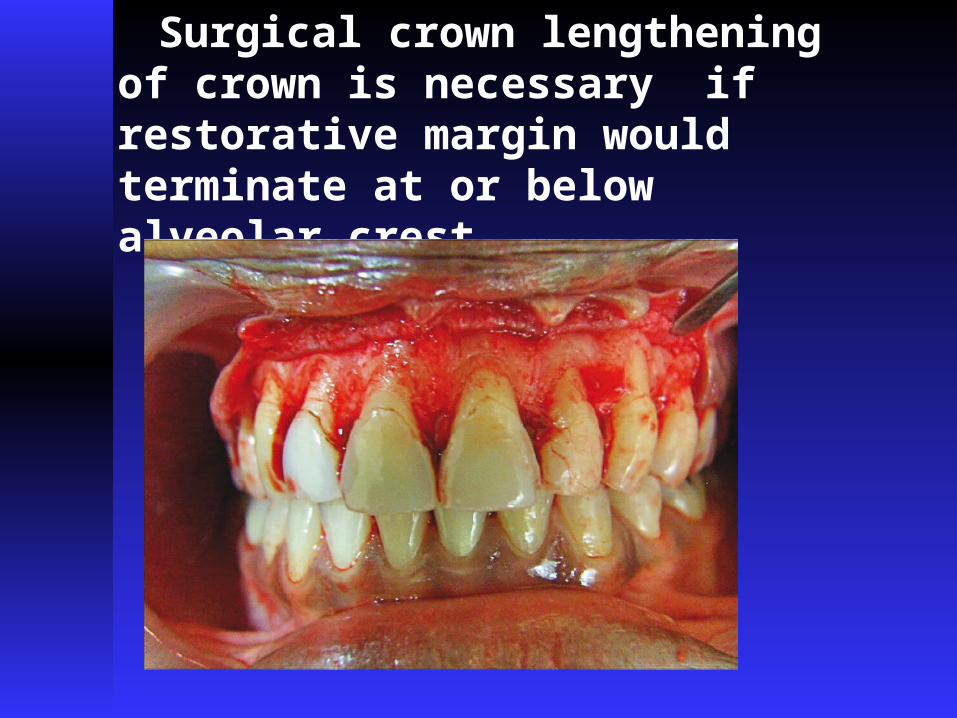
Surgical crown lengthening of crown is necessary if restorative margin would terminate at or below alveolar crest

If restorative margin would terminate at or below alveolar crest surgical lengthening of crown is necessary , the procedure involves reflection of full thickness flap to expose alveolar crest around the teeth in question, the distance from the bone crest to margin of tooth preparation can be measured with periodontal probe, the distance should be 3-4 mm if the bony crest is not reduced. At least 6-8 weeks must elapse to allow for proper healing.


MARGINAL GEOMETRY OR FINISH LINE CONFIGURATION

The margin design depends on:
· type of restoration ,· tooth morphology, · position and alignment of the teeth
in the arch, · esthetics

Guidelines for margin design
· Ease of preparation without overextension or unsupported enamel
· Ease of identification in the impression and on the die· A distinct boundary to which wax pattern can be finished· Conservation of tooth structure· Sufficient bulk of material for esthetic and strength of the
restoration.· The most important consideration in selecting a cervical
margin design is its ability to consistently and predictably provide excellent marginal integrity.

Featheredge Chisel Chamfer Bevel Shoulder Sloped Shoulder
Beveled Shoulder

The shoulderless (featheredge) margin
· Conservation of the tooth structure · Permits an acute margin of the metal· Insufficient removal of tooth structure at
cervical area. (Results in overcontouring )· Impossible to identify the margin of prep.· No control over reduction of cervical tooth
structure· No control in placem. of subgingival margin· Poor resistance to marginal distortion during
firing of porcelain to the gold alloy.· difficult to accurately wax and cast · more susceptible to distortion in the mouth
when the casting is subjected to occlusal forces

Indications for featheredge
· Not recommended· It may have to be
used on the lingual surface of mandibular posterior teeth,
· On teeth with very convex axial surfaces, and on the surface toward which a tooth may have tilted.
· Could be Used for the full metal crown prep

The shoulder (Butt joint) margin
· Adequate
removal of tooth structure at cervical area.
· It is possible to identify the margin of prep.
· Good control over reduction of cervical tooth structure

The shoulder (Butt joint) margin· Control in
placem.of subgingival margin
· Adequate resistance to marginal distortion during firing of porcelain to the metal alloy.
· It does require the destruction of more tooth structure than any other finish line.

The shoulder (Butt joint) margin· Finish line of choice for the all-ceramic
crown and porcelain labial margin· The wide ledge provides resistance to
occlusal forces and minimizes stresses that might lead to fracture of the porcelain. It produces the space for healthy restoration contours and maximum esthetics.

The shoulder (Butt joint) margin· The sharp, 90-degree
internal line angle associated with the classic variety of this finish line concentrates stress in the tooth and is conducive to coronal fracture.

The radial shoulder
· A modified form of shoulder finish line· A small-radius rounded internal angle is
instrumented by an end-cutting parallel sided carbide finishing bur, and finishing is completed with a specially modified bin-angle chisel.
· The cavosurface angle is 90 degrees, and shoulder width is only lessened by the rounded internal angle.


135 sloping shoulder
Very useful preparation to use where the labial shoulder for MC crowns is extending well into the root face, e.g. canines with marked gingival recession. The finishing line can be placed sub-gingivally and the axio-gingival floor line angle left at a higher level.
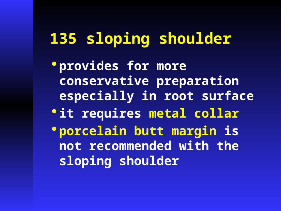
135 sloping shoulder
· provides for more conservative preparation especially in root surface
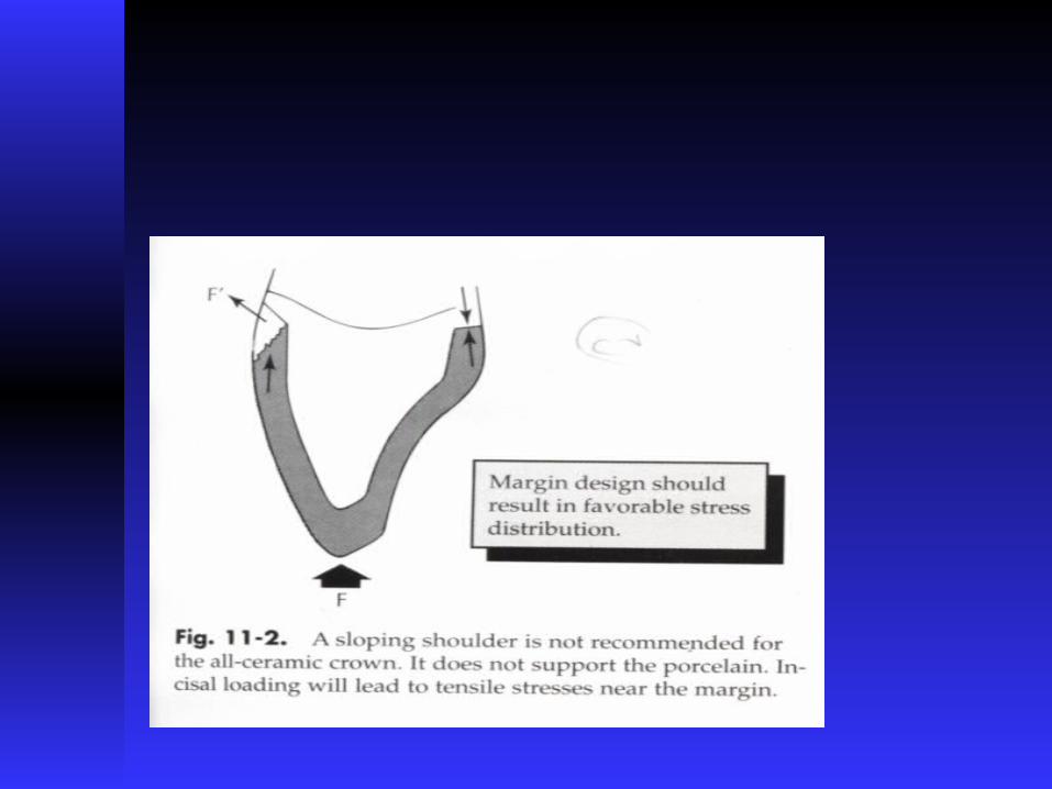
· it requires metal collar· porcelain butt margin is not recommended with the sloping shoulder

The shoulder-bevel· It is obtained by preparing
shoulder at the height of healthy gingival margin, then adding 0.5-1.25 mm bevel.
· It has the same advantages of straight shoulder with optimum opportunity of marginal fit provided by the bevel

· The beveling removes unsupported enamel, may allow some finishing of the metal, & minimize margin discrepency….
· Used for ceramo-metal, full metal

· This design can also be used for the facial finish line of metal-ceramic restorations where gingival esthetics are not critical. It can be used in those situations where a shoulder is already present, either because of destruction by caries or the presence of previous restorations.
· It is also a good finish line for preparations with extremely short walls, since it facilitates axial walls that are nearly parallel.


A shoulder or sloped shoulder is preferred to shoulder with bevel for ceramometal restorations due to biological & esthetic considerations (the metal margin can be thinned to a knife edge & hidden in the sulcus without the need for positioning the margin closer to the epithelial attachment)

Chamfer Conservative
type when compared with shoulder finish line.
control over reduction of cervical tooth structure,
control in placem.of subgingival margin

Chamfer
· Margin of prep.is distinct
· Degree of marginal distortion during firing of porcelain directly related to the thickness of metal at the margin

Chamfer· The preferred gingival
finish line for full veneer metal restoration and the metal only portion of MC crown

Chamfer
· Porcelain margin is not recommended because it will lack mechanical resistance and depth of translucency

√
X
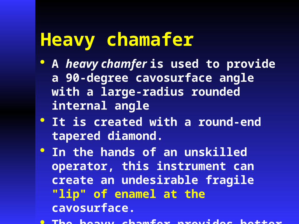
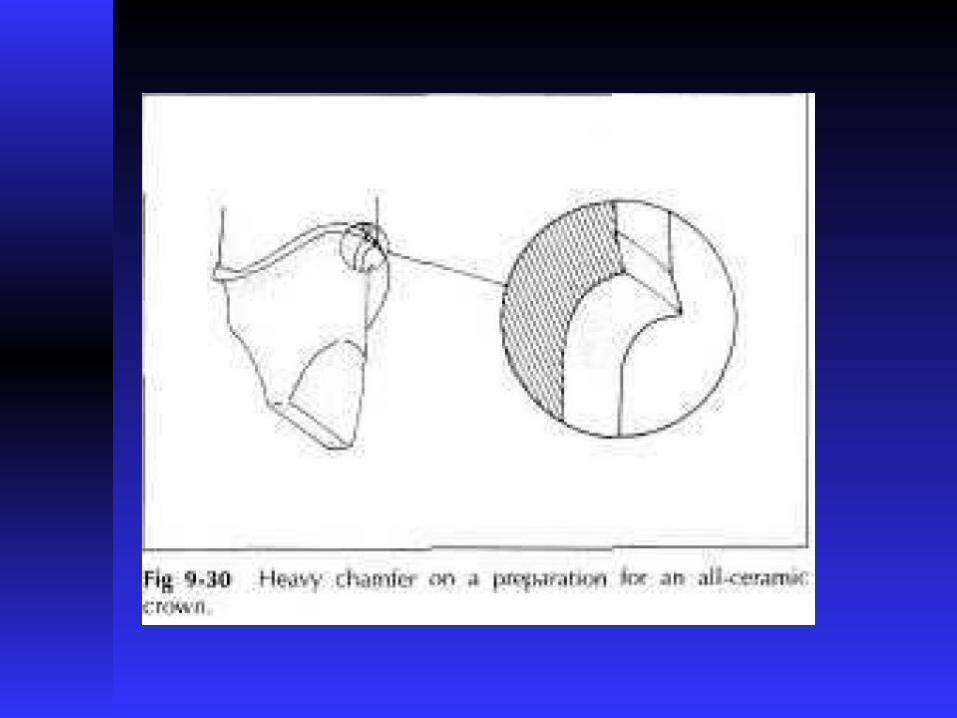
Heavy chamafer· A heavy chamfer is used to provide a 90-degree
cavosurface angle with a large-radius rounded internal angle
· It is created with a round-end tapered diamond.
· In the hands of an unskilled operator, this instrument can create an undesirable fragile "lip" of enamel at the cavosurface.
· The heavy chamfer provides better support for a ceramic crown than does a conventional chamfer, but it is not as good as a shoulder.


Margin forms for MC · Metal collar · Metal feather –edge
(Disappearing margin)· Porcelain margin

Margin forms for MC


Metal collar
Advantages :1. good marginal seal2. preservation of periodontal health3. rigidity during cementation4. wide facial metal collar (.8mm) gives
sufficient rigidity against distortion caused by porcelain shrinkage in comparison of the feather edge collar
5. can be used with any of the finish lines described previously

Disadvantages :
1. difficult to conceal in a shallow crevice or with a thin translucent gingival margin
2. they display in case of gingival recession
3. display of metal becomes very clear with high lip line

Metal feather–edge (Disappearing margin)
· Reduction of the labial metal collar has been described as: feather – edge , triangular formation , hairline collar .
· The metal and opaque layer porcelain meet simultaneously on the external edge of the tooth preparation
· Shoulder preparation is needed for this design to provide rigidity of metal in the cervical area


Disadvantages : 1. the design is technique sensitive and
difficult to achieve without overcontouring the cervical aspect or exposing opaque layer
2. difficult finishing and polishing , microscopically the surface remains rough
3. marginal adaptation after porcelain firing is subject to some distortion ,

Porcelain marginAdvantages:
1. esthetic improvement because of :
a. facial metal collar elimination
b. depth in cervical translucency
c. possibility of light transmission
through the root area
2. less plaque accumulation than metal because of low adhesive forces between plaque and ceramics

· porcelain margin is used with shoulder finish line (1.2mm wide ) internally rounded at a 90-100 angle to root surface with regular and smooth outline

· chamfer and sloping shoulder finish lines are contraindicated with porcelain margin because :
a. porcelain margin would be very thin and prone
to chipping
b. difficult to achieve satisfactory marginal
adaptation because porcelain shrinkage occurs
toward the greatest bulk during firing

Porcelain construction :· various techniques of porcelain margin
construction have been described using :
platinum matrix , refractory dies , separating varnish , wax, or resin binders
· with conventional porcelain margin materials rounded edges with rough and heterogeneous surfaces were more likely to occur using direct lift off technique than platinum matrix substrates
For all metal restorations, chamfer finish line are frequently used the advantages of chamfer finish line
1. They produce the less stress and marginal opening of metal
2. They are easy to form with a tapered round end diamond instrument
3. They posses adequate bulk for restoration4. They are visible on prepared teeth 5. Their depth is sufficient to permit the
concept of normal axial contour .

Standard metal-ceramic crown involving only the anatomical crown and where aesthetics is of primary importance
Labial – Flat shoulder (90°)
Lingual – Deep chamfer with metal collar


ANTERIOR METAL-CERAMIC CROWNS
A UNIFORM
REDUCTION OF
APPROXIMATELY
1.2 MM IS NEEDED
OVER THE ENTIRE
FACIAL SURFACE.

ANTERIOR METAL-CERAMIC CROWNS
TO ACHIEVE ADEQUATE REDUCTION WITHOUT ENCROACHING UPON THE PULP – FACIAL SURFACE PREPARED IN TWO PLANES THAT CORRESPOND ROUGHLY TO THE TWO GEOMETRIC PLANES PRESENT ON THE FACIALSURFACE OF AN UNCUT TOOTH

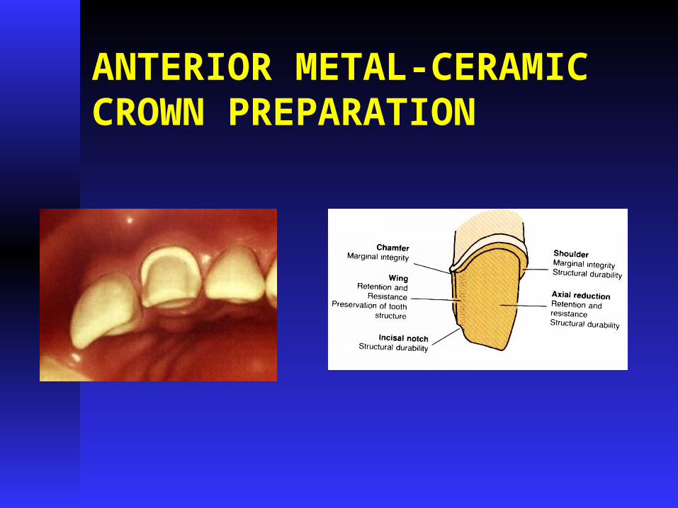
ANTERIOR METAL-CERAMIC CROWNS
FACIAL SURFACE
PREPARED IN ONE
PLANE THAT HAS
ADEQUATE FACIAL
REDUCTION IN THE
GINGIVAL ASPECT:
Inadequate space for a
sufficient thickness of
ceramic material- Poorly
contoured restoration affecting
both esthetic & health of the surrounding gingiva.

ANTERIOR METAL-CERAMIC CROWNS
FACIAL SURFACE
PREPARED IN ONE
PLANE THAT HAS
ADEQUATE FACIAL
REDUCTION IN THE
INCISAL ASPECT- FACIAL
SURFACE OVERTAPERED
AND TOO CLOSE TO THE
PULP.
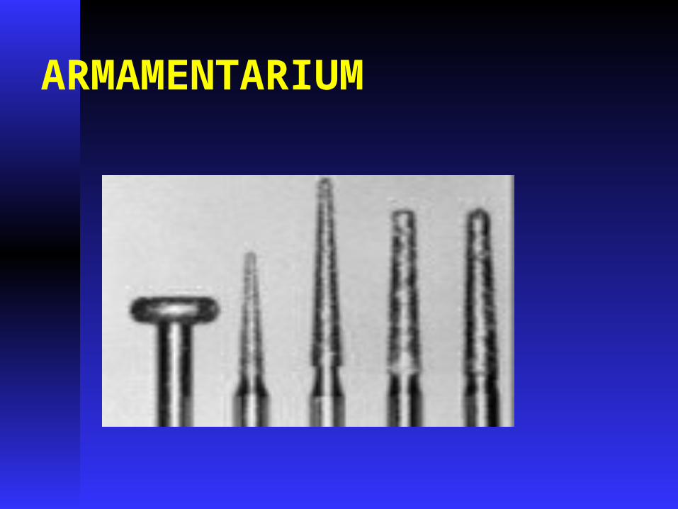
ARMAMENTARIUM

ANTERIOR METAL-CERAMIC CROWNSSILICONE INDEX
MADE BEFORE
TOOTH
PREPARATION
TOOTH BADLY
BROKEN DOWN,
INDEX MADE ON WAXED
UP DIAGNOSTIC CAST.

ANTERIOR METAL-CERAMIC CROWN PREPARATIONPLACEMENT OF DEPTH ORIENTATION GROOVES - ( 1.2MM )THE LABIAL GROOVES CUT IN TWO SETS1. ONE SET PARALLEL
WITH THE GINGIVAL HALF OF LABIAL SURFACE
2. ONE SET PARALLEL WITH THE INCISAL HALF OF LABIAL SURFACE

ANTERIOR METAL-CERAMIC CROWN PREPARATION

ANTERIOR METAL-CERAMIC CROWN PREPARATION

ANTERIOR METAL-CERAMIC CROWN PREPARATION
INCISAL REDUCTION-
(2MM)
ROUND –END
TAPERED DAIMOND.

ANTERIOR METAL-CERAMIC CROWN PREPARATION
Inadequate incisal reduction results in poor incisal translucency

ANTERIOR METAL-CERAMIC CROWN PREPARATION
LABIAL REDUCTION
(INCISAL HALF)
ROUND- END
TAPERED DAIMOND.

ANTERIOR METAL-CERAMIC CROWN PREPARATION
LABIAL REDUCTION
(GINGIVAL HALF)
ROUND-END
TAPERED DAIMOND
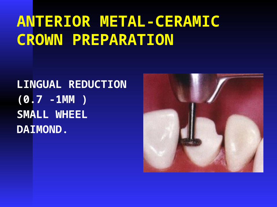
ANTERIOR METAL-CERAMIC CROWN PREPARATION
LINGUAL REDUCTION
(0.7 -1MM )
SMALL WHEEL
DAIMOND.
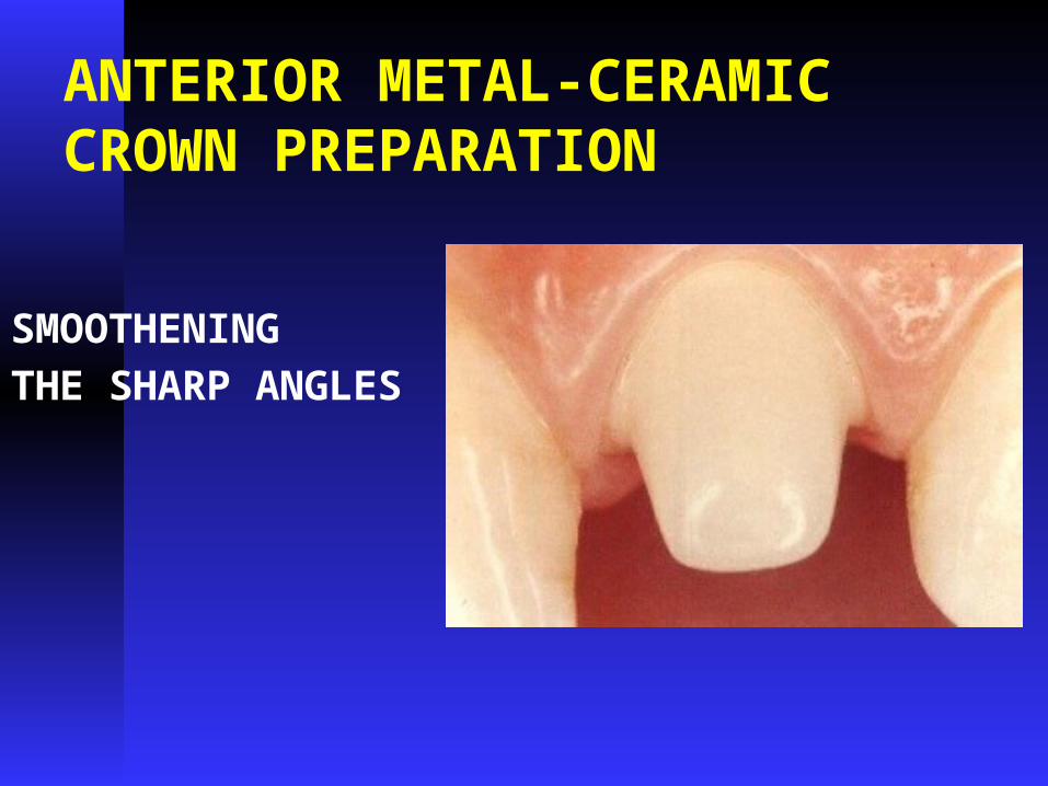
ANTERIOR METAL-CERAMIC CROWN PREPARATION
SMOOTHENING
THE SHARP ANGLES

ANTERIOR METAL-CERAMIC CROWN PREPARATION
RADIAL SHOULDER
MODIFIED FORM OF
SHOULDER
SMALL RADIUS
INTERNAL ANGLE
WITH 90-DEGREE
CAVOSURFACE
ANTERIOR METAL-CERAMIC CROWN PREPARATION
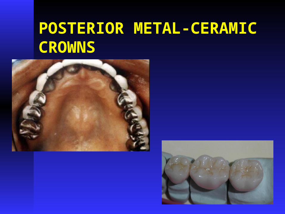
POSTERIOR METAL-CERAMIC CROWNS

POSTERIOR METAL-CERAMIC CROWNS
STEP NO : 1
OCCLUSAL REDUCTION
FOLLOWED BY
FUNCTIONAL CUSP
BEVEL
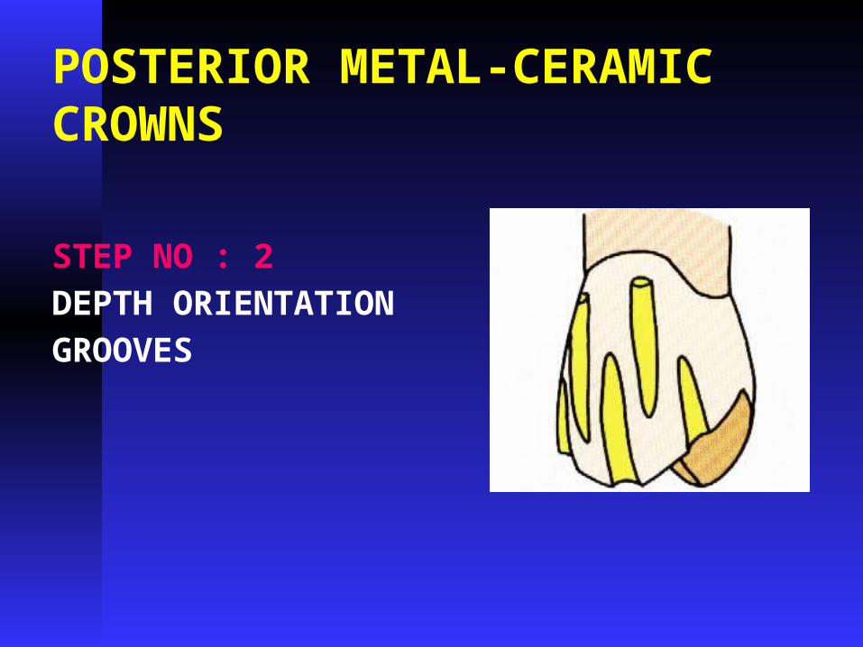
POSTERIOR METAL-CERAMIC CROWNS
STEP NO : 2
DEPTH ORIENTATION
GROOVES

POSTERIOR METAL-CERAMIC CROWNS
STEP NO :3
FACIAL REDUCTION-
OCCLUSAL HALF
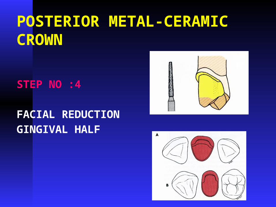
POSTERIOR METAL-CERAMIC CROWN
STEP NO :4
FACIAL REDUCTION
GINGIVAL HALF

POSTERIOR METAL-CERAMIC CROWN
STEP NO: 5
PROXIMAL
AXIAL
REDUCTION

POSTERIOR METAL-CERAMIC CROWN
STEP NO :6
LINGUAL AXIAL
REDUCTION

POSTERIOR METAL-CERAMIC CROWN
STEP NO : 7
AXIAL FINISHING

POSTERIOR METAL-CERAMIC CROWN

POSTERIOR METAL-CERAMIC CROWN

POSTERIOR METAL-CERAMIC CROWN
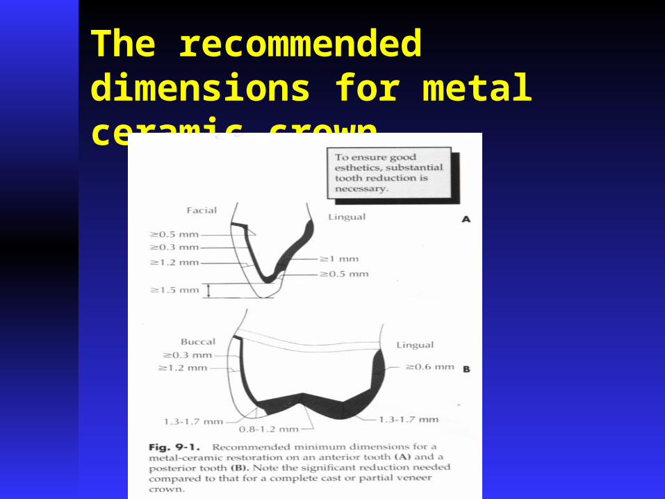
The recommended dimensions for metal ceramic crown

Metal ceramic crown prep.

Remove any unsupported enamel

Avoid traumatizing gingiva during subgingival preparation


Common Faults in Preparation· Insufficient removal of buccal or labial enamel
particularly at the labio-incisal one-third of the preparation
· Insufficient removal of occlusal enamel in posterior teeth particularly at the cusp tips
· Insufficient removal of lingual enamel which may force the ceramist to widen his occlusal table or reduce the gold coping thickness which can increase the risk of metal deformation.

Common Faults in Preparation· Failure to round off all internal line and point
angles, thereby creating stress concentration areas which may cause “pop-off” of the porcelain veneer.
· Flattening occlusal tables in the preparation instead of following the line of the cusp angles.
· Inadequate removal of approximal enamel, particularly on the front teeth, leaving insufficient space for metal and porcelain at the cervical third of the tooth.

Some guidelines for preparing teeth for metal ceramic complete crown:· The total occlusal convergence
should range between 10 and 20 degrees.
· The minimal occlusocervical dimension of molars should be 4 mm when prepared with 10 to 20 degrees total occlusal convergence.
· Many molars need auxiliary grooves or boxes to enhance resistance form.
· Axial grooves/boxes should be used routinely with mandibular molars .
· When tooth conditions and esthetics permit, finish lines should be located supragingivally.
· Check the sub-gingival margins for any deposits of calculus. These must be removed prior to taking the impression. In particular, calculus in the approximal regions is much more easily removed at the time of crown preparation and often can be present despite careful pre-operative prophylaxis.

· Immediate dentine sealing of prepared teeth with a dentine bonding agent (DBA) removes the smear layer, seals patent dentine tubules, halts bacterial ingress, reduces postoperative sensitivity and results in superior bonding of the definitive restoration when using a resin-based cement.

References
· Principles of Tooth Preparations ; Preparations for Full Coverage Crowns, Fundamentals of Fixed prosthodontics, 4th Ed. Shillingburg, Quintessence publishing.


![[PPT]Anatomy of Maxillary Denture Bearing Areadrrolashadid.weebly.com/uploads/1/4/9/4/14946992/lecture... · Web viewAnatomy of Maxillary Denture Bearing Area Rola M. Shadid, BDS,](https://img.dokumen.tips/doc/110x75/5b0197827f8b9a6a2e8e71d0/pptanatomy-of-maxillary-denture-bearing-viewanatomy-of-maxillary-denture-bearing.jpg)