Embed Size (px)
Citation preview

Contents
Preface .......................................................................... vii
Drug dosage and nomenclature ................................... ix
SeCTioN 1 General principles
1. Principles of pharmacology and mechanisms of drug action ................................. 3
2. Pharmacokinetics .............................................. 31 3. Drug discovery, safety and efficacy .................. 63 4. The nervous system, neurotransmission
and the peripheral autonomic nervous system ............................................................... 71
SeCTioN 2 The cardiovascular system
5. Ischaemic heart disease .................................... 91 6. Systemic and pulmonary hypertension ........... 111 7. Heart failure ..................................................... 133 8. Cardiac arrhythmias ........................................ 145 9. Cerebrovascular disease and dementia .......... 16310. Peripheral vascular disease ............................. 17111. Haemostasis .................................................... 177
SeCTioN 3 The respiratory system
12. Asthma and chronic obstructive pulmonary disease ............................................................. 195
13. Respiratory disorders: cough, respiratory stimulants, cystic fibrosis and neonatal respiratory distress syndrome ......................... 211
SeCTioN 4 The renal system
14. Diuretics ........................................................... 21915. Disorders of micturition ................................... 23116. Erectile dysfunction ......................................... 239
SeCTioN 5 The nervous system
17. General anaesthetics ....................................... 24718. Local anaesthetics ........................................... 25719. Opioid analgesics and the management
of pain .............................................................. 26320. Anxiolytics, sedatives and hypnotics .............. 27721. The major psychotic disorders:
schizophrenia and mania ................................ 28722. Depression, attention deficit hyperactivity
disorder and narcolepsy .................................. 29923. Epilepsy ........................................................... 31524. Extrapyramidal movement disorders and
spasticity .......................................................... 32725. Other neurological disorders: multiple
sclerosis, motor neuron disease and Guillain–Barré syndrome ................................. 341
26. Migraine ........................................................... 345
SeCTioN 6 The musculoskeletal system
27. The neuromuscular junction and neuromuscular blockade ................................. 355
28. Myasthenia gravis ............................................ 36129. Non-steroidal anti-inflammatory drugs ........... 36530. Rheumatoid arthritis, other inflammatory
arthritides and osteoarthritis ........................... 37731. Hyperuricaemia and gout ................................ 387
SeCTioN 7 The gastrointestinal system
32. Nausea and vomiting ....................................... 39533. Dyspepsia and peptic ulcer disease ............... 40334. Inflammatory bowel disease ............................ 41535. Constipation, diarrhoea and irritable
bowel syndrome .............................................. 42136. Liver disease .................................................... 42937. Obesity ............................................................. 437

vi Contents
SeCTioN 8 The immune system
38. The immune response and immunosuppressant drugs .............................. 445
39. Antihistamines and allergic disease ................ 455
SeCTioN 9 The endocrine system and metabolism
40. Diabetes mellitus ............................................. 46341. The thyroid and control of metabolic rate ...... 47742. Calcium metabolism and metabolic bone
disease ............................................................. 48343. Pituitary and hypothalamic hormones ............ 49344. Corticosteroids (glucocorticoids and
mineralocorticoids) .......................................... 50545. Female reproduction ....................................... 51546. Androgens and anabolic steroids ................... 53547. Anaemia and haematopoietic
colony-stimulating factors ............................... 54148. Lipid disorders ................................................. 551
SeCTioN 10 The skin and eyes
49. Skin disorders .................................................. 56550. The eye ............................................................ 575
SeCTioN 11 Chemotherapy
51. Chemotherapy of infections ............................ 58752. Chemotherapy of malignancy ......................... 641
SeCTioN 12 General features: toxicity and prescribing
53. Drug toxicity and overdose ............................. 67154. Substance abuse and dependence ................ 68755. Prescribing, adherence and information
about medicines .............................................. 69956. Drug therapy in special situations ................... 703
Index .......................................................................... 715

Preface
n A structured approach to the principles of disease management, outlining core principles of drug choice and planning a therapeutic regimen for many common diseases.
n An updated and reorganised drug compendium giving details of most drugs in the classes discussed in the chapter that have not been used as specific examples. For easy reference these tables set out key similarities and differences among drugs in each class and comple-ment the information provided in the chapter.
n Additional and new style self- assessment exercises and case studies to enable the reader to test their understanding of the principles covered in each chapter.
Chapters covering generic concepts of pharmacology and therapeutics have been extensively updated and simplified and include: how drugs work at a cellular level, drug development, drug metabolism and pharmacokine-tics, the autonomic nervous system, drug toxicity and drug prescribing. Information about genetic variations in both drug handling and drug responses, areas of increasing interest in drug development, has been expanded.
It is our intention that the third edition of this book will encourage readers to develop a deeper understanding of the principles of drug usage that will help them to become safe and effective prescribers and to carry out basic and clinical research and to teach. As medical science advances these principles should underpin the life-long learning essential for the maintenance of these skills.
DGWAGR
KH
The third edition of Medical Pharmacology and Thera peutics has been extensively revised and updated while preserving the widely popular approach of the second edition. The text is structured to reflect the ways that drugs are used in clinical practice. It provides information suitable for all healthcare professionals who require a sound knowledge of the basic science and clinical applications of drugs.
As before, a disease-based approach has been taken wherever possible with the aim of explaining clinical pharmacology and therapeutics and the principles of drug use for the management of common diseases. Medical Pharmacology and Therapeutics provides sound basic pharmacology background material sufficient to underpin the clinical context. New sections on pulmonary hyper-tension, attention deficit hyperactivity disorder, narcolepsy and macular degeneration have been added, reflecting the growing therapeutic options in these conditions. Many of the diagrams are new or modified to further clarify complex areas of pharmacology.
Each chapter in this Third Edition includes:
n An updated and succinct explanation of the major path-ogenic mechanisms of the disease and consequent clinical symptoms and signs, helping the reader to put into context the actions of drugs and the consequences of their therapeutic use.
n An updated comprehensive review of major drugs classes relevant to the disease in question. Example drugs are used to illustrate pharmacological principles and to introduce the reader to drugs currently in wide-spread clinical use.
n Basic pharmacology and mechanisms of drug action, key pharmacokinetic properties and important unwanted effects associated with individual drugs and drug classes.

Drug dosage and nomenclature
Drug nomenclatureIn the past, the non-proprietary (generic) names of some drugs have varied from country to country, leading to potential confusion. Progressively, international agreement has been reached to rationalise these variations in names and a single recommended International Non-proprietary Name (rINN) given to all drugs.
Where the previously given British Approved Name (BAN) and the rINN have differed, the rINN is now the accepted name and is used through this book.
A source of minor irritation, however, is that in most authoritative publications issuing from the UK the internationally accepted name is still being called its BAN or new BAN, and this is likely to continue. For full information on this, the reader is referred to:
http://www.mhra.gov.uk/Howweregulate/Medicines/Namingofmedicines/ChangestomedicinesnamesBANstorINNs/CON009669
A special case has been made for two medicinal substances: adrenaline (rINN – epinephrine) and noradrenaline (rINN – norepinephrine). Because of the clinical importance of these substances and the widespread European use and under-standing of the terms adrenaline and noradrenaline, manufacturers have been asked to continue to dual-label products adrenaline (epinephrine) and noradrenaline (norephinephrine). In this book where the use of these agents as administered drugs is being described dual names are given. In keeping with European convention, however, adrenaline and noradrena-line alone are used when referring to the physiological effects of the naturally occurring substances.
Drug dosagesMedical knowledge is constantly changing. As new information becomes available, changes in treatment, procedures, equipment and the use of drugs become necessary. The authors and the publishers have taken care to ensure that the information given in the text is accurate and up to date. However, readers are strongly advised to confirm that the informa-tion, especially with regard to drug usage, complies with the latest legislation and standards of practice.

1General principles

1Principles of pharmacology and mechanisms of drug action
Studying pharmacology 3
Receptors and receptor-mediated mechanisms of transmitter and drug action 3
Actions of drugs at binding sites (receptors) 4
Major types of receptors 4
Other sites of drug action 12
Properties of receptors 12
Components of drug action 14
Classification of drug action 15
Tolerance to drug effects 17
Pharmacogenomics, pharmacogenetics and drug response 18
Conclusions 19
Appendix – Limited Student Formulary 27
STUDYING PHARMACOLOGY
A drug is a chemical administered in an attempt to prevent, treat or diagnose disease. Natural substances are con-sidered to be drugs when they are administered for such purposes (e.g. herbal medicines). The term ‘drug’ is used for all medicinal substances and not only illicit substances.
Pharmacology is a study of the effect of chemicals on biological systems. This book is confined to pharmacology as it relates to human medicine. Some of the objectives of learning about pharmacology related to human medicine are:
n to realise that medicines should be prescribed safely and effectively; this also requires the development of numeracy skills for accurately calculating drug doses and dilutions
n to understand the ways that drugs work to affect biologi-cal systems
n to appreciate that pharmacology cannot be understood comprehensively without the parallel understanding of related biological and clinical sciences such as bio-chemistry, physiology and pathology
n to provide a suitable framework to allow comparison of the relative benefits and risks of different drugs (drug selection)
n to be able to comprehend and to participate in research studies advancing knowledge of better treatment of patients.
The frequently asked question ‘What do I need to know?’, has no single answer. This will depend upon the individual requirements of the course you are studying and the exami-nations you will be taking. Your year of study (both pre- and post-graduation) is also of relevance; you should be aware that the depth of knowledge required in different areas and topics may vary as you progress through your studies; for example, early in the course you might not be required to have detailed knowledge of drug monitoring, but you should know if the drug has a narrow therapeutic index (when the plasma concentration of the drug that will cause unwanted effects is only slightly higher than the therapeutic plasma concentration). Your personal enthusiasm for phar-macology is also of importance!
We suggest that the following list provides guiding principles. For a drug you are studying in depth, you should know:
n the non-proprietary (generic) drug name (not trade name)n the class to which the drug belongsn whether the drug is available without prescription and,
if so, what problems may be associated with such uncontrolled usage – for example, a patient may not report that he or she is taking a non-prescribed drug; this could interact with a subsequently prescribed drug
n the main reasons for using the drugn the way the drug worksn how the drug is given (route, drug monitoring)n absorption, distribution and elimination of the drugn unwanted effects and unusual characteristics of the
drug.n propensity to cause drug interactionsn are there non-pharmacological treatments that are
effective alternatives or will complement the drug treatment?
The drug formulary that is the bible for prescribers in the UK is the British National Formulary (BNF), available online at http://www.bnf.org/bnf
This contains entries for all drugs licensed for use in the UK. The appendix given at the end of this chapter pro-vides a limited formulary list of core drugs taken from each drug class, which, although exhaustive, should provide stu-dents in the early stages of training with reference points to know which key drugs they should be learning about.
RECEPTORS AND RECEPTOR-MEDIATED MECHANISMS OF TRANSMITTER AND DRUG ACTION
The activities of most cellular processes are closely control-led in order to optimise homeostatic conditions in relation

4 Medical Pharmacology and Therapeutics1
MAJoR TYPeS oF ReCePToRS
Despite the great structural diversity of drug molecules, most act on the following types of protein binding sites (loosely called superfamilies of receptors) to bring about biological change.
n Transmembrane ion channels. These control the passage of ions across membranes and are widely distributed.
n G-protein-coupled receptors. This is a family of trans-membrane receptors named because of their interaction with Guanine nucleotides. Following activation by a drug, second messenger substances are formed which can bring about cellular molecular changes including the opening of transmembrane ion channels.
n Kinase-linked receptors. This is a large family of transmembrane receptors with a cytosolic enzymic component. They signal changes in cells by phosphor-ylating or dephosphorylating enzymes, thereby altering their activity.
n Nuclear receptors. This group of receptors responds to ligands by modification of gene transcription, to increase or decrease the expression of specific cellular proteins.
It should be noted that some mechanisms, such as the opening of ion channels, can be operated by direct interac-tions of drugs with the channel or by the G-protein-coupled mechanisms occurring as a first step and subsequent intracellular events may then activate transmembrane ion channels.
Transmembrane ion channelsChannels (pores) that cross lipophilic membranes (trans-membrane channels) are ubiquitous and allow the transpor-tation of ions into and out of cells. The intracellular concentrations of ions are controlled by a combination of ion pumps, which transport specific ions from one side of the membrane to the other, and ion channels (or gates), which open to allow the selective transfer of ions down their concentration gradients. Based on concentration gradients across the cell membrane:
n both Na+ and Ca2+ will diffuse into the cell if the channels are open, making the cytosol more positive and causing depolarisation of excitable tissues
n K+ will diffuse out of the cell, making the cytosol more negative and inhibiting depolarisation
n Cl− will diffuse into the cell, making the cytosol more negative and inhibiting depolarisation.
The two major types of channel are superfamilies of ligand-gated ion channels (LGICs) and voltage-gated ion channels (VGICs, also called ionotropic receptors). LGICs are opened by the binding of a ligand to an extracellular part of the channel, and VGICs are opened at particular membrane potentials by voltage-sensing segments of the channel. Both channel types are the targets for drug action. Both LGICs and VGICs can control the transportation of a single ion (e.g. K+ channels – see Table 8.1); as an added complex-ity, types of LGICs for K+ exist that function in response to different ligands and different VGICs for K+ exist that respond to different membrane potentials.
Ligand-gated ion channels (LGICs) are a superfamily of receptors including nicotinic acetylcholine receptors,
to physiological and metabolic requirements. Control can be divided into three main areas:
1. The generation of a biological signal. Homeostasis is maintained by communications between cells, tissues and organs in order to optimise bodily functions and responses to external changes. Communication is usually by signals in the form of chemical messengers, such as neurotransmitters or hormones.
2. Cellular recognition sites (receptors). The signal is recognised by responding cells via interaction of the signal with a site of action, binding site or receptor, which may be in the cell membrane, the cytoplasm or the nucleus.
3. Cellular changes. Interaction of the signal and its site of action in responding cells results in functional changes within the cell that give rise to an appropriate biochemi-cal or physiological response to the original homeostatic stimulus.
Each of these three steps provides important targets for drug action and this chapter will outline the principles underlying drug action mainly in areas 2 and 3.
ACTioNS oF DRUGS AT BiNDiNG SiTeS (ReCePToRS)
As shown in the receptor table at the end of this chapter, receptors tend to occur in different families (different recep-tor types). Within any one family of receptors there are different family members (receptor subtypes). As might be expected, distinct families of receptors have different char-acteristics. However, although individual members (recep-tor subtypes) of each family share some common traits inherent in the ‘family’, it is also possible for some subtypes to produce distinct different biological effects.
The search for the ‘holy grail’ in pharmacology is for a ‘perfect drug’ that binds to only one type of receptor/binding site and produces only one desired biological effect without unwanted effects. Although perfection has not been attained, it has proved possible to develop drugs that bind to one type of receptor to produce a desired effect but that have less (but not zero) ability to bind to other receptors which might produce unwanted effects.
Where a drug binds to one type of receptor in preference to another it is said to show selectivity of binding or selectiv-ity of drug action. Selectivity is never absolute but can be high with some drugs and low with others. A drug with a high degree of selectivity is likely to show a greater differ-ence between the dose required for its biological action and the dose required for an unwanted or toxic action. Exam-ples of these are given in later sections.
For most drugs the first step in producing a biological effect is by interaction of the drug with special recognition or binding site(s) [receptors] either on the cell membrane or inside the cell and it is this binding that triggers the cellular response. The chemical signal is termed a ligand, because it ligates to (ties itself to) the specialised cellular macromol-ecule. The cellular macromolecule is termed a receptor, because it receives the ligand.
Receptors may be either in the cell membrane, in order to react with extracellular ligands that cannot readily cross the cell membrane (such as peptides), or in the cytoplasm, for lipid-soluble ligands that can cross the cell membrane.

Principles of pharmacology and mechanisms of drug action 5
channels show fast inactivation after opening; this is pro-duced by an intracellular loop of the channel, which blocks the open channel from the intracellular end. The activity of voltage-gated channels may be modulated by drugs, either indirectly via intracellular events, such as second messen-ger-mediated changes (see Fig. 5.5) or directly, for example by local anaesthetics (see Ch. 18) binding to and blocking activated Na+ channels.
The ability of the transmembrane subunits to exist in a number of configurations leads to the existence of many different subtypes of channel for a single ion. For example, there are many different voltage-gated Ca2+ channels (L, N, P/Q, R and T) and voltage-gated K+ channels (see Table 8.1).
G-protein-coupled receptors (GPCRs) and second messenger systemsThis is an extremely important type of receptor since the human genome has about 1000 sequences for G- protein-coupled receptors. The structure of a hypothetical G-protein-linked transmembrane receptor is shown in Figure 1.2. Most G-protein-coupled transmembrane recep-tors have the N-terminals on the extracellular side and cross the membrane seven times with helical segments (hepta-helical), so that the C-terminal is on the inside of the cell.
gamma-aminobutyric acid (GABA) receptors, glycine recep-tors and 5HT3 receptors. They consist of a number of trans-membrane subunits, which cluster around a central channel. Each peptide subunit is orientated so that hydrophilic chains face towards the channel and hydrophobic chains towards the membrane lipid bilayer. Binding of an agonist to the receptor causes a conformational change in the protein and results in extremely fast opening of the ion channel. The nicotinic acetylcholine receptor is a good example of this type of structure (Fig. 1.1). It requires the binding of two molecules of acetylcholine for channel opening. Channel opening lasts only milliseconds because the ligand rapidly dissociates and is inactivated. Ion chan-nels may be influenced by ligand-operated G-protein- coupled receptors (see below) in two ways:
n indirectly, via the second messenger system affecting the status of the channel
n directly, via the G-protein subunits (α or βγ, see below) interacting with the channel.
Voltage-gated ion channels (VGICs) consist of several sub-units, each of which is a transmembrane protein that crosses the membrane in a number of loops. The central unit contains the pore through which the ions pass and is largely responsible for the selectivity of the channel for a particular ion. Ion selectivity is determined by the amino acid composition of a short segment of the pore, which is different for each type of ion channel. Both Na+ and K+
Acetylcholine attaches to two bindingdomains causing the Na+ channel to open
Na+
Na+
Outside
Inside
Fig. 1.1 Typical ligand-gated transmembrane ion channel. The diagram is based on the acetylcholine nicotinic receptor, which consists of five subunits (each of which comprises four transmembrane segments, M1–M4 – shown as dashed lines) surrounding the central ion channel and has two acetylcholine-binding sites (shown as red circles). For clarity, the transmembrane structure of only one of the five subunits is illustrated. Each acetylcholine-binding site is formed by the extracellular domains of two adjacent α1 subunits. The M2 transmembrane segment of each subunit (shown in blue) is part of the ion channel and undergoes conformational change on ligand binding that allows selective ion flow down its concentration gradient. The ligand-gated GABA-linked Cl− channel is shown in Figure 20.1.

6 Medical Pharmacology and Therapeutics1
Cyclic nucleotide systemOne system is based on cyclic nucleotides such as:
n cyclic adenosine monophosphate (cAMP), which is synthesised from adenosine triphosphate (ATP) via the enzyme adenylyl cyclase, and is involved in numerous cellular functions by activating protein kinase A, which phosphorylates proteins, many of which are enzymes; phosphorylation can either activate or suppress cell activity
n cyclic guanosine monophosphate (cGMP), which is syn-thesised from guanosine triphosphate (GTP) via guanylyl cyclase; cGMP exerts most of its actions through protein kinase G, which, when activated by cGMP, phosphor-ylates target proteins.
There are many isoforms of adenylyl cyclase; these show different tissue distributions and could be important selec-tive sites of drug action in the future, although this has not been exploited successfully to date. The cyclic nucleotide second messenger is inactivated by hydrolysis by phos-phodiesterase enzymes to give AMP or GMP. There are 11 different families of phosphodiesterase enzymes, which provide a potential site for selective drug action and a focus for successful drug development (Table 1.1).
The phosphatidylinositol systemThe other system is based on inositol 1,4,5-triphosphate (IP3) and diacylglycerol (DAG), which are synthesised from the membrane phospholipid phosphatidylinositol 4,5-bisphosphate (PIP2) by the enzyme phospholipase C (Figs 1.3 and 1.5). There are a number of isoenzymes of phospholipase C, which may be activated by the α-subunits of G-proteins or the βγ-subunits of G-proteins (see below). The second messengers produced by phospholipase C (IP3 and DAG) are inactivated and then converted back to PIP2. The main function of IP3 is to mobilise Ca2+ in cells. With the increase in Ca2+ brought about by IP3, DAG is able to activate protein kinase C and phosphorylate target proteins.
The G-protein systemThe G-protein system (Fig. 1.4) consists of three different subunits (i.e. it is a heterotrimer).
n The α-subunit. Twenty-three different types have been identified that belong to four families (αs, αi, αq and α12). The α-subunit is important because it binds GDP/GTP; it also has GTPase activity, which is involved in terminat-ing the activity. When an agonist binds to the receptor, GDP (which is normally present) is replaced by GTP and the α-subunit dissociates from the βγ-subunits. The α-subunit/GTP complex is active while GTP is bound to it, but it is inactivated when the GTP is hydrolysed to GDP.
n The β-subunit. Five closely related forms have been identified. The β-subunit remains associated with the γ-subunit when the receptor is occupied and the com-bined βγ-subunit may activate cellular enzymes, such as phospholipase C.
n The γ -subunit. Ten different forms are known. The γ-subunit remains associated with the β-subunit when the receptor is occupied.
They are therefore universally called seven-transmembrane receptors (7TM receptors). The outer loops produce the active site for ligand binding and the inner loops are involved in coupling to the second messenger system, usually via a G-protein (Figs 1.3 and 1.4). The binding of an appropriate agonist (natural ligand or agonist drug) to the ligand-binding site, on the extracellular side of the membrane, alters the three-dimensional conformation of the receptor protein. The consequences of the change in conformation depend on the nature of the receptor, the intracellular enzymes and other systems to which it is linked, and to some extent on the substrate. The intracellular enzyme systems produce an intracellular signal, the second messenger, which alters the functioning of the cell.
Second messenger systemsSecond messengers can be considered as the ‘foot sol-diers’ of the extracellular signal, since they are released into the cytosol and are responsible for affecting a wide variety of enzymes, ion channels, transporters, etc. There are two complementary second messenger systems (Figs 1.3 and 1.5).
Fig. 1.2 Hypothetical seven-transmembrane receptor. The receptor is a glycoprotein in which sites of glycosylation are present on intracellular loops. The orientation of the receptor within the membrane is achieved by folding of the polypeptide chain resulting in seven transmembrane segments (shown as thick regions), which orientate the peptide within the cell membrane. The receptor is stabilised across the membrane by the presence of polar groups where the chains leave the lipid bilayer. The ligand-binding site represents a small volume in space in which regions of the polypeptide are orientated in such a way as to bind appropriate ligands only. Other possible ligands may be too large for the site or may show much weaker binding characteristics.
Extracellular
Intracellular
Lipid layer
Agonist binding site
Cell membrane
Carboxyl end
Amino end

Principles of pharmacology and mechanisms of drug action 7
P
P P P
P
P
P P
P P
Phospholipase C
DAG
Inactivation byphosphorylation
P
Hydrolysisto inositol
IP3
adenosine ribose
ATP
Adenylylcyclase
adenosine ribose
cAMP
Inactivation byphosphodiesterase
adenosine ribose
5´-AMP
acyl glycerol inositol
acyl
PIP2
acyl
acyl glycerol
inositol
Fig. 1.3 Second messenger systems. Stimulation of transmembrane receptors (see Fig. 1.2 and text) produces intracellular changes by activating or inhibiting cascades of second messengers. Examples are cyclic adenosine monophosphate (cAMP), diacylglycerol (DAG) or inositol triphosphate (IP3) formed from phosphatidyl inositol 4,5 biphosphate (PIP2). (See also Fig. 1.5). As described in the text, second messengers then go on to influence diverse cellular proteins and induce cellular responses.
Fig. 1.4 The functioning of G-protein subunits. Ligand (agonist) binding results in replacement of GDP on the α-subunit by GTP and this is followed by dissociation of the α- and βγ-subunits, which affect a range of intracellular systems (shown as E on the figure) such as second messenger systems (e.g. adenylyl cyclase and phospholipase C), other enzymes and ion channels (see Fig. 1.5). Hydrolysis of GTP inactivates the α-subunit, which then reforms the inactive transmembrane receptor.
β
Agonist
Binding
Replacement
of GDPby GTP E EβαE EβαE Eβ γ
γ γ
γγα
E E
α
Inactive Receptor
GDP GDP GTP
Recombination ofα -GDP- γ -subunits and β
with transmembranereceptor
GTP
Hydrolysis E
Dissociation
GDP
Intracellulareffects
Intracellulareffects
Intracellulareffects
β
Eα
GTP

8 Medical Pharmacology and Therapeutics1
Fig. 1.5 The intracellular consequences of receptor activation and G-protein dissociation. The G-proteins affect the second messenger systems (Fig. 1.3); cAMP, cGMP, diacylglycerol (DAG) and inositol triphosphate (IP3) produce a number of intracellular changes either directly, or indirectly via actions on protein kinases (which change the activities of other proteins by phosphorylation), or by actions on ion channels (Fig. 5.5). The pathways can be activated or inhibited depending upon the type of receptor and G-protein and the particular ligand stimulating the receptor. The effect of the same second messenger can vary enormously depending upon the biochemical functioning of the cell type in different tissues.
Adenylyl cyclase/guanylyl cyclase
cAMP/cGMP
Protein kinases(e.g. A,G)
Receptor-activatedG-protein
Gi
Gs
Gq
G12
Phospholipase C
DAG
IP3
Protein kinase C
Release of Ca2+
from sarcoplasmicreticulum
Intracellular enzymesIon channels (Ca2+ and K+)Contractile proteins
+
+
–
Table 1.1 Isoenzymes of phosphodiesterase (PDE)
Enzyme Main substrate Main site(s) Drug(s) Therapeutic potential
PDE1 cAMP + cGMP Heart, brain, lung, smooth muscle
Under development; (vinpocetine)
Undefined
PDE2 cGMP Adrenal gland, heart, lung, liver, platelets
Under development Undefined
PDE3 cAMP Heart, lung, liver, platelets, adipose tissue, inflammatory cells, smooth muscle
AminophyllineEnoximoneMilrinoneCilostazol
Asthma (Ch. 12)Congestive heart failure (see Ch. 7); peripheral vascular disease (Ch. 10)
PDE4 cAMP Sertoli cells, kidney, brain, liver, lung, inflammatory cells
Aminophylline Asthma (Ch. 12)Inflammation COPD, IBD
PDE5 cGMP Smooth muscle, endothelium, neurons, lung, platelets
SildenafilDipyridamole
Erectile dysfunction (Ch. 16)
PDE6 cGMP Photoreceptors Dipyridamole Undefined
PDE7 cAMP Skeletal muscle, heart, kidney, brain, pancreas, T-lymphocytes
Under development Inflammation (combined with PDE4 inhibitor)
PDE8 cAMP Testes, eye, liver, skeletal muscle, heart, kidney, ovary, brain, T-lymphocytes
Under development Undefined
PDE9 cGMP Kidney, liver, lung, brain Under development Undefined
PDE10 cGMP Testes, brain Under development Schizophrenia?
PDE11 cGMP Skeletal muscle, prostate, kidney, liver, pituitary and salivary glands, testes
Under development Undefined
cAMP, cyclic adenosine monophosphate; cGMP, cyclic guanosine monophosphate; COPD, chronic obstructive pulmonary disease; IBD, inflammatory bowel disease.

Principles of pharmacology and mechanisms of drug action 9
The sequence from receptor binding to activation of second messenger systems is illustrated in Figure 1.4. Binding of an agonist to the receptor results in the replacement of GDP by GTP, and the α- and βγ-subunits of the G-protein are activated. GDP binds more strongly than GTP to the non-activated receptor, but the reverse is true once the ligand binds to the receptor. The subunits dissociate from the receptor protein and exert their intracellular effects via enzymes, ion channels and transporters that respond to the presence of the second messengers. The α-subunit has GTPase activity, which converts the active α-subunit/GTP complex to an inactive α-subunit/GDP complex; the GTPase activity is regulated by a family of proteins, which may provide additional future sites for selective drug actions. The GDPα- and βγ-subunits recombine with the receptor protein to give the inactive form of the receptor/ G-protein complex.
There are three main types of G-proteins, the proper-ties of which are largely determined by the nature of the α-subunit:
n Gs: stimulates membrane-bound adenylyl or guanylyl cyclase to increase cAMP or cGMP
n Gi (and Go): inhibits adenylyl or guanylyl cyclase to decrease cAMP or cGMP
n Gq (and G12): activates phospholipase C.
Activation of the receptor/G-protein complex can produce a number of intracellular events (Fig. 1.5) which affect many cellular processes such as enzyme activity (via the enzyme protein per se or via gene transcription), contractile pro-teins, ion channels (affecting depolarisation of the cell) and cytokine production. The intracellular effects are mediated by the GTP-α-subunit or the βγ-subunit. Depending on the type of G-protein, the GTP-α-subunit may close K+ chan-nels, activate phospholipase C or activate adenylyl cyclase, while the βγ-subunit may open K+ channels, close Ca2+ channels, activate phospholipase A or C, activate or inhibit adenylyl cyclase, activate receptor kinases or activate the transmembrane Ca2+ pump. The effects on ion channels may be:
n direct, for example the βγ-subunit of the acetylcholine muscarinic receptor acts directly to open K+ channels in the sinoatrial node to hyperpolarise the cell, or
n indirect, for example the opening of a K+ channel via phosphorylation of channels through cAMP-mediated activation of protein kinase A.
The intracellular concentration of Ca2+ is important for many processes and this is affected by Gi and Go proteins, which inhibit N and P/Q type Ca2+ channels, Gs proteins, which stimulate L and P/Q channels, and Gq and G12 proteins, which release Ca2+ from intracellular stores via the action of IP3 on its receptors on the endoplasmic reticulum.
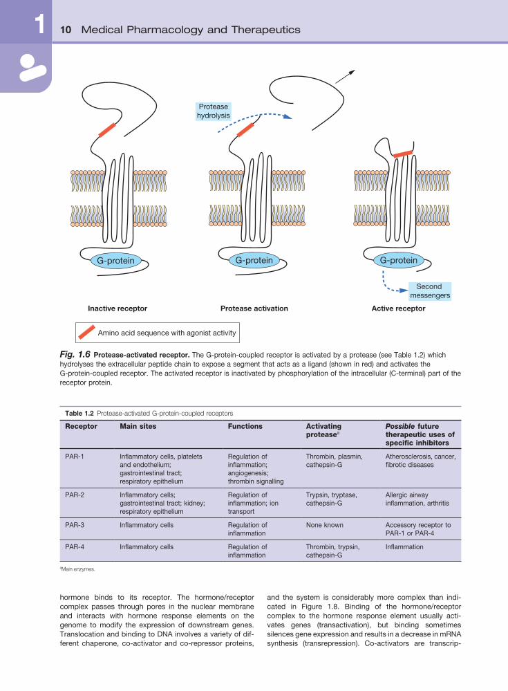
Protease-activated receptors (PARs)Protease-activated receptors (also called proteinase- activated receptors) are recently identified transmembrane G-protein-coupled receptors which are stimulated by cleav-age of the N-terminal of the receptor by a serine protease, rather than by the usual receptor occupancy (as described above). Proteolysis by proteases such as thrombin and trypsin produces a new N-terminal sequence of the recep-tor protein that can act as a ligand, which becomes ‘teth-
ered’ back onto the receptor within extracellular loop-2 (Fig. 1.6). To date, four protease-activated receptors (PAR 1–4) have been identified, each with distinct N-terminal cleavage sites and different tethered ligands (Table 1.2). The receptors appear to play roles in platelet activation and clotting (Ch. 11), and act to transfer information on extracel-lular changes to intracellular functions, such as occurs during inflammation and tissue repair. They may also be involved in brain development, detecting noxious stimuli at sensory nerve endings, and regulation of intestinal secre-tions and permeability. The processes of receptor inactiva-tion and intracellular events have not been fully defined, but are probably similar to those outlined above for other G-protein-coupled receptors. Clinically useful and selective drugs for these receptors are currently under development.
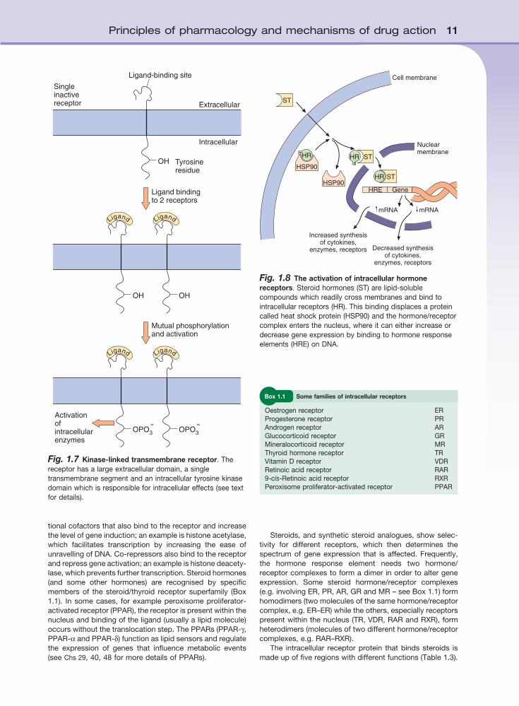
Kinase-linked transmembrane receptorsKinase-linked transmembrane receptors, which bind extra-cellular peptide signalling substances such as insulin, are similar to the G-protein-linked receptors in that they have a ligand-binding domain on the surface of the cell membrane, traverse the membrane and have an intracellular ‘effector’ region (Fig. 1.7). However, they differ in a number of impor-tant respects:
n the extracellular region associated with the ligand-bind-ing domain is very large; this is related to the size of the endogenous ligands, which are peptides such as insulin and cytokines
n there is a single transmembrane helical regionn the intracellular region possesses tyrosine kinase activ-
ity; different receptors have different intracellular effector regions.
Ligand binding is accompanied by dimerisation of two kinase-linked receptors, and these phosphorylate each other. This activated ‘pair of receptors’ then phosphorylates specific intracellular protein(s). The phosphorylated intra-cellular proteins are active enzymes, such as kinases or phospholipases, which can then bring about the relevant intracellular changes appropriate to the biological activities of the extracellular ligand. The phosphorylated intracellular kinases can either act directly on metabolising enzymes or alter the gene transcription of enzymes. Phosphorylation and dephosphorylation are important regulatory steps in the activation/deactivation of numerous intracellular proteins, such as enzymes and transporters.
Intracellular (nuclear) hormone receptorsMany hormones produce long-term changes in cellular activity by altering the genetic expression of enzymes, cytokines or receptor proteins. Such actions on DNA expression are mediated by interactions with intracellular receptors. The sequence of hormone binding and actions for most nuclear receptors are shown in Figure l.8. The cytosolic hormone receptor is usually in an inactive form linked to a protein called heat shock protein (HSP). Binding of the hormone causes dissociation of the HSP and the
10 Medical Pharmacology and Therapeutics1
and the system is considerably more complex than indi-cated in Figure 1.8. Binding of the hormone/receptor complex to the hormone response element usually acti-vates genes (transactivation), but binding sometimes silences gene expression and results in a decrease in mRNA synthesis (transrepression). Co-activators are transcrip-
hormone binds to its receptor. The hormone/receptor complex passes through pores in the nuclear membrane and interacts with hormone response elements on the genome to modify the expression of downstream genes. Translocation and binding to DNA involves a variety of dif-ferent chaperone, co-activator and co-repressor proteins,
Fig. 1.6 Protease-activated receptor. The G-protein-coupled receptor is activated by a protease (see Table 1.2) which hydrolyses the extracellular peptide chain to expose a segment that acts as a ligand (shown in red) and activates the G-protein-coupled receptor. The activated receptor is inactivated by phosphorylation of the intracellular (C-terminal) part of the receptor protein.
G-protein G-protein G-protein
Proteasehydrolysis
Inactive receptor Protease activation Active receptor
Amino acid sequence with agonist activity
Second messengers
Table 1.2 Protease-activated G-protein-coupled receptors
Receptor Main sites Functions Activating proteasea
Possible future therapeutic uses of specific inhibitors
PAR-1 Inflammatory cells, platelets and endothelium; gastrointestinal tract; respiratory epithelium
Regulation of inflammation; angiogenesis; thrombin signalling
Thrombin, plasmin, cathepsin-G
Atherosclerosis, cancer, fibrotic diseases
PAR-2 Inflammatory cells; gastrointestinal tract; kidney; respiratory epithelium
Regulation of inflammation; ion transport
Trypsin, tryptase, cathepsin-G
Allergic airway inflammation, arthritis
PAR-3 Inflammatory cells Regulation of inflammation
None known Accessory receptor to PAR-1 or PAR-4
PAR-4 Inflammatory cells Regulation of inflammation
Thrombin, trypsin, cathepsin-G
Inflammation
aMain enzymes.
Principles of pharmacology and mechanisms of drug action 11
Cell membrane
Nuclearmembrane
mRNAmRNA
Increased synthesisof cytokines,
enzymes, receptors Decreased synthesisof cytokines,
enzymes, receptors
HSP90
HR
HSP90
HR ST
HR ST
ST
HRE Gene
Fig. 1.8 The activation of intracellular hormone receptors. Steroid hormones (ST) are lipid-soluble compounds which readily cross membranes and bind to intracellular receptors (HR). This binding displaces a protein called heat shock protein (HSP90) and the hormone/receptor complex enters the nucleus, where it can either increase or decrease gene expression by binding to hormone response elements (HRE) on DNA.
Fig. 1.7 Kinase-linked transmembrane receptor. The receptor has a large extracellular domain, a single transmembrane segment and an intracellular tyrosine kinase domain which is responsible for intracellular effects (see text for details).
OH
OH
Ligand
OPO3
Ligand
=
Ligand
OPO3
=
OH
Ligand
Singleinactivereceptor
Activationofintracellularenzymes
Ligand-binding site
Extracellular
Intracellular
Tyrosineresidue
Ligand bindingto 2 receptors
Mutual phosphorylationand activation
tional cofactors that also bind to the receptor and increase the level of gene induction; an example is histone acetylase, which facilitates transcription by increasing the ease of unravelling of DNA. Co-repressors also bind to the receptor and repress gene activation; an example is histone deacety-lase, which prevents further transcription. Steroid hormones (and some other hormones) are recognised by specific members of the steroid/thyroid receptor superfamily (Box 1.1). In some cases, for example peroxisome proliferator-activated receptor (PPAR), the receptor is present within the nucleus and binding of the ligand (usually a lipid molecule) occurs without the translocation step. The PPARs (PPAR-γ, PPAR-α and PPAR-δ) function as lipid sensors and regulate the expression of genes that influence metabolic events (see Chs 29, 40, 48 for more details of PPARs).
Oestrogen receptor ERProgesterone receptor PRAndrogen receptor ARGlucocorticoid receptor GRMineralocorticoid receptor MRThyroid hormone receptor TRVitamin D receptor VDRRetinoic acid receptor RAR9-cis-Retinoic acid receptor RXRPeroxisome proliferator-activated receptor PPAR
Box 1.1 Some families of intracellular receptors
Steroids, and synthetic steroid analogues, show selec-tivity for different receptors, which then determines the spectrum of gene expression that is affected. Frequently, the hormone response element needs two hormone/ receptor complexes to form a dimer in order to alter gene expression. Some steroid hormone/receptor complexes (e.g. involving ER, PR, AR, GR and MR – see Box 1.1) form homodimers (two molecules of the same hormone/receptor complex, e.g. ER–ER) while the others, especially receptors present within the nucleus (TR, VDR, RAR and RXR), form heterodimers (molecules of two different hormone/receptor complexes, e.g. RAR–RXR).
The intracellular receptor protein that binds steroids is made up of five regions with different functions (Table 1.3).

12 Medical Pharmacology and Therapeutics1
PRoPeRTieS oF ReCePToRS
Receptor bindingThe binding of the ligand to the receptor is normally revers-ible; consequently, the intensity and duration of the intracel-lular changes are dependent on the continuing presence of the ligand. The extent of drug binding to the receptor (receptor occupancy) is proportional to the drug concentra-tion; the higher the concentration, the greater the occu-pancy. The interaction between the ligand and its receptor does not usually involve permanent covalent chemical bonds but weaker, reversible forces such as:
n ionic bonding between ionisable groups in the ligand (e.g. NH3
+) and in the receptor (e.g. COO−)n hydrogen bonding between amino-, hydroxyl-, keto-
functions, etc, in the drug and the receptorn hydrophobic interactions between lipid-soluble sites in
the ligand and receptorn van der Waals forces, which are very weak interatomic
attractions.
The receptor protein is not a rigid structure: binding of the ligand alters its conformation and the biological properties of the protein, leading to the intracellular changes that are described above.
Receptor selectivityThere are numerous possible extracellular and intracellular chemical signals produced in the body, which can affect different processes. Therefore, a fundamental property of a receptor is its selectivity, i.e. the extent to which it can recognise and respond to only one ligand or a group of related ligands (such as adrenaline and noradrenaline). Some receptors show high selectivity and bind a single endogenous ligand (e.g. acetylcholine is the only endo-genous ligand that binds to N1 nicotinic receptors; see Ch. 4), whereas other receptors are less selective and will bind a number of related endogenous ligands (e.g. the β1-adrenoceptors on the heart will bind noradrenaline, adrenaline and to some extent dopamine, which are all catecholamines).
The ability of receptors to recognise and bind the appro-priate ligand depends on an interaction between the recep-tor molecule and certain characteristics of the chemical structure of the ligand. The formulae of a few representative endogenous ligands that bind to different receptors are shown in Figure 1.9; differences in structure that determine selectivity of action between receptors may be subtle. Receptor selectivity occurs because the three-dimensional organisation of the different sites for reversible binding interactions (such as anion and cation sites, lipid centres and hydrogen bonding sites; see above) corresponds to the three-dimensional structure of the endogenous ligand. Receptors are proteins that are folded into a tertiary struc-ture such that the necessary specific arrangement of bonding centres is brought together within a small volume – the receptor site (Fig. 1.10). Binding of the natural ligand alters the three-dimensional conformation of the protein, which triggers the receptor activity.
There may be a number of subtypes of a receptor, each of which specifically recognises or binds the same ligand. For example, α1-, α2-, β1-, β2- and β3-adrenoceptors all bind adrenaline, but they occur to a different extent in different tissues, and produce different intracellular changes when
Different regions of the receptor are involved in hormone binding, DNA binding and DNA modulation.
Hormone drugs act primarily by mimicking the endogenous hormone (i.e. as an agonist) but often the drug has a longer half-life (Ch. 2) than the endogenous ligand and produces a long-term change. Some drugs act as antagonists by blocking the binding of the normal ligand.
oTHeR SiTeS oF DRUG ACTioN
Probably every protein in the human body has the potential to have its structure or activity altered by foreign com-pounds, so that the list of ‘other sites’ is almost limitless. In addition to the sites and mechanisms of actions dis-cussed above, drugs may also bind to and either activate or inhibit other sites.
n Cell membrane ion pumps. For example, Na+/K+-ATPase in the brain is activated by the anticonvulsant phenytoin whereas that in cardiac tissue is inhibited by digoxin; K+/H+-ATPase in gastric parietal cells is inhibited by proton pump inhibitors (e.g. omeprazole, Ch. 33).
n Enzymes. For example, a number of anticancer drugs inhibit enzymes involved in purine, pyrimidine or DNA synthesis. Some drugs act on the enzymes that synthe-sise or degrade the endogenous ligands for extracellular or intracellular receptors, or second messenger mole-cules (Table 1.1).
n Organelles. For example, some antibiotics interfere with the functioning of the bacterial ribosome.
n Transport proteins. For example, diuretics affect Na+ transport in the renal tubules, and probenecid inhi-bits renal tubular secretion of anions (see Ch. 2 and Ch. 14).
Table 1.3 The structure of steroid hormone receptor proteins
Section of protein
Action Role
N-terminus
A/B Transactivation Activates target genes and gives the specificity of the receptor response
C DNA binding and dimerisation
Binds receptor to DNA by two zinc finger regions
D Nuclear localization
Hinge region to allow correct conformation
E Ligand binding Ligand specificity of receptor; a large complex region; this region also binds heat shock protein
F Unknown Deletion of this region does not alter functioning
C-terminus

Principles of pharmacology and mechanisms of drug action 13
stimulated or blocked (see Ch. 4). The different character-istics of the receptor subtypes allow a drug (or natural hormone) with a particular three-dimensional structure to show selective actions by recognising and then acting pref-erentially on one particular receptor, with fewer unwanted effects from stimulation of related receptors. It should be noted that although ligands may have a high affinity for one receptor subtype, this is never absolute. For example, the neurotransmitter acetylcholine stimulates acetylcholine receptors on ganglia (nicotinic N1 receptor subtype), the neuromuscular junction of skeletal muscle (nicotinic N2 receptor subtype) and at smooth muscle (muscarinic recep-tor subtype); these receptors all respond to acetylcholine at low concentrations but have been shown to be structurally different by using a variety of pharmacological techniques. However, synthetic drugs have been produced which show selectivity of action between these different receptors (due to different affinities/efficacies – see later) and are used for different clinical purposes. This aspect is discussed in detail in Chapter 4. Until recently, receptor subtypes were ‘dis-covered’ when a pharmaceutical company developed a new agonist or antagonist that was found to alter some, but not all, of the activities of a currently known receptor class. Recent developments in molecular biology have enhanced our abilities to detect receptor subtypes. Based on genetic information it is now recognised that there are multiple types of most receptors, and also that there is genetic vari-ation between individuals in the properties or abundance of these receptors (pharmacogenetics – see the end of this chapter). The recognition and cloning of subtypes of recep-tors is important in that it should facilitate the development of drugs showing greater selectivity and hopefully fewer unwanted effects. Greater understanding of genetic differ-ences underlying human variability in drug responses offers the potential for individualisation of the mode of treatment and selection of the correct drug and dosage (see Ch. 3).
Drug stereochemistry and activityReceptors have a three-dimensional organisation in space and, therefore, the ligand has to be presented to the recep-tor in the correct configuration (rather like fitting a right hand into a right-handed glove). Because some drugs are a mixture of stereoisomers (the same chemical structures but with different three-dimensional configurations), the differ-ent isomers may show very different binding characteristics and biological properties. For example, the different stereo-isomers of the α- and β-adrenoceptor antagonist drug labetalol bind to different types of receptor. A drug that is an equal mixture of levo- and dextro-isomers (or S- and R- forms; a racemate) could be a mixture of 50% active compound plus 50% inactive, or, in some cases, a mixture of 50% therapeutic drug and 50% inactive but toxic com-pound. In addition, the different isomers may show different rates of metabolism (see Ch. 2). In consequence, there has been a trend in recent years for the development of single isomers for therapeutic uses; one of the earliest examples was the use of levodopa (the levo-isomer of dopa) in Par-kinson’s disease (Ch. 24).
Receptor numbersThe number of receptors present in a cell is not static, and there is a high turnover of receptors which are being formed and removed continuously. Cell membrane receptor
(a)
(b)
CHCH2NH2
OH
OH
OH
CH2CH2NH2
OH
OH
CH2CH2NH2HO
NH
Dopamine Noradrenaline
5-Hydroxytryptamine (5HT)
CH2CH2NH2N
HN
Histamine
NH2
CH2
COOH
NH2
CH2CH2CH
COOH
O
HO
CH2CH2CH2
NH2
HO
ONH2
C
COOH
O
HO
CH2 CH
Glycine Glutamate
Aspartate - aminobutyrate(GABA)
(c)
O
C O
CH3
Progesterone
OTestosterone
OH
C
C
Fig. 1.9 Groups of chemicals that show preference for different receptors in spite of similar structure. (a) Biogenic amines; (b) amino acids; (c) steroids.

14 Medical Pharmacology and Therapeutics1
COMPONENTS OF DRUG ACTION
Drug actions can show a number of important properties:
n selectivityn potencyn efficacy.
SelectivityMany drugs may act preferentially on particular receptor types or subtypes, such as β1- and β2-adrenoceptors, to different extents (see above) but no drug is specific, i.e. 100% selective, for only one receptor subtype. It is impor-tant to be able to measure the degree of selectivity of a drug and to be able to express numerically the extent to which a drug affects one receptor in relation to another. For example, it is therapeutically important in understanding therapeutic efficacy and unwanted effects that the bron-chodilator salbutamol is approximately ten times more effective in stimulating the β2-adrenoceptors in the airways smooth muscle than the β1-adrenoceptors in the heart.
proteins are synthesised in the endoplasmic reticulum and transported to the membrane; regulation of functioning receptor numbers in the membrane occurs via both trans-port to the membrane (often as homo- or heterodimers) and removal by internalisation. The numbers of receptors within the cell membrane may be altered as a consequence of exposure of the receptor to the drug being used for treat-ment, with either an increase (upregulation) or a decrease (downregulation) in receptor numbers. Changes in receptor numbers following treatment with some drugs can be an important part of the therapeutic response. A well-recog-nised example is the therapeutic benefit of tricyclic antide-pressants (Ch. 22); although they produce an almost immediate increase in the availability of monoamine neuro-transmitters, it is the subsequent relatively slow adaptive downregulation in monoamine receptor numbers that takes many days or weeks which is associated with the time taken to produce a therapeutic response. Tolerance to the effects of some drugs (e.g. opioids) may arise from down-regulation of opioid receptor numbers; as a result, there is the need for increased doses to produce the same analge-sic activity (Ch. 19).
NH2
OH
HO
HO
R
+
O+N
O
OH OH HO
HO
HO
C OHO–O
IV
II
I
VII VI
V
III
H-Bonding Ionic centre
R centre
Aromaticcentre
H-Bonding
–
ADRENOCEPTOR
MUSCARINIC RECEPTOR
Fig. 1.10 Receptor ligand-binding sites. The coloured areas are schematic cross-sections of the seven transmembrane segments of the receptor protein (labelled I to VII). Different segments provide different properties (hydrogen bonding, anionic site, etc.) to make up the active binding site.

Principles of pharmacology and mechanisms of drug action 15
affinity and efficacy (as well as pharmacokinetic variables (Ch. 2) that determine the delivery of the drug to its site of action). Therefore, the in vivo potencies of a series of related drugs may not reflect their in vitro receptor-binding properties.
Potencies of different drugs have to be compared using the ratio of the doses required to produce (or block) the same percentage response. Because dose–response curves are usually parallel in part (for drugs that share a common mechanism of action), the ratio is the same at different response values, e.g. 10%, 20% or 50% response, but not at 100% response.
EfficacyThe efficacy of a drug is its ability to produce the maximal response possible and relates to the extent of functional change imparted to the receptor. For example, agonists can be divided into two groups (Fig. 1.12):
n full agonists, which give an increase in response with increase in concentration until the maximum possible response is obtained (curves A1 and A2)
n partial agonists, which also give an increase in response with increase in concentration but cannot produce the maximum possible response (curve A3).
CLASSIFICATION OF DRUG ACTION
Different types of drug action will be introduced throughout this book. They can be classified as:
n agonistsn antagonistsn partial agonistsn inverse agonistsn allosteric modulatorsn enzyme inhibitors/activatorsn non-specific.
Under many circumstances it is possible to determine for an individual drug the relationship between the dose applied and the biological effect (response) on different receptor subtypes by constructing dose–response curves (Fig 1.11). In modern pharmacological studies it is likely that this type of experiment would be performed by studying the effects of the drugs on isolated cell lines expressing the particular receptor being studied. In Figure 1.11, smaller concentrations of the drug being tested are required to stimulate the β1-adrenoceptor compared with those required to stimulate the β2-adrenoceptor and the drug is therefore said to have selectivity of action at the β1-adrenoceptor. The degree of receptor selectivity is given by the ratio of the levels of response by each receptor type when measured at equimolar doses or concentrations. It is clear from Figure 1.11 that the ratio is highly concentration-dependent and is not apparent at high concentrations, when a maximal stimu-lation at both receptor subtypes occurs.
PotencyThe potency of a drug in vitro is largely determined by the strength of its binding to the receptor, which is a reflection of the receptor affinity, and the ability of the receptor/drug complex to elicit downstream events. The more potent a drug, the lower will be the concentration needed to bind to the receptor and to give a response for an agonist (or to block a response for an antagonist). The potency of a drug in vivo is the amount or dose of drug necessary to produce a specified level of effect, and is dependent on receptor density, efficiency of the stimulus response mechanism,
% m
axim
um r
espo
nse
Increasing dose of β-adrenoceptor agonist
100
50
0D1 D2 D3
Drug action atβ1-adrenoceptor
Drug action atβ2-adrenoceptor
Fig. 1.11 Selectivity of action of a β-adrenoceptor agonist. This illustrates the relative selectivity of action for the β1-adrenoceptor of a hypothetical drug and that selectivity is maintained only over a particular dose range. The drug shows β1-adrenoceptor selectivity, because at low doses it produces dose-related β1-adrenoceptor stimulation with less effect on β2-adrenoceptors. If dose D1 was 10 times less than dose D2, the selectivity ratio for the β1-adrenoceptor is 10. This selectivity diminishes at the higher end of the dose–response curve and is completely lost at doses that produce a maximum response of the drug on both β1- and β2-adrenoceptors (D3).
A2 + RA
A2 + IA
Res
pons
e (%
)
Concentration of agonist
100
50
0
A1 A2
A3
Fig. 1.12 Dose–response curves for agonists in the absence or presence of reversible (competitive) or irreversible (non-competitive) antagonists. A1, A2, two different agonists (A1 more potent than A2); A3, partial agonist; RA, reversible antagonist; IA, irreversible antagonist.

16 Medical Pharmacology and Therapeutics1
AntagonistsAn antagonist binds to the receptor (i.e. has affinity) but does not cause the conformational change that converts the receptor to its active state (i.e. has zero efficacy). The compound will, however, block access to the receptor-binding site of the naturally occurring agonist. The drug effect may only be detectable when the natural agonist is present (e.g. β-adrenoceptor antagonists lower heart rate, particularly when the rate is increased by stimulation of the sympathetic nervous system). The binding of most clinically useful antagonists is reversible and competitive; in conse-quence, the receptor blockade can be overcome by an increase in the concentration of the naturally occurring receptor agonist or by the administration of an agonist drug. Therefore, reversible antagonist drugs move the dose–response curve for an agonist to the right but do not alter the maximum possible response (as shown in curve A2+RA when compared with A2 alone in Fig. 1.12). Antagonists also exhibit selectivity of action. For example, the β-adrenoceptor antagonist propranolol is a non-selective antagonist acting equally on β1- and β2-adrenoceptors, whereas atenolol shows selective antagonism of β1-adrenoceptors and has less effect on β2-adrenoceptors.
Irreversible antagonists, such as phenoxybenzamine, bind covalently to their site of action, and a full response cannot be achieved even by a very large increase in agonist concentration (as shown in curve A2+IA compared with A2 alone in Fig. 1.12).
Partial agonistsA drug showing both agonist and antagonist properties is known as a partial agonist: the activity expressed at any time is dependent on the concentration of the natural ligand or agonist. Partial agonism is responsible for the therapeutic efficacy of several drugs, including buspirone, buprenor-phine, pindolol and salbutamol. Even maximal occupancy of a partial agonist at all available receptors produces a submaximal response, for example because of incomplete amplification of the receptor signal via the G-proteins. A partial agonist will show agonist activity at low concentra-tions of the natural ligand, but the dose–response will not reach the maximal activity even when all receptors are occupied (see Fig. 1.12, drug A3). At high concentrations of the naturally occurring agonist, a partial agonist will behave as an antagonist, because it will prevent access of the natu-rally occurring agonist to the receptor and thereby result in a submaximal response. As a consequence, these drugs can be thought of as stabilisers of cell communication, by enhancing deficient systems while simultaneously blocking excessive activity.
Inverse agonistsThe concept of an inverse agonist arose because some compounds were found to show ‘negative efficacy’ – in other words, they acted on unoccupied receptors to produce a change opposite to that caused by an agonist. The presence of an inverse agonist shifts the receptor equi-librium towards the inactive state, thereby reducing the level of basal activity; in consequence, significant effects will occur only if there is a high level of basal (spontaneous) activity. The role of inverse agonist activity in the therapeutic effects of drugs remains to be fully elucidated, but a number
These represent the classic descriptions of drug actions. It is now recognised that many G-protein-linked receptors can show basal, agonist-independent activity. The G- protein can exist in two different low energy states, inactive and active, which are separated by an energy barrier. Receptors with a low energy barrier show a higher tendency for spontaneous activity, in the absence of a ligand. Because the receptors exist as an equilibrium between active and inactive forms, the basal activity can be either increased or decreased by different types of ligands as follows:
n agonists fully activate the receptorn antagonists have no effect on basal activity, but com-
petitively block the access of other ligandsn partial agonists induce submaximal activation of the
G-protein even at saturating concentrationsn inverse agonists inhibit basal activity.
AgonistsAn agonist whether a therapeutic drug (ligand) or the endogenous agonist (also a ligand), binds to the receptor or site of action, and changes the conformation of the receptor to its active state. An agonist shows both affinity (the strength of binding for the receptor) and efficacy (the extent of functional change imparted to the receptor). Drugs may differ in their affinity and efficacy.
The affinity or strength of binding of the drug to the receptorThis determines the concentration necessary to produce a response and, therefore, is directly related to the potency of the drug. In the examples in Figure 1.12, drug A1 is more potent than drug A2, but both are capable of producing a maximal response (they have the same efficacy). For some compounds a maximal response may require all of the receptors to be occupied, but for most drugs/receptors the maximal response is produced while some receptors remain unoccupied, that is, there may be spare receptors. The presence of spare receptors becomes important when considering changes in receptor numbers owing to adap-tive responses during chronic treatment (tolerance) or caused by irreversible binding of an antagonist (see below).
The rate of binding/dissociationThis is usually of negligible importance in determining the rates of onset or termination of effect in vivo, because these depend mainly on the rates of delivery to and removal from the target organ, that is, on the overall absorption or elimi-nation rate of the drug from the body (see Ch. 2).
Changes in the number of receptorsThe effect of changes in the numbers of receptors on dose–response relationships is complex. With downregulation of receptors, the response obtained depends upon the extent of downregulation and also on the extent of occupancy that is necessary to produce a maximal response. In practice, maximal drug effects are normally produced at concentra-tions that do not give 100% receptor occupancy; with downregulation, the same maximal response may be pro-duced but only with higher percentage occupancy of the reduced number of receptors and hence with higher con-centrations/doses.

Principles of pharmacology and mechanisms of drug action 17
TOLERANCE TO DRUG EFFECTS
Tolerance to drug effects is characterised by a decrease in response with repeated doses. Tolerance may occur through:
n a decrease in the concentrations of drug at the receptor
n a decrease in response produced by the receptor to the same concentration of drug
n a decrease in the number of receptors (so that an increased % occupancy is necessary to produce the same response).
The relationship between drug dosage and the concentra-tions delivered to the receptor is discussed in Chapter 2: some drugs stimulate their own metabolism, and, as a result, they are eliminated more rapidly on repeated dosage and lower concentrations of drug are available to produce a response. However, most clinically important examples of tolerance arise from changes in receptor numbers and concentration–response relationships.
Desensitisation is used to describe both long-term and short-term changes in dose–response relationships arising from a decrease in response of the receptor. Desensitisation can occur by a number of mechanisms:
n decreased receptor numbers (downregulation): a slow process taking hours or days
n decreased receptor binding affinityn decreased G-protein couplingn modulation of the downstream response to the initial
signal.
Extracellular receptors coupled to G-proteins show rapid desensitisation (within minutes) during continued activation, which occurs through three mechanisms.
n Homologous desensitisation. The enzymes activated following ligand binding to a receptor/G-protein complex include G-protein-coupled receptor kinases (GRKs), which interact with the βγ-subunit of the G-protein and inactivate the occupied receptor protein by phosphor-ylation; a related peptide, β-arrestin, enhances the GRK-mediated desensitisation.
n Heterologous desensitisation. The receptor (whether occupied or not) is inactivated through phosphorylation by a cAMP-dependent kinase (protein kinase A or protein kinase C), which causes uncoupling of the G-protein and can be switched on by a variety of signals that increase cAMP.
n Receptor internalisation. Endocytosis of the agonist-coupled receptor can occur within minutes of constant activation of G-protein-coupled receptors and makes the receptor unavailable for further agonist actions by uncoupling the G-protein from the receptor. The phos-phorylated receptor protein may then be internalised and undergo intracellular dephosphorylation prior to re-entering the cytoplasmic membrane.
Downstream modulation of the signal may also occur through feedback mechanisms or simply through depletion of some essential cofactor. An example of the latter is the depletion of the SH groups necessary for the generation of nitric oxide during chronic administration of organic nitrates (see angina, Ch. 5); high doses of indirectly acting
of drugs exhibit this type of activity (Table 1.4). Antagonists (see above) bind to the receptor and block the activity of both agonists and inverse agonists. The mechanism of action of inverse agonists is not well characterised, but they may destabilise the receptor/G-protein coupling, or they may preferentially bind to the inactivated form of the receptor, thereby shifting the equilibrium away from the active form. A final complication is that some drugs, for example β-adrenoceptor antagonists, which act as an antagonist at some tissue receptors, may be an inverse agonist when the receptor is expressed on a different tissue (possibly due to association of the receptor protein with different G-proteins).
Allosteric modulatorsAn allosteric modulator does not act directly on the ligand/receptor site (also called the orthosteric site) but binds to a different (allosteric) site on the receptor. Binding to the allosteric site can change receptor activity by altering the conformation of the protein so as to affect the normal (orthosteric) binding site and thereby enhance or decrease the binding of the natural ligand to its receptor. An example is the benzodiazepine drugs, which alter the affinity of Cl− channels for the neurotransmitter GABA (Ch. 20). Alterna-tively, allosteric modulators may change the conformation of the receptor protein so that it alters the receptor signal-ling (second messenger) domain without affecting the orthosteric site.
Enzyme inhibitors/activatorsSome drugs have a site of action that is an enzyme; the drug acts either on the catalytic site or at an allosteric site. An example is the anticholinesterase group of drugs (see Ch. 4).
Non-specific actionsSome compounds produce their desired therapeutic outcome without interaction with a specific site of action on a protein – for example, the action of osmotic diuretics on the kidney.
Table 1.4 Drugs that show inverse agonist activity
Receptor Drugs
α1-Adrenoceptor Prazosin, terazosin
β1-Adrenoceptor Metoprolol
Muscarinic M1 Pirenzepine (not available for clinical use in the UK)
Histamine H1 Cetirizine, loratadine
Histamine H2 Cimetidine, ranitidine, famotidine
Dopamine D2 Haloperidol, clozapine, olanzapine
Angiotensin II receptor subtype AT1
Losartan, candesartan, irbesartan
Cysteinyl leukotriene CysLT1 Montelukast, zafirlukast

18 Medical Pharmacology and Therapeutics1
than four decades, largely in relation to in vivo variability, and has often used classic genetic techniques such as studies in twins and patterns of inheritance.
Pharmacogenomics has been defined as ‘the investiga-tion of variations of DNA and RNA characteristics as related to drug response’, and relates to genome-wide approaches that define the presence of single-nucleotide polymor-phisms (SNPs) in the genes which affect the activity of the gene product. Molecular biological techniques have allowed recognition of more than 1.4 million SNPs in the human genome. SNPs can be:
n in the upstream regulatory sequence of a coding gene, which can result in increased or decreased expression of the gene in response to the regulatory transcription factors that control the gene product; the gene product will be the same as the normal or ‘wild’ type of gene product
n in the coding region of the gene, which will result in a gene product with an altered amino acid sequence that may have higher activity (although this is unlikely), similar activity, lower activity or no activity at all.
In addition, there can be inactive SNPs because they are in non-coding or silent regions of the genome, or because the base change does not alter the amino acid encoded. In consequence, a major challenge for the future is not in identifying SNPs and the presence of genotypic differences, but rather in defining the functional consequences of the genetic difference and the magnitude of phenotypic differ-ences. Future research will also focus on the importance of different combinations of genetic variants (haplotypes) rather than on single gene differences. The rapid advances in molecular biology have allowed analysis of person-to-person differences in the sequences of the genes involved
sympathomimetic amines may cause depletion of neuronal noradrenaline, which is necessary for their activity (see Ch. 4).
PHARMACOGENOMICS, PHARMACOGENETICS AND DRUG RESPONSES
There are person-to-person variations for any biological property, including the responses to drug administration. The nature of the response is usually similar in all individu-als, because they share the same underlying biology, but the magnitude of the response to the same dose of a drug can differ markedly within a group of individuals. For many responses, this variation is reflected in a single Gaussian distribution (Fig. 1.13a), and such variability is an inherent part of the need to individualise dosage for the person. The presence of a genetic polymorphism in a receptor, site of action, enzyme or transporter (Fig. 1.13b) can give rise to much wider person-to-person variation in response, such that some individuals may have no response to a standard dose while others show toxicity. The genetic origins of many polymorphisms is of increasing importance in relation to drug development (see Ch. 3) and also because it allows the future possibility for genetic screening to be used to optimise drug and dosage selection.
Pharmacogenetics has been defined as ‘the influence of variations in DNA sequence on drug response’, and relates to how genetic differences between individuals affect the response to a drug or the fate of a drug in the body (Ch. 2). Pharmacogenetic research has been undertaken for more
(a) Gaussian distribution of response (b) Polymorphic distribution of response
ResponseResponse
Num
ber
of s
ubje
cts
Num
ber
of s
ubje
cts
Fig. 1.13 inter-individual variation in response to a single dose. The graphs show the numbers of individuals in a population showing a particular level of response to a single dose of a drug against the magnitude of the response. In Figure 1.13a, most individuals show the average response and the overall shape is a normal distribution. In a normal monomorphic distribution (Fig. 1.13a), the magnitude of inter-individual variability is indicated by the coefficient of variation (the dotted line in Fig. 1.13a is for a response showing wider inter-individual variation). Both the coefficient of variation and the magnitude of the difference between phenotypes affect the variation in a polymorphic distribution (Fig. 1.13b).

Principles of pharmacology and mechanisms of drug action 19
of the three main α2-adrenoceptor types (α2A, α2B and α2C), but the consequences for drug activities in clinical practice have not been defined. Nine polymorphisms have been identified in the β2-adrenoceptor, and certain genetic variants have been associated with different asthma phe-notypes and also with differences in drug responses. Poly-morphisms in dopamine D2 receptors have been observed and studied in various conditions, such as alcohol and drug dependency, but, in general, clear relationships of variants of the different dopamine receptors to diseases have not been established, despite the vast amount of research undertaken.
Pharmacogenetic variability will be increasingly impor-tant to optimise drug responses and to minimise adverse effects in individuals. Exploitation of genetic differences will require specific individual genetic information. Until such information is available routinely, careful monitoring of the subject’s response will remain the best guide to successful treatment.
Information on genetic polymorphisms in different receptors can be found on the OMIM® (Online Mendelian Inheritance in Man®; Johns Hopkins University) database, available at http://www.ncbi.nlm.nih.gov/omim
CONCLUSIONS
The selectivity of drugs arises from their ability to interact with and affect certain target sites within a cell. In principle, high selectivity should result in safer drugs with fewer adverse effects. Our increasing knowledge of the complex-ity of receptor pharmacology offers the promise of safer drugs in the future, especially when genetic differences in pharmacokinetics (Ch. 2) and receptors can be taken into account using individual genetic information, prior to the subject being administered any drug. However, it should be remembered that:
n not all effects seen following drug administration are caused by the drug (a ‘placebo effect’ can be produced just by a clinical consultation)
n nearly all drugs show multiple effects no matter how receptor-selective they are
n not all effects produced by a drug will be therapeutically beneficial.
in receptors (pharmacodynamics – this chapter) and in drug metabolism and drug transport (pharmacokinetics – Ch. 2). Knowledge of the precise nature of the differences (SNPs) that result in genetic polymorphisms is beyond the scope of an undergraduate text, and is not necessary to under-stand or appreciate either the current position or possible future developments in the area of pharmacogenomics.
Genetic polymorphisms in receptors and second mes-senger systems are likely to be of increasing importance in the future. While polymorphisms in pharmacokinetics (see Ch. 2) are likely to have an important impact on dosage selection, polymorphisms in receptors may be more impor-tant for drug selection. For example, discovery of a major polymorphism that caused a functional deficit in the angio-tensin (AT2) receptor would mean that neither an angio-tensin-converting enzyme (ACE) inhibitor (Ch. 6) nor an AT2 receptor antagonist would be a good choice to lower blood pressure in such subjects, whereas a β-adrenoceptor antagonist would still be effective. In contrast, a functional deficit in the β1-adrenoceptor could make an ACE inhibitor a better choice than a β-adrenoceptor antagonist for treat-ing cardiovascular conditions. Genomic studies have identi-fied SNPs in nearly every receptor type found in the autonomic system, some of which have been linked to abnormal cardiovascular or respiratory drug responses; however, a genetic variant of major clinical importance has yet to be identified.
Genetic polymorphisms have been reported in many receptor types, and these have been a major focus of research in relation to the aetiology of diseases. To date, few studies have been performed on the influence of genetic differences in receptor structure on drug responses. Polymorphisms of various G-protein receptors have been found in different domains of the protein with a range of different effects (given in parentheses): the N-terminal domain (altered ligand binding or downregulation), the transmembrane domain (altered ligand binding or coupling to G-protein), the intracellular loop (altered ligand binding or coupling to G-protein) and the C-terminal domain (altered coupling to G-protein). However, despite the large number of variants identified, the clinical and therapeutic implica-tions of few of these have been characterised.
Sixteen different human α1A-adrenoceptor isoforms have been identified, including five full-length and 11 truncated versions (which are incapable of ligand binding and signal transduction). Polymorphisms have been reported for each
FURTHeR ReADiNG
Ackerman MJ, Clapham DE (1997) Ion-channels – basic science and clinical disease. N Engl J Med 336, 1575–1586
Alexander SPH, Mathie A, Peters JA (2008) Guide to receptors and channels (GRAC), 3rd edition (2008 revision). Br J Pharmacol 153(suppl 2), S1–S209
Berger JP, Akiyama TE, Meinke PT (2005) PPARs: therapeutic targets for metabolic disease. Trends Pharmacol Sci 26, 244–251
Boswell-Smith V, Spina D, Page CP (2006) Phosphodiesterase inhibitors. Br J Pharmacol 147, S252–S257
Bourguet W, Germain P, Gronemeyer H (2000) Nuclear receptor ligand-binding domains: three-dimensional structures, molecular
interactions and pharmacological implications. Trends Pharmacol Sci 21, 381–388
Catterall WA (1995) Structure and function of voltage-gated ion channels. Annu Rev Biochem 64, 493–531
Catterell WA, Goldin AL, Waxman SG (2003) International Union of Pharmacology. XL. Compendium of voltage-gated ion channels: sodium channels. Pharmacol Rev 55, 575–578
Catterall WA, Striessnig J, Snutch TP et al (2003) International Union of Pharmacology. XL. Compendium of voltage-gated ion channels: calcium channels. Pharmacol Rev 55, 579–581
Chuang TT, Iacovelli L, Sallese M, de Blasi A (1996) G protein-coupled receptors: heterologous regulation of homologous desensitization and its implications. Trends Pharmacol Sci 17, 416–421

20 Medical Pharmacology and Therapeutics1
Costa T, Cotecchia S (2005) Historical review: Negative efficacy and the constitutive activity of G-protein coupled receptors. Trends Pharmacol Sci 26, 618–624
Dohlman HG, Thomer J, Caron MG, Lefkowitz RJ (1991) Model systems for the study of seven-transmembrane-segment receptors. Annu Rev Biochem 60, 653–688
Dolphin A (2003) International Union of Pharmacology. XL. Compendium of voltage-gated ion channels: G protein modulation of voltage-gated calcium channels. Pharmacol Rev 55, 607–627
Essayan DM (2001) Cyclic nucleotide phosphodiesterases. J Allergy Clin Immunol 108, 671–680
Ferguson FFG (2001) Evolving concepts in G protein-coupled receptor endocytosis: the role in receptor desensitization and signaling. Pharmacol Rev 53, 1–24
Fredholm BB, IJzerman AP, Jacobson KA, Klotz KN, Linden J (2001) International Union of Pharmacology. XXV. Nomenclature and classification of adenosine receptors. Pharmacol Rev 53, 527–552
Gudermann T, Kalkbrenner F, Schultz G (1996) Diversity and selectivity of receptor–G-protein interaction. Annu Rev Pharmacol Toxicol 36, 429–459
Gutman GA, Chandy KG, Adelman JP et al (2003) International Union of Pharmacology. XL. Compendium of voltage-gated ion channels: potassium channels. Pharmacol Rev 55, 583–586
Hanoune J, Defer N (2001) Regulation and role of adenylyl cyclase isoforms. Annu Rev Pharmacol Toxicol 41, 145–174
Hawrylyshyn KA, Michelotti GA, Coge F, Guenin S-P, Schwinn DA (2004) Update on human α1-adrenoceptor subtype signaling and genomic organization. Trends Pharmacol Sci 25, 449–455
Hirano K, Yufu T, Hirano M, Nishimura J, Kanaide H (2005) Physiology and pathophysiology of proteinase-activated receptors (PARs): regulation of the expression of PARs. J Pharmacol Sci 97, 31–37
Hofmann F, Biel M, Kaupp UB (2003) International Union of Pharmacology. XL. Compendium of voltage-gated ion channels: cyclic nucleotide modulated channels. Pharmacol Rev 55, 587–589
Hollinger S, Hepler JR (2002) Cellular regulation of RGS proteins: modulators and integrators of G protein signaling. Pharmacol Rev 54, 527–559
Kenakin T (2004) Principles: Receptor theory in pharmacology. Trends Pharmacol Sci 25, 186–192
Kirstein SL, Insel PA (2004) Autonomic nervous system pharmacogenomics: a progress report. Pharmacol Rev 56, 31–52
Kobilka BK, Deupi X (2007) Conformational complexity of G-protein-coupled receptors. Trends Pharmacol Sci 28, 397–406
Koenig JA, Edwardson JM (1997) Endocytosis and recycling of G protein-coupled receptors. Trends Pharmacol Sci 18, 276–287
Lefkowitz RJ (2004) Historical review: A brief history and personal retrospective of seven-transmembrane receptors. Trends Pharmacol Sci 25, 413–422
Lucas KA, Pitari GM, Kazerounian S et al (2000) Guanylyl cyclases and signaling by cyclic GMP. Pharmacol Rev 52, 375–413
McLeod HL, Evans WE (2001) Pharmacogenomics: unlocking the human genome for better drug therapy. Annu Rev Pharmacol Toxicol 41, 101–121
Milligan G, Bond RA, Lee M (1995) Inverse agonism: pharmacological curiosity or potential therapeutic strategy? Trends Pharmacol Sci 16, 10–13
Polson JB (1996) Cyclic nucleotide phosphodiesterases and vascular smooth muscle. Annu Rev Pharmacol Toxicol 36, 403–427
Privalsky ML (2004) The role of corepressors in transcriptional regulation by nuclear hormone receptors. Annu Rev Physiol 66, 315–360
Rana BK, Shiina T, Insel PA (2001) Genetic variations and polymorphisms of G protein-coupled receptors: functional and therapeutic implications. Annu Rev Pharmacol Toxicol 41, 593–624
Simon MI, Strathmann MP, Gautam N (1991) Diversity of G proteins in signal transduction. Science 252, 802–808
Strader CD, Fong TM, Tota MR, Underwood D, Dixon RAF (1994) Structure and function of G-protein-coupled receptors. Annu Rev Biochem 63, 101–132
Strange PG (2003) Mechanisms of inverse agonism at G-protein-coupled receptors. Trends Pharmacol Sci 23, 89–95
Streetman DS (2007) Clinical pharmacogenetics of the major adenosine triphosphate-binding cassette and solute carrier drug transporters. J Pharm Pract 20, 219–233
Sunahara RK, Dessauer CW, Gilman AG (1996) Complexity and diversity of mammalian adenylyl cyclases. Annu Rev Pharmacol Toxicol 36, 461–480
Tsai M-J, O’Malley BW (1994) Molecular mechanisms of action of steroid/thyroid receptor superfamily members. Annu Rev Biochem 63, 451–486
Wei LN (2003) Retinoid receptors and their coregulators. Annu Rev Pharmacol Toxicol 43, 47–72
Wess J (1993) Molecular basis of muscarinic acetylcholine receptor function. Trends Pharmacol Sci 14, 308–313
Wong AHC, Buckle CE, Van Tol HHM (2000) Polymorphisms in dopamine receptors: what do they tell us? Eur J Pharmacol 410, 183–203

Principles of pharmacology and mechanisms of drug action 21
examples of some types of receptors and their properties (some of this information was derived from Alexander, Mathie and Peters 2008)
Type Typical location(s) Principal transduction mechanism
Biological actions Agonists1 Antagonists1
7-transmembrane receptors
Acetylcholine
Muscarinica
M1 CNS and autonomic ganglia (minor role), gastric
Gq Neurotransmission in CNS, gastric secretion
non-selective for all M receptors-bethanecol, pilocarpine, carbachol
Pirenzepine non-selective for all M receptors – atropine, hyoscine, propantheline
M2 Heart, CNS Gi Bradycardia, smooth muscle contraction (GI, airways, bladder)
M3 Smooth muscles, secretory glands
Gq Contraction, secretion
I Darifenacin
Adrenergic
α-Adrenoceptorsb
α1 (α1Aα1Bα1D) CNS and postsynaptic in sympathetic nervous system human prostate (α1A )
Gq Contraction of arterial smooth muscle, decrease in contractions of gut, contraction of prostate tissue
Phenylephrine, methoxamine (tamsulosin α1A)
Prazosin, indoramin
α2 (α2Aα2Bα2C) Presynaptic (in both α- and β-adrenergic neurons)
Gi Decreased noradrenaline release
Clonidine (oxymetazoline α2A)
Yohimbine
β-Adrenoceptorsc
β1 CNS and heart (nodes and myocardium)
Gs Increased force and rate of cardiac contraction
Dobutamine NA > AD Atenolol, metoprololol
β2 Widespread Gs Bronchial dilation, decrease in contraction of gut, metabolic effects
Salmeterol, terbutaline, AD > NA
Butoxamine
β3 Adipocytes Gs Mobilisation of fat stores
AD = NA –
Cannabinoids
CB1, CB2 Cortex,hippocampus, amygdala, basal ganglia, cerebellum
Gi/G0 Behaviour, pain, nausea, stimulation of appetite, depression
N-arachidonoylethanolamine (anandamide), 2-arachidonylglyceryl
Cholecystokinin
CCK1, CCK2 CCK1, primarily GI CCK2 primarily in CNS
Gq/Gs CCK1, gall bladder emptying, inhibits gut motility;CCK2 CNS nociception, anxiety, appetite
CCK-4, CCK-8, CCK-33, gastrin
Calcitonin–gene related peptide
CGRP Sensory nerve endings
Gq/Gs Vasodilation, nociception, inflammation
CGRP

22 Medical Pharmacology and Therapeutics1
Type Typical location(s) Principal transduction mechanism
Biological actions Agonists1 Antagonists1
Dopamined
D1 CNS (N, O, P, S), kidney,d heartd
Gs Vasodilation in kidney (see notes)
Fenoldopam
D2 CNS (C, N, O, SN), pituitary gland, chemoreceptor trigger zone gastrointestinal tract
Gi↑ K+ channels, ↓ Ca2+ channels
Linked to schizophrenia, prolactin secretion, movement control, memory
Bromocriptine, pergolide Butyrophenones, sulpiride, renoxipride, domperidone
D3 CNS (F, Me, Mi) (limbic system)
Gi Cognition emotion Sulpiride
D4 CNS, heart Gi Linked to schizophrenia
Clozapine
D5 CNS (Hi, Hy) Gs
5-Hydroxytryptamine (5HT), serotonin)e
5HT1A CNS Gi Anxiety sleep, appetite, nerve inhibition
Buspirone
5HT1B CNS blood vessels Gi Vasoconstriction presynaptic inhibition
Sumatriptan, eletriptan
5HT1D CNS, blood vessels Gi Vasoconstriction behaviour
Sumatriptan, eletriptan Metergoline
5HT1E CNS Gi
5HT1F CNS Gi
5HT2A CNS platelets, smooth muscle
Gq Schizophrenia, platelet aggregation, vasodilation/vasoconstriction
LSD Ketanserin
5HT2B Stomach Gq Contraction, morphogenesis
5HT2C CNS Gq Satiety
5HT4 CNS, myenteric plexus, smooth muscle
Gs Anxiety, memory, gut motility
Metoclopramide, renzapride
5HT5A CNS Gi
5HT5B CNS ?
5HT6 CNS Gs
5HT7 CNS GI blood vessels
Gs LSD Clozapine
Histamine
H1 CNS, endothelium, smooth muscle
Gq Sedation, sleep, vascular permeability, inflammation
Mepyramine
H2 CNS, cardiac muscle, stomach
Gs Gastric acid secretion
Dimaprit Cimetidine, ranitidine
examples of some types of receptors and their properties (some of this information was derived from Alexander, Mathie and Peters 2008)

Principles of pharmacology and mechanisms of drug action 23
Type Typical location(s) Principal transduction mechanism
Biological actions Agonists1 Antagonists1
H3 CNS (presynaptic), myenteric plexus
Gi Appetite, cognition Thioperamide
H4 Eosinophils, basophils, mast cells
Gi
Gamma-aminobutyric acid receptor type B (GABAB)
GABAB Brain neurons, glial cells, spinal motor neurons and interneurons
Gi effects on Ca2+ channel (closes) and K+ channel (opens)
Widespread reduction of impulse transmission in CNS, suppression of polysynaptic reflexes in spine, generation of spike and wave discharges in absence epilepsy
Baclofen
Glutamate (metabotropic)f
Group I (mglu1 and mglu5)
Gq Multiple possible roles; memory, learning, pain perception, anxiety, modulation of transmission (e.g. DA and NA) ?schizophrenia
Group II (mglu2 and mglu3)
Gi
Group III (mglu4, mglu6, mglu7 and mglu8)
Gi
Peptides
Angiotensin II
AT1 Blood vessels, adrenal cortex, brain
Gq/G0 Vasconstriction, salt retention, aldosterone synthesis, increased noradrenergic activity, cardiac hypertrophy
Losartan, valsartan
AT2 Blood vessels, endothelium, adrenal cortex brain
Tyrosine phosphatases
Weak vasodilation, fetal development, vascular growth. Stimulates NO synthesis in endothelium
Bradykinin
B1 (induced) Widespread (induced by injury, cytokines)
Gq Acute inflammation. Stimulates NO synthesis
ACE inhibitors (indirect via BK stimulation)
B2 (constitutive) Gq Chronic inflammation. Most actions of kinins, vasodilation, pain response
Endothelins
ETA Endothelium Gq Vasoconstriction, proliferation blood vessels
Sitaxentan, bosentan
examples of some types of receptors and their properties (some of this information was derived from Alexander, Mathie and Peters 2008)

24 Medical Pharmacology and Therapeutics1
Type Typical location(s) Principal transduction mechanism
Biological actions Agonists1 Antagonists1
ETB Endothelium (Ch. 6) Gq Release of nitric oxide (indirect vasodilation), direct vasoconstriction, natriuresis
Bosentan
Ghrelin Stomach, endocrine cells; ghrelin receptors are widespread
Gq Ligand for GH secretagogue receptor, induces appetite
[D-Arg1, D-Phe5, D-Trp7,9, Leu11]-substance P
Melanocortin receptor family[MC1-MC5] (Ch. 37)
Gs MSH skin pigmentation, feeding behaviour. ACTH (specifically at MC2) skin pigmentation HPA function
Melanocyte stimulating hormone (MSH)
agouti-related protein (AGRP) (Ch. 37)
Natriuretic peptides
ANPA G-protein, guanylyl cyclase
Increase Na+ excretion
ANPB G-protein, guanylyl cyclase
Increase Na+ excretion
Neuropeptide Y (NPY) receptor family Y1–Y6) (Ch. 37)
Y receptors wide distribution in CNS hypothalamus. NPY secreted into blood from stomach
Gi Increases appetite; stress; memory learning
Tachykinins (neurokinins)
NK1 Gq: slow build-up of response
Nociception (substance P)
Substance P
NK2 Gq: slow build-up of response
Nociception (neurokinin A)
Substance P
NK3 Gq (Neurokinin B) Substance P
Opioids
δ, κ, µ, N/OFQ (nociceptin/orphanin)
Gi
Orexin (OX1 OX2) (also known as hypocretins) (Ch. 37)
Hypothalamus and other areas of brain
Gq Increases food intake, wakefulness, body temperature. Activated by orexin-A and B
Proteinase activated receptors (PAR1, PAR2, PAR3, PAR4)
Platelets, endothelial cells, myocytes, neurons
Gq, Gi Activated by proteolytic cleavage
Trypsin, thrombin
examples of some types of receptors and their properties (some of this information was derived from Alexander, Mathie and Peters 2008)

Principles of pharmacology and mechanisms of drug action 25
Type Typical location(s) Principal transduction mechanism
Biological actions Agonists1 Antagonists1
Vasopressin and Oxytocin
V1a Pituitary, brain Gq Vasoconstriction, platelet aggregation
Arginine vasopressin > oxytocin
V1b Pituitary, brain Gq plus Gi, Modulates ACTH secretion
Arginine vasopressin > oxytocin
V2 Kidney Gs Antidiuretic effect on collecting duct and ascending limb of loop of Henle
Arginine vasopressin > oxytocin
OT Breast, brain uterus Gq Lactation, uterine contraction, CNS actions
Oxytocin > arginine vasopressin
Purinergic receptors (purinoreceptors)
Adenosine
A1 Gi↑ K+ conduction in heart
Decreased glomerular filtration rate, cardiac depression, vasoconstriction, decreased CNS activity, bronchoconstriction
– Methylxanthines
A2A Gs Vasodilation, decreased CNS activity, inhibition of platelet aggregation, bronchodilation
Methylxanthines
A2B Gq Action on intestine and bladder
– Enprofylline
A2B Gs Ca2+ influx Release of mediators from mast cells?
A3 Gi Wide tissue distribution; release of mediators from mast cells
P2Y-receptors (P2Y1, P2Y2, P2Y4, P2Y6)
A family of peptides present in almost all tissues
Gq, Gs Biological effect depends upon G protein coupling
Activated by ATP, ADP, UTP, UDP and UDP-glucose.
Ligand-gated receptors or channels (also called transmitter-gated receptors or channels)
Nicotinic N1 Autonomic ganglia Ligand-gated ion channel
Postganglionic activation
Carbachol, nicotine Trimetaphan, mecamylamine
Nicotinic N2 Neuromuscular junction
Ligand-gated ion channel
Muscle contraction Nicotine Gallamine, vecuronium
5HT3 CNS (A), enteric nerves, sensory nerves
Ligand-gated Na+/K+ channels
Emesis Granisetron, ondansetron
GABAAh Brain neurons, spinal
motor neurons and interneurons
Ligand-gated Cl− channel (open)g
Widespread reduction of impulse transmission in CNS, inhibition of sensory signals at spinal level
Muscimol (benzodiazepines) (zolpidem)
(Picrotoxin) (flumazenil)
examples of some types of receptors and their properties (some of this information was derived from Alexander, Mathie and Peters 2008)

26 Medical Pharmacology and Therapeutics1
Type Typical location(s) Principal transduction mechanism
Biological actions Agonists1 Antagonists1
Glycine Brain neurons, spinal motor neurons and interneurons
Ligand-gated Cl− channel (open)g
Widespread reduction of impulse transmission in CNS, inhibition of sensory signals at spinal level
Intravenous anaesthetics Strychnine, tropisetron, endocannabinoids
Ionotropic glutamate-like receptors (AMPA, NMDA, kainate)f
N-Methyl d-aspartate(NMDA)i
CNS (B, C, sensory pathways)
Ligand-gated Ca2+ channel (slow)
Synaptic plasticity, excitatory transmitter release, excessive amounts may cause neuronal damage
Aspartate Ketamine, phenyclidine (inhibit Ca2+ flux)
Kainate CNS (Hi) Ligand-gated Ca2+ channel (fast)
Synaptic plasticity, transmitter release
Kainate
a-amino-3-hydroxy-5-methyl-4-isoxazole propionic acid (AMPA)
CNS (similar to NMDA receptors)
Ligand-gated Ca2+ channel (fast)
Synaptic plasticity, transmitter release
Purinergic P2X family (P2X1-P2X7)
P2X1 smooth muscle; P2X2-3 ganglia
Ligand-gated ion channels (Na+, Ca2+ and K+ and exceptionally Cl−)
Neuronal depolarisation, influx of Na+ and Ca2+ > efflux of K+
Endogenous ligand ATP
Ryanodine receptors (RyR1, RyR2, RyR3)
RyR (skeletal muscle), RyR2 (heart), RyR3 (widespread, brain)
Cytosolic Ca2+, ATP
Intracellular Ca2+ concentrations,?myasthenia gravis
Ryanodine, caffeine >100 mM Ca2+, Mg2+
dantrolene
examples of some types of receptors and their properties (some of this information was derived from Alexander, Mathie and Peters 2008)
1Only selected agonists and antagonists are included. Many of these are investigational tools and have no clinical use.
The transduction processes are Gs = ↑ adenylyl cyclase, increased protein kinase A; Gi = ↓ adenylyl cyclase, decreased protein kinase A; Gq = ↑ phospholipase C, increased IP3, diacylglycerol.
AD – adrenaline; NA – noradrenaline; LSD – lysergic acid diethylamide.aAdditional muscarinic receptors (M4 (Gi) and M5 (Gq)) have been identified recently but their clinical importance is unclear.bThree α1-adrenoceptors have been identified (α1A, α1B, α1D), all of which act via Gq proteins: α1A-adrenoceptors have a higher affinity for noradrenaline than for adrenaline, α1B- and α1D-adrenoceptors do not show selectivity.cThe β-adrenoceptors differ in their affinities for noradrenaline (NA) and adrenaline (AD): β1, NA > AD; β2, AD > NA: β3, NA = AD.dCommonly divided into D1-like (D1 and D5) and D2-like (D2, D3,D4). The type of receptor in heart and kidney remains questionable as the mRNA for D1 receptors has not been found (it could be D5). CNS areas: A, area postrema; B, basal ganglia; C, caudate putamen; Ch, choroid; Co, cortex; F, frontal cortex; G, globus pallidus; Hi, hippocampus; Hy, hypothalamus; Me, medulla; Mi, midbrain: N, nucleus accumbens; 0, olfactory tubercle; P, putamen; R, raphe nucleus; S, striatum; SN, substantia nigra; T, thalamus.eThere are many experimental drugs that are selective agonists and antagonists for receptors subtypes and some are undergoing clinical trials for various conditions. Only clinically useful examples are given. The identification and classification of 5HT receptors is a complex and rapidly changing field. 5HT is involved in numerous pathways within the CNS and the roles of the different receptor types has not been fully characterised. Effects associated with the different receptors are not well established, with the exception of 5HT3 and emesis.fGlutamate produces increased neuronal activity in many regions of the CNS; the different receptors are formed from subunits that can exist in different isoforms; a number of variants have been demonstrated for each.gPart of response to vascular damage.hGABA produces inhibition of neurotransmission widely throughout the brain.The GABAA receptor Cl− channel has a binding site for GABA and also adjacent binding sites for benzodiazepines (which affect the response to GABA), barbiturates, picrotoxin and steroids.

Principles of pharmacology and mechanisms of drug action 27
APPENDIx
LiMiTeD STUDeNT FoRMULARY
This formulary has been derived from the formulary put together by Maxwell and Walley (2003), the Southampton University Hospitals Trust formulary and the Southampton Medical School Pharmacology Course documents.
Its purpose is to introduce students in the early years of their undergraduate course in medicine to representative examples of core drugs and their uses in major areas of medicine. It is not exhaustive.
gastrointestinal system
Therapeutic problem Core drugs
Dyspepsia and gastro-oesophageal reflux disease Antacids e.g. magnesium and aluminium-containing antacids, eg aluminium hydroxide and magnesium carbonate
Acid secretion inhibition e.g. proton pump inhibitors and H2 receptor antagonists (see peptic ulcer disease drugs);
Anti-reflux drugs, e.g. gaviscon
Motility stimulants These may be used in gastrointestinal reflux disease e.g. metoclopramide
Peptic ulcer disease Proton-pump inhibitors, e.g. omeprazoleH2 receptor antagonists, e.g. ranitidine, cimetidine; other drugs e.g.
sucralfate, misoprostil
Helicobacter pylori eradication Antibiotics, e.g. clarithromycin, amoxicillin, metronidazole
inflammatory bowel disease (ulcerative colitis, Crohn’s disease)
Corticosteroids, e.g. prednisoloneOther drugs e.g. sulfasalazine, mesalazineCytokine inhibitors, e.g. infliximab
Clostridium difficile colitis Antibiotics for C. difficile, metronidazoleVancomycin
Diarrhoea Oral rehydration therapy;Antimotility drugs, e.g. loperamide, codeine
Constipation Bulk forming laxatives, e.g. ispaghulaStimulant laxatives, e.g. sennaOsmotic laxatives, e.g. magnesium hydroxide, lactuloseFaecal softeners, e.g. docusate
Bowel cleansing Sodium picosulfate
Antispasmodics Antimuscarinics, e.g. hyoscineOther antispasmodics, mebeverin
cardiovascular system
Therapeutic problem Core drugs
Hypertension β-adrenoceptor antagonists, e.g. atenololα-adrenoceptor antagonists, e.g. doxazosin. Centrally-acting
antihypertensives, e.g. clonidine, moxonidineAngiotensin-converting enzyme inhibitors, e.g. captoprilAngiotensin-II receptor antagonists, e.g. candesartan, losartanThiazide and related diuretics, e.g. bendroflumethazide, metolazone. Loop
diuretics, e.g. furosemidePotassium-sparing diuretics, e.g. amiloride, spironolactoneCalcium channel blockers, e.g. amlodipinePotassium channel openers, e.g. minoxidil, nicorandil
Heart failure Many drugs used for hypertension are of benefit, in addition positive inotropic drugs:
cardiac glycosides, e.g. digoxin. Phosphodiesterase inhibitors, e.g. milrinone
β-adrenoceptor antagonists, e.g. bisoprolol

28 Medical Pharmacology and Therapeutics1
cardiovascular system
Therapeutic problem Core drugs
Acute coronary syndrome(angina, myocardial infarction)
Many drugs listed under hypertension; in addition:Glyceryl trinitrate, isosorbide mononitrateinhibitors of platelet aggregation, e.g. aspirin, dipyridamole, clopidogrel,
abciximabThrombolytics, e.g. streptokinase, tenecteplaseHeparins, unfractionated heparins, low molecular weigh heparinsOral anticoagulants, e.g. warfarinLipid-regulating drugs, e.g. simvastatin
Arrhythmias Antiarrhythmic drugs, e.g. digoxin, adenosine, amiodarone, lidocaine, β-adrenoceptor antagonists, calcium channel blockers
Cardiac arrest, deep vein thrombosis, pulmonary embolus,
Pulmonary oedema, stroke treatment and prevention, hyperlipidaemia
Appropriate drugs chosen from the classes already described for the Cardiovascular System
respiratory system
Therapeutic problem Core drugs
Asthma (acute and chronic), chronic obstructive pulmonary disease, respiratory failure
Oxygen; Bronchodilators: β2-adrenoceptor agonists, e.g. salbutamol, salmeterol
Antimuscarinc bronchodilators, e.g. ipratropiumLeukotriene receptor antagonists, e.g. montelukastOther drugs, e.g. aminophylline, cromoglicate, magnesium sulphateCorticosteroids, e.g. beclometasone
central nervous system
Therapeutic problem Core drugs
insomnia, anxiety Benzodiazepines and other drugs, e.g. diazepamOther drugs, e.g. buspirone, propranolol
Schizophrenia, mania Antipsychotic drugs, e.g. chlorpromazine, clozapineMood stablisers, e.g. lithium
Depression Tricyclic and related antidepressants, e.g. amitriptylineSelective serotonin re-uptake inhibitors, e.g. fluoxetineMonoamine oxidase inhibitors, e.g. phenelzine
Analgesia Simple analgesics without anti-inflammatory properties, e.g. paracetalmol. Nonsteroidal analgesics – see section on Musculoskeletal and Joint Diseases
Compound analgesics e.g. co-codamolOpioid analgesics, e.g. codeine, morphine, diamorphine
Nausea and vertigo Dopamine antagonists, e.g. metoclopramideSerotonin type 3 (5HT3) receptor antagonists, e.g. ondansetronMuscarinic receptor antagonists, e.g. hyoscineOther agents, betahistine
Acute and chronic migraine Acute migraine Prophylaxis of chronic migraine
Serotonin type 1 receptor agonists, e.g. sumatriptanSerotonin type 2 receptor antagonists, e.g. pizotifen
epilepsy Anticonvulsant therapy, e.g. diazepam, phenytoin, carbamazepine, sodium valproate, gabapentin
Status epilepicus e.g. diazepam
Parkinson’s disease Drugs enhancing dopaminergic activity, e.g. L-dopa and dopa decarboxylase inhibitor combinations, e.g. co-careldopa
Other drugs influencing dopamine or dopamine receptors, e.g. bromocriptine, entacapone, ropinirole
Antimuscarinic drugs, e.g. procyclidine
Dementia (Alzheimer’s disease) Anticholinesterases, e.g. donepizilNMDA receptor antagonists, e.g. memantine

Principles of pharmacology and mechanisms of drug action 29
infectious diseases
Therapeutic problem Core drugs
Community and hospital acquired infections (bacteria, fungi, viruses)
Penicillins, e.g. benzyl penicillin, amoxicillin, co-amoxiclav, flucloxacillinCephalosporins, e.g. cefalexinTetracyclines e.g. oxytetracyclineTrimethoprimAminoglycosides, e.g. gentamicinVancomycinMacrolides, e.g. erythromycinChloramphenicolQuinolones, e.g. ciprofloxacinMetronidazoleAntituberculosis drugs, e.g. isoniazid, rifampicin, ethambutolAntifungal drugs, e.g. amphotericin, fluconazoleAntiviral drugs, e.g. acyclovirNucleoside reverse transcriptase inhibitor, e.g. abacavirProtease inhibitors, e.g. saquinavirAntimalarial drugs, e.g. mefloquine, proguanil
endocrine system
Therapeutic problem Core drugs
Diabetes mellitus, thyroid disease, osteoporosis, hormone deficiencies and excess
Diabetes, e.g. long and short-acting insulinsSulphonylureas, e.g. gliclazideBiguanides, e.g. metforminThe glitazones, e.g. rosiglitazoneThyroid disease, e.g. levothyroxine, propranolol, carbimazoleBisphosphonates, e.g. alendronic acid, calcium, vitamin DHypothalamic, pituitary hormones and anti-oestrogens:Anti-oestrogens, e.g. clomifeneAnterior pituitary hormones, e.g. somatotropinHypothalamic hormones, e.g. gonadorelin (LHRH)Posterior pituitary hormones and antagonists, e.g. desmopressin,
vasopressin
Genito-urinary disorders
Urinary retention α-blockers, e.g. doxazosin
Benign prostatic hypertrophy 5α-reductase inhibitors, e.g. finasteride
Urinary frequency and incontinence Antimuscarinic drugs, e.g. oxybutynin
Erectile dysfunction Phosphodiesterase inhibitors, e.g. sildenafil
obstetrics and gynaecology
Therapeutic problem Core drugs
Steroid oral contraception Combined oral contraceptives, progestogen-only contraceptivesEmergency contraception, progestin
Injectable contraception Injectable steroidal contraceptives, e.g. medroxyprogesterone acetate
Intrauterine contraception Intra-uterine progestogen containing device
Menstrual disorders
Dysmenorrhoea Mefenamic acid
Menorrhagia ProgestogensAntifibrinolytic agent, e.g. tranexamic acid
Endometriosis Combined contraceptive; danazol
Hormone replacement therapy (menopause) Oestrogens (natural and synthetic), progestins
Induction of labour Oxytocics, e.g. prostaglandins, oxytocin
Prevention of pre-term labour and myometrial relaxation
Calcium channel blockers, e.g. nifedipineβ-adrenoceptor agonists, e.g. terbutaline
Induction of abortion Oxytocics, mifepristone
Post-partum haemorrhage Oxytocics, ergometrine

1 30 Medical Pharmacology and Therapeutics
malignant disease and immunosuppression
Therapeutic problem Core drugs
Anti-cancer and immunosuppression Anti-cancer drugs:alkylating agents, e.g. cyclophosphamideCytotoxic antibiotics, e.g. doxorubicinAntimetabolites, e.g. methotrexateVinca alkaloids, e.g. vinblastinOther drugs, e.g. asparaginase, cisplatinAnti-oestrogens, e.g. tamoxifen, anastrazole, herceptinImmunosuppressant agents acting in a variety of ways, e.g. azathioprine,
corticosteroids, cyclosporineAnti CD20 monoclonal antibody, e.g. rituximabInterferon alfa
Musculoskeletal and joint disease
Rheumatoid arthritis Simple analgesic e.g. paracetamol, Nonsteroidal anti-inflammatory drugs, e.g. indometacin, diclofenac
Corticosteroids e.g. prednisoloneDisease modifying drugs, e.g. methotrexate, gold, penicillamine, anti-
malarials (hydroxychloroquine), azathioprine, sulfasalazine, cytokine inhibitors, e.g. infliximab
Impaired neuromuscular transmission, e.g. myasthenia gravis
Anticholinesterases, e.g. pyridostigmine
Spasticity Skeletal muscle relaxants, e.g. baclofen
ophthalmology
Glaucoma β-Blockers, e.g. timololProstaglandin analogues, e.g. latenoprostSympathomimetics, e.g. brimonidineCarbonic anhydrase inhibitors, e.g. acetazolamideMiotics, e.g. pilocarpine
Mydriatics and cycloplegics Mydriatics, e.g. phenylephrineMydriatics and cycloplegics, e.g. atropine, tropicamide
Surgery, anaesthetics and intensive care Many drugs listed from other sections would be used including opioids, sympathomimetics, anti-emetics
Intravenous anaesthetics for induction, e.g. thiopental, propofolInhalation anaesthetics, e.g. isofluraneMuscle relaxants, e.g. suxamethonium, atracuriumAntimuscarinics, e.g. atropine, glycopyrroniumAnticholinesterases, e.g. neostigmineLocal anaesthesia, e.g. lidocaine, bupivacaineAnalgesics, e.g. morphine, fentanyl
Maxwell S, Walley T. Teaching safe and effective prescribing in UK medical schools: a core curriculum for tomorrow’s doctors. Br J Clin Pharmacol 2003;55:496-503.


![Introduction to Pharmacology 1. Define the terms: pharmacology, drug… · 2013. 9. 5. · [DRUG] - Receptors at Target Site. 4 13 Simplification of drug action Drug Biological Alteration](https://img.dokumen.tips/doc/110x75/5fe194536710e40acd1b517e/introduction-to-pharmacology-1-define-the-terms-pharmacology-drug-2013-9-5.jpg)


![Pharmacology Drug Dosage Calculations.ppt [Read-Only]hy.health.gov.il/.../Pharmacology_Drug_Dosage_Calculations.pdf · Pharmacology Drug Dosage Calculations Shelby County EMS Training](https://img.dokumen.tips/doc/110x75/5b5411317f8b9a5a578cc70a/pharmacology-drug-dosage-read-onlyhyhealthgovilpharmacologydrugdosagecalculationspdf.jpg)