Embed Size (px)
Citation preview
Principles of Human Anatomy and Physiology, 11e 1
Chapter 19
The Cardiovascular System: The Blood
Lecture Outline

Principles of Human Anatomy and Physiology, 11e 2
INTRODUCTION
• Blood inside blood vessels, interstitial fluid around body cells, and lymph inside lymph vessels constitute one’s internal environment.
• To obtain nutrients and remove wastes, cells must be serviced by blood and interstitial fluid.
• Blood, a connective tissue, is composed of plasma and formed elements.
• Interstitial fluid bathes body cells (Figure 19.1).• The branch of science concerned with the study of blood,
blood-forming tissues, and the disorders associated with them is called hematology.
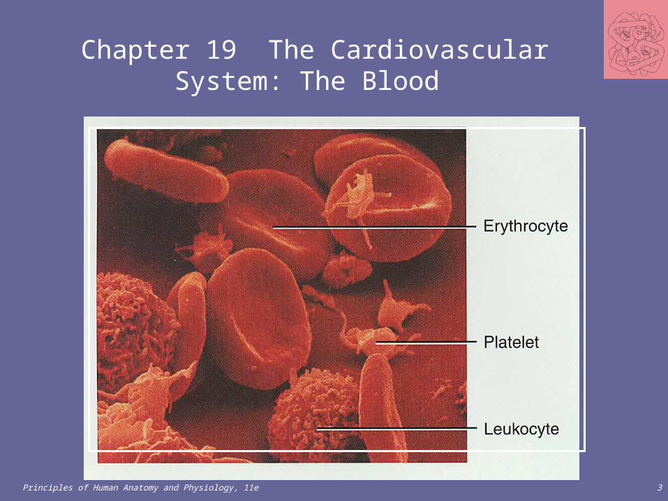
Principles of Human Anatomy and Physiology, 11e 3
Chapter 19 The Cardiovascular System: The Blood

Principles of Human Anatomy and Physiology, 11e 4
• Cells of the body are serviced by 2 fluids– blood
• composed of plasma and a variety of cells• transports nutrients and wastes
– interstitial fluid• bathes the cells of the body
• Nutrients and oxygen diffuse from the blood into the interstitial fluid & then into the cells
• Wastes move in the reverse direction• Hematology is study of blood and blood disorders
Fluids of the Body
Principles of Human Anatomy and Physiology, 11e 5
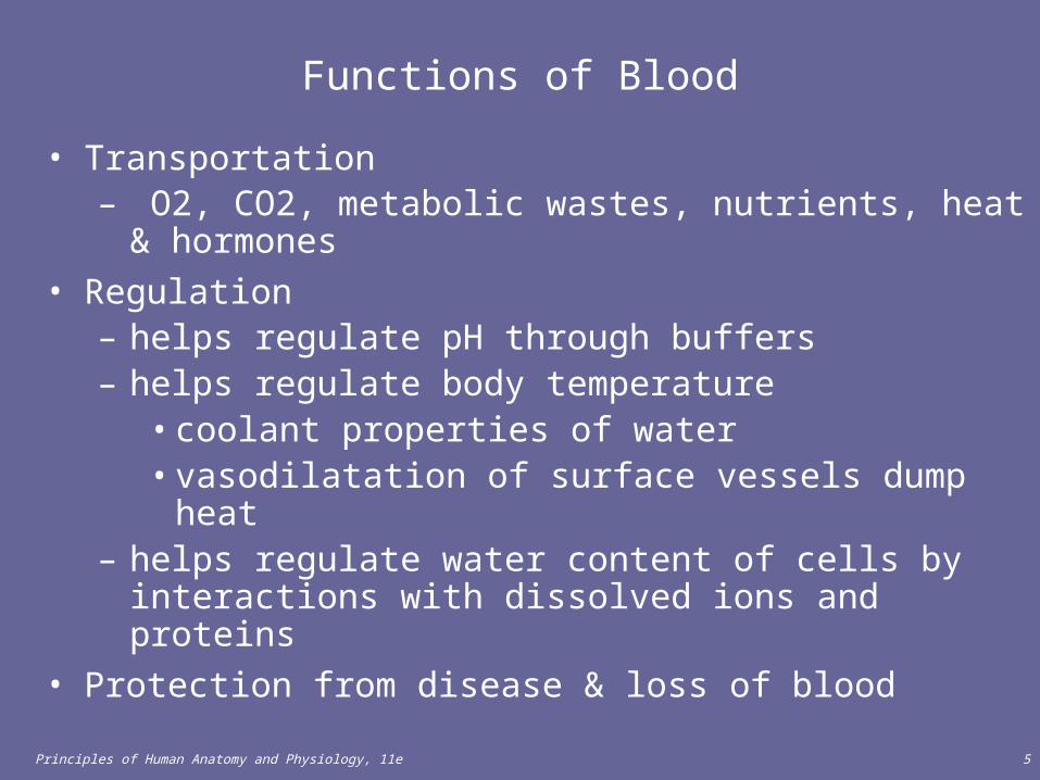
Functions of Blood
• Transportation– O2, CO2, metabolic wastes, nutrients, heat & hormones
• Regulation– helps regulate pH through buffers– helps regulate body temperature
• coolant properties of water • vasodilatation of surface vessels dump heat
– helps regulate water content of cells by interactions with dissolved ions and proteins
• Protection from disease & loss of blood
Principles of Human Anatomy and Physiology, 11e 6
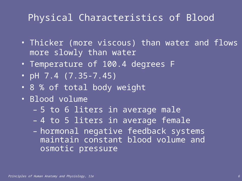
Physical Characteristics of Blood
• Thicker (more viscous) than water and flows more slowly than water
• Temperature of 100.4 degrees F• pH 7.4 (7.35-7.45)• 8 % of total body weight• Blood volume
– 5 to 6 liters in average male– 4 to 5 liters in average female– hormonal negative feedback systems maintain constant
blood volume and osmotic pressure

Principles of Human Anatomy and Physiology, 11e 7
Techniques of Blood Sampling
• Venipuncture– sample taken from vein with hypodermic needle &
syringe– median cubital vein (see page 717)– why not stick an artery?
• less pressure• closer to the surface
• Finger or heel stick– common technique for diabetics to monitor daily
blood sugar– method used for infants

Principles of Human Anatomy and Physiology, 11e 8
COMPONENTS OF BLOOD
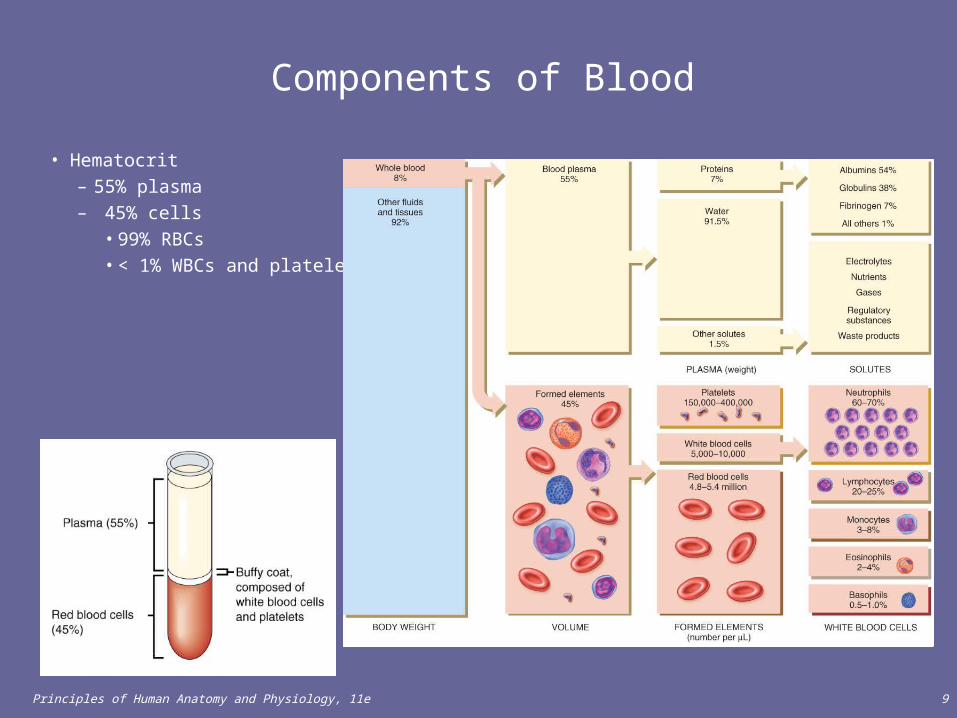
• Blood consists of 55% plasma and 45% formed elements (Figure 19.1).
• Blood plasma consists of 91.5% water and 8.5% solutes.• Principal solutes include proteins (albumins, globulins,
fibrinogen), nutrients, enzymes, hormones, respiratory gases, electrolytes, and waste products.
• Table 19.1 summarizes the chemical composition of plasma.
Principles of Human Anatomy and Physiology, 11e 9
Components of Blood
• Hematocrit– 55% plasma– 45% cells
• 99% RBCs• < 1% WBCs and platelets

Principles of Human Anatomy and Physiology, 11e 10
Blood Plasma
• 0ver 90% water• 7% plasma proteins
• created in liver• confined to bloodstream
– albumin• maintain blood osmotic pressure
– globulins (immunoglobulins)• antibodies bind to foreign
substances called antigens• form antigen-antibody complexes
– fibrinogen• for clotting
• 2% other substances – electrolytes, nutrients, hormones, gases, waste products

Principles of Human Anatomy and Physiology, 11e 11
Formed Elements of Blood• Red blood cells ( erythrocytes )• White blood cells ( leukocytes )
– granular leukocytes• neutrophils, eosinophils, basophils
– agranular leukocytes• lymphocytes = T cells, B cells, and natural killer cells• monocytes
• Platelets (special cell fragments)
Principles of Human Anatomy and Physiology, 11e 12
FORMATION OF BLOOD CELLS
• Blood cells are formed from pluripotent hematopoietic stem cells (Figure 19.3).
• Bone marrow may be obtained through aspiration or biopsy. The sample is then sent to pathology for examination.
• Originating from the pluripotent stem cells are the myeloid stem cells and lymphoid stem cells.

Principles of Human Anatomy and Physiology, 11e 13
Hematocrit
• Percentage of blood occupied by cells– female normal range
• 38 - 46% (average of 42%)– male normal range
• 40 - 54% (average of 46%)• testosterone
• Anemia – not enough RBCs or not enough hemoglobin
• Polycythemia– too many RBCs (over 65%)– dehydration, tissue hypoxia, blood doping in athletes

Principles of Human Anatomy and Physiology, 11e 14
Blood Doping
• Injecting previously stored RBC’s before an athletic event– more cells available to deliver oxygen to tissues
• Dangerous – increases blood viscosity– forces heart to work harder
• Banned by Olympic committee

Principles of Human Anatomy and Physiology, 11e 15
Formation of Blood Cells
• Most blood cells types need to be continually replaced– die within hours, days or weeks– process of blood cells formation is hematopoiesis or
hemopoiesis• In the embryo
– occurs in yolk sac, liver, spleen, thymus, lymph nodes & red bone marrow
• In adult– occurs only in red marrow of flat bones like sternum, ribs,
skull & pelvis and ends of long bones

Principles of Human Anatomy and Physiology, 11e 16
Hematopoiesis

Principles of Human Anatomy and Physiology, 11e 17
Stages of Blood Cell Formation• Pluripotent stem cells
– .1% of red marrow cells– replenish themselves as they differentiate into either myeloid or lymphoid
stem cells• Myeloid stem cell line of development continues:
– progenitor cells(colony-forming units) no longer can divide and are specialized to form specific cell types
• example: CFU-E develops eventually into only red blood cells– next generation is blast cells
• have recognizable histological characteristics • develop within several divisions into mature cell types
• Lymphoid stem cell line of development – pre-B cells & prothymocytes finish their develop into B & T lymphocytes
in the lymphatic tissue after leaving the red marrow

Principles of Human Anatomy and Physiology, 11e 18
Hemopoietic Growth Factors
• Regulate differentiation & proliferation• Erythropoietin (EPO)
– produced by the kidneys increase RBC precursors• Thrombopoietin (TPO)
– hormone from liver stimulates platelet formation• Cytokines are local hormones of bone marrow
– produced by some marrow cells to stimulate proliferation in other marrow cells
– colony-stimulating factor (CSF) & interleukin stimulate WBC production

Principles of Human Anatomy and Physiology, 11e 19
Medical Uses of Growth Factors
• Available through recombinant DNA technology– recombinant erythropoietin (EPO) very effective in
treating decreased RBC production of end-stage kidney disease
– other products given to stimulate WBC formation in cancer patients receiving chemotherapy which kills bone marrow
• granulocyte-macrophage colony-stimulating factor• granulocyte colony stimulating factor
– thrombopoietin helps prevent platelet depletion during chemotherapy

Principles of Human Anatomy and Physiology, 11e 20
Blood Cells
• Myeloid stem cells give rise to RBCs, platelets, and all WBCs except for lymphocytes.
• Lymphoid stem cells give rise to lymphocytes.• Myeloid stem cells differentiate into progenitor cells or
precursor cells (blast cells) which will develop into the actual formed elements of blood.
• Lymphoid stem cells differentiate into pre-B and prothymocytes which develop into B-lymphocytes and T-lymphocytes, respectively.
• This process of hemopoiesis (or hematopoiesis) is stimulated by several hematopoietic growth factors. These hematopoietic growth factors stimulate differentiation and proliferation of the various blood cells.
Principles of Human Anatomy and Physiology, 11e 21
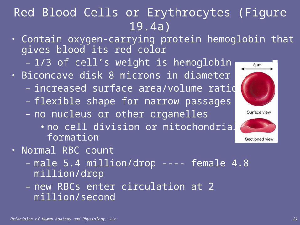
• Contain oxygen-carrying protein hemoglobin that gives blood its red color– 1/3 of cell’s weight is hemoglobin
• Biconcave disk 8 microns in diameter– increased surface area/volume ratio – flexible shape for narrow passages– no nucleus or other organelles
• no cell division or mitochondrial ATP formation• Normal RBC count
– male 5.4 million/drop ---- female 4.8 million/drop– new RBCs enter circulation at 2 million/second
Red Blood Cells or Erythrocytes (Figure 19.4a)

Principles of Human Anatomy and Physiology, 11e 22
Hormones
• Erythropoietin increases the number of RBC precursors.• Thrombopoietin increases the number of platelet precursors.• Cytokins (colony-stimulating factors and interleukins)
increase the number of WBC precursors.• Growth factors, available through recombinant DNA
technology, hold great potential for use in patients who cannot normally form the blood cells.

Principles of Human Anatomy and Physiology, 11e 23
Hemoglobin
• Globin protein consisting of 4 polypeptide chains• One heme pigment attached to each polypeptide chain
– each heme contains an iron ion (Fe+2) that can combine reversibly with one oxygen molecule

Principles of Human Anatomy and Physiology, 11e 24
Transport of O2, CO2 and Nitric Oxide
• Each hemoglobin molecule can carry 4 oxygen molecules from lungs to tissue cells
• Hemoglobin transports 23% of total CO2 waste from tissue cells to lungs for release– combines with amino acids in globin portion of Hb
• Hemoglobin transports nitric oxide & super nitric oxide helping to regulate BP– iron ions pick up nitric oxide (NO) & super nitric oxide
(SNO)& transport it to & from the lungs– NO causing vasoconstriction is released in the lungs– SNO causing vasodilation is picked up in the lungs

Principles of Human Anatomy and Physiology, 11e 25
RBCs
• Production of abnormal hemoglobin can result in serious blood disorders such as thalassemia and sickle cell anemia. (Figure 19.15)
• The blood test, hemoglobin A1c, can be used to monitor blood glucose levels in diabetics

Principles of Human Anatomy and Physiology, 11e 26
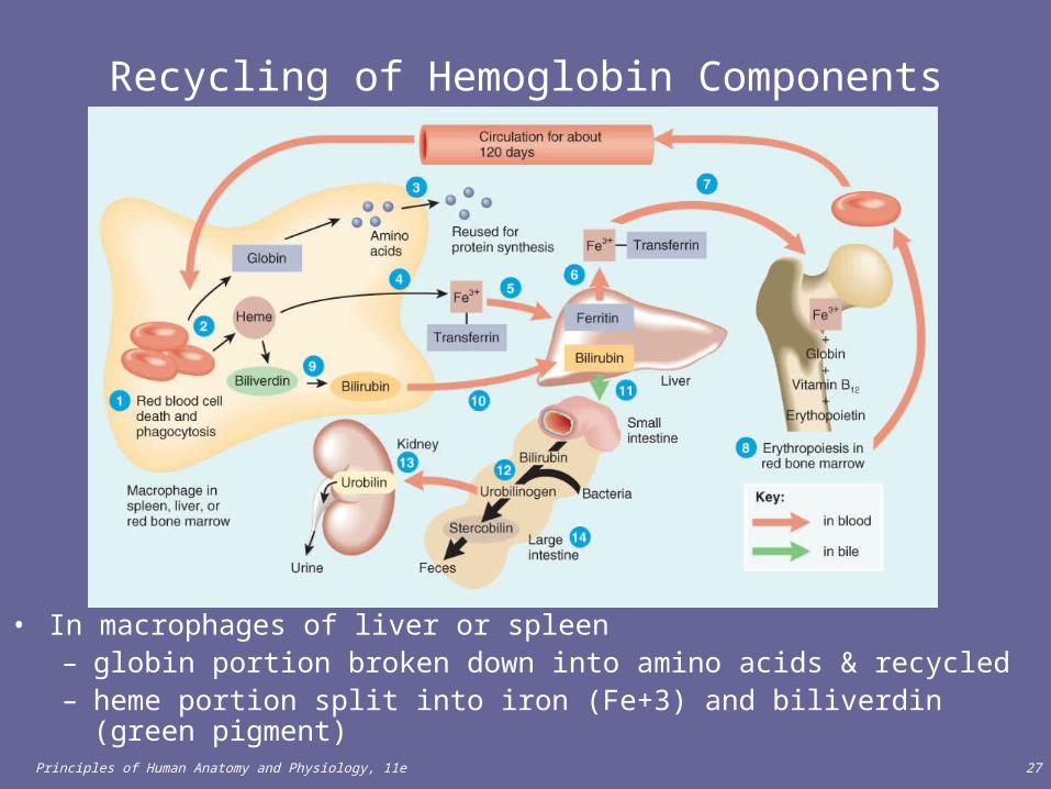
RBC Life Cycle
• RBCs live only 120 days– wear out from bending to fit through capillaries– no repair possible due to lack of organelles
• Worn out cells removed by fixed macrophages in spleen & liver
• Breakdown products are recycled
Principles of Human Anatomy and Physiology, 11e 27
Recycling of Hemoglobin Components
• In macrophages of liver or spleen – globin portion broken down into amino acids & recycled– heme portion split into iron (Fe+3) and biliverdin (green pigment)
Principles of Human Anatomy and Physiology, 11e 28
Fate of Components of Heme
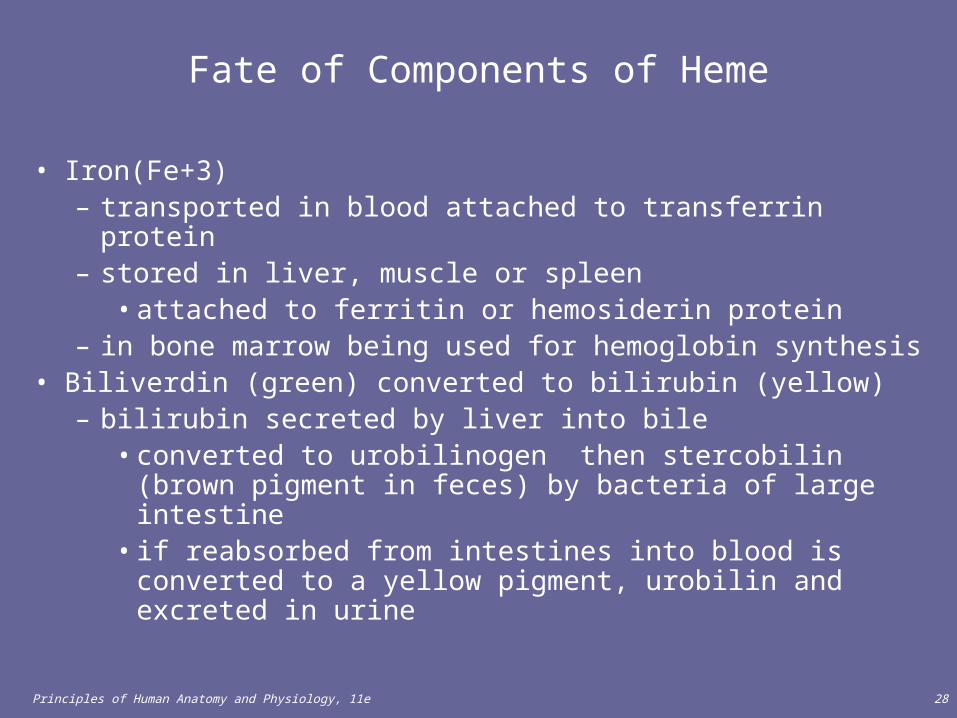
• Iron(Fe+3) – transported in blood attached to transferrin protein– stored in liver, muscle or spleen
• attached to ferritin or hemosiderin protein– in bone marrow being used for hemoglobin synthesis
• Biliverdin (green) converted to bilirubin (yellow)– bilirubin secreted by liver into bile
• converted to urobilinogen then stercobilin (brown pigment in feces) by bacteria of large intestine
• if reabsorbed from intestines into blood is converted to a yellow pigment, urobilin and excreted in urine

Principles of Human Anatomy and Physiology, 11e 29
Erythropoiesis: Production of RBCs
• Erythrocyte formation, called erythropoiesis, occurs in adult red bone marrow of certain bones (Figure 19.3).
• The main stimulus for erythropoiesis is hypoxia (Figure 19.6).
• Proerythroblast starts to produce hemoglobin• Many steps later, nucleus is ejected & a reticulocyte is
formed – orange in color with traces of visible rough ER
• Reticulocytes escape from bone marrow into the blood• In 1-2 days, they eject the remaining organelles to become
a mature RBC

Principles of Human Anatomy and Physiology, 11e 30
Feedback Control of RBC Production
• Tissue hypoxia (cells not getting enough O2)– high altitude since air has less O2– anemia
• RBC production falls below RBC destruction
– circulatory problems• Kidney response to hypoxia
– release erythropoietin– speeds up development of
proerythroblasts into reticulocytes

Principles of Human Anatomy and Physiology, 11e 31
Normal Reticulocyte Count
• Should be .5 to 1.5% of the circulating RBC’s• Low count in an anemic person might indicate bone
marrow problem– leukemia, nutritional deficiency or failure of red bone
marrow to respond to erythropoietin stimulation• High count might indicate recent blood loss or successful
iron therapy

Principles of Human Anatomy and Physiology, 11e 32
WHITE BLOOD CELLS
• Leukocytes (white blood cells or WBCs) are nucleated cells and do not contain hemoglobin. Two principal types are granular (neutrophils, eosinophils, basophils) and agranular (lymphocytes and monocytes) (Figure 19.7).– Granular leukocytes include eosinophils, basophils, and
neutrophils based on the straining of the granules.– Agranular leukocytes do not have cytoplasmic granules
and include the lymphocytes and monocytes, which differentiate into macrophages (fixed and wandering).
• Leukocytes have surface proteins, as do erythrocytes. They are called major histocompatibility antigens (MHC), are unique for each person (except for identical siblings), and can be used to identify a tissue.
Principles of Human Anatomy and Physiology, 11e 33
WBC Physiology
• Less numerous than RBCs– 5000 to 10,000 cells per drop of blood– 1 WBC for every 700 RBC
• Leukocytosis is a high white blood cell count– microbes, strenuous exercise, anesthesia or surgery
• Leukopenia is low white blood cell count– radiation, shock or chemotherapy
• Only 2% of total WBC population is in circulating blood at any given time– rest is in lymphatic fluid, skin, lungs, lymph nodes &
spleen

Principles of Human Anatomy and Physiology, 11e 34
Function of WBCs • Different WBCs combat inflammation and infection in different
ways.
– Neutrophils and wandering or fixed macrophages (which develop from monocytes) do so through phagocytosis.
– Eosinophils combat the effects of histamine in allergic reactions, phagocytize antigen-antibody complexes, and combat parasitic worms.
– Basophils develop into mast cells that liberate heparin, histamine, and serotonin in allergic reactions that intensify the inflammatory response.
– B lymphocytes, in response to the presence of foreign substances called antigens, differentiate into tissue plasma cells that produce antibodies.
– T lymphocytes destroy foreign invaders directly.

Principles of Human Anatomy and Physiology, 11e 35
Function of WBCs
• WBCs leave the blood stream by emigration (Figure 19.8).• Some WBCs, particularly neutrophils and macrophages, are
active in phagocytosis.• The chemical attraction of WBCs to a disease or injury site
is termed chemotaxis.

Principles of Human Anatomy and Physiology, 11e 36
WBC examination
• A differential white blood cell count is a diagnostic test in which specific white blood cells are enumerated. Because each type of WBC plays a different role, determining the percentage of each type in the blood assists in diagnosing the condition.
• Table 19.2 shows the significance of elevated or depressed counts of the various WBCs.
• Bone marrow transplants may be used to treat several types of anemia, leukemia, and numerous other blood disorders. (Clinical Application)
Principles of Human Anatomy and Physiology, 11e 37
WBC Anatomy and Types
• All WBCs (leukocytes) have a nucleus and no hemoglobin• Granular or agranular classification based on presence of
cytoplasmic granules made visible by staining– granulocytes are neutrophils, eosinophils or basophils– agranulocytes are monocyes or lymphocytes

Principles of Human Anatomy and Physiology, 11e 38
Neutrophils (Granulocyte)
• Polymorphonuclear Leukocytes or Polys• Nuclei = 2 to 5 lobes connected by thin strands
– older cells have more lobes– young cells called band cells because of horseshoe
shaped nucleus (band)• Fine, pale lilac practically invisible granules • Diameter is 10-12 microns • 60 to 70% of circulating WBCs
Principles of Human Anatomy and Physiology, 11e 39
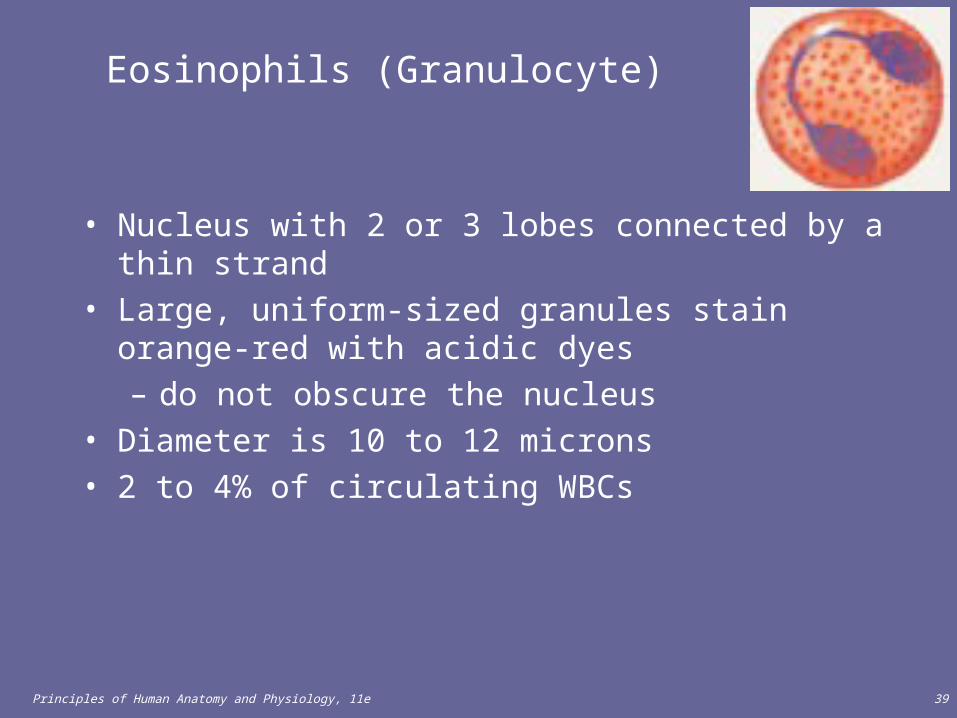
Eosinophils (Granulocyte)
• Nucleus with 2 or 3 lobes connected by a thin strand• Large, uniform-sized granules stain orange-red with
acidic dyes– do not obscure the nucleus
• Diameter is 10 to 12 microns• 2 to 4% of circulating WBCs

Principles of Human Anatomy and Physiology, 11e 40
Basophils (Granulocyte)
• Large, dark purple, variable-sized granules stain with basic dyes– obscure the nucleus
• Irregular, s-shaped, bilobed nuclei • Diameter is 8 to 10 microns• Less than 1% of circulating WBCs

Principles of Human Anatomy and Physiology, 11e 41
Lymphocyte (Agranulocyte)
• Dark, oval to round nucleus• Cytoplasm sky blue in color
– amount varies from rim of blue to normal amount• Small cells 6 - 9 microns in diameter• Large cells 10 - 14 microns in diameter
– increase in number during viral infections• 20 to 25% of circulating WBCs

Principles of Human Anatomy and Physiology, 11e 42
Monocyte (Agranulocyte)
• Nucleus is kidney or horse-shoe shaped• Largest WBC in circulating blood
– does not remain in blood long before migrating to the tissues– differentiate into macrophages
• fixed group found in specific tissues– alveolar macrophages in lungs– kupffer cells in liver
• wandering group gathers at sites of infection• Diameter is 12 - 20 microns• Cytoplasm is a foamy blue-gray • 3 to 8% o circulating WBCs

Principles of Human Anatomy and Physiology, 11e 43
Emigration & Phagocytosis in WBCs
• WBCs roll along endothelium, stick to it & squeeze between cells.– adhesion molecules (selectins)
help WBCs stick to endothelium• displayed near site of injury
– molecules (integrins) found on neutrophils assist in movement through wall
• Neutrophils & macrophages phagocytize bacteria & debris– chemotaxis of both
• kinins from injury site & toxins

Principles of Human Anatomy and Physiology, 11e 44
Neutrophil Function
• Fastest response of all WBC to bacteria• Direct actions against bacteria
– release lysozymes which destroy/digest bacteria– release defensin proteins that act like antibiotics & poke
holes in bacterial cell walls destroying them– release strong oxidants (bleach-like, strong chemicals )
that destroy bacteria

Principles of Human Anatomy and Physiology, 11e 45
Monocyte Function
• Take longer to get to site of infection, but arrive in larger numbers
• Become wandering macrophages, once they leave the capillaries
• Destroy microbes and clean up dead tissue following an infection

Principles of Human Anatomy and Physiology, 11e 46
Basophil Function
• Involved in inflammatory and allergy reactions• Leave capillaries & enter connective tissue as mast
cells• Release heparin, histamine & serotonin
– heighten the inflammatory response and account for hypersensitivity (allergic) reaction

Principles of Human Anatomy and Physiology, 11e 47
Eosinophil Function
• Leave capillaries to enter tissue fluid• Release histaminase
– slows down inflammation caused by basophils• Attack parasitic worms• Phagocytize antibody-antigen complexes
Principles of Human Anatomy and Physiology, 11e 48
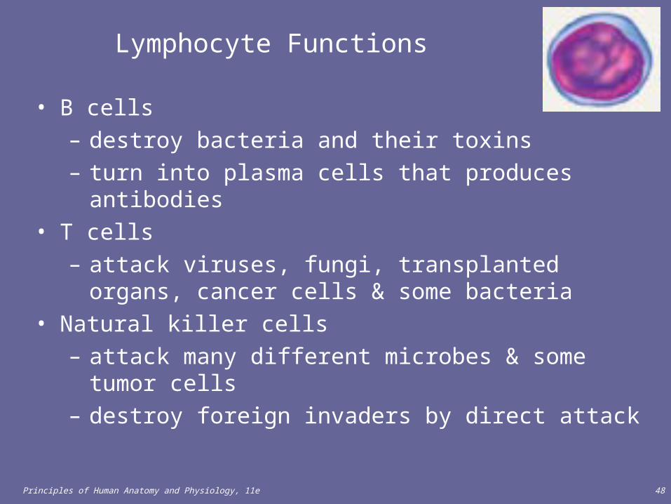
Lymphocyte Functions
• B cells– destroy bacteria and their toxins– turn into plasma cells that produces antibodies
• T cells– attack viruses, fungi, transplanted organs, cancer cells &
some bacteria• Natural killer cells
– attack many different microbes & some tumor cells– destroy foreign invaders by direct attack
Principles of Human Anatomy and Physiology, 11e 49
Complete Blood Count
• Screens for anemia and infection• Total RBC, WBC & platelet counts; differential WBC;
hematocrit and hemoglobin measurements• Normal hemoglobin range
– infants have 14 to 20 g/100mL of blood– adult females have 12 to 16 g/100mL of blood– adult males have 13.5 to 18g/100mL of blood

Principles of Human Anatomy and Physiology, 11e 50
Differential WBC Count
• Detection of changes in numbers of circulating WBCs (percentages of each type)– indicates infection, poisoning, leukemia, chemotherapy,
parasites or allergy reaction• Normal WBC counts
– neutrophils 60-70% (up if bacterial infection)– lymphocyte 20-25% (up if viral infection)– monocytes 3 -- 8 % (up if fungal/viral infection)– eosinophil 2 -- 4 % (up if parasite or allergy reaction)– basophil <1% (up if allergy reaction or hypothyroid)

Principles of Human Anatomy and Physiology, 11e 51
Bone Marrow Transplant
• Intravenous transfer of healthy bone marrow • Procedure
– destroy sick bone marrow with radiation & chemotherapy– donor matches surface antigens on WBC– put sample of donor marrow into patient's vein for
reseeding of bone marrow– success depends on histocompatibility of donor & recipient
• Treatment for leukemia, sickle-cell, breast, ovarian or testicular cancer, lymphoma or aplastic anemia
Principles of Human Anatomy and Physiology, 11e 52
PLATELETS
• Thrombopoietin stimulates myeloid stem cells to produce platelets.• Myeloid stem cells develop into megakaryocyte-colony-forming cells that
develop into megakaryoblasts (Figure 19.2).• Megakaryoblasts transform into megakaryocytes which fragment.• Each fragment, enclosed by a piece of cell membrane, is a platelet
(thrombocyte).• Normal blood contains 250,000 to 400,000 platelets/mm3. Platelets have
a life span of only 5 to 9 days; aged and dead platelets are removed by fixed macrophages in the spleen and liver.

Principles of Human Anatomy and Physiology, 11e 53
PLATELETS
• Platelets help stop blood loss from damaged vessels by forming a platelet plug. Their granules also contain chemicals that promote blood clotting.
• A complete blood count (CBC) is a test that screens for anemia and various infections. It usually includes counts of RBCs, WBCs, and platelets per μL of whole blood; hematocrit and differential white blood cell count. The amount of hemoglobin in grams per ml is also determined.
• Table 19.3 summarizes the formed elements in blood.

Principles of Human Anatomy and Physiology, 11e 54
Platelet (Thrombocyte) Anatomy
• Disc-shaped, 2 - 4 micron cell fragment with no nucleus
• Normal platelet count is 150,000-400,000/drop of blood
• Other blood cell counts– 5 million red & 5-10,000 white blood cells

Principles of Human Anatomy and Physiology, 11e 55
Platelets--Life History
• Platelets form in bone marrow by following steps: – myeloid stem cells to megakaryocyte-colony forming
cells to megakaryoblast to megakaryocytes whose cell fragments form platelets
• Short life span (5 to 9 days in bloodstream)– formed in bone marrow– few days in circulating blood– aged ones removed by fixed macrophages in liver
and spleen

Principles of Human Anatomy and Physiology, 11e 56
STEM CELL TRANSPLANT FROM BONE MARROW AND CORD-BLOOD
• Bone marrow transplant replaces diseased marrow with healthy marrow.
• Patient’s diseased marrow is destroyed.• Healthy marrow is supplied by a donor or the patient.• There are several problems with this method.

Principles of Human Anatomy and Physiology, 11e 57
Cord-blood transplant
• Stem cells are taken from the umbilical cord and frozen• This method offers several advantages over marrow
transplant.

Principles of Human Anatomy and Physiology, 11e 58
HEMOSTASIS
• A clot is a gel consisting of a network of insoluble protein fibers (fibrin) in which formed elements of blood are trapped (Figure 19.10).
• The chemicals involved in clotting are known as coagulation (clotting) factors; most are in blood plasma, some are released by platelets, and one is released from damaged tissue cells (Table 19.4).
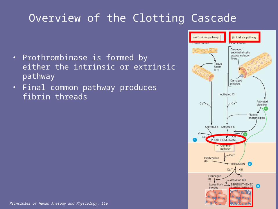
• Blood clotting involves a cascade of reactions that may be divided into three stages: formation of prothrombinase (prothrombin activator), conversion of prothrombin into thrombin, and conversion of soluble fibrinogen into insoluble fibrin (Figure 19.11).
Principles of Human Anatomy and Physiology, 11e 59
HEMOSTASIS
• The clotting cascade can be initiated by either the extrinsic pathway or the intrinsic pathway.
• Normal coagulation requires vitamin K and also involves clot retraction (tightening of the clot) and fibrinolysis (dissolution of the clot).
• The fibrinolytic system dissolves small, inappropriate clots and clots at a site of damage once the damage is repaired.
• Plasmin (fibrinolysin) can dissolve a clot by digesting fibrin threads and inactivating substances such as fibrinogen, prothrombin, and factors V, VIII, and XII.

Principles of Human Anatomy and Physiology, 11e 60
Hemostasis
• Stoppage of bleeding in a quick & localized fashion when blood vessels are damaged
• Prevents hemorrhage (loss of a large amount of blood)• Methods utilized
– vascular spasm– platelet plug formation– blood clotting (coagulation = formation of fibrin threads)

Principles of Human Anatomy and Physiology, 11e 61
Vascular Spasm
• Damage to blood vessel produces stimulates pain receptors• Reflex contraction of smooth muscle of small blood vessels• Can reduce blood loss for several hours until other
mechanisms can take over• Only for small blood vessel or arteriole

Principles of Human Anatomy and Physiology, 11e 62
Platelet Plug Formation• Platelets store a lot of chemicals in granules needed for
platelet plug formation– alpha granules
• clotting factors • platelet-derived growth factor
– cause proliferation of vascular endothelial cells, smooth muscle & fibroblasts to repair damaged vessels
– dense granules• ADP, ATP, Ca+2, serotonin, fibrin-stabilizing factor, &
enzymes that produce thromboxane A2• Steps in the process
– (1) platelet adhesion (2) platelet release reaction (3) platelet aggregation

Principles of Human Anatomy and Physiology, 11e 63
Platelet Adhesion
• Platelets stick to exposed collagen underlying damaged endothelial cells in vessel wall

Principles of Human Anatomy and Physiology, 11e 64
Platelet Release Reaction• Platelets activated by adhesion• Extend projections to make contact with each
other • Release thromboxane A2 & ADP activating
other platelets• Serotonin & thromboxane A2 are
vasoconstrictors decreasing blood flow through the injured vessel

Principles of Human Anatomy and Physiology, 11e 65
Platelet Aggregation
• Activated platelets stick together and activate new platelets to form a mass called a platelet plug
• Plug reinforced by fibrin threads formed during clotting process

Principles of Human Anatomy and Physiology, 11e 66
Blood Clotting
• Blood drawn from the body thickens into a gel– gel separates into liquid (serum) and a clot of insoluble fibers
(fibrin) in which the cells are trapped• If clotting occurs in an unbroken vessel is called a thrombosis• Substances required for clotting are Ca+2, enzymes synthesized by
liver cells and substances released by platelets or damaged tissues• Clotting is a cascade of reactions in which each clotting factor activates
the next in a fixed sequence resulting in the formation of fibrin threads– prothrombinase & Ca+2 convert prothrombin into thrombin– thrombin converts fibrinogen into fibrin threads
Principles of Human Anatomy and Physiology, 11e 67
Overview of the Clotting Cascade
• Prothrombinase is formed by either the intrinsic or extrinsic pathway
• Final common pathway produces fibrin threads

Principles of Human Anatomy and Physiology, 11e 68
Extrinsic Pathway
• Damaged tissues leak tissue factor (thromboplastin) into bloodstream
• Prothrombinase forms in seconds• In the presence of Ca+2, clotting factor
X combines with V to form prothrombinase

Principles of Human Anatomy and Physiology, 11e 69
Intrinsic Pathway
• Activation occurs– endothelium is damaged &
platelets come in contact with collagen of blood vessel wall
– platelets damaged & release phospholipids
• Requires several minutes for reaction to occur
• Substances involved: Ca+2 and clotting factors XII, X and V

Principles of Human Anatomy and Physiology, 11e 70
Final Common Pathway
• Prothrombinase and Ca+2 – catalyze the conversion of prothrombin
to thrombin• Thrombin
– in the presence of Ca+2 converts soluble fibrinogen to insoluble fibrin threads
– activates fibrin stabilizing factor XIII – positive feedback effects of thrombin
• accelerates formation of prothrombinase
• activates platelets to release phospholipids

Principles of Human Anatomy and Physiology, 11e 71
Clot Retraction & Blood Vessel Repair
• Clot plugs ruptured area of blood vessel• Platelets pull on fibrin threads causing
clot retraction – trapped platelets release factor XIII
stabilizing the fibrin threads• Edges of damaged vessel are pulled
together• Fibroblasts & endothelial cells repair the
blood vessel

Principles of Human Anatomy and Physiology, 11e 72
Role of Vitamin K in Clotting
• Normal clotting requires adequate vitamin K– fat soluble vitamin absorbed if lipids are present– absorption slowed if bile release is insufficient
• Required for synthesis of 4 clotting factors by hepatocytes– factors II (prothrombin), VII, IX and X
• Produced by bacteria in large intestine

Principles of Human Anatomy and Physiology, 11e 73
Hemostatic Control Mechanisms
• Fibrinolytic system dissolves small, inappropriate clots & clots at a site of a completed repair– fibrinolysis is dissolution of a clot
• Inactive plasminogen is incorporated into the clot– activation occurs because of factor XII and thrombin– plasminogen becomes plasmin (fibrinolysin) which digests fibrin
threads• Clot formation remains localized
– fibrin absorbs thrombin– blood disperses clotting factors– endothelial cells & WBC produce prostacyclin that opposes
thromboxane A2 (platelet adhesion & release)• Anticoagulants present in blood & produced by mast cells
–

Principles of Human Anatomy and Physiology, 11e 74
Intravascular Clotting
• Thrombosis– clot (thrombus) forming in an unbroken blood vessel
• forms on rough inner lining of BV• if blood flows too slowly (stasis) allowing clotting factors
to build up locally & cause coagulation– may dissolve spontaneously or dislodge & travel
• Embolus – clot, air bubble or fat from broken bone in the blood
• pulmonary embolus is found in lungs• Low dose aspirin blocks synthesis of thromboxane A2 &
reduces inappropriate clot formation– strokes, TIAs and myocardial infarctions

Principles of Human Anatomy and Physiology, 11e 75
Anticoagulants and Thrombolytic Agents
• Anticoagulants suppress or prevent blood clotting– heparin
• administered during hemodialysis and surgery– warfarin (Coumadin)
• antagonist to vitamin K so blocks synthesis of clotting factors• slower than heparin
– stored blood in blood banks treated with citrate phosphate dextrose (CPD) that removes Ca+2
• Thrombolytic agents are injected to dissolve clots– directly or indirectly activate plasminogen– streptokinase or tissue plasminogen activator (t-PA)

Principles of Human Anatomy and Physiology, 11e 76
Hemostatic Control Mechanisms
• Clots are generally localized due to fibrin absorbing thrombin into the clot, clotting factors diffusing through blood, and the production of prostacyclin, a powerful inhibitor of platelet adhesion and release.
• Substances that inhibit coagulation, called anticoagulants, are also present in blood. An example is heparin.
• Patients who are at increased risk of forming blood clots may receive an anticoagulant drug such as heparin or warfarin. To prevent clots in donated blood, a substance that removes Ca+2 such as EDTA or CPD may be added to the blood.
• Despite the anticoagulating and fibrinolytic mechanisms, blood clots sometimes form within the cardiovascular system.

Principles of Human Anatomy and Physiology, 11e 77
HEMOSTASIS
• Clotting in an unbroken blood vessel is called thrombosis.• A thrombus (clot), bubble of air, fat from broken bones, or
piece of debris transported by the bloodstream that moves from its site of origin is called an embolus.
• At low doses aspirin inhibits vasoconstriction and platelet aggregation thereby reducing the chance of thrombus formation. Thrombolytic agents are injected into the body to dissolve clots that have already formed. Streptokinase or tissue plasminogen activator (TPS) are thrombolytic agents.
Principles of Human Anatomy and Physiology, 11e 78
ABO Group
• In the ABO system, agglutinogens (antigens) A and B determine blood types (Figure 19.12).
• Plasma contains agglutinins (antibodies), designated as a and b, that react with agglutinogens that are foreign to the individual.
• Table 19.5 indicates the incidence of ABO and Rh blood types.

Principles of Human Anatomy and Physiology, 11e 79
Blood Groups and Blood Types
• RBC surfaces are marked by genetically determined glycoproteins & glycolipids – agglutinogens or isoantigens– distinguishes at least 24 different blood groups
• ABO, Rh, Lewis, Kell, Kidd and Duffy systems

Principles of Human Anatomy and Physiology, 11e 80
ABO Blood Groups
• Based on 2 glycolipid isoantigens called A and B found on the surface of RBCs
– display only antigen A -- blood type A
– display only antigen B -- blood type B
– display both antigens A & B -- blood type AB
– display neither antigen -- blood type O
• Plasma contains isoantibodies or agglutinins to the A or B antigens not found in your blood
– anti-A antibody reacts with antigen A
– anti-B antibody reacts with antigen B

Principles of Human Anatomy and Physiology, 11e 81
RH blood groups
• Antigen was discovered in blood of Rhesus monkey• People with Rh agglutinogens on RBC surface are Rh+.
Normal plasma contains no anti-Rh antibodies• Antibodies develop only in Rh- blood type & only with exposure
to the antigen– transfusion of positive blood– during a pregnancy with a positive blood type fetus
• Transfusion reaction upon 2nd exposure to the antigen results in hemolysis of the RBCs in the donated blood

Principles of Human Anatomy and Physiology, 11e 82
Hemolytic Disease of Newborn
• Rh negative mom and Rh+ fetus will have mixing of blood at birth• Mom's body creates Rh antibodies unless she receives a RhoGam shot soon after
first delivery, miscarriage or abortion– RhoGam binds to loose fetal blood and removes it from body before she reacts
• In 2nd child, hemolytic disease of the newborn may develop causing hemolysis of the fetal RBCs

Principles of Human Anatomy and Physiology, 11e 83
Transfusions
• Knowledge of blood types is essential to safe transfusion of blood and may also be used in proving or disproving paternity, linking suspects to crimes, or as a part of anthropology studies to establish a relationship among races.
• The interactions of the blood types of the ABO system are summarized in Table 19.6.

Principles of Human Anatomy and Physiology, 11e 84
Transfusion and Transfusion Reactions
• Transfer of whole blood, cells or plasma into the bloodstream of recipient– used to treat anemia or severe blood loss
• Incompatible blood transfusions
– antigen-antibody complexes form between plasma antibodies & “foreign proteins” on donated RBC's (agglutination)
– donated RBCs become leaky (complement proteins) & burst– loose hemoglobin causes kidney damage
• Problems caused by incompatibility between donor’s cells and recipient’s plasma
• Donor plasma is too diluted to cause problems

Principles of Human Anatomy and Physiology, 11e 85
Universal Donors and Recipients
• People with type AB blood called “universal recipients” since have no antibodies in plasma– only true if cross match the blood for other
antigens• People with type O blood cell called “universal
donors” since have no antigens on their cells– theoretically can be given to anyone

Principles of Human Anatomy and Physiology, 11e 86
Typing and Cross-Matching Blood for Transfusion
• The Rh and ABO blood groups may be detected by a simple medical test, blood typing, in which a sample of blood is mixed with serum containing agglutinins to each of the major agglutinogens (AB, B, and Rh) (Figure 19.14).
• Typing is the determination of blood types, whereas cross-matching is the mixing of donor and recipient blood for compatibility.

Principles of Human Anatomy and Physiology, 11e 87
DISORDERS: HOMEOSTATIC IMBALANCES
• Anemia • Sickle-cell • Hemophilia • Disseminated intravascular clotting • Acute leukemia• chronic leukemia

Principles of Human Anatomy and Physiology, 11e 88
Anemia = Not Enough RBCs
• Symptoms– oxygen-carrying capacity of blood is reduced– fatigue, cold intolerance & paleness
• lack of O2 for ATP & heat production• Types of anemia
– iron-deficiency =lack of absorption or loss of iron– pernicious = lack of intrinsic factor for B12 absorption– hemorrhagic = loss of RBCs due to bleeding (ulcer)– hemolytic = defects in cell membranes cause rupture– thalassemia = hereditary deficiency of hemoglobin– aplastic = destruction of bone marrow (radiation/toxins)

Principles of Human Anatomy and Physiology, 11e 89
Sickle-cell Anemia (SCA)
• Genetic defect in hemoglobin molecule (Hb-S) that changes 2 amino acids – at low very O2 levels, RBC is deformed by changes in
hemoglobin molecule within the RBC• sickle-shaped cells rupture easily = causing anemia &
clots
• Found among populations in malaria belt– Mediterranean Europe, sub-Saharan Africa & Asia
• Person with only one sickle cell gene– increased resistance to malaria because RBC membranes
leak K+ & lowered levels of K+ kill the parasite infecting the red blood cells
Principles of Human Anatomy and Physiology, 11e 90
Hemophilia
• Inherited deficiency of clotting factors – bleeding spontaneously or after minor trauma– subcutaneous & intramuscular hemorrhaging– nosebleeds, blood in urine, articular bleeding & pain
• Hemophilia A lacks factor VIII (males only)– most common
• Hemophilia B lacks factor IX (males only)• Hemophilia C (males & females)
– less severe because alternate clotting activator exists• Treatment is transfusions of fresh plasma or concentrates of
the missing clotting factor

Principles of Human Anatomy and Physiology, 11e 91
Disseminated Intravascular Clotting
• Life threatening paradoxical presence of blood clotting and bleeding at the same time throughout the whole body– so many clotting factors are removed by widespread
clotting that too few remain to permit normal clotting• Associated with infections, hypoxia, low blood flow rates,
trauma, hypotension & hemolysis• Clots cause ischemia and necrosis leading to
multisystem organ failure

Principles of Human Anatomy and Physiology, 11e 92
Leukemia
• Acute leukemia– uncontrolled production of immature leukocytes– crowding out of normal red bone marrow cells by
production of immature WBC– prevents production of RBC & platelets
• Chronic leukemia– accumulation of mature WBC in bloodstream because
they do not die– classified by type of WBC that is predominant---
monocytic, lymphocytic.

Principles of Human Anatomy and Physiology, 11e 93
end