Embed Size (px)
Citation preview
Principles of BiochemistryFourth Edition
Chapter 6Mechanisms of Enzymes
Copyright © 2006 Pearson Prentice Hall, Inc.
Horton • Moran • Scrimgeour • Perry • Rawn
The Terminology of Mechanistic Chemistry
A. Nucleophilic Substitutions
B. Cleavage Reactions
C. Oxidation—Reduction Reactions

Nucleophilic substitution reaction
Electron-rich, or nucleophilic Electron-poor, or electrophilic
unstable, high-energy state
Many chemical reactions have ionic intermediates.
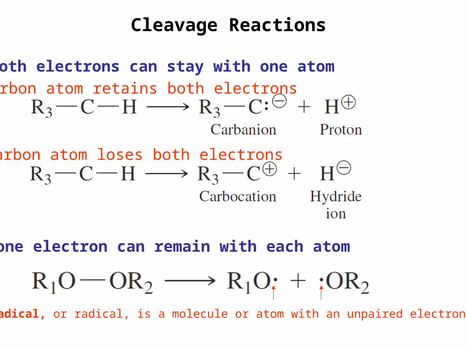
Cleavage Reactions
carbon atom retains both electrons
carbon atom loses both electrons
*one electron can remain with each atom
*both electrons can stay with one atom
A free radical, or radical, is a molecule or atom with an unpaired electron.
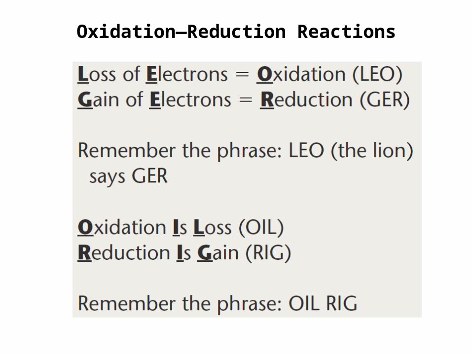
Oxidation—Reduction Reactions

Energy diagram for a single-step reaction
short lifetimes: 10-14 to 10-13 sec
lower activation barriermore stable transition state more often reaction proceed
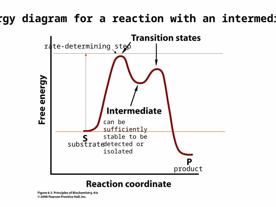
Energy diagram for a reaction with an intermediate
can be sufficiently stable to be detected or isolated
rate-determining step
substrate
product
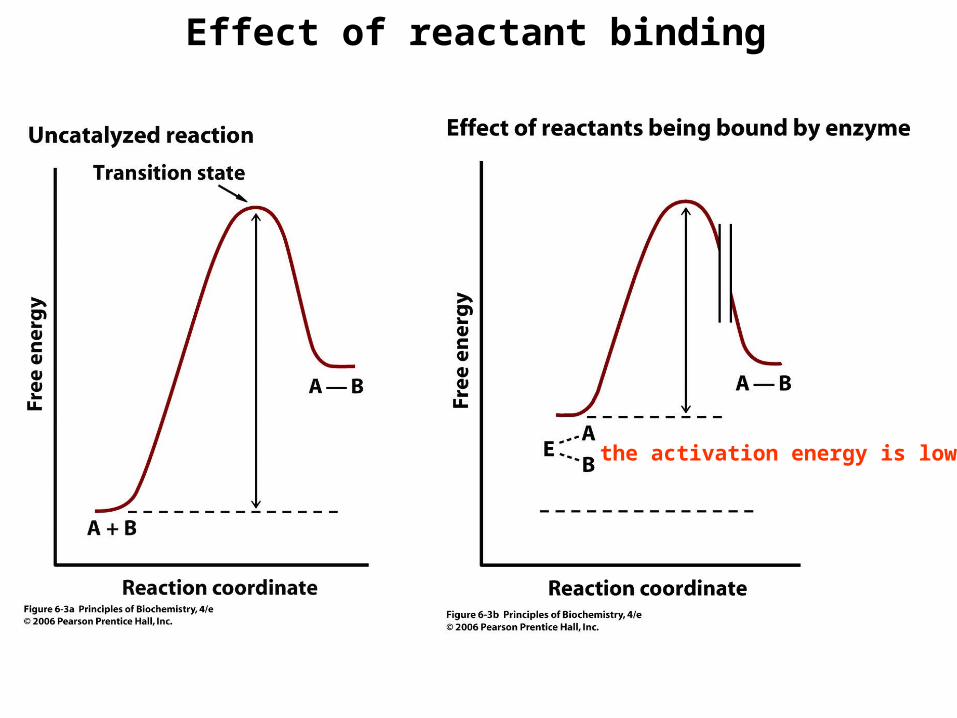
Effect of reactant binding
the activation energy is lowered
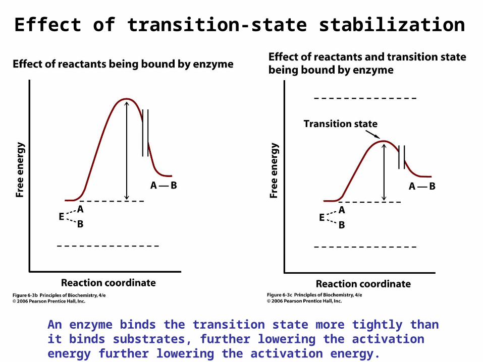
Effect of transition-state stabilization
An enzyme binds the transition state more tightly than it binds substrates, further lowering the activation energy further lowering the activation energy.
Chemical Modes of Enzymatic Catalysis
The formation of an ES complex places reactants in proximity to reactive amino acid residues in the enzyme active site.
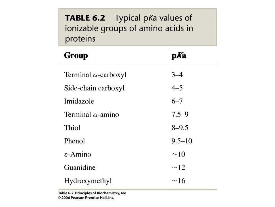
Ionizable side chains participate in two kinds of chemical catalysis: acid–base catalysis and covalent catalysis. These are the two major chemical modes of catalysis.
In addition to reactive amino acid residues, there may be metal ions or coenzymes in the active site.
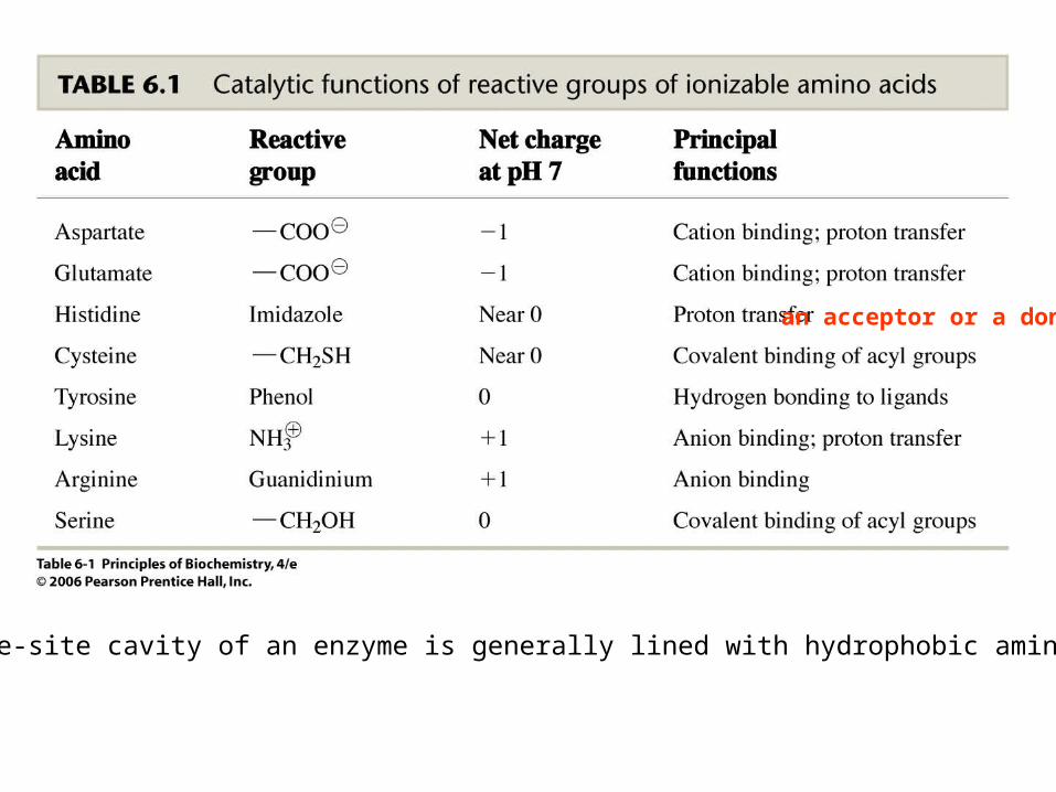
The active-site cavity of an enzyme is generally lined with hydrophobic amino acidresidues.
an acceptor or a donor
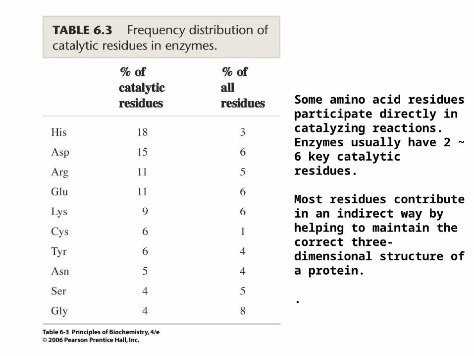
Some amino acid residues participate directly in catalyzing reactions. Enzymes usually have 2 ~ 6 key catalytic residues.
Most residues contribute in an indirect way by helping to maintain the correct three-dimensional structure of a protein.
.
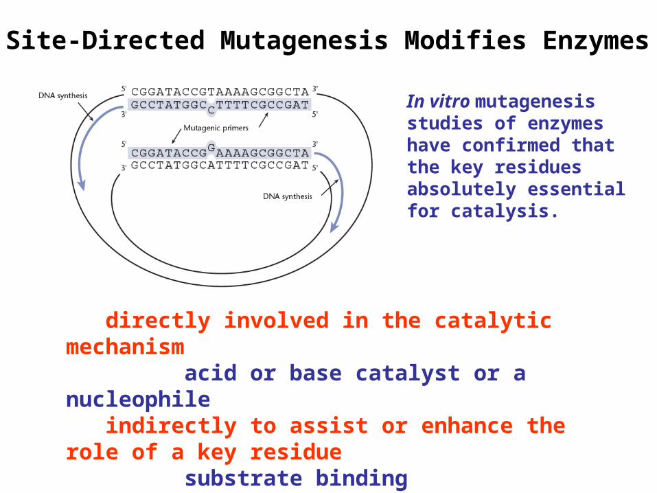
Site-Directed Mutagenesis Modifies Enzymes
directly involved in the catalytic mechanism acid or base catalyst or a nucleophile indirectly to assist or enhance the role of a key residue substrate binding stabilization of the transition state interacting with essential cofactors.
In vitro mutagenesis studies of enzymes have confirmed that the key residues absolutely essential for catalysis.
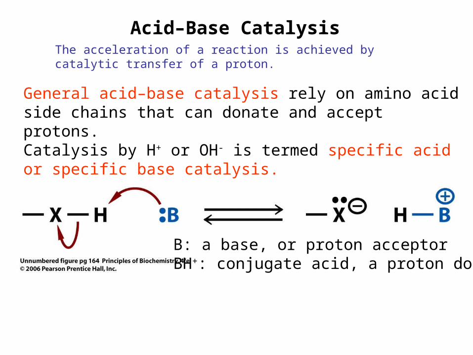
Acid–Base CatalysisThe acceleration of a reaction is achieved by catalytic transfer of a proton.
General acid–base catalysis rely on amino acid side chains that can donate and accept protons.Catalysis by H+ or OH- is termed specific acid or specific base catalysis.
B: a base, or proton acceptorBH+: conjugate acid, a proton donor
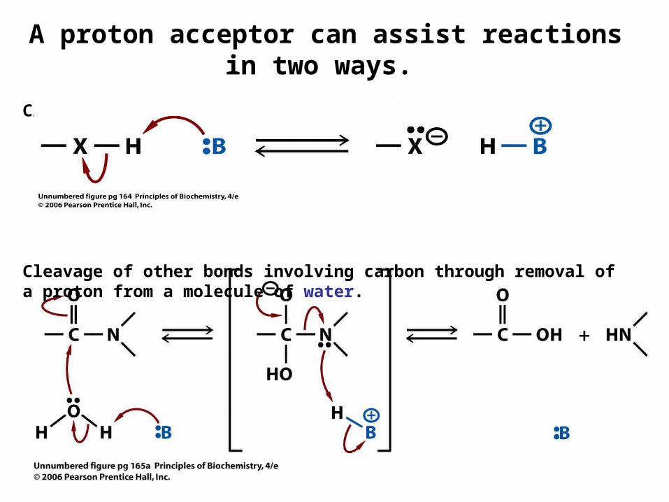
A proton acceptor can assist reactions in two ways.
Cleave O-H, N-H or C-H bonds by removing a proton.
Cleavage of other bonds involving carbon through removal of a proton from a molecule of water.
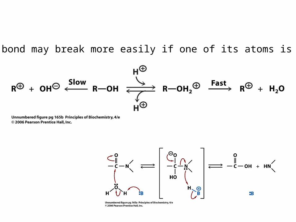
A covalent bond may break more easily if one of its atoms is protonated.
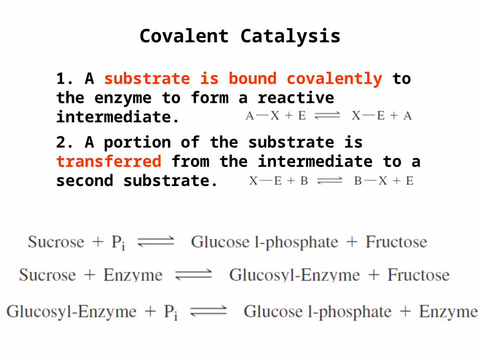
Covalent Catalysis
1. A substrate is bound covalently to the enzyme to form a reactive intermediate.
2. A portion of the substrate is transferred from the intermediate to a second substrate.
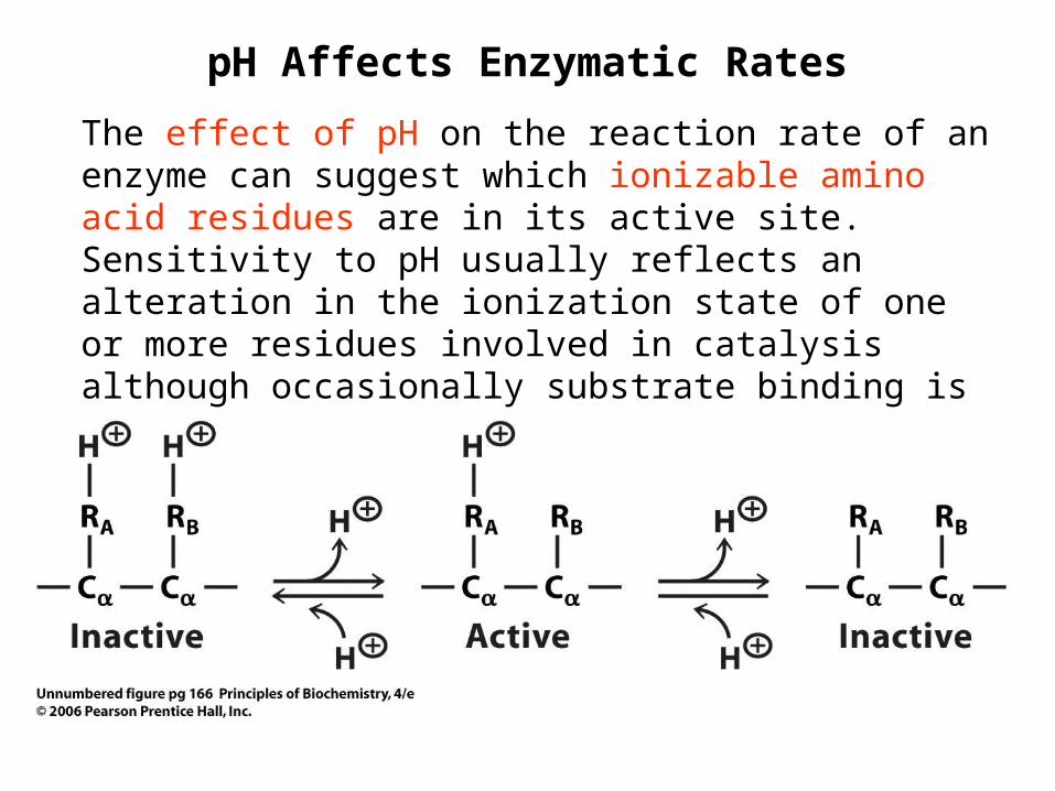
pH Affects Enzymatic Rates
The effect of pH on the reaction rate of an enzyme can suggest which ionizable amino acid residues are in its active site. Sensitivity to pH usually reflects an alteration in the ionization state of one or more residues involved in catalysis although occasionally substrate binding is affected.

pH–rate profile for papain
A plot of reaction velocity versus pH most often yields a bell-shaped curve provided the enzyme is not denatured when the pH is altered.
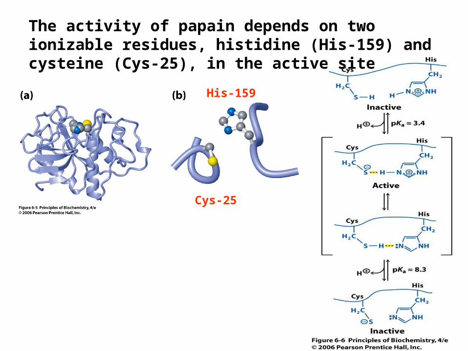
The activity of papain depends on two ionizable residues, histidine (His-159) and cysteine (Cys-25), in the active site
His-159
Cys-25
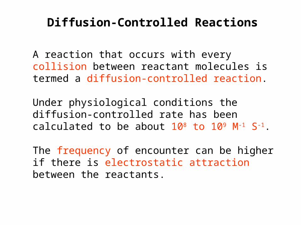
Diffusion-Controlled Reactions
A reaction that occurs with every collision between reactant molecules is termed a diffusion-controlled reaction.
Under physiological conditions the diffusion-controlled rate has been calculated to be about 108 to 109 M-1 S-1.
The frequency of encounter can be higher if there is electrostatic attraction between the reactants.

The binding of a substrate to an enzyme is a rapid reaction. If the rest of the reaction is simple and fast, the binding step may be the rate-determining step, and the overall rate of the reaction may approach the upper limit for catalysis.

Triose phosphate isomerase catalyzes the rapid interconversion of dihydroxyacetone phosphate (DHAP) and glyceraldehyde 3-phosphate (G3P) in the glycolysis and gluconeogenesis pathways.

General acid–base catalysis mechanism proposed for the reaction catalyzed by triose phosphate isomerase.
When dihydroxyacetone phosphate binds, the carbonyl oxygen forms a hydrogen bond with the neutral imidazole group of His-95. The carboxylate group of Glu-165 removes a proton from C-1 of the substrate to form an enediolate intermediate.
His-95 forms a strong hydrogen bond to the C-2 oxygen atom of the enediolate, and protonates this oxygen atom.

General acid–base catalysis mechanism proposed for the reaction catalyzed by triose phosphate isomerase.
Next, the imidazolate form of His-95 abstracts a proton from the hydroxyl group at C-1 and shuttles the proton between oxygen atoms, producing another unstable enediolate intermediate.
Glu-165 donates a proton to C-2, producing D-glyceraldehyde 3-phosphate.

Structure of yeast (Saccharomyces cerevisiae) triose phosphate isomerase
transition state analogue
His-95
Glu-165
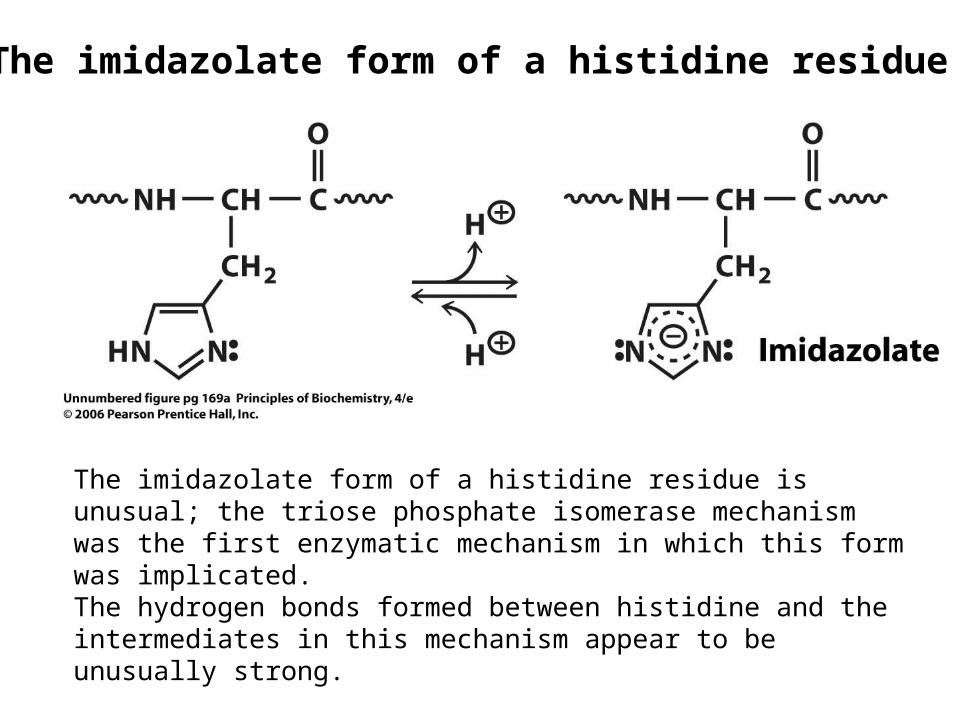
The imidazolate form of a histidine residue is unusual; the triose phosphate isomerase mechanism was the first enzymatic mechanism in which this form was implicated.The hydrogen bonds formed between histidine and the intermediates in this mechanism appear to be unusually strong.
The imidazolate form of a histidine residue

In the mid-1970s, Jeremy Knowles and his coworkers determined the rate constants of all four kinetically measurable enzymatic steps in both directions.
The reaction coordinate of triose phosphate isomerase
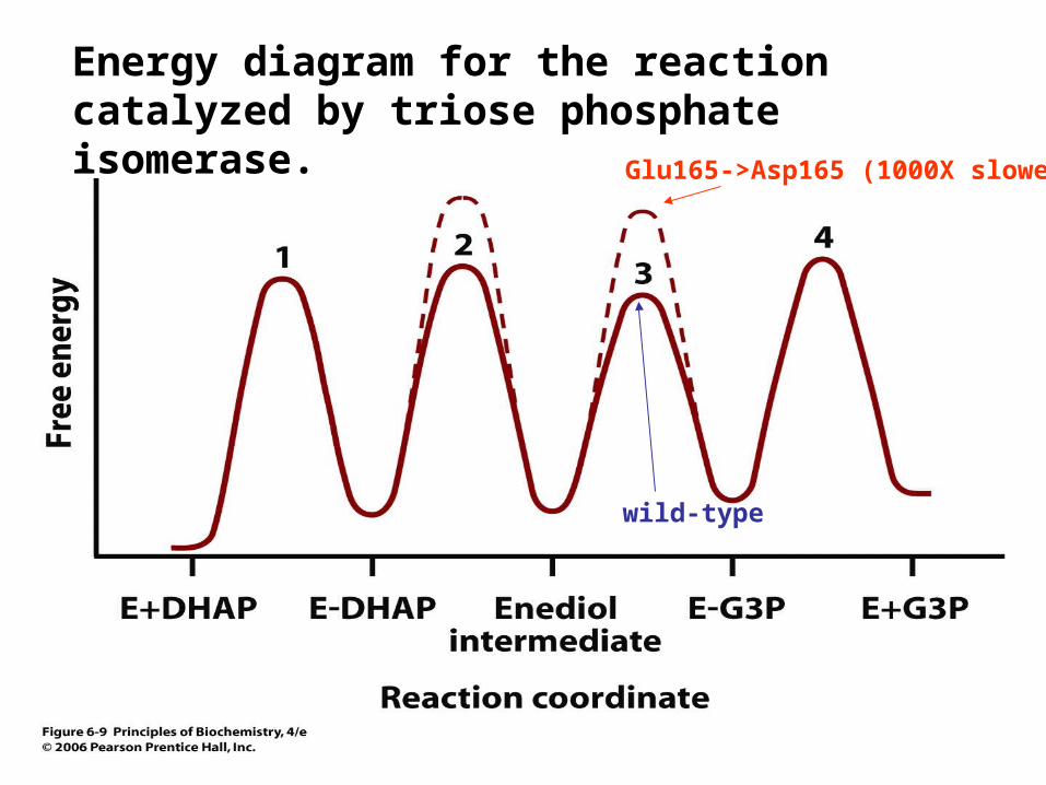
Energy diagram for the reaction catalyzed by triose phosphate isomerase.
wild-type
Glu165->Asp165 (1000X slower)
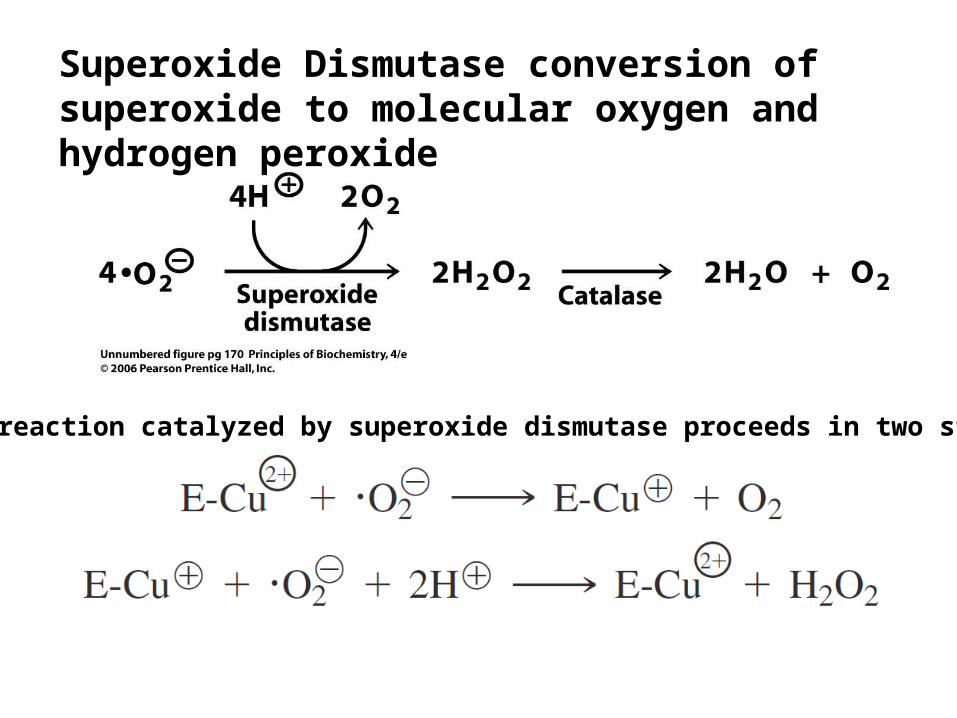
Superoxide Dismutase conversion of superoxide to molecular oxygen and hydrogen peroxide
The reaction catalyzed by superoxide dismutase proceeds in two steps
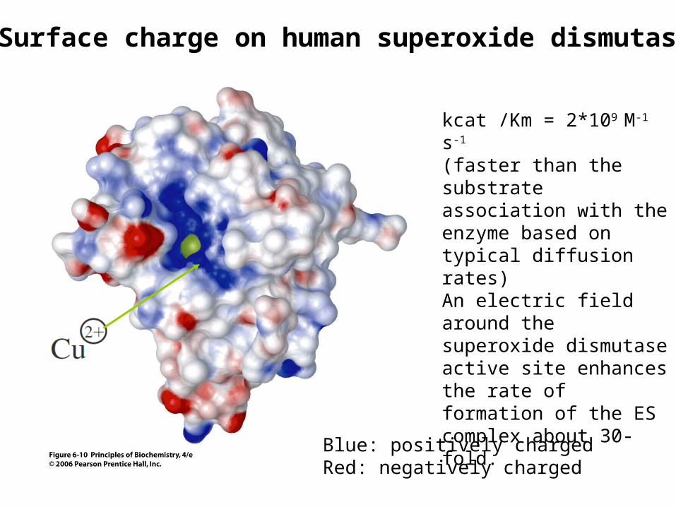
Surface charge on human superoxide dismutase
Blue: positively chargedRed: negatively charged
kcat /Km = 2*109 M-1 s-1
(faster than the substrate association with the enzyme based on typical diffusion rates)An electric field around the superoxide dismutase active site enhances the rate of formation of the ES complex about 30-fold.

The Proximity Effect
For entropy traps:decreasing their entropy and increasing the probability of their interaction.
The Proximity Effect:William Jencks and his colleagues two molecules at the active site work as an intramolecular (unimolecular)The acceleration is expressed in terms of the enhanced relative concentration, called the effective molarity, of the reacting groups in the unimolecular reaction.
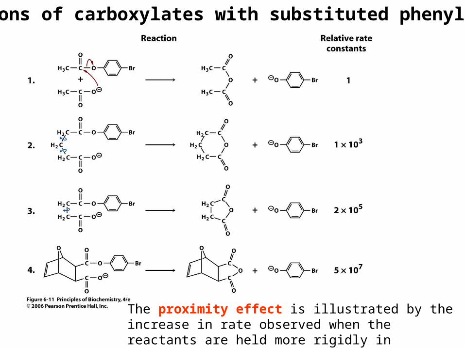
Reactions of carboxylates with substituted phenyl esters
The proximity effect is illustrated by the increase in rate observed when the reactants are held more rigidly in proximity.

Energy of substrate binding
Enzyme-substrate tight binding
Enzyme-substrate weak binding

Yeast hexokinase contains two structural domains connected by a hinge region (induced fit)
Open conformation Closed conformation
glucose
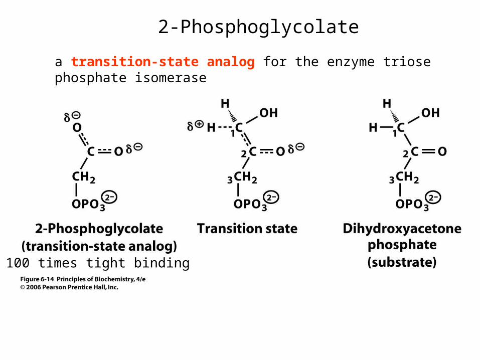
2-Phosphoglycolate
a transition-state analog for the enzyme triose phosphate isomerase
100 times tight binding
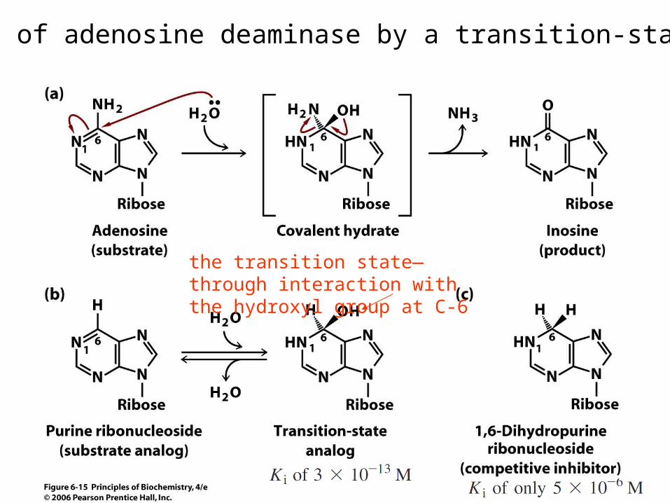
Inhibition of adenosine deaminase by a transition-state analog
the transition state—through interaction with the hydroxyl group at C-6
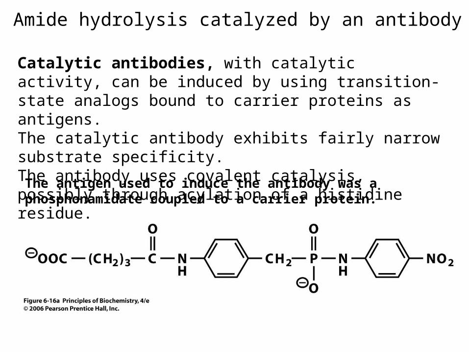
Amide hydrolysis catalyzed by an antibody
The antigen used to induce the antibody was a phosphonamidate coupled to a carrier protein.
Catalytic antibodies, with catalytic activity, can be induced by using transition-state analogs bound to carrier proteins as antigens.The catalytic antibody exhibits fairly narrow substrate specificity.The antibody uses covalent catalysis, possibly through acylation of a histidine residue.
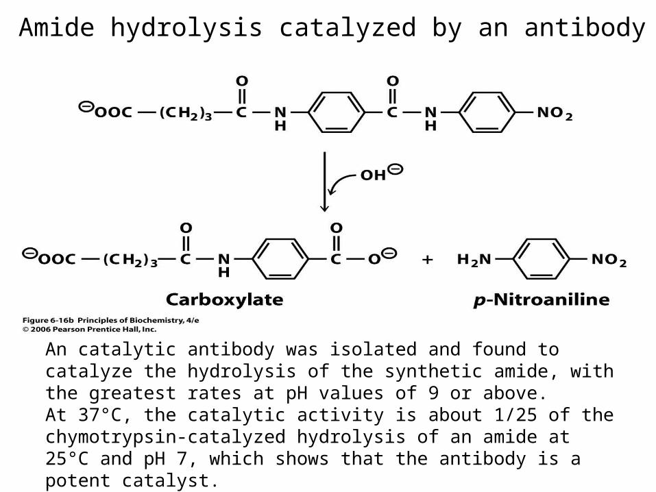
Amide hydrolysis catalyzed by an antibody
An catalytic antibody was isolated and found to catalyze the hydrolysis of the synthetic amide, with the greatest rates at pH values of 9 or above. At 37°C, the catalytic activity is about 1/25 of the chymotrypsin-catalyzed hydrolysis of an amide at 25°C and pH 7, which shows that the antibody is a potent catalyst.
Lysozyme
Lysozyme catalyzes the hydrolysis of some polysaccharides, especially those that make up the cell walls of bacteria. Lysozyme causes lysis, or disruption, of bacterial cells.Many secretions such as tears, saliva, and nasal mucus contain lysozyme activity to help prevent bacterial infection.
Lysozyme specifically catalyzes hydrolysis of the glycosidic bond between C-1 of a MurNAc residue and the oxygen atom at C-4 of a GlcNAc residue.
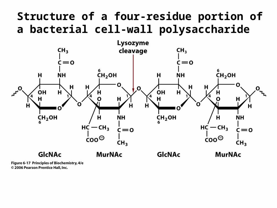
Structure of a four-residue portion of a bacterial cell-wall polysaccharide

Lysozyme from chicken with a trisaccharide molecule
trisaccharide molecule
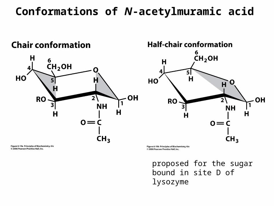
Conformations of N-acetylmuramic acid
proposed for the sugar bound in site D of lysozyme
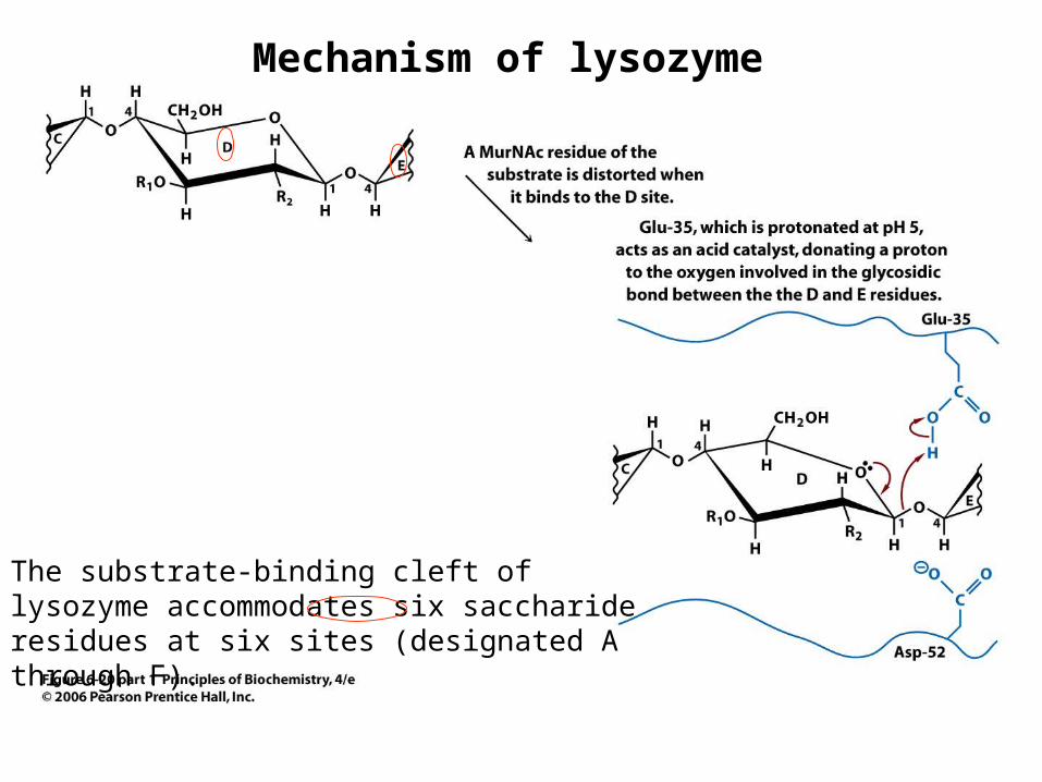
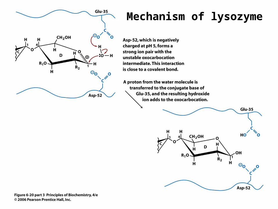
Mechanism of lysozyme
The substrate-binding cleft of lysozyme accommodates six saccharide residues at six sites (designated A through F).
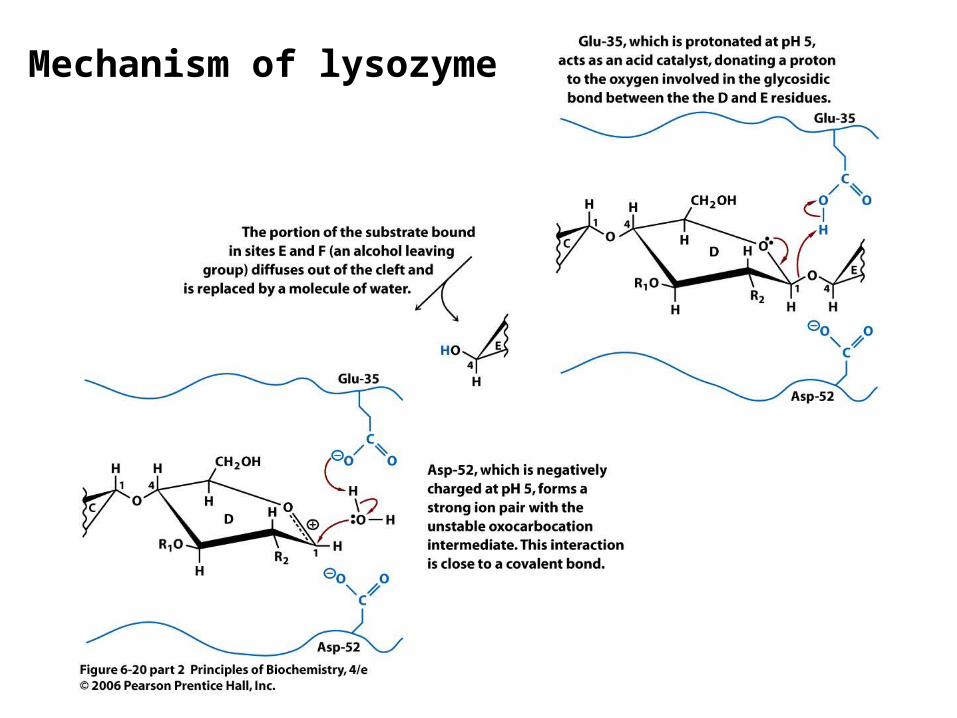
Mechanism of lysozyme
Mechanism of lysozyme
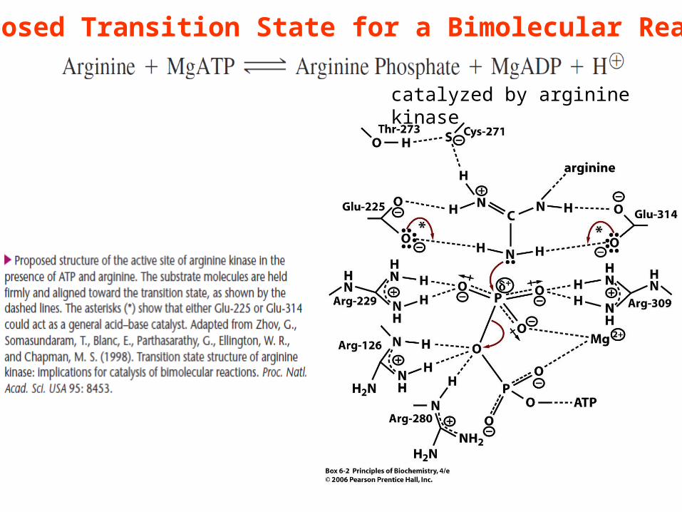
Proposed Transition State for a Bimolecular Reaction
catalyzed by arginine kinase
Properties of Serine Proteases
Serine proteases Cleave the peptide bond of proteinsSerine residue in their active sitesEx: trypsin, chymotrypsin, and elastase
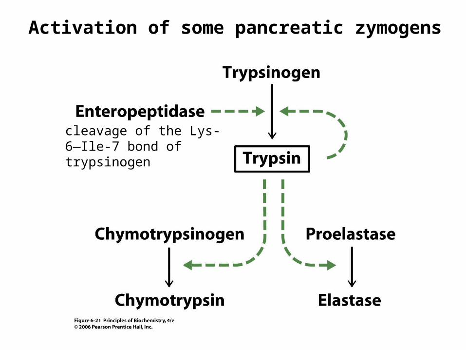
Activation of some pancreatic zymogens
cleavage of the Lys-6—Ile-7 bond of trypsinogen
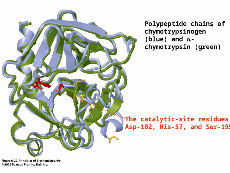
Polypeptide chains of chymotrypsinogen (blue) and -chymotrypsin (green)
The catalytic-site residues Asp-102, His-57, and Ser-195
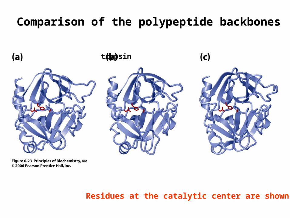
Comparison of the polypeptide backbones
chymotrypsin trypsin elastase
Residues at the catalytic center are shown in red
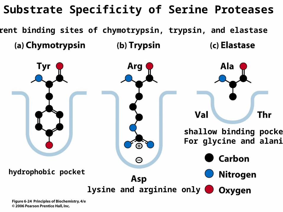
Different binding sites of chymotrypsin, trypsin, and elastase
hydrophobic pocket
shallow binding pocketFor glycine and alanine
lysine and arginine only
Substrate Specificity of Serine Proteases
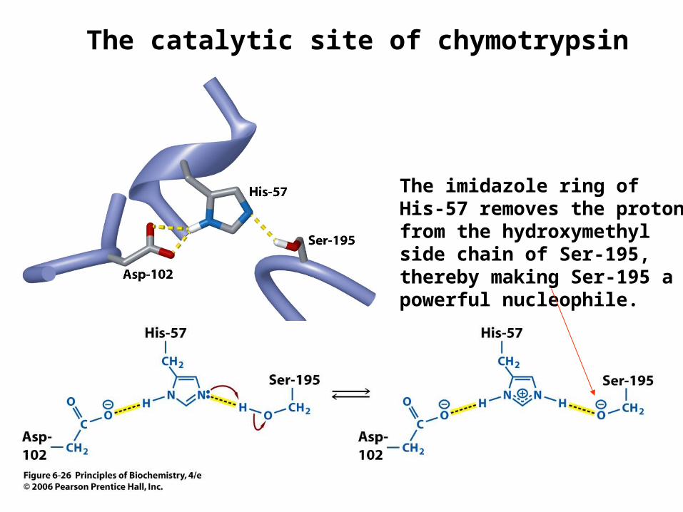
The catalytic site of chymotrypsin
The imidazole ring of His-57 removes the proton from the hydroxymethyl side chain of Ser-195, thereby making Ser-195 a powerful nucleophile.
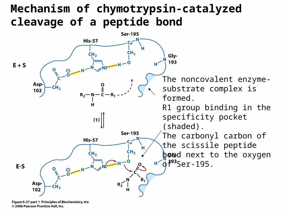
Mechanism of chymotrypsin-catalyzed cleavage of a peptide bond
The noncovalent enzyme-substrate complex is formed.R1 group binding in the specificity pocket (shaded). The carbonyl carbon of the scissile peptide bond next to the oxygen of Ser-195.
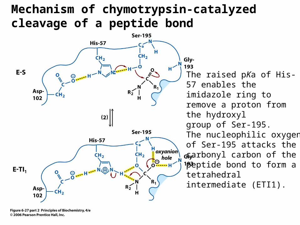
Mechanism of chymotrypsin-catalyzed cleavage of a peptide bond
The raised pKa of His-57 enables the imidazole ring to remove a proton from the hydroxylgroup of Ser-195. The nucleophilic oxygen of Ser-195 attacks the carbonyl carbon of the peptide bond to form a tetrahedral intermediate (ETI1).
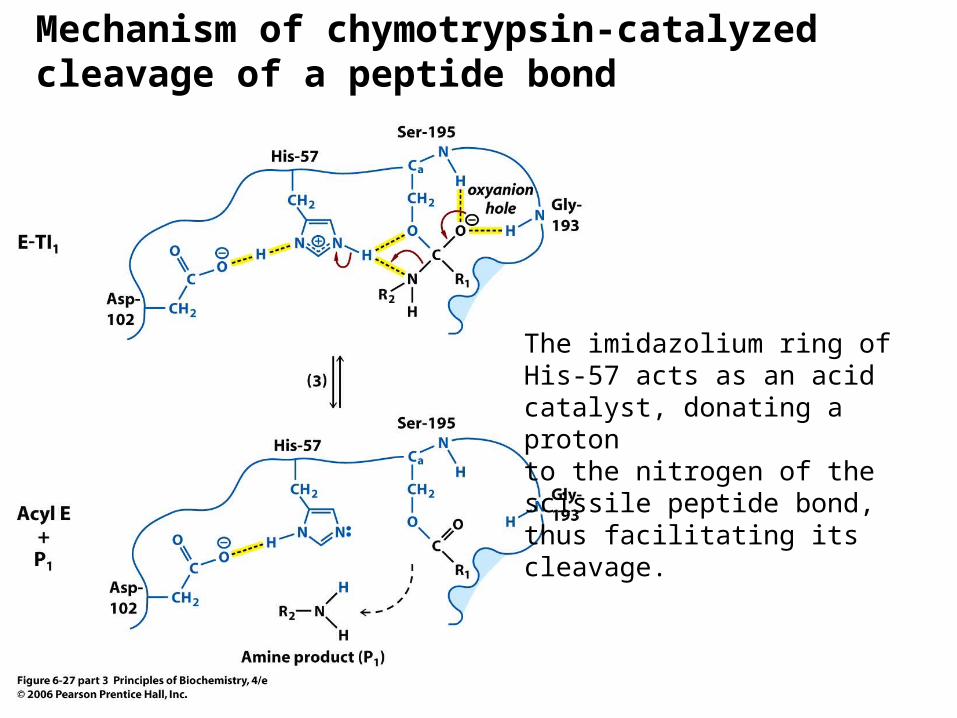
Mechanism of chymotrypsin-catalyzed cleavage of a peptide bond
The imidazolium ring of His-57 acts as an acid catalyst, donating a protonto the nitrogen of the scissile peptide bond, thus facilitating its cleavage.
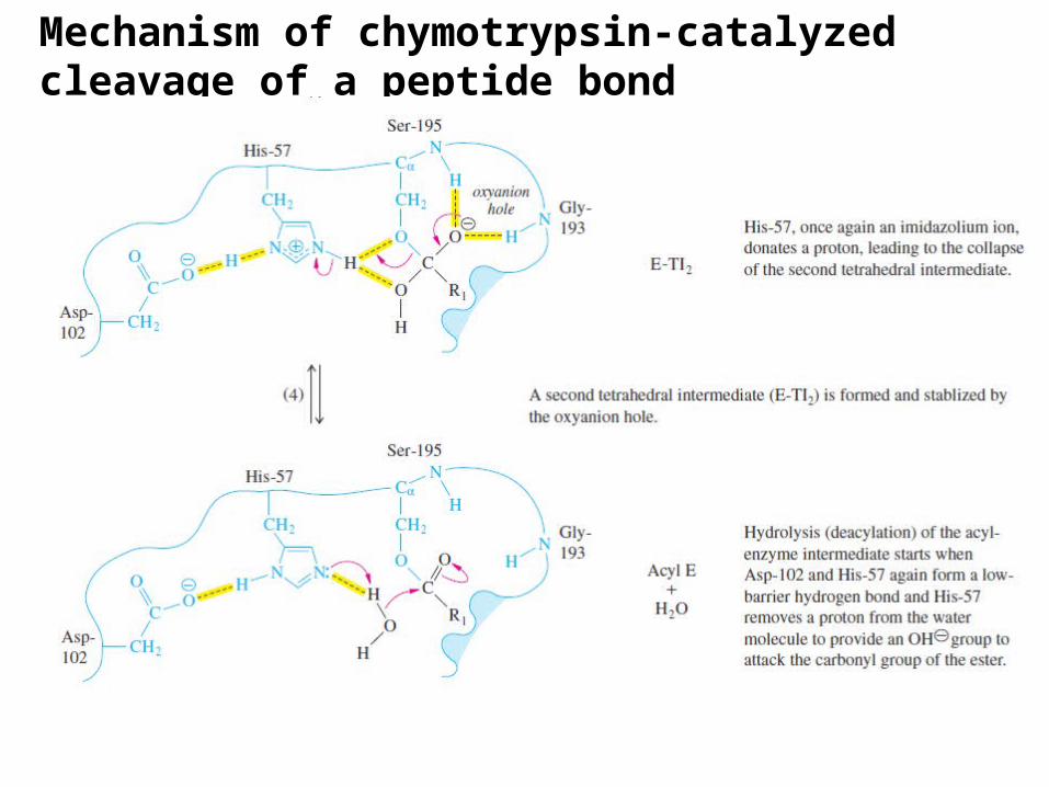
Mechanism of chymotrypsin-catalyzed cleavage of a peptide bond
Mechanism of chymotrypsin-catalyzed cleavage of a peptide bond
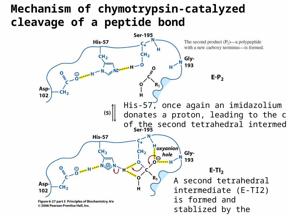
His-57, once again an imidazolium ion,donates a proton, leading to the collapseof the second tetrahedral intermediate.
A second tetrahedral intermediate (E-TI2) is formed and stablized by the oxyanion hole.
Mechanism of chymotrypsin-catalyzed cleavage of a peptide bond
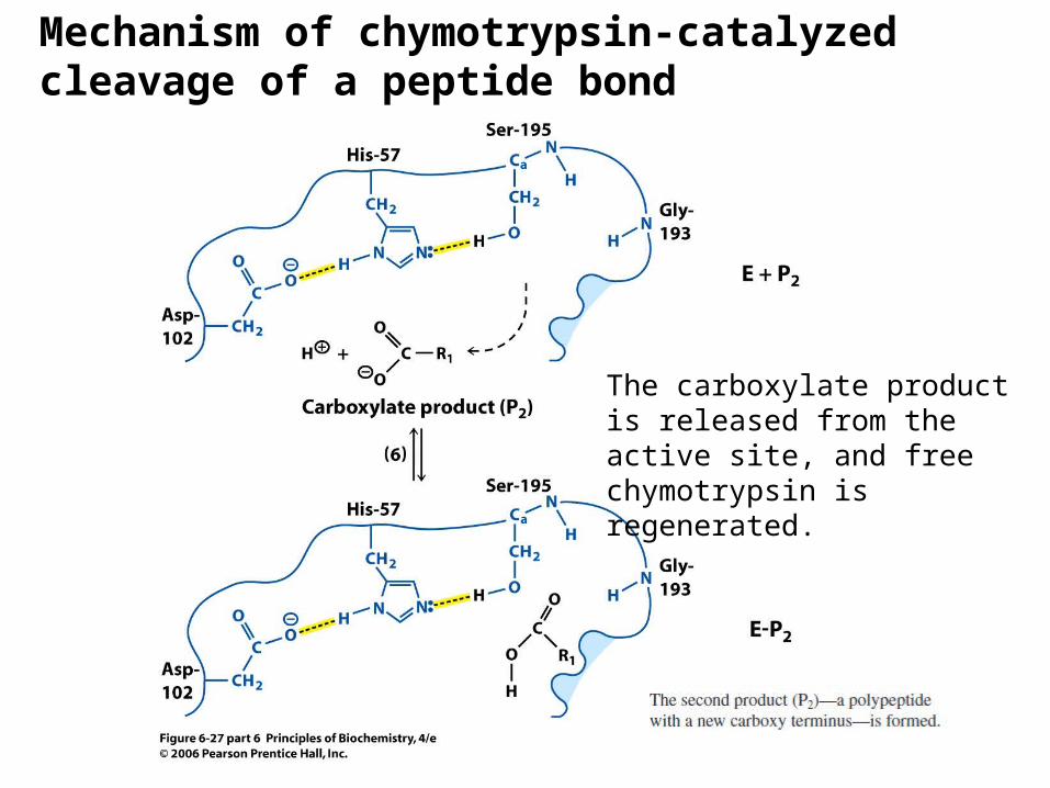
The carboxylate product is released from the active site, and free chymotrypsin is regenerated.