Embed Size (px)
Citation preview

Concepts of Botany, (page 1 of 10)
BIOL 221 – Concepts of Botany
Primary Plant Body: Embryogenesis and the Seedling
(Photo Atlas: Figures 1.29, 9.147, 9.148, 9.149, 9.150, 9.1, 9.2) A. Introduction Plants are composed of fewer cell types, fewer tissues, and fewer organs than animals, yet they exhibit a remarkable variety of forms which are adapted to an equally wide variety of environments. Unlike animals, plants exhibit much flexibility in their structure, have modular organization, and have indeterminate growth (post-embryonic development of organs and tissues, etc.) due to the presence of meristems. All of the cells of the plant body are derived from the meristematic regions in the shoot and the root. Following fertilization, the EMBYRO is formed during EMBRYOGENESIS and all of the primary organ regions are established. Following embryogenesis, GERMINATION, which is the resumption of growth in the seed ensues and the RADICLE or primary root emerges first. The beginning of the plant, as with most organism starts from the ZYGOTE which rapidly develops first into an EMBRYO with the SEED. In many seeds the embryo, once formed, stops growing and moves into a state of DORMANCY. Later, if the seed is in favorable conditions, it GERMINATES during which the embryo resumes rapid growth and development and becomes the mature plant. Development follows a fairly standard pattern. The embryo can be divided into two main regions, the HYPOCOTYL that develops into the root and the EPICOTYL that develops into the shoot system. Plants differ from animals in their mechanisms of growth. Unlike animals, plants continue to grow as long as they live. They exhibit open growth by retaining localized areas of embryonic cells called MERISTEMS. Animals also have meristems but they are more or less diffuse throughout the organ. Meristem tissue consists of cells which do two things: produce cells and produce hormones. The cells produced by meristems DIFFERENTIATE to become tissues which have certain functions such as photosynthesis, support, conduction, protection and specialized structures. Meristems found at the tips of root and stems are called APICAL MERISTEMS. A plant composed of cells originating from apical meristems is HERBACIOUS and makes up the PRIMARY PLANT BODY. Cells produced by LATERAL MERISTEMS add girth to stems and roots. Cells produced by lateral meristems are called SECONDARY TISSUES and make up the SECONDARY PLANT BODY. In complex land plants various plant cell types combine in characteristic ways to produce simple and complex tissues. These in turn form organs such as leaves, flowers and modified organs including thorns, spines, tendrils, and tubers.
Concepts of Botany, (page 2 of 10)
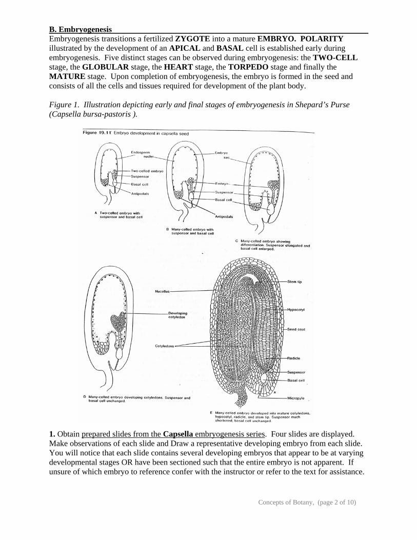
B. Embryogenesis Embryogenesis transitions a fertilized ZYGOTE into a mature EMBRYO. POLARITY illustrated by the development of an APICAL and BASAL cell is established early during embryogenesis. Five distinct stages can be observed during embryogenesis: the TWO-CELL stage, the GLOBULAR stage, the HEART stage, the TORPEDO stage and finally the MATURE stage. Upon completion of embryogenesis, the embryo is formed in the seed and consists of all the cells and tissues required for development of the plant body. Figure 1. Illustration depicting early and final stages of embryogenesis in Shepard’s Purse (Capsella bursa-pastoris ). 1. Obtain prepared slides from the Capsella embryogenesis series. Four slides are displayed. Make observations of each slide and Draw a representative developing embryo from each slide. You will notice that each slide contains several developing embryos that appear to be at varying developmental stages OR have been sectioned such that the entire embryo is not apparent. If unsure of which embryo to reference confer with the instructor or refer to the text for assistance.
Concepts of Botany, (page 3 of 10)
Draw and Label the embryos including the following: For each drawing, label the structures present using the following terms: two-celled proembryo, basal cell, suspensor, protoderm, procambium, ground meristem, endosperm, cotyledons, hypocotyl, shoot apical meristem, seed coat, root tip, and radicle (note, not all structures will be visible in all stages). Also label the stage each drawing best fits: Each of the prepared slides below may have embryos in either of the following 5 stages. For each prepared slide, label to which stage it belongs.
1. Globular stage 2. Heart stage 3. Torpedo stage 4. Bending cotyledons stage 5. Mature embryo stage.
a) PREPARED SLIDE: PRECOTYLEDON Is the basal cell apparent at this stage? How many cells are in the suspensor? Approximately how many cells are in the embryo? b) PREPARED SLIDE: EARLY COTYLEDONS Is the basal cell present? Is the suspensor present? Is this a dicot or monocot? How do you know? c) PREPARED SLIDE: BENDING COTYLEDONS Can you find evidence of a basal cell? Can you recognize the primary meristems? Is a root or shoot apex apparent? Why do the cotyledons bend? d) PREPARED SLIDE: MATURE EMBRYO How has the embryo changed? Is the basal cell connected to the embryo? Is the suspensor present? If not, WHY not?
Concepts of Botany, (page 4 of 10)
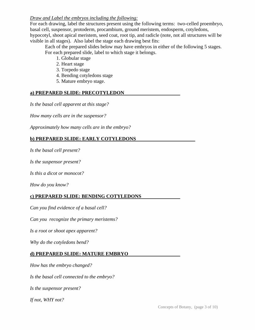
C. Embryo Strucuture At maturity the embryo often consists of an EPICOTYL, COTYLEDONS, and a HYPOCOTYL. The epicotyl, together with its juvenile leaves, is called the PLUMULE. If the tip of the hypocotyl is distinctly root-like, it is termed the RADICLE. In monocotyledonous plants such as the grasses, specialized terms are used to describe the embryonic parts: the single massive cotyledon is termed the SCUTELLUM, while the plumule and radicle are enclosed by protective structures termed the COLEOPTILE and the COLEORHIZA respectively. You will be expected to know all of the structures that are characteristic of both a dicot and a monocot seed and seedling. Figure 3. Illustration representing the long-section through a mature dicot (garden bean) and monocot (maize) seed. In figure 3 above, be able to identify the HOMOLOGOUS structures for monocots and dicots. 1. Using an imbibed BEAN seed, carefully break the seed open (you will have to remove the outer shell first) and examine the embryo with the naked eye AND with the dissecting scope. Draw and label the parts of the embryo and the seed. Is this an example of a dicot or a monocot? How do you know? Is a radicle apparent? What is the food source? Is this different from the maize?
Concepts of Botany, (page 5 of 10)
2. Using a dry MAIZE kernel, cut the kernel in half lengthwise to view the internal seed and embryo structures. Make a thin section of the kernel for use with the dissecting scope. ADD a drop of Potassium Iodine onto the cut section. This will stain the starch and improve viewing. Draw and label the parts of the embryo and the seed. Is this an example of a dicot or a monocot? How do you know? Is a radicle apparent? What tissue is staining positive for starch? That is, what will be the young seedling’s food source? Is this different from the bean? Be sure to be able to distinguish the young embryonic maize plant from the endosperm. 3. PREPARED SLIDE: ZEA (CORN OR MAIZE) EMBRYO, LONGITUDINAL SECTION. Be sure you can spot all structures in Figure 3 (right side) in this prepared section of an corn seed. Do this with the compound light microscope. 4. Now obtain a maize kernel and bean seed that have been imbibed for at least 3 days and appears to be germinating as recognized by the emergence of the radicle (primary root). Prepare a section for use with the dissecting scope as in step 4. Be careful with the seed so that the root (and any shoot) are bisected (cut longitudinally in half). This will allow you to see the growth of the organs from the internal regions of the embryo. Identify and Draw the organs and tissue recognizable:
Concepts of Botany, (page 6 of 10)
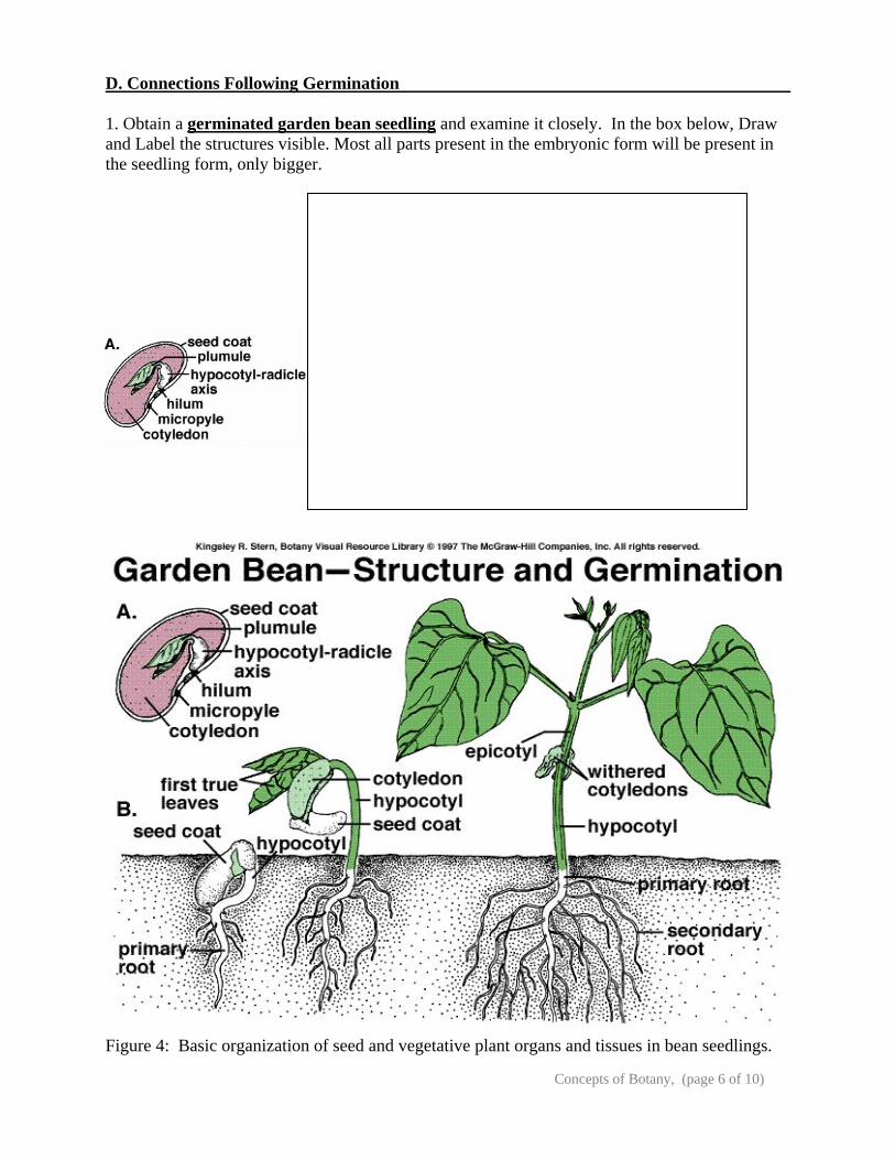
D. Connections Following Germination 1. Obtain a germinated garden bean seedling and examine it closely. In the box below, Draw and Label the structures visible. Most all parts present in the embryonic form will be present in the seedling form, only bigger.
Figure 4: Basic organization of seed and vegetative plant organs and tissues in bean seedlings.
Concepts of Botany, (page 7 of 10)
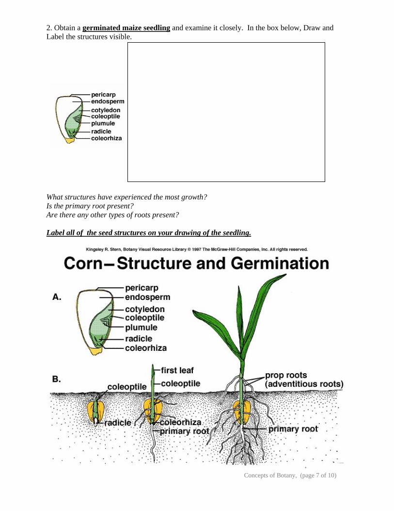
2. Obtain a germinated maize seedling and examine it closely. In the box below, Draw and Label the structures visible. What structures have experienced the most growth? Is the primary root present? Are there any other types of roots present? Label all of the seed structures on your drawing of the seedling.
Concepts of Botany, (page 8 of 10)
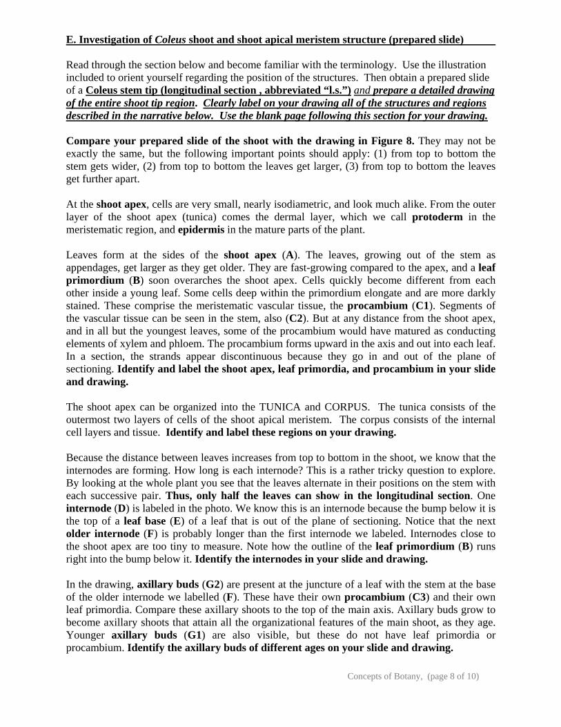
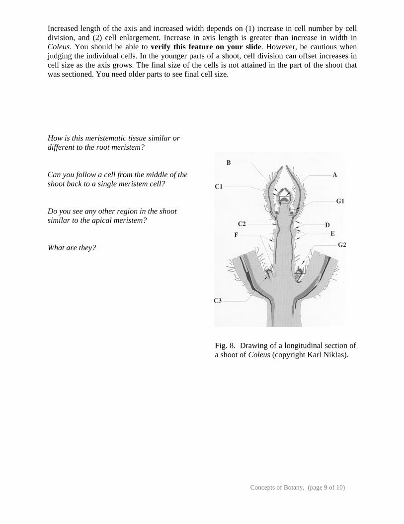
E. Investigation of Coleus shoot and shoot apical meristem structure (prepared slide) Read through the section below and become familiar with the terminology. Use the illustration included to orient yourself regarding the position of the structures. Then obtain a prepared slide of a Coleus stem tip (longitudinal section , abbreviated “l.s.”) and prepare a detailed drawing of the entire shoot tip region. Clearly label on your drawing all of the structures and regions described in the narrative below. Use the blank page following this section for your drawing. Compare your prepared slide of the shoot with the drawing in Figure 8. They may not be exactly the same, but the following important points should apply: (1) from top to bottom the stem gets wider, (2) from top to bottom the leaves get larger, (3) from top to bottom the leaves get further apart. At the shoot apex, cells are very small, nearly isodiametric, and look much alike. From the outer layer of the shoot apex (tunica) comes the dermal layer, which we call protoderm in the meristematic region, and epidermis in the mature parts of the plant. Leaves form at the sides of the shoot apex (A). The leaves, growing out of the stem as appendages, get larger as they get older. They are fast-growing compared to the apex, and a leaf primordium (B) soon overarches the shoot apex. Cells quickly become different from each other inside a young leaf. Some cells deep within the primordium elongate and are more darkly stained. These comprise the meristematic vascular tissue, the procambium (C1). Segments of the vascular tissue can be seen in the stem, also (C2). But at any distance from the shoot apex, and in all but the youngest leaves, some of the procambium would have matured as conducting elements of xylem and phloem. The procambium forms upward in the axis and out into each leaf. In a section, the strands appear discontinuous because they go in and out of the plane of sectioning. Identify and label the shoot apex, leaf primordia, and procambium in your slide and drawing. The shoot apex can be organized into the TUNICA and CORPUS. The tunica consists of the outermost two layers of cells of the shoot apical meristem. The corpus consists of the internal cell layers and tissue. Identify and label these regions on your drawing. Because the distance between leaves increases from top to bottom in the shoot, we know that the internodes are forming. How long is each internode? This is a rather tricky question to explore. By looking at the whole plant you see that the leaves alternate in their positions on the stem with each successive pair. Thus, only half the leaves can show in the longitudinal section. One internode (D) is labeled in the photo. We know this is an internode because the bump below it is the top of a leaf base (E) of a leaf that is out of the plane of sectioning. Notice that the next older internode (F) is probably longer than the first internode we labeled. Internodes close to the shoot apex are too tiny to measure. Note how the outline of the leaf primordium (B) runs right into the bump below it. Identify the internodes in your slide and drawing. In the drawing, axillary buds (G2) are present at the juncture of a leaf with the stem at the base of the older internode we labelled (F). These have their own procambium (C3) and their own leaf primordia. Compare these axillary shoots to the top of the main axis. Axillary buds grow to become axillary shoots that attain all the organizational features of the main shoot, as they age. Younger axillary buds (G1) are also visible, but these do not have leaf primordia or procambium. Identify the axillary buds of different ages on your slide and drawing.
Concepts of Botany, (page 9 of 10)
Increased length of the axis and increased width depends on (1) increase in cell number by cell division, and (2) cell enlargement. Increase in axis length is greater than increase in width in Coleus. You should be able to verify this feature on your slide. However, be cautious when judging the individual cells. In the younger parts of a shoot, cell division can offset increases in cell size as the axis grows. The final size of the cells is not attained in the part of the shoot that was sectioned. You need older parts to see final cell size. How is this meristematic tissue similar or different to the root meristem? Can you follow a cell from the middle of the shoot back to a single meristem cell? Do you see any other region in the shoot similar to the apical meristem? What are they?
Fig. 8. Drawing of a longitudinal section of a shoot of Coleus (copyright Karl Niklas).

Concepts of Botany, (page 10 of 10)
Coleus shoot tip observations and structures: