Embed Size (px)
Citation preview
J Korean Radiol Soc 1999; 40 : 107 -109
Primary Hepatic Leiomyosarcoma A Case Report l
Hak-Soo Lee, M.D., Byung-Hee Koh, M .D. , Yong-Soo Kim , M.D. , Hyun-Chul Rhim, M.D. On-Koo Cho, M .D. , Heung-Suk Seo, M .D. , Chang-Kok Hahm, M.D. , Kwang-Su Lee, M.D 2
Primary hepatic leiomyosarcoma is a rare tumor, most frequently occurring in liver parenchyma. We recently encountered an exophytic hepatic leiomyosarcoma; CT scans indicated an indistinct border, with the parenchyme of the liver and parenchymal beaking suggesting a primary hepatic mass . We present an unusual case ofprimary leiomyosarcoma which showed exophytic growth.
Index word : Liver neoplasm , CT
Primary hepatic leiomyosarcoma is a rare tumor and may arise from hepatic veins or bile ducts (1) ; it should be distinguished from leiomyosarcoma of the ligamentum teres or inferior vena cava(1 - 3). To our knowledge, there is no description of exophytic growth of this tumor in the radiologic literature(1 - 5). We present an unusual case of primary leiomyosarcoma of the liver exhibiting exophytic growth.
Case Report
A 62-year-old woman was admitted to our hospital with an abdominal mass. Abdominal computed tomography (CT) performed at another institution a low density mass projecting into the porta hepatis. Fineneedle biopsy of the mass indicated leiomyosarcoma. The patient was transferred to our hospital for further evaluation and treatment.
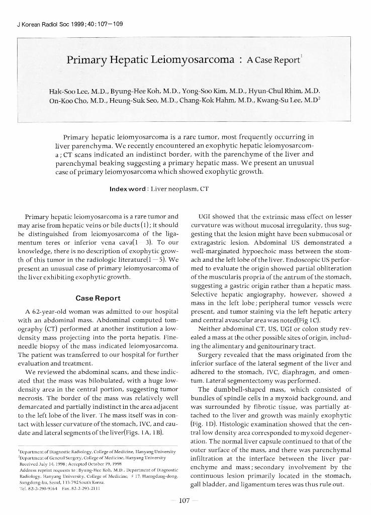
We reviewed the abdominal scans , and these indicated that the mass was bilobulated , with a huge lowdensity area in the central portion, suggesting tumor necrosis. The border of the mass was relatively well demarcated and partially indistinct in the area ad jacent to the left lobe of the liver. The mass itself was in contact with lesser curvature ofthe stomach, rvc, and caudate and lateral segments ofthe liver(Figs. 1A, 1B).
' Dcparlment of Diagnostic Radiology , CoIIege ofMedicine, Hanyang Un ivers ity
' Department ofGenera I S l1 rge ry , CoII ege of Medi c in e, Hanya ng University
Recc ived J l1 Iy 14, 1998 ; Accepted October 19 , 1998 Addrcss repr int rcquests 10: By ung. Hee Ko h, M.D. , Depa rtmcnt of Diagnostic
Radi o logy , Hanyang Univcrsit y, CoIIcge of Medi ci nc, 17, 1-lae ng d ang-do ng, Sl1 ng d ong-ku , Scou L 133-792 So uth Korca
TeI. 8 2-2- 290-9 164 Fax.82-2-293- 2111
UGr showed that the extrinsic mass effect on lesser curvature was without mucosal irregularity, thus sug gesting that the lesion might have been submucosal or extragastric lesion. Abdominal US demonstrated a well-marginated hypoechoic mass between the stomach and the left lobe ofthe liver. Endoscopic US performed to evaluate the origin showed partial obliteration ofthe muscularis propria ofthe antrum ofthe stomach,
suggesting a gastric origin rather than a hepatic mass Selective hepatic angiography, however, showed a mass in the left lobe; peripheral tumor vessels were present, and tumor staining via the left hepatic artery and central avascular area was noted(Fig 1 C).
Neither abdominal Cτ US , UGr or colon study revealed a mass at the other possible sites of origin, includ ing the alimentary and genitourinary tract.
Surgery revealed that the mass originated from the inferior surface of the lateral segment of the liver and adhered to the stomach, rvc, diaphragm, and omentum. Lateral segmentectomy was performed .
The dumbbell-shaped mass , which consisted of bundles of spindle cells in a myxoid background , and was surrounded by fibrotic tissue , was partially attached to the liver and growth was mainly exophytic (Fig. 1D). Histologic examination showed that the centrallow density area corresponded to myxoid degeneration. The normalliver capsule continued to that of the outer surface of the mass, and there was parenchymal infiltration at the interface between the liver parenchyme and mass; secondary involvement by the continuous lesion primarily located in the stomach ,
gall bladder, and ligamentum teres was thus rule out.
- 107 -

Hak-Soo Lee , et al : Primary Hepatic Leiomyosarcoma
A B
C D
Fig. 1. A. Contrast-enhanced CT scan shows a lobulated low density mass projecting into porta hepatis, which has parenchymal beaking (arrows) with the left lobe ofthe liver, and also indistinct border(open arrows). B. On CT scan below than Fig. 1a, the mass is abutting the lesser curvature ofthe stomach(arrows), and seen in extrahepatic 10-cation. CHA (common hepatic artery , open arrow) is displaced by the mass. C. Selective hepatic arteriography shows a large hypervascular mass, which has central necrotic area and dislocate the stretched left hepatic artery (open arrows) latera lJy. There are contrast filling in the inferior aspect of the mass suggesting tumor vessel or tumor staining(arrows). D. Cut gross specimen shows a lobulated mass with peripheral solid portion (arrows) and central myxoid area(not shown). The remnant left lobe ofthe liver had a triangular shape corresponding to the parenchymal beaking on CT. The capsule ofthe mass is continuous to the normal capsule ofthe liver in microscopic examination(not shown)
Discussion
Leiomyosarcoma of the liver may arise within the liver (primary hepatic leiomyosarcoma) or from the ligamentum teres (ligamentum teres leiomyosarcoma) (4). Diagnosis of a smooth muscle tumor arising in the liver requires that certain criteria be met. The tumor should not originate from an adjacent structure, nor represent a single metastasis from another primary origin. The most frequent primary sites, including the ali-
mentary tract(stomach, small bowel. colon), genitourinary tract(uterus, bladder, prostate, kidney) , and the
retroperitoneum(inferior vena cava), should be evaluated(2 - 6). Grossly, primary leiomyosarcoma of the
liver usually presents as a single large mass, of firm consístency.
Primary hepatic leiomyosarcoma should be dífferentiated from that of the ligamentum teres; the 10-cation of the latter is unique, the prognosis is better, and it is encapsulated and clearly demarcated from the liver(4), which is mainly compressed by the mass in its
- 108 -

J Korean Radiol Soc 1999; 40: 107-109
ligamentum teres rather than by infiltration(7). To ascertain that the tumor did not originate from an ad
jacent structure, a diagnosis of primary leiomyos arcoma ofthe liver thus requires a careful search.
The common signs and symptoms are hepatomegaly,
an abdominal or right upper quadrant mass, abdominal distention, and weight loss(3 , 4).
The Iiterature contains few descriptions of the CT findings o f leiomyosarcoma of the liver(2) . Most primary hepatic leiomyosarcomas are located in Iiver parenchy ma(3, 4), and to our knowledge, no descrip tion of exophytic growth of this tumor, as in our case, is to be found in the radiologic literature(3 - 5). The tumor has described as a large w ell-delineated mass with a predominantly peripherally enhanced wall or a mainly cystic appearance. Findings depend on the tumor’s pathologic presentation; as in our case , central low d ensity corresponds to predominantly central necrosis, hemorrhage or amorphous gelatinous tissue (2).
1n our case, the main portion of the mass projected into the porta hepatis, and on CT scans was found to show mainly a mass effect, with displacement of sur rounding organs rather than inv asion. Selective hepatic arteriography of leiomyosarcoma of the IVC may show a mass supplied by the left hepatic artery(8,
9) . Be cause parasitic tumor supply is possible, the extrah - epatic mass may still involve the left lobe.
Leiomy osarcoma of the liver is a slow growing tumor, and the survival period ranges from several months to y ears from initial diagnosis(3, 4). Excision of the bulk of the primary tumor is the treatment of choice . 1n the previous literature, consideration of the
possibility of primary hepatic leiomyosarcoma was seen as important ; in such cases, because of aggressive surger y , the prognosis was good(l , 3, 4).
W e h ave described an exophytic hepatic leiomyosarcoma which was shown by CT scanning to have an indistinct border with the par enchyme of the liver,
and paren chymal beaking that suggested primary hep atic mass . It was differentiated by imaging modalities which include d abdominal CT and visceral angiograp hy.
References
1. Masur H. Sussman EB, Molander DW. Primary hepatic leiomyosarcoma : a report of two cases. Gastroenterology 1975; 69
994- 997
2. Hepatic leiomyosa rcomas : CT features with pathologic correlat ion. Soyer P, Bluemke DA. Riopel M, Hruban RH , Fishman EK. EurJ RadioI 1995; J9: 11 7- 182
3. Ba ur M, Potzi R, Lochs H, Neuhold N, Neuhold N, Walgram M, Ga ngl A. Primary leiomyosarcoma of the liver-a case report Z Gastroenterol 1993; 3 1 : 20 - 23
4. 0 ’ Leary MR, Hi ll RG, Lev ine RA. Peritoneoscopic diagnosis of pri mary leiomyosarcoma of li ve r. Hum Pathol 1982; 13: 76-78
5. Bae K K, Cho JH , Chang JC. Pr imary hepatic leiomyosarcoma: a case report. J Korean Radiol Soc 1996; 34 : 405-408
6. Haw kins EP, Jordan GL, McGavran MH. Primary leimyosarcoam of the li ver. Am J Surg Pathol 1980 ; 4: 301 -304
7 . Mital RN, Bazaz-Malik G. Leiomyosa rcoma of ligamentum teres of the li ver. Am J Gastroenterol 197 1 ; 56 : 48-5 1
8. Griffi n AS , Sterchi JM. Primary leiomyosarcoma of the inferior vena cava: a case report and review of the literature. J Surg
Oncol 1987; 34 : 53-60
9. Tegtmeyer CJ , Buschi A. The angiogra phic diagnosis of leiomyosarcoma of the infe rior vena cava. Radiology 1977; 122: 683-
685
대한빔사선의학회지 1999; 40: 107-109
원발성 간 평활근육종 : 1예 보고l
l 한양대학병원 진단방사선과
2한양대학병원 일반외과
이학수 · 고병희 · 김용수 · 임현철 · 조온구 · 서흥석 · 함창곡 · 이광수2
원발성 깐평활근육종은 매우드물며,대부분은간내에 위치한다.
저자들은 최근 간의 돌출성 종괴로 보였으나, 전산화 단층 촬영 소견상 간 실질과 경계가 불분명하고 예각을
보여, 원발성 간종괴로 여겨졌던, 돌출성 성장을 보인 원발성 간i생활근육종 1여l를 보고하는 바이다.
- 109 -

」F 1999년도 춘계 전공의연수교육 안내
1999년도 춘계 전공의 연수교육을 다음과 같이 안내하오니 참고하시기 바라오며 특히 믿건부터 사전등록제를 실시하고
자 하오니 많은 협조와 참여를 부탁드립니다
펴 일 시 : 1쨌년 4월 24일(토.) 13:30 - 16:50
장 소 : 서울힐튼호텔 Grand Ballroom A Tel (82-2) 317-3200 Fax (82-2) 317-3203
@ 샤전퉁록
우리 학회에서는 전공의 연수교육의 효과적인 운영을 위해 사전등록제를 시행합니다 사전등록을 챔1 회원에게는 전공
의연수교육의 교재를 사전에 우송할 예정이며 받으신 교재는 전공의연수교육 참가시 지참하셔야 하며 만약 지참하지 않
았을 경우 등록처에서 5, 000원에 구입하실 수 있습니다.
l 등록비
구 분
전공의회원
비전공의회원
사전등록(1999년 3월 10일 까지)
10 , 000원
15 , 000원
현장등록(1999년 3월 10일 이후)
15 , 000원
20, 000원
’ 사전등록을 취소할 경우 3월 치일(수) 까지는 등록비의 환불이 가능하나 그 이후는 환불이 되지 않습니다 l샤전등록 f뱅
사전등록은 아래 구좌로 송금이 완료된 경우만 인정하며 , 송금하신 후 학회 Home Page를 이용하여 신청하시거나, 별지의 사전등록 신청서를 작성하신 후 신청서를 우편 또는 Fax로 발송해1면 됩니다.
, 거래은행 : 평화은행 구좌번호 : 025-01-0001-042 예금주 : 대한방사선의학회
책 신청l앙법
Home Page를 이용하는 방법 등 춘계학술대회 등록방법과 동일합니다
피 주 제 : 방사선과 영역의 의료정보 활용
시 간
1:00 -1:30 - 2:00 2:00 - 2:30 2:30 - 3:00 3:00 - 3: 20 3:20 - 3:50 3:50 - 4:20 4:20-4:50
연 제 연 사
등록
의료정보연구동향소개
Intemet에 대한 이해
Medline 검색 및 참고문헌 관리 휴식
WordProc없잉r 사용법
스프레드 쉬트를 이용한 자료관리
효과적인 slide 및 전시작품 만들기
김성현(보건의료기술연구 기획평가단)
정성훈(경상의대)
정은철(성균관의대)
치순주(인제의대)
최병길(가톨릭의대)
한준구(서울의대)
- 110 -