Embed Size (px)
Citation preview

Medical University of South Carolina Medical University of South Carolina
MEDICA MEDICA
MUSC Theses and Dissertations
2005
Primaquine-Induced Hemolytic Anemia: Hemotoxicity of Primaquine-Induced Hemolytic Anemia: Hemotoxicity of
5-Hydroxyprimaquine (5-HPQ) 5-Hydroxyprimaquine (5-HPQ)
Zachary Scott Bowman Medical University of South Carolina
Follow this and additional works at: https://medica-musc.researchcommons.org/theses
Recommended Citation Recommended Citation Bowman, Zachary Scott, "Primaquine-Induced Hemolytic Anemia: Hemotoxicity of 5-Hydroxyprimaquine (5-HPQ)" (2005). MUSC Theses and Dissertations. 143. https://medica-musc.researchcommons.org/theses/143
This Dissertation is brought to you for free and open access by MEDICA. It has been accepted for inclusion in MUSC Theses and Dissertations by an authorized administrator of MEDICA. For more information, please contact [email protected].

PRIMAQUINE-INDUCED HEMOLYTIC ANEMIA:
HEMOTOXICITY OF S-HYDROXYPRIMAQUINE (5-HPQ)
by
Zachary Scott Bowman
A dissertation submitted to the faculty of the Medical University of South Carolina in partial fulfillment of the requirement for the degree of Doctor of Philosophy in the
College of Graduate Studies.
Department of Cell and Molecular Pharmacology
2005
Chairman, Advisory Committee

11
ACKNOWLEDGEMENTS
This body of work could not have been completed without the help of many people, to
whom I wish to express my gratitude:
Dr. David McMillan, not only for the stipend support, laboratory space, and materials
required for this project, but also for his friendship and guidance, and his commitment to
foster an enviroment designed to stimulate and nurture intellectual and personal growth.
Dr. David Jollow, for his valuable insight and discussion, which have been an integral
component of my education.
Dr. Kevin Schey, Dr. Craig Beeson, and Dr. Brad Schulte, for their willingness to serve
on my committee, and their guidance, genuine interest, and support throughout my
training.
Current lab members: Dr. JoEllyn McMillan, for her constant support and guidance, and
Jennifer Schulte, for her invaluable technical assistance and patience in teaching me the
various techniques applied in this dissertation, and her constant support and
encouragement.
Dr. John Oatis and the NMR facility, for help in the synthesis and indentification of 5-
hydroxyprimaquine.
My wife, Julie, and my family, for their continuing love, patience, and understanding,
which have contributed more to my success than I could ever begin to express.

111
COPYRIGHT INFORMATION
Portions of this dissertation have been previously published with copyright ownership as follows:
Chapter 2.
Bowman ZS, Oatis JE, Jr., Whelan JL, Jollow OJ and McMillan OC (2004) Primaquineinduced hemolytic anemia: susceptibility of normal versus glutathione-depleted rat erythrocytes to 5-hydroxyprimaquine. J Pharmacol Exp Ther 309: 79-85.
Chapter 3.
Bowman ZS, Jollow OJ, and McMillan OC (2005) Primaquine-induced hemolytic anemia: role of splenic macrophages in the fate of 5-hydroxyprimaquine-treated rat erythrocytes. Manuscript submitted.
Chapter 4.
Bowman ZS, Morrow JO, Jollow OJ, and McMillan OC (2005) Primaquine-induced hemolytic anemia: role of membrane lipid peroxidation and cytoskeletal protein alterations on the hemotoxicity of 5-hydroxyprimaquine. J Pharmacol Exp Ther 314: Manuscript in press.

TABLE OF CONTENTS
KEY TO ABBREVIATIONS LIST OF FIGURES
VI
VB
X
Xl
LIST OF TABLES ABSTRACT
CHAPTERS:
1.. Introduction and Specific Aims 1
Normal Red Cell Senescence -Physiology 2 -Oxidati ve Stress in the Red Cell 3
Proposed mechanisms of Red Cell Senescence 4 -Phospholipid Asymmetry 8 -Protein-Protein Interactions and Recognition of "Self' 11
Drug-Induced Hemolytic Anemia 15 Glucose-6-Phosphate Dehydrogenase Deficiency
-Historical 19 -The Enzyme 20 -Genetics 21
Malaria 22 Anti-malarial Therapy
-Historical 24 -Malaria Chemotherapy and Drug Resistance 26
Primaquine 27 .. Primaquine Metabolism 29
5-Hydroxyprimaquine 32 Rationale and Specific Aims 35
2. Primaquine-Induced Hemolytic Anemia: Susceptibility of Normal versus GSH-Depleted Rat Erythrocytes to 5-Hydroxyprimaquine 39
Introduction 40 Materials and Methods 43 Results 52 Discussion 61
IV

3. Primaquine-Induced Hemolytic Anemia: Role of Splenic Macrophages in the Fate of Rat Erythrocytes Treated with 5-HPQ
Introduction Materials and Methods Results Discussion
4. Primaquine-Induced Hemolytic Anemia: Role of Membrane Lipid Peroxidation and Skeletal Protein Alterations on the Hemotoxicity of 5-HPQ
Introduction Materials and Methods Results Discussion
5. Summary, Discussion and Future Directions
REFERENCES
Summary, Discussion and Future Directions Conclusion
66
67 69 72 82
90
91 92 100 111
122
123 143
146
v

RES
Hb
NADH
NADPH
ROS
PS
GSH
G6PD
PQ-CX
MAQ
MAQ-NOH
5-HPQ
HPLC-EC
PBSG
GSSG
PSSG
DEM
DCFDA
NEM
HBSS
CH
DIT
KEY TO ABBREVIATIONS
reticuloendothelial system
hemoglobin
reduced nicotinamide adenine dinucleotide
reduced nicotinamide adenine dinucleotide phosphate
reactive oxygen species
phosphatidylserine
reduced glutathione
glucose-6-phosphate dehydrogenase
carboxyprimaquine
6-methoxy -8-aminoquinoline
6-methoxy -8-hydroxy laminoquinoline
5-hydroxyprimaquine
high-performance liquid chromatography with electrochemical detection
isotonic phosphate-buffered saline with glucose
oxidized glutathione (glutathione disulfide)
protein-glutathione mixed disulfides
diethy 1 maleate
2', 7' -dichlorodihydrofluorescein diacetate
N -ethy I maleimide
Hank's buffered saline solution
cumene hydroperoxide
dithiothreitol
VI

Vll
LIST OF FIGURES
1.1 Normal production of red cell ROS 5
1.2 Pentose phosphate pathway (HMP shunt) 6
1.3 Asymmetrical distribution of RBC membrane phospholipids 9
1.4 Membrane lipid transporters 10
1.5 CD47-SIRPa model for regulation of erythrophagocytosis 13
1.6 Schematic diagram of the band 3 macrocomplex 14
1.7 Effect of G6PD deficiency on primaquine and dapsone hemolytic activity (Degowin Figure) 18
1.8 Life cycle of the malaria parasite 23
1.9 Putative pathways of primaquine metabolism 31
1.10 Types of primaquine-metabolite redox pairs 33
1.11 Working hypothesis for quinoneimine induced oxidative stress 34
2.1 Putative metabolism of primaquine to 5-HPQ 42
2.2 Schematic representation of the synthesis of 5-HPQ 44
2.3 LC-MS analysis of 5-methoxyprimaquine reaction products 46
2.4 IH NMR spectrum of 5-HPQ 47
2.5 Stability of 5-HPQ in blood versus buffer 53
2.6 Cyclic voltammogram of 5-HPQ 55
2.7 5-HPQ concentration-dependent direct hemolytic activity 56
2.8 5-HPQ concentration-dependent methemoglobin formation 58
2.9 Effect of 5-HPQ on rat erythrocyte sulfhydryl status. 60
2.10 5-HPQ concentration-dependent direct hemolytic activity in aSH-depleted rat erythrocytes 62

vIn
3.1 Disposition of SICr in saline-pretreated rats receiving slCr-labeled 5-HPQ-treated rat erythrocytes 74
3.2 Disposition of 5ICr in clodronate liposome-pretreated rats receiving slCr-labeled 5-HPQ-treated rat erythrocytes 75
3.3 Effect of 5-HPQ on clodronate liposome- and saline-pretreated rat organ weights 76
3.4 Disposition of 5ICr in rats receiving sier-labeled broken cells 78
3.5 Effect of 5-HPQ on J774A.l erythrophagocytosis 80
3.6 Visualization of J774A.l phagocytosis of 5-HPQ-treated rat Erythrocytes 81
3.7 Effect of aSH-depletion on 5-HPQ-induced erythrophagocytosis by J774A.1 macrophages 83
4.1 Effect of 5-HPQ on rat erythrocyte ROS generation 102
4.2 Effect of 5-HPQ on rat erythrocyte F2-isoprostane formation 103
4.3 Effect of 5-HPQ on rat erythrocyte phosphatidylserine asymmetry 105
4.4 Effect of 5-HPQ on rat erythrocyte membrane proteins 107
4.5 Effect of DTT on 5-HPQ-induced Hb binding to rat erythrocyte membrane proteins 108
4.6 Effect of aSH-depletion on 5-HPQ-induced Hb binding to rat erythrocyte membrane proteins 110
4.7 Effect of DTT on 5-HPQ-induced Hb adduct formation in intact red cells 112
4.8 Effect of DTT on the 5-HPQ-induced hemolytic activity 113
4.9 Working hypothesis for 5-HPQ-induced hemolytic activity in rat erythrocytes 121
5.1 Schematic representation of 5-HPQ autooxidation 128
5.2 Effect of 5-HPQ on ferrylhemoglobin formation 131

5.3
5.4
5.5
Schematic representation of possible RSS formation
J774A.l phagocytosis of 5-HPQ-treated GSH-depleted rat erythrocytes
Effect of DTT on the phagocytosis of 5-HPQ treated rat erythrocytes
IX
133
142
144

LIST OF TABLES
1 ... 1 Drugs and chemicals that cause hemolytic anemia
1 ... 2 Current drugs for antimalarial treatment and prevention
16
28
x

Xl
ABSTRACT
Primaquine is an important antimalarial agent because of its activity against
exoerythrocytic forms of Plasmodium sp. Methemoglobinemia and hemolytic anemia,
however, are dose-limiting side effects of primaquine therapy. These hemotoxic effects
are believed to be mediated by metabolites, though the identity of the toxic specie( s) and
the mechanism underlying hemotoxicity have remained unclear. Previous studies
showed that an N-hydroxylated metabolite of primaquine, 6-methoxy-8-
hydroxylaminoquinoline, was capable of mediating primaquine-induced hemotoxicity.
The present studies were undertaken to investigate the hemolytic mechanism of 5-
hydroxyprimaquine (5-HPQ), a phenolic metabolite that has been detected in
experimental animals.
5-HPQ was synthesized, isolated by flash chromatography and characterized by NMR
spectroscopy and mass spectrometry. In vitro exposure of 51er-Iabeled erythrocytes to 5-
HPQ induced a concentration-dependent decrease in erythrocyte survival (TC50 ~40 ~M)
when the exposed cells were returned to the circulation of isologous rats. 5-HPQ also
induced methemoglobin formation and depletion of glutathione (GSH) when incubated
with suspensions of rat erythrocytes. Furthermore, when red cell aSH was depleted
(>95%) by titration with diethyl maleate to mimic aSH instability in human glucose-6-
phosphate dehydrogenase deficiency, a 5-fold enhancement of hemolytic activity was
observed. These data indicate that 5-HPQ also has the requisite properties to contribute
to the hemotoxicity of primaquine.

XlI
To investigate the fate of erythrocytes in vivo after in vitro exposure to 5-HPQ, rat
sler-Iabeled erythrocytes were incubated with hemolytic concentrations of 5-HPQ and
then re-administered intravenously to rats. The time-course of loss of radioactivity from
blood and uptake into the spleen and liver was measured. In rats given 5-HPQ-treated
erythrocytes, an increased rate of removal of radioactivity from the circulation was
observed as compared to the vehicle control. The loss of blood radioactivity was
accompanied by a corresponding increase in radioactivity appearing in the spleen but not
in the liver. When rats were pretreated with clodronate-Ioaded liposomes to deplete
splenic macrophages, there was a decreased rate of removal of radioactivity from the
circulation and a markedly diminished uptake into the spleen. A role for phagocytic
removal of 5-HPQ-treated red cells was confirmed in vitro using the J774A.l
macrophage cell line. Furthermore, depletion of red cell aSH with diethyl maleate
significantly enhanced in vitro phagocytosis of 5-HPQ-treated red cells. These data
indicate that splenic macrophages are responsible for removing 5 .. HPQ-treated red cells
and support a role for depletion of aSH as a key event in the process leading to
macrophage recognition and phagocytosis of 5-HPQ-damaged erythrocytes.
To investigate the mechanism underlying the hemolytic activity of 5-HPQ, we have
examined the effect of hemolytic concentrations of 5-HPQ on ROS formation within rat
erythrocytes using the cellular ROS probe, 2' ,7' ... dichlorodihydrofluoresein diacetate
(DCFDA). In addition, we examined the effect of 5-HPQ on membrane lipids and
cytoskeletal proteins. The data indicate that 5-HPQ causes a prolonged, concentration
dependent generation of ROS within erythrocytes. Interestingly, 5-HPQ-generated ROS
was not associated with the onset of lipid peroxidation or an alteration in

Xlll
phosphatidylserine asymmetry. Instead, 5-HPQ induced oxidative injury to the
erythrocyte cytoskeleton, as evidenced by changes in the normal electrophoretic pattern
of membrane ghost proteins. Immunoblotting with an anti-hemoglobin antibody revealed
that these changes were due primarily to the formation of disulfide-linked hemoglobin
skeletal protein adducts. These data suggest that cytoskeletal protein damage, rather than
membrane lipid peroxidation or loss of phosphatidylserine asymmetry, underlies the
process of removal of erythrocytes exposed to 5-HPQ.
Cummulati vely, the data presented in this dissertation describe a mechanism by
which exposure to 5-hydroxyprimaquine leads to the hemolytic removal of red cells; i.e.
the generation of intracellular oxidative stress causes protein oxidation and hemoglobin
binding to the membrane and subsequent phagocytosis by macrophages. A relationship
between drug-induced hemolytic anemia and mechanisms of red cell senescence is
discussed, as are possible implications for the antimalarial therapeutic effect of
primaquine. The relative contribution of N-hydroxy vs. phenolic metabolites to the
overall hemotoxicity of primaquine remains to be assessed.

CHAPTER!
Background and Introduction

Normal Red Cell Senescence
Physiology
2
The mature red cell is a "marvel of functional design" that has been stripped of all
organelles and nuclear material, leaving only a "wafer-shaped membranous bag of
protein," 8}lm in diameter (Jandl, 1996). Human red cells are formed from progenitor
cells in the bone marrow prior to being released into the circulation, where they begin an
arduous journey of 100 to 200 total miles, which lasts approximately 120 days (-60 days
in the rat). During this journey, each "membranous bag of protein" travels at an
incredible rate, such that it passes through the heart once every 1 to 3 minutes, while
squeezing through capillary channels as small as 3Jlm across and managing constant
exposure to oxidative stress (Jandl, 1996).
The red cell is perfectly engineered and well equipped to handle these normal
stresses. However, certain changes are thought to accumulate on the red cell surface over
its lifespan, which define the cell as "senescent" and lead to its uptake and degradation by
resident macrophages of the reticuloendothelial system (RES) by 120 days (Rifkind,
1966). Given the massive number of red cells our circulation (-43 trillion), the removal
of 1/120th of the total number circulating erythrocytes every day represents a truly
remarkable feat. Approximately 360 billion senescent red cells are removed from the
circulation every day, amounting to almost 5 million red cells every second (Bratosin et
aI., 1998). However, despite representing a very significant physiological event, the
3
specific changes that occur to the red cell and the mechanism(s) by which it is recognized
and removed from the circulation are not well defined.
Oxidative Stress in the Red Cell
Red cells are ideally designed to deliver oxygen to the tissues. The hemoglobin (Hb)
content of the red cell is approximately 5mM ("",,34 g/dl) with a heme-iron content of "",,20
mM, representing "",,95% of total cellular protein, (Jandl, 1996). Such high hemoglobin
concentrations allow the red cell to be highly specialized for the transport of oxygen and
carbon dioxide between the lungs and tissues. However, it is this specialized capacity
that also renders the red cell susceptible to chronic oxidative stress.
Oxidative stress occurs in the red cell, because the exposure of its iron-rich contents
to oxygen-rich environments leads to auto-oxidation of hemoglobin and subsequent
formation of methemoglobin and hydrogen peroxide (Fig. 1.1). Thanks to the activities
of several anti-oxidant enzymes, including superoxide dismutase, catalase, glutathione
peroxidase and glutathione reductase, the red cell is adequately equipped to handle this
stress under normal conditions. Methemoglobin is readily reduced to hemoglobin by the
NADH-dependent methemoglobin reductase (Passon and Hultquist, 1972), and
superoxide is converted to hydrogen peroxide by superoxide dismutase (Fridovich, 1975),
which is then acted on by the catalase and glutathione peroxidase/reductase systems
(Eaton, 1991) as follows:

4
° .!. + ° .!. + 2H+ SOD
)II- H20 2 + 02 2 2
2H20 2 Catalase 11- 2H20 + 02
2GSH + 2H20 2 GSH Px .. 2H20 + GSSG
GSSG GSH Red .. 2GSH
Importantly, the actions of each of these enzymes (with the exception of superoxide
dismutase) require the presence of NADPH as a cofactor. The only source of NADPH in
the red cell is by generation from the pentose phosphate pathway, also called the hexose
monophosphate (HMP) shunt (Fig. 1.2). The shunt acts by shuttling glucose-6-phosphate
through several steps, the first two of which are oxidations that generate NADPH from
NADP+. Normally the HMP shunt operates at only 3-5% of its maximum capacity, but
when confronted with excess ROS generation through the pathway shown above or
otherwise, the red cell responds by stimulating shunt activity (discussed below) and
producing sufficient NADPH to deal with the stress. Any deficiency in this system or
with the production of NADPH has devastating consequences for the ability of red cells
to handle oxidative stress.
Proposed Mechanisms of Red Cell Senescence.
The mechanisms by which aged red cells are recognized and removed from the
circulation are unclear. However, it is currently thought that oxidative damage in the red
cell accumulates over time and leads to specific cell-surface changes, which mark the red
cell for removal from the circulation, defining it as "senescent" (Bartosz, 1991). Many

0+2- 02 l1li(
Hb (oxy)
t
~ Oe+3 -02- l1li( » Oe+3 + O!!. 2
MetHb Superoxide
MetHb Reductase
NADH
Fe+2
• OH + HO- ....... -----
GPx/GR Catalase
SOD
NADPH
5
Fig. 1.1. Normal production of active oxygen species within the red cell as a consequence of ferric iron-superoxide anion dissociation from oxyhemoglobin. Hb, oxyhemoglobin; MetHb, methemoglobin; SOD, superoxide dismutase; Gpx/GR, glutathione peroxidase/glutathione reductase.
(Jollow and McMillan, 2001)

6
pentose Phosphate pathway
t IG6Pq G6P ---r....!~;;;;;;;;jjjyr=---41 .. ~ 6PGL ~ ~ PENTOSE PHOSPHATE
GLUCOSE (HMP Shunt)
NADP NADPH
IGSH Reductas~
(2)GSH GSSG
IGSH Peroxidas~
LACTATE
Fig. 1.2. Pentose phosphate pathway, or hexose monophosphate (HMP) shunt, of red cells. This cyclic pathway is activated by changes to cellular NADPINADPH ratios that favor NADP and accelerate the regeneration of NADPH, which is necessary for detoxification of ROS and the maintenance of GSH and cellular enzymes in a reduced state. The first, rate-limiting enzyme of the HMP shunt is G6PD.

7
studies have attempted to identify the precise target(s) of oxidative damage and to
determine the mechanism(s) by which they lead to erythrophagocytosis by macrophages.
The major hypotheses are: 1) that specific modifications to the red cell membrane result
in the exposure of "senescence antigens" (protein, lipid, or carbohydrate) on the red cell
surface that are recognized by circulating "senescence antibodies," which opsonize
senescent red cells for phagocytosis (Kay, 1994); 2) that enzymatic removal of sialic
acid residues from red cell glycolipids or glycoproteins results in recognition by a
macrophage receptor (Bratosin et a!., 1998); 3) that red cell aging is analogous to the
apoptotic response observed in other (nucleated) cell types, i.e. oxidative stress results in
caspase activation and alterations to the normal asymmetric distribution of phospholipids
in the membrane (MandaI et a!., 2002; Kuypers and de Jong, 2004); or 4) that protein
protein interactions are necessary for recognition of "self," and oxidative damage alters
these interactions among the cytoskeletal, integral membrane, and cell surface proteins
(Oldenborg,2004).
While each of these hypotheses is appealing, there is no consensus at present as to
which represents the final erythrocyte death signal. It has been suggested that the
mechanism of red cell senescence might not be able to be reduced to a single hypothesis,
but that "when a metabolism or a cellular phenomenon is of vital importance it is always
protected by multiple pathway systems" (Bratosin et aI., 1998). Much work remains to
solve the molecular and cellular mechanism of erythrophagocytosis, but two hypotheses
have arisen very recently that appear to be particularly relevant.

8
Phospholipid Asymmetry
Phospholipid asymmetry was first established in the red cell, and most of what is
known about its regulatory process was also discovered in erythrocytes. In all cell types,
including normal circulating blood cells, an asymmetric conformation of membrane
phospholipids is tightly regulated. Phosphatidylserine (PS) is actively and exclusively
maintained within the inner leaflet of the plasma membrane and only becomes exposed to
the outer leaflet in situations that require recognition and removal of damaged cells, Le.
apoptosis, or initiation of the coagulation cascade (Kuypers and de Jong, 2004).
Phosphatidy lethanolamine (PE) is also found within the inner leaflet, but it is not as
tightly regulated as PS (Fig. 1.3). On the other hand, the choline-containing
phospholipids, phosphatidylcholine and sphingomyelin, are found mainly in the outer
leaflet of the plasma membrane. Three classes of integral membrane enzymes have been
shown to be responsible for regulation of phospholipid asymmetry. Specifically, an ATP
driven aminophospholipid translocase, also called "flippase," very efficiently transports
PS and PE from the outer leaflet to the inner leaflet of the plasma membrane (Fig. 1.4),
Additionally, a calcium-activated "phospholipid scramblase" causes non-specific
bidirectional movement of multiple phospholipids, and an ATP-dependent "floppase,"
outwardly directs PC and cholesterol.
It has been shown that PS exposure is one of the earliest detectable features in all
apoptotic cells and facilitates recognition and removal of apoptotic bodies by
macrophages expressing a PS receptor (Kuypers and de Jong, 2004). Additionally,
transport of PS to the outer leaflet of cell membranes has been shown to occur as a result

9
50
Outer monolayer
PC SM
~
~ "'C "--'" a. -0 0 --0.. ..c .- a. - (/)
0 0 ..c ..c 0.. a.. (J) ro 0 .f-J
0 ..c t-o..
PE
Inner monolayer
50
Fig. 1.3. Asymmetrical distribution of phospholipids in RBC membranes, shown as a percentage of total membrane phospholipid. PC, phosphatidylcholine; SM, sphingomyelin; PE, phosphatidylethanolamine; PS, phosphatidylserine.

10
PS PL
Outer Leaflet
PC,SM
PS, PE
Inner Leaflet
PL PC
Flippase Scramblase Floppase
Fig. 1.4. Schematic representation of the action of lipid transporters in the plasma membrane. PC, phosphatidylcholine; SM, sphingomyelin; PS, phosphatidylserine; PE, phosphatidylethanolamine. "Flippase" (aminophospholipid translocase) is an ATPdependent transporter, which inwardly directs PS and PE. "Floppase" represents a class of ATP-dependent transporter, which transports PC and SM outwardly. "Scramblase" is an ATP-independent, Ca+2-activated transporter, which non-specifically disrupts phospholipids asymmetry via bidirectional transport.

11
of accumulation of malonyldialdehyde (MDA) following lipid peroxidation or damage to
spectrin (Jain, 1984), as well as from non-specific Ca++·induced cell membrane
scramblase activation (Bevers et aI., 1998). Interestingly, senescent red cells appear to
retain portions of the apoptotic machinery and to also share the common apoptotic
endpoint of PS exposure. MandaI et a1. demonstrated that procaspase 3 was converted to
caspase 3 in red cells oxidatively damage by t-butyl hydroperoxide, which resulted in the
exposure of PS and subsequent phagocytosis by macrophages (MandaI et aI., 2002).
Although red cells lack mitochondria, it has been hypothesized that intracellular oxidative
stress in the erythrocyte mimics the apoptotic response observed in nucleated cells, such
that activation of caspase 3 negatively regulates flippase and causes extracellular
exposure of PS (Bratosin et aI., 1997; MandaI et aI., 2002). These studies led to the
suggestion that there may be a significant correlation between red cell senescence,
oxidative stress and loss of phospholipid asymmetry.
Protein-Protein Interactions and Recognition of "self"
It has been proposed that normal protein-protein interactions among the cytoskeletal
and integral membrane proteins of erythrocytes are necessary to confer the recognition of
"self' to macrophages (Oldenborg et aI., 2000). CD47, i.e. integrin-associated protein
(lAP), is a ubiquitously expressed transmembrane glycoprotein with five hydrophobic
transmembrane-spanning domains and a short, alternatively spliced, hydrophobic tail. It
was first identified in the placenta and neutrophil granulocytes as a protein that associates
with u v f33 integrins, regulates integrin function and signals leukocyte responses to RGD
containing extracellular matrices (Oldenborg, 2004). Like other cell types, red cells

12
express CD47; however, unlike other cell types, red cells do not express integrins,
suggesting that there may be a unique role for this protein in erythrocytes.
CD47 was demonstrated to be a marker of "self' in red cells, and this was
hypothesized to represent a potential pathway for the expression of hemolytic anemia. In
their initial study, Oldenborg et ai. showed that red cells from CD47 knockout mice were
rapidly cleared when administered to the circulation of wild type mice (Oldenborg et aI.,
2000). Furthermore, they showed that CD47 on normal blood cells could prevent
elimination by binding to the macrophage receptor, signal regulatory protein alpha
(SIRPa), which is thought to send an inhibitory signal to the macrophages (Fig. 1.5).
SIRPa contains intracellular immunoreceptor tyrosine-based inhibitory motifs (ITIMs),
which become tyrosine-phosphorylated in response to CD47. ITIM phosphorylation
causes the recruitment of the SH2 containing phosphatase (SHP-1) to the membrane,
which subsequently inhibits tyrosine kinase-dependent signaling pathways thereby
preventing phagocytosis (Oldenborg, 2004). Based on the finding that CD47 functions
as a "marker of self' on erythrocytes, a role for CD47 in senescent red cells was
explored. Several studies showed that aged murine red cells and stored or banked
erythrocytes, which have been associated with increased clearance in vivo, both had
reduced CD47 expression, supporting the hypothesis that phagocytosis of senescent red
cells may be regulated by CD47-SIRPa interaction (Anniss and Sparrow, 2002;
Oldenborg, 2004).
CD47 is a member of the band 3 protein-based macrocomplex of integral and
peripheral proteins in the red cell membrane (Bruce et aI., 2003). In its tetrameric form,
band 3 (anion exchanger 1 or AE1) binds to protein 2.1 (ankyrin), protein 4.2, and CD47,

13
in
---Red Cell----
out
SIRPa
out
- Macrophage- - - - - - - - - - - - - - - - --
in
Fig. 1.5. Model for the regulation of erythrophagocytosis by the CD47/SIRPa system. Interaction of normal red cell CD47 with the macrophage SIRPa receptor results in tyrosine phosphorylation and subsequent recruitment of SHP-1 to the membrane, which negatively regulates phagocytosis by inhibiting downstream signaling.

14
out
Fig. 1.6. Schematic diagram of the band 3 macrocomplex. Tetramers of band 3 are attached to the spectrin cytoskeleton through ankyrin (band 2.1). Protein 4.2 binds ankyrin, band 3, and CD47, providing vertical attachment of CD47 to the cytoskeleton. CD47 is also horizontally associated with band 3 through the Rh proteins.
(Bruce et aI., 2003)
15
and is the major attachment site of the red cell membrane to the cytoskeleton (Fig. 1.6).
Given its importance for self-recognition, it is conceivable that oxidative damage to
members of the band 3 macrocomplex, or CD47 itself, perhaps in the form of free
sulfhydryl modification, could alter normal CD47-presentation to macrophages, resulting
in a loss of self-recognition and phagocytosis.
Drug-Induced Hemolytic Anemia
Anemia, defined as a reduction in the concentration of circulating red cells, can occur
from either a decrease in red cell production or from an increase in red cell destruction.
Hemolytic anemia is the generic term used to describe the disease process that results
from an increased destruction of red cells. Despite what the term "hemolytic anemia"
might imply, frank red cell lysis in the circulation, i.e. "intravascular hemolysis," is
relatively rare. Rather, the more common mechanism of destruction of red cells is due
the recognition and phagocytosis of intact red cells by splenic macrophages, i.e.
"extravascular hemolysis" (Jandl, 1996). Clinically, a decreased hematocrit causes
hemolytic anemia patients to present acutely with fatigue, pallor, shortness of breath, and
a rapid heart rate. As a result of hemoglobin catabolism, jaundice and dark urine also
occur. Additional signs may include an increased spleen size, an increased reticulocyte
count, and/or hemoglobinuria.
A variety of drugs and environmental chemicals are known to cause hemolytic
anemia, some of the more common of which are listed in Table 1-1. The "drug-induced
hemolytic anemias" are collectively referred to as the Heinz body hemolytic anemias
because of the "spheroidal inclusions of denatured hemoglobin that are a late sign of prior

(JandI, 1996)
Table 1-1. Drugs and chemicals that cause hemolytic anemia
Categories
Antimalarials
Sulfonamides
Sulfones
Analgesics
Antipyretics
Nitrofurans
Miscellaneous
N aturall y -occuring compounds
R~presentative compounds
Primaquine Pamaquine Pentaquine
Sulfanilamide Sulfapyradine Sulfacetamide Sulfamethoxazole
Dapsone Thiazolesulfone Diaphenylsulfone
Acetanilid
Phenylhydrazine
Nitrofurantoin
Naphthalene Toluidine blue Methylene Blue Trinitrotoluene Nalidixic Acid Niridazole Pyridium Phosphine
Vicine/Convicine (fava beans) Lawsone (henna)
16

17
oxidation" (Borges and Desforges, 1967). While the exact mechanism by which the drugs
listed in Table 1-1 cause the premature removal of damaged, but intact, red cells is
unclear, the Heinz body hemolytic anemias tend to share some common characteristics:
1) they are thought to occur following an initial oxidative stress, which results from free
radical metabolites; 2) this oxidative stress is handled by the normal defense mechanisms
of the cell, i.e. aSH peroxidase and catalase, which protect free sulfhydryl groups on Hb,
SH-containing enzymes, membrane proteins, and membrane lipids; 3) aSH peroxidase
and catalase, which are both dependent on NADPH, are eventually overwhelmed by this
oxidative stress; 4) free radicals then attack various cellular targets, including
hemoglobin, SH-containing enzymes, membrane protein, and/or membrane lipids; and 5)
intracellular oxidative damage leads to cellular changes that are recognized by splenic
macrophages and result in erythrocyte phagocytosis. While the precise mechanism by
which intracellular oxidative damage is translated into an external cell-surface signal that
marks the cell for removal is not clear, the common findings of oxidative stress and
splenic sequestration have led to the hypothesis that drug-induced hemolytic anemia is
due to an acceleration of the normal physiologic aging processes, whatever those
mechanisms might be (Azen and Schilling, 1963; Tizianello et aI., 1968).
Interestingly, there is some evidence available to suggest that there may be more than
one mechanism by which hemolytic agents impose oxidative damage. Clinical studies by
Degowin et al. (De go win et al., 1966) made an important distinction between the
hemolytic effect of primaquine and another hemolytic drug, dapsone (Fig. 1.7). In their
studies, equivalent doses of primaquine or dapsone were administered to normal and
18
PQ (G6PD-Def) DDS (G6PD-Def)
(J) 70 t1 9 Q)
~ / U / • 0
60 /
~
/ ; ..c • " ..... / " ~ '- / " Q) " '0
/ ,,". Q) 50 /
" / " Q) .0 / ,," DDS (Normal) co / " -. 40 / • " '- " () / 0 " • / " u:;
I " - ,," . 0 30 I 0/ " - " -;:R.. / ." "
0 - / " (J) ~ " 'w 20 / " .. ~
0 I " E ", Q) " I 10 PQ (Normal)
0 I
0 1 2 3 4 5
Dose (mg base/kg body-weight)
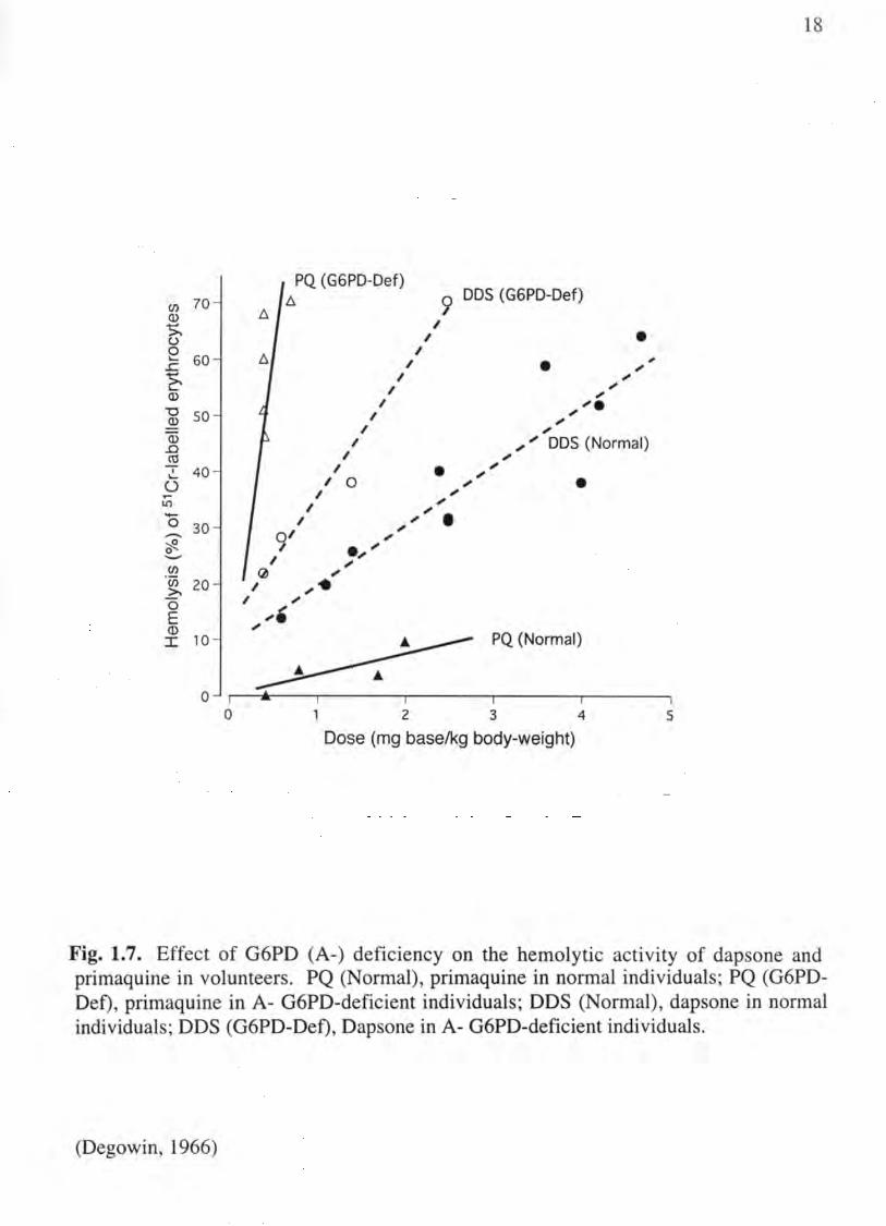
Fig. 1.7. Effect of G6PD (A-) deficiency on the hemolytic activity of dapsone and primaquine in volunteers. PQ (Normal), primaquine in normal individuals; PQ (G6PDDef), primaquine in A- G6PD-deficient individuals; DDS (Normal), dapsone in normal individuals; DDS (G6PD-Def), Dapsone in A- G6PD-deficient individuals.
(Degowin, 1966)

19
African (A-) variant G6PD deficient volunteers. In normal volunteers, dapsone was
shown to be the more potent hemolytic agent. However, in the G6PD-deficient
individuals (see below), primaquine was the much more potent hemolytic agent.
Dapsone showed only a two-fold increase in toxicity in G6PD-deficient individuals, but
the toxicity of primaquine was multiplied 30-fold. While the reason for the difference is
unclear, it may be related to the fact that dapsone is metabolized to a single
hydroxylamine-type metabolite that is hemolytic, whereas primaquine metabolism is
complex with multiple non-toxic and toxic pathways (see below). Thus the hemolytic
activity of primaquine may be mediated by the synergistic action of multiple toxic
metabolites, including N-hydroxy-, quinoneimine- and quinone-types (see below). At a
minimum though, the data show that there is marked variation in the hemolytic response
to different agents, which suggests that more than one mechanism may be involved in
drug-induced hemolytic anemia.
Glucose-6-Phosphate Dehydrogenase Deficiency
Historical
The potential risk of hemolytic anemIa from use of the 8-aminoquinoline
antimalarial-drug, pamaquine, was first reported in 1926. Observations over the next 30
to 40 years, showed that pamaquine, and then later primaquine, were not universally
toxic, but only caused hemolytic anemia in a subset of uniquely susceptible individuals,
who were termed "primaquine sensitive." For normal individuals, pamaquine and
primaquine (as well as many of the other drugs listed in Table 1-1) would cause
hemolytic anemia only when given in very large doses, well above the therapeutic

20
window. However, "primaquine sensitive" individuals presented with hemolytic anemia
even when receiving conventional therapeutic doses of primaquine. In response to these
initial observations, extensive work was done in the 1950s and 1960s to elaborate the
mechanism of "primaquine sensitivity," and those studies showed that the sensitivity was
associated with an intrinsic abnormality in the red cell, which was later identified as an
enzymatic defect of glucose-6-phosphate dehydrogenase, i.e. G6PD (Beutler et aI., 1955).
Glucose-6-Phosphate Dehydrogenase, the enzyme
G6PD is a cytosolic "housekeeping" enzyme that catalyzes the first and rate-limiting
step of the pentose phosphate pathway (i.e. the hexose monophosphate shunt or HMP
shunt), and in erythrocytes, this pathway is the sale source for NADPH. NADPH, in tum,
is an important reducing cofactor for a variety of antioxidant enzymes, including catalase
and GSH peroxidase/reductase (Fig. 1.2), which are necessary for the detoxification of
ROS in the cell.
The human G6PD monomer consists of 515 amino acids with a molecular weight of
59.2 kDa (Beutler, 1994), but the active enzyme exists as a dimer that has binding sites
for both glucose-6-phosphate and NADP. While the precise mechanisms of control for
this important pathway are not yet completely understood, there appear to be at least two
distinct roles for the cellular NADP/NADPH ratio (Jollow and McMillan, 2001). In
addition to being a cofactor for the enzyme, NADP is necessary for conversion of the
inactive G6PD monomer to the active dimer. Consequently, in the presence of high
NADP concentrations, the conversion of monomer to dimer promotes G6PD, and hence
HMP shunt activity (Jollow and McMillan, 2001). Conversely, NADPH provides a

21
potent inhibitory feedback control that is dependent on substrate (glucose-6-phosphate)
concentration, such that the inhibition is greatest when the concentration of G6P is lowest
(Yoshida and Lin, 1973). Under quiescent conditions, the NADPH/NADP ratio favors
NADPH, hence these controls are estimated to limit the activity of normal G6PD to less
than 1 % of the V max value of the enzyme (Yoshida and Lin, 1973). However, under
enhanced oxidative stress conditions, NADPH is rapidly consumed and converted to
NADP by the activities of catalase and GSH peroxidase/reductase. As the
NADPH/NADP ratio drops, the inhibitory control of NADPH is diminished, and the
activity of G6PD increases sufficiently to generate enough NADPH to handle the stress.
Genetics
G6PD-deficiency is an X-linked disorder, which, with several classes of variants, is
considered to be the most common genetic deficiency in the world, affecting over 400
million individuals worldwide (Beutler, 1994). Most of these variants are point
mutations that result in a single amino acid change, which leads to loss of activity as the
red blood cell ages (Beutler, 1990; Jollow and McMillan, 1998). Thus aged cells are
thought to have decreased levels of active G6PD and are hence more susceptible to
oxidative damage. G6PD deficient individuals include approximately 10% of African-
American males (A-variant) who have a residual G6PD activity that is approximately
10% of normal. A- red cells that are older than 55 days are considered to have lost all
activity. About 15% of Caucasians of Mediterranean descent (Med- variant), and 70% of
Kurdish Jews, have a much more severe deficiency and red cells older than only 20 days
are considered to have lost all activity. There are also population groups in Asia, India

22
and the Middle East that may express the same or other variants (Jollow and McMillan,
1998).
Forty-five years ago, several studies suggested that G6PD deficiency actually confers
a resistance to falciparum malaria infection (Beutler, 1994), which has been postulated to
have created a selective advantage for the G6PD deficiency in areas where malaria is
endemic. The geographical correlation between a high prevalence of G6PD deficiency
and areas where malaria is historically endemic is striking (Ruwende et aI., 1995). That
G6PD deficiency is so common in those countries most affected by malaria complicates
and significantly limits the use of drugs like primaquine in those countries where it is
most needed.
Malaria
Malaria is a life threatening parasitic disease of epidemic proportions that is a major
public health concern for developing countries. Every year, infection is responsible for
300-500 million acute illnesses and an estimated 1.5-2.7 million deaths worldwide (Kain
and Keystone, 1998). Tropical and subtropical regions with dense populations, typically
the poorest in the world, are most affected but tend to be the least economically equipped
to handle the severe disease burden or to contribute to the development of malarial
treatment and prevention. Areas where malaria is considered to be endemic include:
Africa, parts of Asia, Central and South America, Oceania and certain Caribbean Islands
(Kain and Keystone, 1998).
Malaria is a protozoan disease that is caused in humans by four different species of
the genus Plasmodium: P. falciparum (falciparum malaria), P. vivax (vivax malaria), P.

/ /,/ +-I (/)
o :c c: ro E :::l :c //
/ ,/
Infected Hepatocyte
23
~ o <b
~ Sa
Hypnozoite ~ ,\. (P. vivax; P. ova/e) ,..... """" n'
Schizont
- - - - - - - - - - - - --- -
Schizont
Merozoite Release
Late Trophozoite
9 c:--~-: ~-
Gametocytes
Merozoite
-------
Erythrocyte
Ring Form (Early Trophozoite)
Q (')
Ci> ? ~ ,..... <b ~ ,.....
::i v)' V) c: <b
~ ~ V) <b '-
- .
Fig. 1.8. The life cycle of the malaria parasite (see text for details).

24
ovale, and P. malariae. The disease is characterized by extreme exhaustion associated
with paroxysms of high fever, sweating, shaking chills and anemia. Malarial
transmission occurs when an infected mosquito injects the malarial sporozoites into the
bloodstream of a human host while taking a blood meal (Fig. 1.8). After a brief period of
about 30 minutes, the sporozoites disappear from the bloodstream as they either migrate
to the liver, where they infect hepatocytes, or are destroyed by phagocytes. Those
sporozoites that infect hepatocytes form cyst-like structures and continue to develop and
multiply during what is referred to as the "exoerythrocytic phase" of the parasite's
development. This period can vary significantly depending on the malarial species
involved. In particular, P. falciparum and P. malariae schizonts will develop directly into
merozoites, but P. vivax and P. ovale schizonts are able to develop into hypnozoites as
well as merozoites. The hypnozoite forms can remain dormant in the liver for months or
even years before being reactivated into merozoites to continue the cycle, causing
malarial fever spikes and chills long after the initial infection (Le. vivax malaria or
"relapsing malaria"). The mechanism responsible for the reactivation of hypnozoites is
unknown; regardless, the sporozoites eventually develop into merozoites and multiply
until they overload the cell and force it to rupture. When rupture occurs, the merozoites
are released into the bloodstream, where they infect red blood cells and initiate the
"erythrocytic cycle." It is this cycle that is responsible for the severe fever spikes and
chills typically associated with malaria infection. Similar to the exoerythrocytic cycle,
the erythrocytic schizonts will eventually overload and rupture the red blood cell,
releasing malarial forms that either reinfect other red cells to continue the cycle of fever
spikes or develop into gametocytes, which are taken up by a new mosquito taking a blood

25
meal. The sexual phase of the malarial life cycle occurs in the mosquito and is completed
when the mosquito injects sporozoites into a new host during another blood meal.
Antimalarial Therapy.
Historical
Until the middle of the 20th century, malaria was endemic in parts of Europe and
North America. Prior to the 18th century, the most accepted treatment for malaria was the
so-called Gaelen method, which aimed to "expulse" the disease through "bleeding and
purging". The first attempts at specifically treating malaria date back to the early 17th
century when the bark of Cinchona trees from Peru was used. By the early 18th century,
its use had become widespread, and in 1820, two French pharmacists isolated quinine as
the active Cinchona bark ingredient. Quinine quickly replaced the crude bark for
treatment of malaria and is credited with destroying the traditional expulsion methods of
Gaelen. Over the next 100 years, demand for quinine increased, but the drug still had to
be extracted from the Cinchona bark. As the world's overwhelming need for natural
sources of quinine expanded, so did the search for more readily available synthetic drugs
(Wernsdorfer and McGregor, 1988; Wiesner et al., 2003).
In 1891, Paul Ehrlich cured two malaria patients with methylene blue, based on the
observation that the dye was taken up by malarial parasites. By 1925, modification of the
basic structure of methylene blue resulted in the synthesis of the first 8-aminoquinoline
antimalarial compound, pamaquine. Unfortunately, pamaquine proved to be much too
toxic and largely ineffective against P. falciparum. However, it did prove to be the first
drug that was capable of preventing malarial relapse due to P. vivax. In 1932, mepacrine

26
(atebrine, quinacrine) was developed by attaching the basic side chain of pamaquine to
acridine instead of quinoline. Mepacrine proved to be active against the erythrocytic
stages of P. falciparum and was extensively used in combination with pamaquine during
World War II. Studies by German scientists in the 1930s led to the synthesis of
chloroquine, a 4-aminoquinoline blood schizontocide structurally derived from quinine
that is effective for the treatment of both P. falciparum and P. vivax. Chloroquine
quickly became the most effective and important antimalarial drug and was even used in
several programs aimed at global eradication of malaria. Extensive studies in the 1940s
then resulted in the development of most of the antimalarial therapies used today
(Wernsdorfer and McGregor, 1988; Wiesner et aI., 2003).
Malaria Chemotherapy and Drug Resistance
Therapeutic regimens against P. falciparum, which typically consist of various
combinations of blood schizontocides, have been very successful in the past. However
extensive use these drugs, in particular the 4-aminoquinoline, chloroquine, has lead to
increasing resistance throughout the world to almost all blood schizontocidal antimalarial
agents currently used (Bjorkman and Phillips-Howard, 1990; White, 1998). Plasmodium
falciparum resistance to chloroquine was first encountered within 15 years of its
introduction and has steadily expanded ever since (Peters, 1987). Today, chloroquine is
used primarily to treat malaria only in Central America, Haiti, the Dominican Republic
and most of the Middle East; while malarial infection in every other endemic region of
the world (Africa, Asia, and parts of the Middle East) is considered to be chloroquine
resistant. Additionally, chloroquine-resistant parasites have become resistant to other,

27
sometime chemically unrelated drugs, such as qUInIne, mefloquine, proguanil, and
pyrimethamine, among others. Because of this increased resistance, quinine has actually
re-emerged as a drug of choice, but only in combination with other drugs. The latest
search for an effective drug has lead to artemisinin and its derivatives, compounds
derived from the ancient Chinese herbal qinghao and its active ingredient qinghaosu. No
resistance has yet developed against the artemisinins, but the potential is thought to exist
(Krishna et aI., 2004). Importantly, none of these drugs have been shown to be effective
against relapsing P. vivax and P. ovale malaria.
As is shown in Table 1-2, the development of antimalarial therapies has mainly
concentrated against the erythrocytic cycle, while primaquine remains the only tissue
schizontocide approved for the radical cure of latent tissue stages of malarial parasites.
Additionally, no significant resistance has developed against primaquine; it continues to
be effective against the latent tissue stage of all four human malarial species and for the
treatment and prophylaxis of the most lethal species, P. falciparum (Shanks et aI., 2001).
Primaquine thus has the potential to provide an important tool to combat malarial
transmission and resistance, if something can be done to control its side-effects.
Primaquine
Primaquine (6-methoxy-8-[4-amino-1-methylbutylamino]quinoline; Fig. 1.9) is the
prototype 8-aminoquinoline antimalarial drug. It was one of three drugs (pentaquine,
isopentaquine and primaquine) that were developed during the Second World War by the
antimalarial research initiative of the US Army that were shown to be more effective
against vivax malaria than pamaquine but less toxic. Of these three drugs, primaquine

Table 1-2. Current Drugs for Antimalarial Treatment and Prevention
Target
Erythrocytic Cycle (Blood Schizontocides)
Exo-erythrocytic Cycle (Tissue Schizontocides)
Mosquito (Vector Control)
Drug
Chloroquine Hydroxychloroquine Quinine Quinidine Mefloquine Proguanil Atovaquone Sulfadoxine-pyrimethamine CycloguaniI Clindamycin Tetracyclines Sulfonamides Dapsone Promin Artemether Artemisinin
Primaquine
Insecti ci des Netting
28

29
remains the most effective available compound for elimination of the hypnozoite stages
of P. vivax and P. ovale (Wernsdorfer and McGregor, 1988). However, despite its unique
importance for antimalarial therapy for over fifty years, the mechanism of therapeutic
action of primaquine remains unknown. Limited data suggests that possible targets might
include the parasitic inner mitochondrial membrane, dihydroorotate synthase, or DNA
(Howells et aI., 1970; Boulard et aI., 1983; Peters, 1984). It has also been suggested that
primaquine is converted to redox active metabolites in the liver, which contribute to the
antimalarial activity of primaquine by generating reactive oxygen species (Bates et aI.,
1990), although direct evidence for this postulate is lacking ..
Primaquine is effective against the latent tissue forms of all four human malarial
species and has been gaining use for both the treatment and prophylaxis of P.falciparum
in addition to the relapsing species (Shanks et at, 2001). Interestingly, primaquine is also
useful when combined with clindamycin for the treatment of Pneumocystis carinii
pneumonia (PCP), an infection that is common in immunosuppressed individuals and
definitive for the conversion of HIV to AIDS, another worldwide epidemic (Toma et aI.,
1998). However, despite its importance and unique therapeutic effectiveness, use of
primaquine is limited by its toxicity in G6PD deficient individuals, which, along with
multi-drug resistance to most of the blood schizontocides, highlights the need for more
effective antimalarial drugs with higher therapeutic ratios.
Primaquine Metabolism.
Previous studies have shown that redox-active metabolites rather than the parent drug
are responsible for causing the major toxicities of primaquine, and possibly its

30
therapeutic effects as well. Primaquine is rapidly absorbed with a bioavailability of 96%
in humans but is rapidly and extensively metabolized, resulting in removal of less than
2% of the administered dose by renal excretion (Mihaly et al., 1985). However, little is
known about the metabolic fate of primaquine, since the extraction, identification and
quantification of the major redox-active metabolites has been significantly complicated
by their instability and poor organic solubility (Idowu et aI., 1995).
Despite the difficulty in identifying metabolites, the metabolism of primaquine is
thought to be complex and involve oxidation to both the aminoquinoline ring and the
alkylamino side-chain (Fig. 1.9). Carboxyprimaquine (PQ-CX) is formed by oxidation
of the terminal carbon of the side chain, and was found in the plasma of humans and rats
in concentrations significantly higher than the parent compound (Mihaly et aI., 1985).
Although PQ-CX is the most abundant metabolite detected, it is neither redox active
itself, which was shown by its inability to stimulate HMP shunt activity or form
methemoglobin in red cells (Link et aI., 1985; Baird et aI., 1986); nor does it appear in
the urine, which indicates further metabolism prior to excretion (Mihaly et aI., 1984).
The only other human metabolite of primaquine that has been detected is the N
dealkylated derivative, 6-methoxy-8-aminoquinoline or MAQ (Baty et aI., 1975), which
was also unable to stimulate HMP shunt activity or cause methemoglobin formation
(Link et aI., 1985; Baird et aI., 1986). Recently, our laboratory used human and rat liver
microsomes to show that MAQ could be metabolized in vitro to MAQ-NOH, which was
subsequently shown to have significant hemolytic activity (Bolchoz et aI., 2001). Other
than these three metabolites (PQ-CX, MAQ, and MAQ-NOH), no other human
metabolites have been observed to be formed either in vivo or in vitro.

PO-NOH
OH
5-0H-6-MAO ".
/S-MAQ
HO
" .
/ /
OH
5,6-DHAO
, ~ HO
,
HN~COOH ~ -:O~
po-ex
OH 5-HPQ
\ / ! I NH ": HN~ 2 /
OH ,/ 5,6-DHPO .... '
" .. ' ..' .'
" ~ .. ,fI'"
.
..... Sulfate/Glucuronide ~.
Conjugation
31
Fig. 1.9. Putative pathways of primaquine metabolism. PQ-NOH, N-hydroxyprimaquine; PQ-CX, 8-(3-carboxy-1-methylpropylamino )-6-methoxyquinoline; MAQNOH, 6-methoxy-8-hydroxylaminoquinoline; 6-DesM-PQ, 6-desmethylprimaquine; 5-HPQ, 5-hydroxyprimaquine; 6-MAQ, 6-methoxy-8-aminoquinoline; 5-0H-6-MAQ, 5-hydroxy-6-methoxy-8-aminoquinoline; 5,6-DHAQ, 5,6-dihydroxy-8-aminoquinoline; 5,6-DHPQ, 5,6-dihydroxyprimaquine.
(Bolchoz et al., 200 1)

32
5-Hydroxyprimaquine
Following the suggestions of Tarlov et a!. (Tarlov et a!., 1962), much attention has
been given to the putative phenolic metabolites of primaquine (5-HPQ, 5,6-DHPQ, 5-
OH-6-MAQ, and 5,6-DHAQ), which are thought to support quinone- or quinoneimine
type redox-cycling and generation ofROS as shown in Fig. 1.10 (Link et a!., 1985). To
examine the hemotoxic potential of these putative metabolites, several of the phenolic
metabolites were synthesized in the 1980s and made available to investigators under a
contract funded by the WHO. Studies with these derivatives in isolated erythrocytes
showed that they were able to oxidize hemoglobin, stimulate hexose monophosphate
shunt activity and deplete erythrocytic GSH (Allahyari et a!., 1984; Link et aI., 1985;
Agarwal et a!., 1988; Fletcher et a!., 1988). Additionally, more recent studies from our
laboratory assessed the hemotoxicity of MAQ-NOH, a hydroxylamine-type metabolite,
which was shown to be directly hemotoxic to red cells (Bolchoz et a!., 2001).
Collectively, these studies have led our laboratory to propose that the complex
metabolism of primaquine can be simplified into three types of reactive metabolites: N
nitroso, p-quinoneimine-, and o-quinone-type metabolites (Fig. 1.10). While the
quantitati ve importance of each of these metabolites is not yet known, each type is
thought to have the ability to redox cycle and thereby generate reactive oxygen species
according to the scheme shown in Fig. 1.11. While the initial experiments effectively
demonstrated the oxidative activity of phenolic metabolites in vitro, they failed to
establish a relevant model to assess whether their activities in vitro were relevant to the
hemotoxicity observed in vivo. Thus, little is known about the capacity of phenolic
metabolites to affect red cell survival in vivo, and efforts to detect these metabolites in

N-nitroso
O~ N
p-quinoneimine
OH
o
33
o-qulnone
HO
OH
o
Fig. 1.10. Structures of types of primaquine reactive metabolite redox pairs. R=alkyl side-chain or H.

HMP SHUNT
NADP+
NADPH
°2 5-HPQ Hb++
QI Hb+++
• H20 2 02--
HO·
! 1 ntracellular
Oxidative Damage
NAD+
NADH
Fig. 1.11. Working hypothesis for quinoneimine-induced oxidative stress in red cells. 5-HPQ = 5-hydroxyprimaquine;QI = 5-hydroxyprimaquine-quinoneimine; Hb++ = ferrohemoglobin; Hb+++ = ferrihemoglobin; HMP SHUNT = hexose monophosphate shunt
V.)
~

35
primaquine-treated humans have not been successful. Furthermore, the compounds
initially offered by the WHO are no longer available, are difficult to synthesize, and are
extremely unstable.
5-HPQ, which is formed by hydroxylation in the 5-position of the quinoline ring, is
the most simple of the putative phenolic metabolites. Although it has not been found in
humans, formation of 5-HPQ from primaquine is expected, since it would arise by a
single metabolic step and has been shown to be formed in several experimental animals,
including rats, dogs and monkeys (Strother et aI., 1981; Fletcher et aI., 1984; Ni et aI.,
1992). Additional studies have shown that 5-HPQ can generate H20 2 and hydroxyl
radicals within the red cell. It was suggested that this formation of ROS was the result of
an iron-catalyzed reaction between the semiquinone intermediate and H20 2 (Vasquez
Vivar and Augusto, 1992). It is conceivable that this generation of intracellular ROS
could mediate the hemolytic damage of primaquine in G6PD-deficient individuals, who
are unable to sufficiently replenish GSH.
Similar to the other phenolic metabolites mentioned above, 5-HPQ has been shown
to deplete glutathione, form methemoglobin, and stimulate HMP-shunt activity.
However, there is no evidence that 5-HPQ has a direct hemolytic effect, and it is not
known whether the conditions under which 5-HPQ was shown to cause oxidative
damage in vitro are relevant to those that cause hemolytic damage in vivo.
Rationale and Specific Aims.
The mechanism underlying primaquine-induced hemolytic injury is unknown.
Current thinking is that primaquine itself is not responsible, but rather cellular injury

36
results from an oxidative stress generated within the red cell by redox-active metabolites.
A classical approach to identify the toxic metabolites (Le. administration of primaquine in
vivo in order to match toxicity to the formation of a metabolite) cannot be used, because
rats (and humans with normal G6PD activity) do not respond to primaquine with toxicity,
as noted above. In addition, the reactivity of certain metabolites in blood, i.e. 5-HPQ,
does not allow for the correlation of blood levels with toxicity. Thus an alternative
approach must be used.
Three types of redox-active metabolites are known or postulated to arise during the
hepatic metabolism of primaquine: an N-hydroxy derivative (MAQ-NOH); and two
phenolic derivatives, 5-HPQ and 5-hydroxy-6-desmethyl primaquine. Previous studies in
our lab focused on MAQ-NOH, a hydroxylamine-type metabolite. MAQ-NOH was
shown to be a direct-acting hemolytic agent in rats and to have the requisite properties to
contribute to the overall hemotoxicity of primaquine (Bolchoz et al., 2001). The
following studies are focused on the more simple of the two putative phenolic
metabolites, 5-HPQ, since it would arise by a single metabolic step and hence may be
formed in larger amounts. Previous studies showed that 5-HPQ could redox cycle to its
quinoneimine analog, generate methemoglobin and cause loss of GSH when added to red
cells in vitro (Allahyari et aI., 1984; Link et aI., 1985; Agarwal et ai., 1988; Fletcher et
ai., 1988). Thus, 5-HPQ appears to be an redox-active metabolite that might be able to
mediate the overall hemotoxicity of primaquine.
The long-term objective of these studies is to design a "better primaquine"; that is, a
derivative that retains antimalarial activity but has little or no hemolytic capacity. Our
fundamental contention is that an improved drug can only be developed if the mechanism

37
of toxicity is understood. To this end, the present studies will seek to establish: 1) the
direct hemolytic activity of 5-HPQ; 2) the nature of injury induced within the red cell by
5-HPQ, and 3) the relationship between these internal oxidative changes with the
premature sequestration of 5-HPQ-damaged red cells. The information obtained should
be useful in modulating metabolism and/or RBC in the rat to confer "primaquine
sensitivity" and to examine the pattern of oxidative damage in order to determine which
type(s) of metabolites contribute. These studies will also significantly advance our
understanding of the general mechanism of drug-induced hemolytic anemia, i.e.
examination of the nature of the cellular oxidative damage will allow us to assess the
relevance of specific lesions to the premature removal of damage cells, which could also
provide insight into the normal, physiologic removal of senescent erythrocytes.
Hypothesis: 5-HPQ causes the hemolytic removal of damaged erythrocytes in rats by
initiating an intracellular oxidative stress that specifically alters membrane components
and leads to recognition and uptake of intact cells by splenic macrophages.
Specific Aim 1. To characterize the chemical properties, hemolytic response and pattern
of oxidative injury induced in rat erythrocytes by 5-HPQ.
Specific Aim 2. To determine the role of splenic macrophages in the fate of 5-HPQ
damaged rat erythrocytes.

38
Specific Aim 3. To examine the role of membrane lipid peroxidation and cytoskeletal
protein alterations in the hemotoxicity of 5-hydroxyprimaquine.

CHAPTER 2
Primaquine-Induced Hemolytic Anemia: Susceptibility of Normal versus GSH-Depleted Rat Erythrocytes to 5-Hydroxyprimaquine

40
Introduction
Malaria is a widespread, life-threatening parasitic disease that is responsible for 300-
500 million acute illnesses and an estimated 1.5-2.7 million deaths worldwide each year
(Kain and Keystone, 1998). Primaquine, an 8-aminoquinoline anti-malarial drug, is
effective against the exoerythrocytic forms of all four of the malarial species that infect
humans and is the only radically curative drug for the latent tissue forms of Plasmodium
vivax and P. ovale (Tracy and Webster, 2001). Primaquine is also used in combination
with chloroquine to combat the problem of multiple drug resistance in P. Jalciparum
(Shanks et aI., 2001). Despite its clinical importance and effectiveness, use of primaquine
has long been known to be limited by its capacity to induce hemolytic anemia,
particularly in individuals with a hereditary deficiency in erythrocytic glucose-6-
phosphate dehydrogenase (G6PD) activity (Dern et aI., 1955; Degowin et aI., 1966).
Since G6PD deficiency is prevalent in malarial areas, this dose-limiting toxicity can have
a major impact on the usefulness of this drug in these populations.
Importantly, primaquine is not directly toxic to erythrocytes at clinically relevant
concentrations. Although the hemotoxicity of primaquine has long been considered to be
dependent on metabolism, the metabolite(s) responsible and the underlying mechanism(s)
have remained unclear. We have reported recently that 6-methoxy-8-
hydroxylaminoquinoline (MAQ-NOH), an N-hydroxylated metabolite of primaquine, is a
direct-acting hemolytic and methemoglobinemic agent in rats, and therefore may be a

41
contributor to the hemotoxicity observed in primaquine-treated humans (Bolchoz et aI.,
2001).
Metabolism of primaquine, however, is relatively complex, and a variety of known
and putative phenolic metabolites have also been considered to be capable of mediating
primaquine hemotoxicity. In particular, hydroxylation of primaquine at the 5-position of
the quinoline ring (Fig. 2.1) is known to yield redox active derivatives that are capable of
inducing oxidative stress within normal and G6PD-deficient human erythrocytes. Several
of these compounds, including 5-hydroxyprimaquine (5-HPQ), 5-hydroxy-6-
desmethylprimaquine and their N-dealkylated derivatives, were synthesized in the 1980s
and made available to investigators by the World Health Organization. Studies with these
compounds in isolated suspensions of red cells have shown that they can induce
methemoglobin formation, glutathione (GSH) depletion and stimulation of hexose
monophosphate shunt activity (Allahyari et aI., 1984; Link et aI., 1985; Baird et aI., 1986;
Agarwal et aI., 1988; Fletcher et aI., 1988; Vasquez-Vivar and Augusto, 1994). However,
there is a notable lack of evidence for their hemolytic activity in vivo.
Progress towards understanding the role of phenolic metabolites in primaquine
induced hemolytic anemia has been hampered because they are no longer available, the
synthetic methods to prepare them are relatively difficult, and the products are highly
unstable. As a first step in our investigation of the potential contribution of phenolic
metabolites to primaquine-induced hemolytic anemia, we have re-synthesized 5-HPQ and
examined its stability and redox behavior. In addition, we have assessed the hemolytic
potential of 5-HPQ in aSH-normal and GSH-depleted rat red cells. In view of the critical
role proposed for oxidative stress in the mechanism underlying primaquine-induced

HN
Primaquine (PO)
HN
OH
5-HPO
Fig. 2.1. Putative metabolism of primaquine to 5-HPQ.
42

43
hemolytic anemia, we measured the formation of methemoglobin and monitored red cell
sulfhydryl status under hemolytic conditions in order to correlate the hemolytic response
with these indicators of intracellular oxidative damage. We report that 5-HPQ is an
extremely potent direct-acting hemolytic agent in rats, and that hemolytic activity is
associated with methemoglobin formation and a marked depletion of erythrocytic GSH.
When GSH was depleted from rat red cells to mimic aSH instability of human G6PD
deficient red cells (Gaetani et aI., 1979), the hemolytic activity of 5-HPQ was markedly
enhanced. The significance of the data with regard to the overall contribution of
metabolites to primaquine-induced hemolytic anemia is discussed.
Materials and Methods
Chemicals and Materials.
6-Methoxy-8-nitroquinoline, ferrous bromide, sodium metal, stannous chloride,
potassium trifluoroacetate and GSH were obtained from Sigma-Aldrich (St. Louis, MO).
Na251Cr04 in sterile saline (1 mCi/ml, pH 8) was obtained from New England Nuclear
(Billerica, MA). All other chemicals and reagents were of the best grade commercially
available.
5 -Methoxyprimaquine (5, 6-dimethoxy -8-[4-amina-l-methy lbuty lamina ]quinoline)
was pre-pared from 6-methoxy-8-nitroquinoline as described previously and shown in
Fig. 2.2 (Elderfield et aI., 1955). 5-HPQ (5-hydroxy-6-methoxy-8-[4-amino-l-methyl
but ylamino] quinoline) was synthesized from 5-methoxyprimaquine by HBr-catalyzed
hydrolysis using a modification of an established method (Allahyari et aI., 1984). The
composition of the reaction mixture was monitored as a function of time via LC-MS. The

Br2F e, CaC03 .. H20, CHC13 .
H3CO
Br
conc HCl
o
Br~N
o
HN~N
HO
o EtOH
OCH3
HN~NH2
9
o
6
48% HBr
7
+ HO
10
NaOMe, Me0t! pyndme
+
8
44
Fig. 2.2. Schematic representation of the synthesis of 5-HPQ. 6-methoxy-8-nitroquinoline (1) was brominated by electrophilic substitution to form 5-bromo-6-methoxy-8-nitroquinoline (2). Nucleophilic aromatic substitution of 2 with sodium methoxide produced 5,6-dimethoxy-8-nitroquinoline (3), which was then reduced with tin(II) chloride to form 5,6-dimethoxy-8-aminoquinoline (4). 4-bromo-l-phthalimidopentane (5) was used to alkylate 4 to form 5,6-dimethoxy-8-(4-phthalimido-l-methylbutylamino)-quinoline (6). Hydrazinolysis of 6 formed 5-methoxyprimaquine (7), which yielded 5-HPQ (8) and a mixture of products (9,10) after selective hydrolysis with HBr.

45
reaction mixture contained four major components (Fig. 2.3): 5-HPQ (m1z 275.5-276.5;
4.60 min, 21.0%), 5-HPQ quinoneimine (273.5-274.5; 4.41 min, 42.8%), 5-
methoxyprimaquine (m/z 289.5-290.5; 9.58 min, 21.1%) and 5-hydroxy-6-
desmethylprimaquine (mlz 259.5-260.5; 4.09 min, 15.1 %). The yield of 5-HPQ was
optimized by adjusting the reaction temperature to 120° C and the reaction time to 20
min. The yield was increased further by reducing the quinoneimine to the hydroquinone
using sodium dithionite and maintaining the reaction mixture under argon to minimize
oxidation of the hydroquinone. The reaction mixture was then purified in two steps using
SepPak Plus C18 cartridges (Waters Corporation, Milford, MA). 5-HPQ was eluted from
the first cartridge with 5% acetonitrile/0.05% aqueous trifluoroacetic acid, lyophilized
and then applied to a second cartridge. 5-HPQ was eluted from the second cartridge with
5% acetonitrile in water containing 5 mM HBr. After removal of the solvent by
lyophilization, elemental analysis confirmed the presence of the trihydrobromide salt of
5-HPQ (purity >99% as judged by HPLC and NMR analysis). IH NMR (in D20; Fig.
2.4): [) 8.62 (dd, 1=1.4, 4.4 Hz, 1, H-2), 8.43 (dd, J=8.5, 1.4 Hz, 1, H-4), 7.46 (s, 1, H-7),
4.40 (dd, 4.3, 8.5 Hz, 1, H-3), 3.76 (m, 2, H-l '), 3.82 (s, 3, OCH3), 2.77 (m, 2, H-4'),
1.65 (m, 1, H-2'), 1.65 (m, 1, H=3'), 1.56 (m, 1, H-2'), 1.52 (m, 1, H-3'), 1.19 (d, J=6.6
Hz, 3, H-5'). 13C NMR (in D20): [) 148.6 (C-2), 142.7 (C-6), 139.7 (C-5), 134.9 (C-9),
132.4 (C-4), 122.4 (C-8), 121.8 (C-3), 120.4 (C-I0), 112.8 (C-7), 57.9 (C-l '), 57.6
(OCH3), 38.9(C-4'), 29.7 (C-2'), 23.1 (C-3'), 15.9 (C-5'). Because 5-HPQ is unstable,
even when stored in the dark under argon at -80°C, it was routinely prepared for
immediate use (i.e., within 24-48 hr) from its more stable precursor, 5-
methoxyprimaquine, as described above.

CD 100 0
e ~ 3030 )\C1i
Total Ion Current as 9.58 "0
[150.00-1000.00] c-::l?f. 50
1\ s:J - 6.67E6 as 14.22 - --Cii a::
0
~ 100
fij j - - J \ mlz=275.5-276.5 "C 5-Hydroxyprimaquine (ID c- 3.27ES ::l?f. 50
~--Cii a::
0
100
~ ] 4.41 1\ Primaquine quinoneimine (ID c ~ mlz=273.5-274.5 c- 6.67E6 ::l?f. 50
s:J -as c; a::
0
100
~ ~ /\9058 S-Methoxyprimaquine (6) as
m/z=289.5-290.S "0 c-::J?f. 50 3.29ES s:J -co Cii a:
0
100
~ 1 Iroo9 S-Desmethylprimaquine (10) c as "2- mlz=259.5-2S0.5 ::l?f. 50
10.89 ~- A 2.35ES c; a::
0 ••• 'I" .," I" 1'1" I'" "',. • I' '" I"" I
1 2 3 4 5 S 7 8 9 10 11 12 13 14 15 16 17
Retention time (min)
Fig. 2.3. LC-MS analysis of key reaction products from the selective hydrolysis of 5-methoxyprimaquine. ~ 0-..

5' °20 -OCH3
NH2 HN
4'
OH
H5'
H7
H2'(?)/H3'(2)
H4'
H3
-~--.------.-- -, T ------.-------.----------
8 7 6 5 4 3 2 1 ppm
Fig. 2.4. IH NMR spectrum of 5-HPQ ~ -.l

48
HPLC Analysis.
Chromatography was performed on a Waters HPLC system (Milford, MA) consisting
of a model 510 pump, a Rheodyne injector (5-mlloop), and a 250-mm Alltech Platinum
EPS C18 reverse phase column. 5-HPQ was eluted with 10% acetonitrile in water
containing 0.05% trifluoroacetic acid at a flow rate of 1.1 ml/min, and was detected on a
Waters model 481 UV -Vis variable wavelength detector set at 254 nm. For stability
studies, a Bioanalytical Systems H-PLC system (West Lafayette, IN) consisting of a
model PM-80 pump, a Rheodyne 7125 injector (20 JlI loop), and a 150-mm Alltech
Platinum EPS CI8 reverse phase column was used. 5-HPQ was eluted with 7%
acetonitrile in water containing 0.05% trifluoroacetic acid and 50 mM potassium
trifluoroacetate at a flow rate of 1.0 ml/min, and was detected using a Bioanalytical
Systems Epsilon electrochemical detector equipped with a glassy carbon working
electrode (oxidation mode, +0.35 V) and a Ag/AgCI reference electrode.
NMR Spectroscopy and Mass Spectrometry.
Proton and carbon NMR spectra were obtained on a Varian Inova spectrometer
operating at 400 and 100 MHz, respectively_ Proton assignments were made by employing
the double quantum filtered COSY experiment acquired in the phase sensitive mode. 2x256
fids were acquired. Digital resolution in FI was increased by linear prediction to 1024
points, processed using the Gaussian weighting function, then Fourier transformed. The
chemical shifts of unresolved multiplets were based on the chemical shifts of the cross
peaks. Carbon resonances were assigned using gradient versions of the heteronuclear single
quantum coherence (HSQC) and heteronuclear multi-bond correlation (gHMBC)

49
experiments. In the HSQC 128 fids were acquired. Linear prediction increased the points in
Fl to 512, Gaussian weighted, then Fourier transformed. In the HMBC 400 fids were
acquired, linear prediction increased the points in Fl to 1200, sinebell weighted, then
Fourier transformed. The nuclear Overhauser effect spectroscopy (NOESY) experiment
was acquired in the phase sensitive mode by collecting 2x256 fids. Digital resolution in Fl
was increased by linear prediction to 1024 points, processed using the Gaussian weighting
function, then Fourier transformed. Presence of a methoxy group in the 6-position of 5-
HPQ was verified by the NOESY experiment.
Mass spectra were obtained using a Finnigan LCQ ion trap mass spectrometer
(Thermo-Finnigan Instrument Systems Inc., San Jose, CA). A 150-mm Alltech Platinum
EPS C18 reverse phase column was used. The sample was eluted with 10% acetonitrile in
water containing 0.05% trifluoroacetic acid at a flow rate of 0.5 mllmin. The column
effluent was split and 10% was directed to the ESI source. Instrument parameters were as
follows: ESI needle voltage, 4.5 kV; ESI capillary temperature, 200°C; ion energy, 45%;
isolation window, 1 amu; scan range, 150.0-1000.0 amu. MS and MS/MS data were
acquired automatically using Xcalibur software (version 1.2).
Electrochemical Activity of 5-HPQ.
Cyclic voltammetry was performed using a Bioanalytical Systems (West Lafayette,
IN) CV-27 voltammograph, C-IA/B cell stand, and a Model RXY recorder. Stock
solutions of 5-HPQ (245 ~M) were prepared in argon-purged isotonic phosphate-buffered
saline (pH 7.4) supplemented with 10 mM D-glucose (PBSG). Samples were scanned at a

50
rate of 150 mV/s at room temperature under an argon atmosphere using a carbon-paste
working electrode, a platinum auxiliary electrode and an AgI AgCI reference electrode.
Animals and Erythrocyte Incubation Conditions.
Male Sprague-Dawley rats (75-100 g) were purchased from Harlan Laboratories
(Indianapolis, IN), and maintained on food and water ad libitum. Animals were
acclimated for 1 week to a 12-h light-dark cycle prior to their use. Blood from the
descending aorta of anesthetized rats was collected into heparinized tubes and washed
three times with PBSG to remove the plasma and buffy coat. The cells were resuspended
to a 40% hematocrit in PBSG and used the same day they were collected. Stock solutions
of 5-HPQ in argon-purged water were prepared to deliver the appropriate concentration
of 5-HPQ in 10 JlI to erythrocyte suspensions (1-3 ml).
Measurement of Hemolytic Activity.
The survival of rat s1Cr-Iabeled red cells was determined in vivo after in vitro
incubation with various concentrations of 5-HPQ (25-300 JlM). After incubation for 2 hr
at 37°C, the erythrocytes were washed once and resuspended in PBSG (40% hematocrit).
Aliquots (0.5 ml) were administered intravenously to isologous rats. To blood samples
were taken from the orbital sinus 30 min after administration of labeled red cells.
Additional blood samples were taken every 48 hr for 14 days. At the end of the
experiment, the samples were counted in a well-type gamma counter, and the data were
expressed as a percentage of the To blood sample. The hemolytic response was quantified
by calculating the fraction of radiolabeled red cells that were removed from the

51
circulation within the first 48 hr for each animal by linear regression as described
previously (McMillan et aI., 2001). Statistical significance was determined with the use
of Student's t test.
Determination of Methemoglobin Formation and Sulfhydryl Status.
Methemoglobin levels in erythrocyte suspensions treated with 5-HPQ (25-1000 ~M)
were measured using a modification of the spectrophotometric technique of Evelyn and
Malloy (Evelyn and Malloy, 1938) as described previously (Harrison and Jollow, 1987).
For determination of sulfhydryl status, aliquots (200 f..tl) of the erythrocyte
suspensions were removed at various intervals after addition of 5-HPQ and assayed for
GSH, oxidized glutathione (GSSG), and glutathione-protein mixed disulfides (PSSG) by
HPLC with electrochemical detection as described previously (Grossman et aI., 1992).
The amount of sulfhydryl present in the samples was determined by comparison of peak
areas to prepared standards.
GSH Depletion of Erythrocyte Suspensions.
Diethyl maleate (DEM) was used to deplete GSH in red cell suspensIons as
previously described (Bolchoz et aI., 2002b). Briefly, DEM (750 f..tM) dissolved in
acetone was added to packed red cells. Following a 15-minute incubation at 37°C, the red
cells were analyzed for aSH content by HPLC with electrochemical detection as
described above. Under these conditions, GSH was reduced to about 5% of initial levels.
The cells were resuspended to a 40% suspension in PBSa and used on the same day that
they were collected.

52
Results
Stability and Electrochemistry of 5-HPQ.
NMR studies undertaken as part of the characterization of the newly synthesized 5-
HPQ indicated that it was stable for over 24 hr when maintained at low pH under strictly
anaerobic conditions. This indicated that it could be prepared and kept as a solution
without significant degradation before its experimental use in erythrocyte suspensions.
On the other hand, previous work had shown 5-HPQ to be unstable in the presence of
oxygen (at pH 8.5) due to its facile conversion to its quinoneimine form (Vasquez-Vivar
and Augusto, 1990).
Therefore, to determine the stability of the 5-HPQ hydroquinone/quinoneimine redox
pair under our experimental conditions, 5-HPQ (500 JlM) was added to aerobic PBSG
(pH 7.4) in the absence and presence of red cells. Aliquots were withdrawn at intervals,
treated with an excess of sodium dithionite, and then assayed for 5-HPQ by HPLC-EC
(Fig. 2.5). Rapid loss of 5-HPQ occurred in both situations with a half-life of about 45
sec in the absence of red cells and about 30 sec in their presence. Since the hydroquinone
and quinoneimine forms of 5-HPQ were not well separated on the HPLC-EC column,
LC-MS analysis (in which both halves of the redox pair could be detected independently
by selected ion monitoring) confirmed that the disappearance of 5-HPQ was not due
simply to its oxidation to the quinoneimine during chromatographic analysis, but instead
was due to the complete degradation of the redox pair (data not shown).
Previous studies have shown that in the presence of an excess of NADPH and a
catalyst (ferredoxin:NADP+ oxidoreductase), 5-HPQ can generate greater than
stoichiometric amounts of hydrogen peroxide (Vasquez-Vivar and Augusto, 1992), which

100
ctS ~ 10 ctS ~ ctS Q) a..
Buffer
I /
Blood
1-t----,..-.....---.,.------.-...,......--,..-.....,.....---.
o 0.5 1 1.5 2 2.5 Time (min)
53
Fig. 2.5. Stability of 5-HPQ in blood versus buffer. 5-HPQ (500jlM) was added to buffered saline in the absence and presence of erythrocytes (40% hematocrit) and allowed to incubate aerobically at 37°C. Aliquots were removed at designated intervals, treated with dithionite, and assayed for 5-HPQ concentration using HPLC with electrochemical detection. Values are expressed as a percentage of the concentration at To and are means ± S.D. (n = 3).

S4
suggests that this compound has the ability to redox cycle. To determine directly whether
oxidation of 5-HPQ to its quinoneimine form is reversible, we examined its
electrochemical activity in argon-purged PBSG (pH 7.4) using cyclic voltammetry. As
shown in Fig. 2.6, when an excitation potential scan was initiated in the positive
direction, two oxidation products (peaks A and B) were observed. When the potential
scan was reversed (in the negative direction), a reduction product (peak C) was observed.
These data are consistent with a concerted oxidation of 5-HPQ hydroquinone to 5-HPQ
quinoneimine (peak B) via a semiquinone radical intermediate (peak A), as suggested by
previous work (Vasquez-Vivar and Augusto, 1992). Peak C corresponds to the reduction
of the oxidized products formed on the forward scan back to 5-HPQ hydroquinone. These
electrochemical data support the concept that 5-HPQ can undergo cycling as a fully
reversible redox couple at physiological pH.
Direct Hemolytic Activity of 5-HPQ.
Although a variety of studies on the oxidative activity of 5-HPQ in red cells have
been published, its direct hemolytic activity has not been established. To investigate the
hemolytic potential of 5-HPQ, rat sler-Iabeled erythrocytes were incubated with various
concentrations of 5-HPQ for 2 h at 37°C. The cells were then washed and returned to the
circulation of isologous rats. A To blood sample was taken from the orbital sinus 30 min
after administration of the labeled red cells, and then serial blood samples were taken at
48 hr intervals for 14 days. As shown in Fig. 2.7 A, exposure of the labeled cells to 5-
HPQ caused a concentration-dependent increase in the rate of removal of radioactivity
from the circulation as compared with controls. Fig. 2.7B shows the concentration

55
c 0.8
Reduction
E(+)VOLTS
Oxidation
0.8
B ia
Fig.2.6. Cyclic voltammogram of 5-HPQ (245 f.lM) in argon-purged PBSG (pH 7.4) at room temperature. Working electrode, carbon paste; reference electrode, Ag/ AgCl; auxiliary electrode, platinum. Scan rate, 150 mY/sec. A and B, anodic (oxidation) peaks; C, cathodic (reduction) peak.

56
A B 100 • Control
100
90 • 40 J!M 90 .....-~ 80 • 50 J!M ..-.. 80 0 ~ '-"" • 100 ~M
0 "'0 ----0 70 Q) 70 0 C> .c 60 co 60 c E 0'> 50
co 50 0 . . . . . . - . . . - .... - - -c
c () 40 .~ 40 +=
E ~
Q) 30 0 30 t.- E t.-
O 20 Q)
20 ~ J: Lt)
10 10
0 0 0 2 4 6 8 10 12 14 1 10 100 1000
Time (days) [5-HPQ] (PM)
Fig. 2.7. A, survival of rat 51Cr-Iabeled erythrocytes in vivo after in vitro exposure to 5-HPQ. Radiolabeled erythrocytes were incubated for 2 hr at 37°C with the indicated concentrations of 5-HPQ; control cells were incubated with vehicle (10 f..tl H20). The erythrocytes were then washed and re-administered intravenously to isologous rats. To blood samples were taken 30 min after administration of labeled cells. Data points are means ± S.D. (n=4). B, concentration dependence for the hemolytic response following 5-HPQ exposure. Values are means ± S.D. (n=4).

57
response curve for the hemolytic activity of 5-HPQ. The concentration-response curve for
5-HPQ was extremely sharp, with an apparent threshold concentration of about 25 J..lM, a
TC50 of approximately 40 t-tM, and a maximal response at about 100 t-tM.
Methemoglobin Formation by 5-HPQ.
5-HPQ has been previously shown to deplete red cell GSH and induce
methemoglobin formation (Allahyari et aI., 1984; Link et aI., 1985; Baird et aI., 1986;
Agarwal et aI., 1988; Fletcher et aI., 1988; Vasquez-Vivar and Augusto, 1994). To
determine the relationship between these endpoints and the hemolytic response, we
examined the time- and concentration-dependence of methemoglobin formation in rat
erythrocyte suspensions exposed to 5-HPQ. As shown in Fig. 2.8A, incubation of a rat
red cell suspension with a maximal hemolytic concentration of 5-HPQ (100 t-tM) resulted
in the rapid formation of methemoglobin. This concentration produced peak
methemoglobin level of only about 20%, which nevertheless remained constant over the
2 hr incubation period. Fig. 2.8B depicts the concentration dependence of the
methemoglobinemic response to 5-HPQ at 30 min post-exposure. Methemoglobin levels
ranged from approximately 3.5% at 25 t-tM 5-HPQ to a maximum of about 40% at 300
t-tM (TC50 ca. 100 t-tM).
Effect of 5-HPQ on Rat Erythrocyte Sulfhydryl Status.
To examine the fate of red cell GSH following treatment with hemolytic
concentrations of 5-HPQ, aliquots were taken at various intervals and analyzed for GSH,
GSSG, and PSSG levels by HPLC-EC. As shown in Fig. 2.9A, addition of 100 ~M 5-

58
A B
25 5-HPQ 60 .........
I ..........
.0 ..c I :r:
50 as 20 as ..... ..... 0 0 ..... .....
..... ..... 40 i 0 0
~ 15 ~ • 0 0 --- • "'--""
c: c: 30 .c
10 E 0 0 C) M 20 0 ..... E Control «1 Q) 5 I .0 .r:. :r: 10 ..... Q) ..... ~
(])
~ 0 0
0 10 20 30 40 50 60 10 100 1000 Time (min) [5-HPQ] (J1M)
Fig. 2.8. Effect of 5-HPQ on methemoglobin formation in rat erythrocytes. A, rat erythrocytes were treated with 5-HPQ (100 j.lM) and assayed for methemoglobin levels over time; control cells were incubated with vehicle (10 j.ll H20). Data points are means ± S.D. (n=3). B, concentration-dependence for methemoglobin formation. Aliquots of the incubation mixture were assayed for methemoglobin 30 min after exposure to 5-HPQ (25-1000 j.lM). Data points are means ± S.D. (n=3).

59
HPQ to rat red cells resulted in a complete depletion of GSH within 15 min. The loss of
GSH was matched by an increase in PSSG; GSSG remained low throughout the
incubation period. The concentration dependence of the 5-HPQ-induced depletion of
GSH is shown in Fig. 2.9B. As with the hemolytic response (Fig. 2.7B), a sharp
concentration response curve was observed, with a TC50 of approximately 40 J.1M.
Hemolytic Activity of 5-HPQ in GSH-Depleted Erythrocytes.
The enhanced susceptibility displayed by G6PD-deficient individuals to primaquine
induced hemolytic anemia is thought to be due to an inability to maintain sufficient levels
of NADPH, and thus reduced glutathione, in response to the oxidative stress. To
reproduce in rat erythrocytes the instability of GSH known to occur in human G6PD
deficient erythrocytes, 51Cr-Iabeled red cells were titrated with DEM to deplete GSH by
>95%. The aSH-depleted red cells were then exposed to various concentrations of 5-
HPQ in vitro for 2 hr at 37°C, and their survival was determined in vivo. As shown in Fig.
2.10A, the survival of untreated GSH -depleted red cells (T 50 = 11.0 ± 1.9 days) was not
significantly different from the survival of GSH-normal red cells (Fig. 4A; T50 = 9.8 ± 0.8
days). As expected from the previous experiment, the rate of removal of GSH-normal red
cells exposed to a sub-hemolytic concentration of 5-HPQ (10 J.1M) was also not
significantly different from the controls (Fig. 2.10A). In contrast, exposure of GSH
depleted red cells to a 10 J.1M concentration of 5-HPQ provoked a dramatic increase in
their rate of removal. Quantitation of the hemolytic response for GSH-depleted red cells
(Fig. 2.10B) revealed the concentration-response curve to be shifted significantly to the

60
A B
2.5 100 • GSH
• GSSG 90 ,-... 2 .. PSSG 80 ~ ,-...
E • Total ~ 70 ........... 0 ........... en c ..- 1.5 ............ 60 c 0 Q) :;:; <t3 .S!2 50 .> c. :::s 1
Q) 40 C'" "0
Q) :c :c en 30 en ~ ~ 0.5 20
10
0 0 0 30 60 90 120 150 180 1 10 100 1000
Time (min) [5-HPQ] (JiM)
Fig. 2. 9. Effect of 5-HPQ on rat erythrocyte sulfbydryl status. A, rat red cells were incubated at 37°C in PBSG containing 5-HPQ (100 ~M). At the indicated time points, aliquots were withdrawn and assayed for GSH, GSSG, and glutathione-protein mixed disulfides (PSSG). Data points are means ± S.D. (n=3). B, concentration dependence for GSH depletion by 5-HPQ (5-100 ~M) in rat erythrocytes. GSH concentration was determined before addition of 5-HPQ and again at 15 min post-exposure. Values are expressed as a percentage of the initial level and are means ± S.D. (n=3).

61
left of the response curve for aSH-normal cells (Fig. 2.7B), with a TCso under these
conditions of about 7.5 f.lM.
Discussion
Oxidative metabolism has long been known to playa critical role in the onset of
primaquine-induced hemotoxicity, and phenolic metabolites have been considered the
most likely candidates for mediating both the hemolytic and methemoglobinemic
responses that have been observed during the course of therapy with this antimalarial
drug. Considerable attention has been given to the 5-hydroxy- and 5,6-dihydroxy
metabolites of primaquine because they have the potential to redox cycle (via
quinoneimine and 5,6-quinone formation, respectively) and generate reactive oxygen
species. Support for the importance of phenolic metabolites has come from a variety of in
vitro studies which showed that these compounds were able to induce oxidative changes
within red cells, such as stimulation of hexose monophosphate shunt activity, GSH
depletion and hemoglobin oxidation. What has been missing from these efforts, however,
is evidence that links these biochemical changes observed in vitro to loss of erythrocyte
viabili ty in vivo.
The present results demonstrate that a redox active phenolic metabolite of
primaquine, 5-HPQ, is a direct-acting hemolytic agent in the rat (Fig. 2.7). This loss of
erythrocyte viability in vivo was correlated with a rapid and extensive depletion of GSH
(Fig. 2.9A), which exhibited a concentration dependence that coincided with that of the
hemolytic response (Fig. 2.9B). The disappearance of GSH was matched by the

62
A B 100 • Control 100
• 10 J.lM
-0 90 0 Cant -GSJ:i 90 0 ....--. 0 80 l::i. 5 J.lM -GSH ~ 80 0
CO ......,.
0 10 J,tM -GSH Q) c::: 70 0> 70
0> ~
c::: 60 E 60 c::: ~
~ 50 Cl 50 E (,)
Q) 40 ~ 40 a: 0 ~ 30 E 30 0 Q) ~ :c LO 20 20 ~ 0
10 10
0 0 0 2 4 6 8 10 12 14 0.1 1 10 100
Time (days) 5-HPQ (f.tM)
Fig. 2.10. A, survival of normal vs. GSH-depleted rat 51Cr-Iabeled erythrocytes in vivo after in vitro exposure to 5-HPQ. Radiolabeled red cells were treated with DEM to deplete intracellular GSH (>95%). The cells were incubated for 2 hr at 37°C with the indicated concentrations of 5-HPQ; GSH-depleted control cells were incubated with vehicle (10 f.!1 H20). Data points are means ± S.D. (n=4). B, concentration-dependence for the hemolytic response in GSH-depleted red cells. Values are means ±S.D. (n=3).

63
formation of mixed disulfides between GSH and the soluble protein of the red cell. The
importance of GSH status in determining the sensitivity of rat red cells to this hemolytic
agent is illustrated by the data in Fig. 2.10A, which shows that depletion of GSH with
DEM prior to 5-HPQ exposure caused a marked enhancement of the hemolytic response.
These data strongly support the concept that the hemolytic response has a discrete dose
threshold and that this threshold is dependent on the presence GSH in the red cell.
Although the in vitro exposure/in vivo survival data presented in Fig. 2.7 do not allow
for a direct assessment of the role of 5-HPQ in primaquine hemotoxicity, this assay does
permit the hemolytic damage observed in vivo to be reproduced in vitro under controlled
conditions during a 2-hr incubation period before the red cells are returned to the
circulation of rats, and thus serves as a useful indicator of the relative potency among
direct-acting hemolytic agents. Interestingly, 5-HPQ is the most potent hemolytic agent
we have examined to date. The TCso of 5-HPQ (ca. 40 f.tM) was about 3.5-fold lower than
that of dapsone hydroxylamine (TCso ca. 150 f.tM), an N-hydroxy metabolite known to be
the sole mediator of the hemolytic activity of dapsone, and about 8.5-fold lower than that
of MAQ-NOH (TCso ca. 350 f.tM), an N-hydroxy metabolite shown recently by our
laboratory to have the requisite properties to mediate primaquine hemotoxicity. Of
interest, the potency of 5-HPQ was increased by more than 5-fold in GSH-depleted red
cells (TCso ca. 7.5 f.tM).
As shown in Fig. 2.8A, hemolytic concentrations of 5-HPQ were associated with the
formation of methemoglobin, however, the concentration response curve for
methemoglobin formation (Fig. 2.8B) was shifted well to the right of the hemolytic
concentration response curve. In addition, the methemoglobinemic efficacy of 5-HPQ

64
was limited to about 40% of the maximum response, even when extremely high (1 mM)
concentrations were used. Although the reason for this lack of efficacy and low relative
potency is unknown and requires further investigation, it may be related to the marked
instability of 5-HPQ in the presence of red cells (Fig. 2.5). Alternatively, 5-HPQ may
interfere with the normal reduction of methemoglobin, either by depletion of reducing
cofactors (NADH/NADPH) andlor inhibition of cellular reductases, or by generating
more stable oxidants that continue to generate methemoglobin at a rate that exceeds its
reduction. In any case, the concentration response data suggest that the mechanisms
underlying methemoglobin formation and hemolytic activity of 5-HPQ may be unrelated.
Taken together, these data strongly support a role for 5-HPQ in primaquine-induced
hemolytic anemia, and furthermore, may provide an explanation for the dramatic
difference in primaquine sensitivity between G6PD-deficient and G6PD-normal
individuals. Data published by Degowin et al. (1966) showed that doses of primaquine
necessary to provoke a hemolytic response in G6PD-deficient humans are about 20-fold
lower than those required to elicit a similar response in G6PD-normal individuals,
whereas the doses of dapsone required to induce similar responses in G6PD-deficient vs.
normal differed by only a factor of 2. Although the reason for the difference in
susceptibility between dapsone and primaquine is not yet understood, it may be related to
the fact that dapsone hemotoxicity is mediated by a single hydroxylamine metabolite,
whereas primaquine hemotoxicity may be mediated by the synergistic action of multiple
toxic metabolites, including N-hydroxy, quinoneimine and quinone . . In summary, we have demonstrated that a phenolic metabolite of primaquine, 5 .. HPQ,
is directly hemotoxic to the rat red cell. We have also shown that the hemotoxicity is

65
highly dependent on the level of GSH in the red cell, which suggests that GSH status may
underlie the apparent threshold for primaquine hemotoxicity in G6PD deficiency. The
actual contribution of this metabolite, however, to primaquine hemotoxicity remains to be
assessed.

CHAPTER 3
Primaquine-Induced Hemolytic Anemia: Role of Splenic Macrophages in the Fate of
5-Hydroxyprimaquine-Treated Rat Erythrocytes

67
Introduction
The hemolytic anemia induced by the antimalarial drug, primaquine, has long been
known to be due to an accelerated uptake of oxidatively damaged erythrocytes into the
spleen (Beutler, 1969). Individuals who have a deficiency in erythrocytic glucose-6-
phosphate dehydrogenase (G6PD) activity have an enhanced susceptibility to primaquine
because they are unable to generate sufficient NADPH, and hence reduced GSH, to
detoxify reactive oxygen species (ROS) generated by the drug. It is also well accepted
that primaquine itself is not biologically active in erythrocytes at toxicologically relevant
concentrations, but that redox active metabolites, produced by CYP-catalyzed ring- and
N-hydroxylation, are responsible for the hemolytic injury that occurs after administration
of the parent compound.
One of the ring-hydroxylated metabolites, 5-hydroxyprimaquine (5-HPQ), has long
been postulated to be capable of mediating primaquine hemotoxicity in vivo. In support
of this postulate, we recently demonstrated that 5-HPQ is directly hemolytic in rat red
cells; that is, when rat 51Cr-Iabeled-red cells are incubated with 5-HPQ in vitro for 2 hrs at
37°C and then given intravenously to isologous rats, the tagged red cells are removed
rapidly from the circulation (Bowman et aI., 2004). Loss of erythrocyte viability in vivo
was associated with oxidative stress responses in vitro, including methemoglobin
formation, depletion of erythrocytic GSH and formation of glutathione-protein mixed
disulfides. Furthermore, when erythrocytic GSH was partially depleted (by 95%) by

68
titration with diethyl maleate (OEM) to mimic human G6PO deficiency, the hemolytic
activity of 5-HPQ was enhanced in the rat erythrocytes by more than 5-fold.
The potency and pro-oxidant nature of the hemolytic activity of 5-HPQ in rats
supports its role as a major contributor to primaquine-induced hemolytic anemia;
however, little is known about the fate of these erythrocytes in vivo; i.e., do they undergo
intravascular lysis and uptake of the cell fragments into the liver, or do the cells remain
intact and undergo selective uptake into the spleen? In addition, the role of macrophages
in the process of removal of damaged erythrocytes has been presumed, yet no data exist
that demonstrate a requirement for phagocytic activity in order to provoke a hemolytic
response.
The present studies were undertaken to determine the fate of 5-HPQ-treated erythrocytes
in rats, and to clarify the role of macrophages in this process. Experimentally, we have
compared uptake of 5-HPQ-treated erythrocytes into the spleens and livers of normal rats
with that of rats pretreated with clodronate-Ioaded liposomes. This pretreatment is known to
deplete splenic macrophages and hepatic Kupffer cells (Van Rooijen and Sanders, 1994). We
report that 5-HPQ-treated slCr-Iabeled erythrocytes are rapidly taken up into the spleen in
preference to the liver when re-administered intravenously to untreated rats. In clodronate
loaded liposome pretreated rats, the survival of the 5-HPQ damaged red cells in the
circulation was markedly prolonged, coinciding with a virtual abolition of splenic uptake and
a much delayed hepatic uptake. Parallel studies using J774A.1 macrophage cultures
demonstrated the uptake of 5-HPQ-damaged, but visually intact, rat red cells into "jackpot"
cells in the culture (Cocco and Ucker, 2001). The uptake was concentration-dependent in
regard to 5-HPQ, and was enhanced markedly by prior depletion of erythrocytic GSH by
DEM titration. The data indicate that the premature removal of 5-HPQ-damaged red cells in

69
the rat results from their selective recognition and uptake by splenic macrophages. Of
importance, the reproduction of the in vivo macrophage-dependent toxicity in vitro indicates
that the 1774A.l macrophage culture can be used as a model to look for external cell surface
markers that initiate splenic sequestration of erythrocytes inflicted with hemolytic injury.
Materials and Methods.
Chemicals and materials.
5-HPQ was synthesized by HBr-catalyzed hydrolysis of 5-methoxyprimaquine as
described previously (Bowman et aI., 2004). Na/1Cr04 in sterile saline (ImCi/ml, pH 8)
was obtained from New England Nuclear (Billerica, MA). Phosphatidylcholine (1,2-
diacyl-sn-glycero-3-phosphocholine), cholesterol and clodronate (dichloromethylene
bisphosphonate) were purchased from Sigma-Aldrich (St. Louis, MO). 1774A.l cells are
a monocyte-derived murine macrophage cell line that was obtained from the ATCC
(Manassas, VA). All other reagents were of the best grade commercially available.
Animals and erythrocyte incubation conditions.
Male Sprague-Dawley rats (75-100 g) were purchased from Harlan (Indianapolis, IN)
and maintained on food and water ad libitum. Animals were acclimated for 1 week to a
12-h light/dark cycle before their use. Blood from the descending aorta of anesthetized
rats was collected into heparinized tubes and washed three times with HBSS (with Ca2+
and Mg2+) to remove the plasma and buffy coat. The cells were resuspended to a 40%
hematocrit in HBSS and used the same day they were collected. Stock solutions of 5-
HPQ in argon-purged water were prepared to deliver the appropriate concentrations of 5-
HPQ in 10 pI to erythrocyte suspensions (1-3 ml).

70
In some experiments, diethyl maleate (DEM) was used to deplete aSH (>95%) in
erythrocyte suspensions as described previously (Bolchoz et aI., 2002b). Briefly, DEM
(750 pM) dissolved in acetone was added to packed red cells. After 15 min of incubation
at 37°C, the content of erythrocytic aSH was determined from an aliquot by HPLC-EC.
Preparation and administration of clodronate-loaded liposomes.
Clodronate-Ioaded liposomes were prepared as described previously (Van Rooijen
and Sanders, 1994). The liposomes were washed once and centrifuged to remove free
clodronate, and the liposomes were administered intravenously (0.1 mllIO g body wt) via
the tail vein to rats 24 hr before administration of the radiolabeled erythrocytes.
Intravenous administration of saline served as a control to ensure that these animals had
normal, non-blocked, non-suppressed and non-activated macrophages.
Measurement of erythrocyte disposition in vivo.
The survival and fate of erythrocytes was determined in saline-treated or clodronate
loaded liposome-treated rats after in vitro exposure to 5-HPQ as described previously
(Bowman et aI., 2004). Briefly, StCr-Iabeled erythrocytes were incubated with 5-HPQ (40
pM) for 2 h at 37°C. After incubation the erythrocytes were washed once and
resuspended in HBSS (40% hematocrit). Aliquots (0.5 ml) of the labeled cells were
administered intravenously to isologous rats. To blood samples (75 pI) were taken from
the orbital sinus 1 hr after administration of labeled red cells. Additional blood samples
were taken at 48 hr intervals for 7 days. At the end of the experiment, the samples were

71
counted in a well-type gamma counter and the data are expressed as a percentage of the
To blood sample.
To determine the disposition of the SICr label, livers and spleens were excised at
designated intervals from groups of rats that had received the labeled erythrocytes as
described above. The tissues were weighed and then counted concurrently in a well-type
gamma counter, and the values were corrected for 5ICr activity of residual erythrocytes
within the tissue vasculature. In experiments to determine the disposition of broken red
cells, untreated 5ICr-Iabeled erythrocytes (40% hematocrit) were subjected to three
freeze-thaw cycles using a dry ice-acetone bath to induce complete cell lysis. Aliquots of
the (unwashed) broken cell preparation were administered intravenously to rats in the
same manner as described above for the intact erythrocytes.
Measurement olin vitro erythrophagocytosis.
J774A.1 macrophages were plated in 12-well culture plates at 2 x 10⁵ cells/well to
prepare semiconfluent monolayers. Twenty-four hours after plating the macrophages,
aliquots (50 JlI) of control and 5-HPQ-treated 5Ier-Iabeled packed erythrocytes (treated as
described above) were added to the macrophage cultures and allowed to incubate for up
to 24 hr at 37°C. After incubation, non-ingested erythrocytes were removed by washing
once with NH4CI (0.8% w/v) followed by washing three times with HBSS. The adherent
macrophages were released from the plates with 0.5 ml NaOH (0.5 N), and the extent of
phagocytosis was assessed by counting the radioactivity in the macrophages using a well
type gamma counter.

72
Results
Disposition of51Cr-labeled erythrocytes after in vitro exposure to 5-HPQ.
Although the direct hemolytic activity of 5-HPQ in rats has been established
(Bowman et aI., 2004), the fate of 5-HPQ-damaged red cells in vivo; i.e., intravascular
lysis vs. splenic sequestration, is unknown. To determine the anatomic site(s) of
sequestration of 5-HPQ-damaged red cells and the role of macrophages in this process,
rat 5lCr-Iabeled erythrocytes were incubated with 5-HPQ (40 ltM) for 2 hr at 37°C. After
the incubation, the erythrocytes were washed and returned to the circulation of saline
pretreated or clodronate-Ioaded liposome-pretreated rats. At designated intervals a blood
sample was taken from each rat followed by excision of the spleen and liver for
determination of uptake of the radiolabeI.
Upon readministration to saline-pretreated rats, 5-HPQ-treated erythrocytes showed
an initial rapid disappearance from the circulation (Fig. 3.1A). This loss of blood
radioactivity coincided with a marked increase in splenic radioactivity (Fig. 3.1B), which
was not observed in rats that received saline-treated control cells. The splenic
radioactivity reached an apparent steady state by day 1 corresponding to processing of the
tagged red cells and subsequent release of the radioactivity for urinary excretion (data not
shown). The levels of radioactivity in the livers of rats given 5-HPQ-treated erythrocytes
were slightly higher than those in the controls (Fig. 3.1B), and these levels remained
constant over the course of the experiment. On a whole organ basis, splenic radioactivity
in saline-pretreated rats was 4 to 5 times that of the liver and 50 to 60 times higher when
expressed per gram of tissue.

73
In contrast to the rapid disappearance of blood radioactivity in saline-pretreated rats,
the rate of removal of 5-HPQ-treated erythrocytes in rats pretreated with clodronate
loaded liposomes was markedly reduced (Fig. 3.2A), and was not accompanied by
significant uptake of radioactivity into the spleen (Fig. 3.2B). As shown in Fig. 3.3A, the
decreased rate of removal of blood radioactivity was associated with a time-dependent
decrease in the spleen/body weight ratio (40% decrease at day 7), which presumably
reflects extensive depletion of macrophages from this tissue. Administration of
clodronate-Ioaded liposomes had no effect on liver/body weight ratio (Fig. 3.3B). The
amount of 5ICr detected in the livers of rats pretreated with saline or clodronate-Ioaded
liposomes was relatively low, suggesting that under normal conditions, hepatic Kupffer
cells do not have an active role in removing 5-HPQ-damaged erythrocytes. Of note, the
clodronate-Ioaded liposome-pretreated rats showed a significant increase in liver
radioactivity on day 7 (Fig. 3.2B), which suggests that Kupffer cells may have begun to
repopulate the Ii ver and remove damaged erythrocytes in the absence of a functional
splenic uptake mechanism. This observation is in agreement with previous studies which
showed that Kupffer cells repopulate the liver after liposome treatment more rapidly than
macrophages repopulate the spleen (Van Rooijen et aI., 1990). Although uptake into the
liver may be considered to reflect intravascular lysis of erythrocytes, it has been shown
that Kupffer cells will take on the role of removing senescent and damaged erythrocytes
in splenectomized animals (Ganick et aI., 1977).

74
,-.100 (A) Blood
~ 0 ......... "'C 0
80 0 .c c 0')
60 c c
"ctS E Q) 40 ~
~ +-'
"S +=i
20 (.) ctS 0 "0 ctS c:: 0
0 1 2 3 4 5 6 7
(8) Spleen and Liver
30
25
Q) 20 (J)
0 "'C Spleen ~
() 15 .,... Liver LO
~
0 ~ 10 0
5 .... _.- - - -.- - - - - --II
-----~---.-.--- ----. 0
0 1 2 3 4 5 6 7 Time (days)
Fig. 3.1. Disposition of the SICr label in (A) blood and (B) spleens and livers of rats infused with slCr-Iabeled erythrocytes exposed in vitro to the vehicle (.) or 40 }lM 5-HPQ (_) for 2 hr at 37°C. Data points are means ± S.D. (n = 3).

75
............. (A) Blood ~ e.....100 "0 0 0 .c 80 c C') c:
60 c: ro E Q)
40 '-~
+-' .> :;::; u 20 ro 0 "0 ro 0 c::
0 1 2 3 4 5 6 7
35 (B) Spleen and Liver
30
Q) 25 Spleen (J) 0 ---- Liver -0 20 '-() ~
LO 15 'to-
0
-~ -;:f!. 10 0 ...-...-...-...-
E:]----I!t' 5 /
-----0
0 1 2 3 4 5 6 7 Time (days)
~\~. ~:l. £\\~c\ ~1 pleUea\men\ Dj ra'ls wl'ln sallDe tC1Dsea sym'oo)s) or c)oGronate-)oaaea liposomes (open symbols) on the disposition of the 51Cr label in (A) blood and (B) spleens and livers of rats infused with 51Cr-labeled erythrocytes exposed in vitro to the vehicle (circles) or 40 JlM 5-HPQ (squares) for 2 hr at 37°C. Data points are means ± S.D. (n = 3).

76
(A) Spleen weight
0.5
.-.. 0.4 ~ 0
"'-" -+oJ .r::. C')
Q) 0.3 ~ >. -0 0
0.2 .0 '-C Q) Q)
c. 0.1 (f)
0 0 1 2 3 4 5 6 7
(8) Liver weight 6
5 .-.. ~ 0 "'-" -+oJ 4 .r::. C')
Q)
~ 3 >. -0 0 .0
2 -.::: Q)
> ~
1
0 0 1 2 3 4 5 6 7
Time (days)
Fig. 3.3. Effect of pre-treatment of rats with clodronate-Ioaded liposomes on (A) spleen and (B) liver weight/body weight ratios. Rats were given saline (e) or clodronate-Ioaded liposomes (0) intravenously. At the indicated time points, the spleens and livers were excised and weighed. Data points are means ± S.D (n = 3). *Significantly different from PBS-pretreated rats (p < 0.05).

77
Disposition 0/51 Cr-Iabeled broken cells in vivo.
To determine if intravascular lysis could have contributed to the hemolytic response
to 5-HPQ, the disposition of untreated 5ter-Iabeled broken red cells was examined.
Broken erythrocytes were prepared by subjecting 51Cr-tagged cells to multiple freeze
thaw cycles. As shown in Fig. 3.4A, rapid and complete disappearance of the 5ter-Iabel
from the blood occurred within 2 days, and the loss of blood radioactivity was
accompanied by a modest increase in liver radioactivity that probably reflects uptake of
red cell fragments by Kupffer cells (Fig. 3.4B). In contrast to 5-HPQ-treated erythrocytes,
only a minor amount of radiolabel from the broken cells was taken up into the spleen.
About 30% of the administered radioactivity appeared in the urine during the seven-day
observation period; the remainder was unaccounted for and was presumed to be occluded
within the microcirculation.
Role o/macrophages on the hemotoxicity of 5-HPQ in vitro.
To determine whether splenic macrophage-dependent uptake of 5-HPQ-treated
erythrocytes could be reproduced in vitro, ingestion of 5lCr-labeled rat erythrocytes was
assessed using 1774A.1 cells, a mouse monocyte-derived macrophage cell line that has
been shown previously to bind and ingest both necrotic and apoptotic cells (Cocco and
Ucker, 2001). Radiolabeled erythrocytes were incubated with hemolytic concentrations
of 5-HPQ for 2 hr at 37°C. The erythrocytes were then washed and incubated with
macrophages for various time periods. At the end of each incubation period, the
macrophages were rinsed with HBSS to remove non-adherent erythrocytes, and then
treated with ammonium chloride to remove adherent (but non-ingested) erythrocytes.
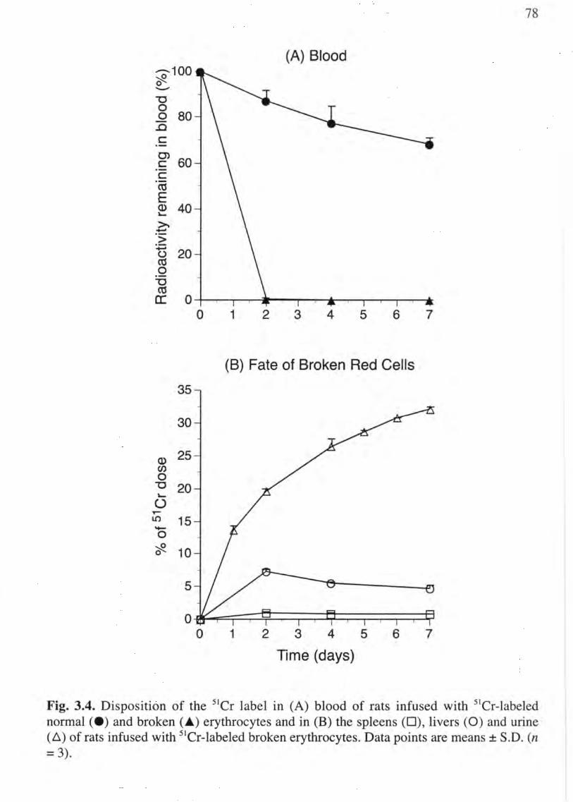
78
(A) Blood ---100 ~ 0 '-"
-0 0 0 80 .0 c: C>
60 c: c:
"co E Q) 40 ~
~ +"""
"> +=i 20 () CO 0 -0 ctS a: 0
0 1 2 3 4 5 6 7
(B) Fate of Broken Red Cells
35
30
Q) 25 en 0 -0 20 ~
() T'""
LO 15 "t-o ~ 0 10
5
0 0 1 2 3 4 5 6 7
Time (days)
Fig. 3.4. Disposition of the Sier label in (A) blood of rats infused with Ster-Iabeled normal (e ) and broken (~) erythrocytes and in (B) the spleens (D), livers (0) and urine (~) of rats infused with Ster-Iabeled broken erythrocytes. Data points are means ± S.D. (n = 3).

79
As shown in Fig. 3.5, there was a concentration-dependent increase in radioactivity
associated with the macrophages after 18 hr of incubation. The level of radioactivity
associated with the macrophages at earlier time points (6 and 12 hr) was not significantly
different from the controls (data not shown). Of importance, the concentration
dependence for this response coincided with the concentration dependence for hemolytic
acti vi ty in vivo.
Visual confirmation of the phagocytic response was carried out by microscopic
examination of the macrophages after 18 hr of incubation and prior to red cell lysis with
ammonium chloride. In cultures containing control erythrocytes, only an occasional red
cell could be observed within some of the macrophages (Fig. 3.6A). In contrast, large
numbers of intact erythrocytes could be observed within many of the macrophages that
received 5-HPQ-treated erythrocytes (Fig. 3.6B). Of note, a subset of the J774A.l
macrophages appeared to account for a majority of the uptake of the damaged red cells.
This phenomenon has been observed previously and suggests that within the clonal
population, some macrophages, designated "jackpot cells," are more competent at
erythrocyte recognition than are others. The mean values for macrophage radioactivity
incorporate macrophages that have ingested no erythrocytes and others that contain
several ingested erythrocytes.

80
1600 *
E 1400 c. ~ 1200 en ~ 1000 «S .c c. 800 0 '-U «S 600 E c 400 '-() ,.-
200 U')
0 0 25 50 75
[5-HPQ] (tJM)
Fig. 3.5. Effect of 5-HPQ on the uptake of rat 51Cr-Iabeled red cells by 1774A.1 macrophages. 51Cr-Iabeled erythrocytes were incubated with the indicated concentrations of 5-HPQ for 2 hr at 37°C. The erythrocytes were then washed and added to 1774A.l macrophage cultures for 24 hr. The cultures were washed with saline and ammonium chloride to remove unbound and bound (but non-ingested) erythrocytes, respectively. The macrophages were then scraped from the plates and the radioactivity was counted. Data points are means ± S.D. (n=3). *Significantly different from controls (p < 0.01).

81
Fig. 3.6. Light micrographs of J774A.l macrophage cultures after incubation with (A) control and (B) 5-HPQ-treated rat erythrocytes. Rat erythrocytes were treated with the vehicle or 5-HPQ (50 JlM) for 2 hr at 37°C before being incubated with the macrophages for 18 hr. The arrows indicate the presence of intact 5-HPQ-treated erythrocytes within jackpot cells." Magnification 63x.

82
Effect of GSH depletion on phagocytic uptake.
G6PD-deficient individuals have long been known to have an enhanced susceptibility
to primaquine-induced hemolytic anemia. We have shown previously that this enhanced
susceptibility can be reproduced by prior depletion of red cell aSH (Bolchoz et aI., 2002;
Bowman et aI., 2004). In the case of 5-HPQ, the hemolytic susceptibility of GSH
depleted rat erythrocytes is about 5-fold greater than aSH-normal red cells in response to
treatment with 5-HPQ (Bowman et aI., 2004). To determine the effect of aSH depletion
on phagocytic removal in vitro, 5lCr-Iabeled erythrocytes were titrated with diethyl
maleate to deplete aSH (ca. 95%) prior to exposure to a low concentration of 5-HPQ.
After 2 hr of incubation at 37°C, control and aSH-depleted erythrocytes were incubated
with J774A.l macrophages for 18 hr. As shown in Fig. 3.7, treatment of aSH-normal
erythrocytes with 25 llM 5-HPQ did not induce a significant increase in phagocytic
uptake by macrophages as compared with the control. However, the aSH-depleted
erythrocytes showed a significant increase in their phagocytic uptake as compared with
the control. This observation is in agreement with the previous in vitro exposure/ in vivo
erythrocyte survival study (Fig. 2.10), which showed that 25 llM 5-HPQ was sub
hemolytic in aSH-normal erythrocytes but was hemolytic in aSH-depleted erythrocytes
(Bowman et aI., 2004).
Discussion
Human erythrocytes have a predetermined lifespan in the circulation of
approximately 120 days. During this time, damage is thought to accumulate within
erythrocytes to the point that these cells are recognized as senescent and are removed
from the circulation by macrophages of the reticuloendothelial system. Although 360

800
700 ..--... E c. 600 u
'-""
500
400
300
200
100
83
*
o 25 o 25
GSH normal GSH depleted
[5-HPQ] (PM)
Fig. 3.7. Effect of depletion of erythrocytic GSH on uptake of 5-HPQ-treated erythrocytes by J774A.l macrophages. slCr-Labeled erythrocytes were titrated with DEM to deplete GSH by 95%. GSH-normal and GSH-depleted erythrocytes were then incubated with the vehicle or 5-HPQ (25 pM) for 2 hr at 37°C. The erythrocytes were then washed and incubated with J774A.l macrophages for 18 hr. Data points are means ± S.D. (n=4). *Significantly different from controls (p < 0.05).

84
billion senescent erythrocytes are removed from the circulation per day by this system,
surprisingly little is known about the changes red cells undergo as they age or the mechanism
by which these cells are recognized and removed from the circulation (J andl, 1996).
Studies supporting specific mechanisms are numerous and often conflicting, but most
have in common the idea that an accumulation of damage to the membrane (in some form
or another) occurs as a consequence of normal chronic, low-level oxidative stress (Clark
et aI., 1984). How intracellular oxidative stress might provide a signal for phagocytic
uptake is not understood, however, a number of theories have been proposed.
On the one hand, oxidative stress may stimulate erythrocyte phagocytosis through a
non-specific mechanism referred to as "culling". In this process, oxidative stress is
thought to alter the membrane cytoskeleton leading to a decrease in erythrocyte
deformability, thereby restricting progress of erythrocytes through the splenic red pulp
and increasing their exposure to resident macrophages (Chadbum, 2000). On the other
hand, oxidative stress may induce discrete changes to the external cell surface that
stimulate specific phagocytic receptors on macrophages. Although what constitutes a
recognition ligand on the erythrocyte surface is not yet known, extensive studies have
supported several competing viewpoints, including: 1) unmasking of a cryptic antigenic
site leading to opsonization by circulating autologous IgG (Kay, 1994); 2) unmasking of
carbohydrate epitopes by desialization of glycoproteins, such as glycophorin A (Kay,
1993); 3) extemalization of the membrane phospholipid, phosphatidylserine (MandaI et
aI., 2002); and 4) loss of self recognition caused by alterations in the cell surface
expression or lateral distribution of the macrophage inhibitory ligand, CD47 (Oldenborg
et aI., 2000).

85
As part of ongoing studies in our laboratory to identify redox-active metabolites of
primaquine that contribute to its hemotoxicity, we have demonstrated that a phenolic
metabolite, 5-HPQ, is a direct-acting hemolytic agent in the rat (Bowman et aI., 2004).
More specifically, we have shown that treatment of erythrocytes with 5-HPQ in vitro
induces a GSH-dependent acceleration in the removal of the treated red cells in vivo. The
present studies were undertaken to determine the fate of these erythrocytes and to
ascertain whether splenic macrophages playa significant role in this process.
To determine the fate of 5-HPQ-treated erythrocytes in vivo, we compared the extent
of uptake of control, broken and 5-HPQ-treated erythrocytes into the spleen and liver,
which are the two major organs for erythrocyte clearance. Broken red cells and
erythrocytes exposed to 40 }lM 5-HPQ in vitro were both removed rapidly from the
circulation as compared to the control. However, radioactivity from the broken cells was
distributed to the liver and was ultimately recovered in the urine (Fig. 3.4B), whereas the
radiolabel from 5-HPQ-treated erythrocytes was distributed primarily to the spleen (Fig.
3.1 B). These data indicate that the spleen is the primary anatomic site of sequestration for
erythrocytes exposed to 5-HPQ, and suggest that intravascular lysis does not contribute
significantly to the loss of erythrocytes from the circulation. A similar observation was
reported in G6PD-deficient individuals receiving primaquine (Degowin et aI., 1966).
Those studies demonstrated that the radiolabel was taken up into both the spleen and the
liver; however, the role of the spleen was significantly greater when uptake was
expressed relative to organ weight. Importantly, these observations lend further support
to the hypothesis that 5-HPQ has the requisite properties to mediate primaquine toxicity
in vivo.

86
The selective depletion of macrophages from specific tissues has been shown to be
beneficial for the determining their role in defined biological processes (Van Rooijen and
Sanders, 1994). Macrophages have long been thought to be responsible for the splenic
sequestration that is observed following administration of hemolytic agents. If
macrophages are the key players and erythrocyte removal is not due simply to non
specific trapping in the splenic cords, then substances that cause a loss of macrophage
viability or otherwise interfere with the recognition mechanism should have an inhibitory
effect on splenic sequestration. To test the dependence of the hemolytic response on the
presence of functional macrophages, rats were given clodronate-Ioaded liposomes 24 hr
before administration of the radioalabeled erythrocytes. After being ingested the
liposomes are degraded within macrophages by lysosomal phospholipase, releasing
clodronate into the cytosol where it induces macrophage cell death by apoptosis (Van
Rooijen and Sanders, 1994). A dose of 0.1 ml of clodronate-liposome suspension per 10
g body weight has been reported to be sufficient for depletion of splenic macrophages
and hepatic Kupffer cells, and the decline in spleen/body weight ratio observed over the
course of the experiment (Fig. 3.3) is consistent with selective loss of macrophages from
the spleen.
The time it takes to remove 50% of the sler-Iabeled erythrocytes for 5-HPQ-treated
erythrocytes in clodronate-Ioaded liposome-pretreated rats was significantly delayed (by
-3 days) in relation to that of the saline-pretreated rats (Fig. 3.2A). In addition, the
accumulation of radioactivity that was observed in the spleens of saline-pretreated rats
was markedly reduced in clodronate liposome-pretreated rats (Fig. 3.2B). Interestingly,
uptake of the label into the livers of clodronate liposome-pretreated animals increased

87
significantly 4 days after administration of the labeled cells (Fig. 3.2B). This observation
is in agreement with previous studies which showed that Kupffer cells repopulate the
liver more rapidly than macrophages repopulate the spleen following treatment with the
liposomes (Van Rooijen and Van Kesteren-Hendrikx, 2003), and it reflects the ability of
Kupffer cells to increase their phagocytic activity towards erythrocytes in the absence of
a functional splenic uptake mechanism (Ganick et aI., 1977).
An in vitro assay for hemolytic anemia has long been desired. Although the rat 51Cr_
labeled erythrocyte in vitro exposure/in vivo survival assay (e.g., Fig. 3.1A) is a useful
experimental tool for identifying direct-acting hemolytic agents and for studying the
mechanism underlying the hemolytic response, this assay is limited by the need for large
numbers of animals, it does not easily permit erythrocyte-macrophage interactions to be
examined in detail, and it offers no way to investigate the hemolytic response in human
erythrocytes. Measurement of drug-induced erythrocyte lysis in the test-tube (i.e.,
osmotic fragility) is not a valid substitute for the hemolytic response in vivo because of
the recognition that the toxic endpoint is splenic sequestration. Efforts to develop cell
culture assays based on isolated splenic macrophages or Kuppfer cells are hindered by
the low numbers of cells that can be harvested from these tissues.
In an effort to reproduce in vitro the uptake of 5-HPQ-treated erythrocytes that was
observed in vivo, the phagocytic activity of murine monocyte-derived macrophage cell
lines towards control and 5-HPQ-treated erythrocytes was examined. In initial
experiments we utilized RAW 264.7 macrophages, but they did not respond to 5-HPQ
treated erythrocytes (data not shown), though these cells did ingest erythrocytes that had
been treated with neuraminidase (which cleaves sialic acid residues). In contrast, 1774A.l

88
macrophages were found to respond selectively to 5-HPQ-treated erythrocytes. Uptake of
the StCr label by these macrophages was dependent on the concentration of 5 ... HPQ (Fig.
3.5), and this concentration-dependence correlated closely with that of the hemolytic
response (Bowman et aI., 2004). Importantly, uptake of the SICr label was due to
phagocytosis of intact erythrocytes (Fig. 3.6), which supports the conclusion that
erythrocyte removal in vivo is due to uptake of intact erythrocytes rather than to
intravascular lysis. Moreover, the data suggest that restrictive passage of erythrocytes
through the splenic architecture may not be necessary for removal of the damaged cells
from the circulation.
To test the validity of this cell culture system as an experimental model for 5 ... HPQ
induced hemolytic anemia, the effect of depletion of erythrocyte aSH on phagocytic
uptake was examined. In agreement with previous studies (Bowman et aI., 2004), aSH
depletion induced the uptake of erythrocytes that were treated with a sub-hemolytic
concentration of 5-HPQ in GSH-normal erythrocytes (Fig. 3.7). Taken together, these
data indicate that 1774A.l macrophages can be used as a valid model to identify cell
surface changes that signal macrophage recognition and phagocytosis.
For many years, "pitting" and "culling" have been commonly accepted mechanisms
for the functional removal of oxidatively damaged red cells by the spleen. "Culling" is
thought to occur as damage to the red cell results in increased transit time through the
tight meshwork of the splenic red pulp, which increases the likelihood of exposure to and
recognition by cordal macrophages (Chadburn, 2000). Possible mechanisms of trapping
that have been suggested include: 1) that the hemoconcentration that occurs in the splenic
red pulp creates an acidic, hypotonic environment that results in cell swelling and

89
trapping within the splenic meshwork; or 2) that removal is directly related to profound
morphological alterations and/or damage to the red cell membrane that renders them less
flexible and thus more susceptible to becoming trapped (Chadburn, 2000). "Pitting" is
thought to lead to the removal of damaged cells as cell inclusions or appendages, such as
Heinz bodies, cause parts of the red cell to be trapped in the sinus wall and subsequently
amputated by macrophages, allowing the pliant part of the cell to slip through. Although
the cell survives, it does so with a reduction of membrane surface, which can ultimately
lead to spherocytosis and trapping in the spleen. Erythrophagocytosis by J774A.l
macrophages in vitro provides evidence that "pitting" and "culling" are not necessary for
the recognition of 5-HPQ-damaged red cells. Although a role for culling cannot be ruled
out, since it could still work by increasing the time of exposure of damaged red cell to
macrophages, it is likely that the macrophages recognize a specific signal on the red cell
surface rather than a general structural alterations.
In summary, we have demonstrated that splenic macrophages play a crucial role in
the removal of 5-HPQ-treated rat red cells from the circulation, and that this response can
be reproduced in vitro using J774A.l macrophages. This observation further supports the
notion that 5-HPQ is an important contributor to the hemolytic response induced by
primaquine. Future studies are warranted to determine the nature of the signal that
commits the damaged erythrocytes to premature splenic sequestration ..

CHAPTER 4
Primaquine-Induced Hemolytic Anemia: Role of Membrane Lipid Peroxidation and Cytoskeletal Protein Alterations on the
Hemotoxicity of 5-Hydroxyprimaquine

91
Introduction
Malaria is a life-threatening parasitic disease of epidemic proportions, and it is a
major public health concern for developing countries. Each year malarial infection is
responsible for an estimated 300-500 million acute illnesses and 2 million deaths in
tropical and subtropical regions worldwide (Kain and Keystone, 1998). Primaquine, the
prototype 8-aminoquinoline tissue schizontocide, is effective against all four malarial
species that infect humans and is the only drug approved for the radical cure of relapsing
malaria. As resistance has developed from extensive use of blood schizontocides, such as
chloroquine, the importance of primaquine in combating multiple drug resistance has
increased, and it is now used in combination with several blood schizontocides for the
treatment and prophylaxis of all forms of malaria, including the most lethal malarial
species, Plasmodiumfalciparum (Shanks et aI., 2001). Primaquine is also effective in the
treatment of Pneumocystis carinii pneumonia, a common infection in AIDS patients
(Toma et aI., 1998). The clinical utility of primaquine, however, is restricted by its
hemotoxicity, particularly in patients who are genetically deficient in erythrocytic
glucose-6-phosphate dehydrogenase (G6PD) activity (Beutler, 1990).
The hemolytic activity of primaquine has long been known to be due to an intra
erythrocytic oxidative stress that is mediated by redox-active metabolites rather than by
the parent drug itself. The oxidatively damaged erythrocytes are thought to be recognized
by splenic macrophages as the equivalent of senescent red cells, resulting in their
selective removal from the circulation (Rifkind, 1966). 5-Hydroxyprimaquine (5-HPQ) is

92
a putative human metabolite of primaquine that forms a redox pair with its quinoneimine
form; continuous cycling of this redox pair is thought to generate reactive oxygen species
(ROS) within erythrocytes (Vasquez-Vivar and Augusto, 1992). We have shown that
when 5-HPQ is incubated with rat red cells in vitro, damage occurs within the cells such
that they are removed rapidly from the circulation of isologous rats as compared to
untreated controls (Bowman et aI., 2004). While it is clear that ROS attack on the
cytosolic surface of the membrane leads to a signal for removal of damaged erythrocytes
from the circulation, neither the nature of the crucial target(s), whether lipid or protein~
nor the mechanism of transfer of the signal across the membrane is known.
The mechanism(s) by which senescent or damaged (but intact) erythrocytes are
selected for splenic sequestration are not known. Two current hypotheses are: 1) that red
cell damage is analogous to the apoptotic response observed in other (nucleated) cell
types, resulting in changes to the normal asymmetric distribution of phospholipids
(MandaI et aI., 2002); and 2) that protein oxidation interferes with normal protein-protein
interactions among the cytoskeletal, integral membrane and cell surface proteins that
confer the recognition of "self' (Oldenborg et aI., 2000; Bruce et aI., 2003). The present
study examines the role of the postulated truncated apoptotic pathway in the hemolytic
activity of 5-HPQ, specifically in regard to potential roles for membrane lipid
peroxidation and loss of phospholipid (i.e., phosphatidylserine) asymmetry. We also
address the alternate hypothesis, that alterations to skeletal membrane proteins in 5-HPQ
damaged red cells provide a crucial link between internal oxidative stress and external
recognition and removal of erythrocytes from the circulation.

93
The data indicate that under in vitro incubation conditions that provoke premature
splenic sequestration of red cells in vivo, 5-HPQ generates intracellular ROS but does not
induce lipid peroxidation or cause loss of phosphatidylserine asymmetry in the plasma
membrane of the red cell. In contrast, profound alterations occur to certain proteins of the
cytoskeleton in 5-HPQ-treated erythrocytes, and these alterations are due primarily to the
formation of disulfide-linked hemoglobin-skeletal protein adducts. Further studies are
warranted to determine the specific signal for erythrocyte phagocytosis, however, the
present data are consistent with the concept that cytoskeletal protein damage, rather than
alterations in the lipid bilayer, underlies the process of splenic uptake of red cells
damaged during the course of primaquine therapy.
Materials and Methods
Chemicals and materials.
5-HPQ was synthesized by HBr-catalyzed hydrolysis of 5-methoxyprimaquine as
described previously (Bowman et aI., 2004). Bovine factor V, bovine factor Xa and
bovine prothombin were obtained from Enzyme Research Laboratories (South Bend, IN).
Chromogenic substrate S-2238 was purchased from Chromogenix (DiaPharma Group,
Inc., Westchester, OH). Alexafluor 647-conjugated annexin V and 2' ,7'-dichloro
dihydrofluorescein diacetate (DCFDA) were obtained from Molecular Probes, Inc.
(Eugene, OR). Calcium ionophore A23187, N-ethylmaleimide (NEM) , cumene
hydroperoxide (CH) and rabbit anti-rat hemoglobin antibodies were purchased from
Sigma-Aldrich (St. Louis, MO). All other reagents were of the highest grade
commercially available.

94
Erythrocytes and incubation conditions.
Male Sprague-Dawley rats (75-100 g) were obtained from Harlan Laboratories
(Indianapolis, IN) and maintained on food and water ad libitum. Animals were
acclimated for 1 week to a 12-h light/dark cycle before their use. Blood was collected
from the descending aorta of anesthetized rats into heparinized tubes and washed 3 times
with Hanks' Balanced Salt Solution with calcium (HBSS, Invitrogen, Carlsbad, CA) to
remove the plasma and buffy coat. The cells were resuspended to a 40% hematocrit in
HBSS and used on the same day they were collected. Erythrocytes were washed 3 times
with HBSS and resuspended to a 40% hematocrit.
Stock solutions of 5-HPQ in argon-purged water were prepared to deliver the
appropriate concentration of 5-HPQ in 10 pI to erythrocyte suspensions (1-2 ml). The
erythrocyte suspensions (40% hematocrit) were allowed to incubate for up to 2 hr at
37°C, and then washed once with HBSS prior to biochemical analyses. The effects of
DTT on intact red cells were determined as follows: 1.) red cells in PBSG were
incubated for the indicated times up to forty minutes in the presence or absence of 50pM
5-HPQ; 2.) Cells were washed once with PBSG and reincubated for 1 additional hour in
the presence or absence of 5 mM DTT; 3.) at the end of the incubation with DTT, the red
cells were washed once again with PBSG and prepared for the appropriate assay as
described below.
Measurement of ROS formation in erythrocytes.
Rat erythrocytes were suspended in isotonic phosphate-buffered saline (pH 7.4)
supplemented with 10 mM D-glucose (PBSG) to a 10% hematocrit. DCFDA (600 pM),

95
dissolved in DMSO, was added to the erythrocyte suspension and allowed to incubate for
15 min at 37°C. Immediately after the addition of various concentrations of 5-HPQ,
fluorescence was measured at 2 min intervals for 20 min (excitation 488 nrn, emission
529 nm) on a Molecular Devices SprectraMAX Gemeni XS Fluorescence Microplate
Reader (Molecular Devices, Sunnyvale, CA).
Preparation of erythrocyte membrane ghosts.
Red cell ghosts were prepared from vehicle- and 5-HPQ-treated red cells as described
previously with modification (Grossman et aI., 1992). Briefly, washed red cells were
centrifuged and the packed cells were lysed in 30 ml of ice-cold phosphate buffer (5 mM,
pH 8.0). The membrane ghosts were pelleted by centrifugation at 20,000 x g for 10 min.
The supernatant was removed by aspiration and the ghosts were repeatedly washed with
phosphate buffer until the control cells yielded white ghosts, typically 3-4 washes.
Determination of lipid peroxidation in erythrocytes.
Lipid peroxidation was assessed by measuring the content of F2-isoprostanes in red
cell ghosts prepared from vehicle- and 5-HPQ-treated red cells as described previously
(Bolchoz et aI., 2002b). Briefly, the lipids were extracted from membrane ghost
suspensions (250 }ll), prepared with argon-purged phosphate buffer, with
chloroform/methanol (2: 1, v/v). The extracted lipids were then subjected to alkaline
hydrolysis to release the esterified F2-isoprostanes. The work-up procedure for
quantifying F2-isoprostanes by GC/MS is described in detail elsewhere (Morrow and
Roberts, 1999). Treatment of red cell suspensions for 1 hr at 37°C with the lipid-soluble

96
peroxide, cumene hydroperoxide (1 mM), was used as a positive control for lipid
peroxidation (van den Berg et aI., 1992).
NEM and calcium ionophore treatment of erythrocytes.
As a positive control for translocation of phosphatidylserine from the inner to the
outer leaflet of the lipid bilayer, rat erythrocytes were treated with NEM to inhibit
aminophospholipid translocase, and calcium ionophore A23187 was added to induce
membrane lipid scrambling as described previously (Kuypers et aI., 1996). Briefly, rat
erythrocytes were washed three times with HBSS (with calcium) and reconstituted to
40% hematocrit with HBSS. The red cell suspension (1 ml) was allowed to incubate with
10 mM NEM for 30 min at 37°C, and then washed twice with HBSS. Calcium ionophore
A23187 (4 }lM) in DMSO was then added and allowed to incubate for 1 hour at 37°C.
After incubation, the red cells were washed with 2.5 mM EDTA (1 ml) to remove the
calcium, and then washed twice more with calcium-free HBSS containing 1 % bovine
serum albumin (BSA) to remove the ionophore. The cells were then re-suspended in 1 ml
of annexin binding buffer (10 mM HEPES, 140 mM NaCI, and 2.5 mM CaCI2, pH 7.4),
and analyzed for annexin V labeling and prothrombinase activity as described below.
Determination of phosphatidylserine translocation in erythrocytes.
Erythrocyte labeling with annexin V was performed according to a modification of a
flow cytometric technique (Kuypers et aI., 1996). Following incubation with the vehicle,
the positive control, or 5-HPQ (2 hr at 37°C), aliquots (50 }ll) of the suspensions were
removed and washed twice with annexin binding buffer (1 ml). After the last wash,

97
aliquots of the packed red cells (2 Jil) were combined with annex in binding buffer (997
Jil), and AlexaFluor 647 -conjugated annexin-V solution (1 Jil) was added to the
erythrocyte suspensions to bring the total volume to 1 ml. After 30 min incubation in the
dark at room temperature, the samples were washed and re-suspended in annexin binding
buffer (1 ml), and analyzed on a Becton Dickinson FACSCalibur analytical flow
cytometer (BD Biosciences, San Jose, CA).
Phosphatidylserine translocation was also assessed by measuring the conversion of
prothrombin to thrombin using a modification of a method described previously (Kuypers
et aI., 1996). Briefly, after incubation with the vehicle or 5-HPQ, erythrocyte suspensions
were washed once with HBSS, and 1 pI of packed cells was added to 1 ml of Tris buffer
(pH 7.4) at 37°C. Bovine factor V (0.33 D/ml) and bovine factor Xa (0.33 D/ml) were
added, followed by 0.13 mg of prothrombinase. After 4 min, the reaction was quenched
with 15 mM EDTA. The erythrocytes were pelleted by centrifugation and 75 }ll of the
supernatant was added to 1 ml of chromogenic substrate S-2238 working solution. The
increase in absorbance at 405 nm was determined over 1 min at 37°C in a Shimadzu UV-
160 Spectrophotometer (Shimadzu Corporation, Kyoto, Japan) equipped with software
for enzyme kinetics. The amount of thrombin formed per unit time was determined by
comparison with thrombin standards.
Electrophoretic analysis o/membrane cytoskeletal proteins.
Red cell ghosts from control and 5-HPQ-treated erythrocytes were washed
exhaustively to remove unbound hemoglobin (4 washes). The ghost protein was then
solubilized in NuPAGE LDS Sample Buffer (Invitrogen, Carlsbad, CA), with or without

98
40 mM dithiothreitol (DTT), and heated at 70°C for 10 min. Solubilized proteins (15 jlg)
were loaded and resolved on 4-12% NuPAGE Bis-Tris gels with MOPS Running Buffer
(Invitrogen) at 200 V (constant) for 50 min. Resolved proteins were transferred to PVDF
membranes for immunoblot analysis according to the Invitrogen protocol. For SDS
PAGE analysis, solubilized membrane ghosts were resolved on continuous gels under
non-reducing conditions as described previously (McMillan et aI., 1995), then stained
with Gel Code Blue Stain Reagent (Pierce, Rockford, IL) and destained with water.
Rat red cell membrane proteins were identified according to molecular weight
(Fairbanks et aI., 1971) with the use of molecular weight protein standards (Invitrogen).
Blotted proteins were blocked in TBST (pH 7.5) containing 5% non-fat dry milk and
incubated in TBST containing 1 % BSA and primary antibody (rabbit anti-rat
hemoglobin, 1: 10,000 vol/vol). After washing and incubation with the peroxidase
conjugated secondary antibody (anti-rat IgG), the immunoblots were developed using
ECL detection (Amersham Biosciences, Piscataway, NJ).
GSH Depletion of Erythrocyte Suspensions.
Diethyl maleate (DEM) was used to deplete aSH (>95%) in red cell suspensions as
described previously (Bolchoz et aI., 2002b). Briefly, DEM (750}lM) dissolved in
acetone was added to packed red cells. After a 15-min incubation at 37°C, the red cells
were analyzed for aSH content by HPLC-EC. The cells were re-suspended to a 40%
hematocrit in HBSS and used on the same day that they were collected.

99
Measurement of Hemolytic Activity.
The survival of rat 51Cr-labeled red cells was determined in vivo after in vitro
incubation with 50}lM 5-HPQ with or without 5mM DTT as described above. After
incubation at the specified times at 37°C, the erythrocytes were washed once and
resuspended in PBSG (40% hematocrit). Aliquots (0.5ml) were administered
intravenously to isologous rats. To blood samples were taken from the orbital sinus xx
min after administration of labeled red cells. Additional blood samples were at sequential
times over 14 days. At the end of the experiment, the samples were counted in a well
type gamma counter, and the data were expressed as a percentage of the To blood sample.
The hemolytic response was quantified by calculating the fraction of radiolabeled red
cells that were removed from the circulation within the first 48 hr for each animal by
linear regression as described previously (McMillan et aI., 2001). Statistical significance
was determined with use of Student's t test.
Erythrophagocytosis by J774 macrophages.
J774A.l macrophages (ATCC) were plated at 200,000 cells/well in 12 well culture
plates and allowed to adhere. After 24 hr, 50}l1 Cr-Iabeled, vehicle- or 5-HPQ-treated
packed red cells were added to the macrophage monolayer and incubated for 24 hr at
37°C. Non-ingested RBCs were removed by lysis with NH4CI and washed three times
with buffer. Adherent macrophages were released with NaOH and phagocytosis was
assessed by measuring radioactivity in a well-type gamma counter. Data were expressed
as either counts per minute (CPM) or as a percent of control.

100
Results
Effect of 5-HPQ on ROS generation in rat erythrocytes.
DCFDA is a lipid-soluble probe that can detect the intracellular formation of several
types of ROS, including hydroxyl, peroxyl, alkoxyl, and nitroxyl free radicals, as well as
peroxynitrite (Halliwell and Whiteman, 2004). To assess the formation of ROS in 5-HPQ
treated rat erythrocytes, erythrocyte suspensions were pre-incubated with DCFDA for 15
min followed by addition of 5-HPQ. Fluorescence was then measured at 2 min intervals
for 20 min using a fluorescence microplate reader. Preliminary experiments indicated that
the concentration of red cells in the suspensions had to be reduced from 40% to a 10%
hematocrit to avoid significant quenching of the fluorescence signal by the cells. 5-HPQ
concentrations were reduced accordingly (by 4-fold) to maintain the same 5-HPQ-to-red
cell ratio that was used in the other experiments.
As shown in Fig. 4.1, incubation of erythrocytes with DCFDA in the absence of 5-
HPQ resulted in a slight but measurable increase in fluorescence over a 20 min time
period, consistent with the known low-level, steady production of ROS in normal red
cells (Jandl et aI., 1960). Inclusion of 5-HPQ (5-30 pM) in the erythrocyte suspension
caused a marked and concentration-dependent increase in the generation of fluorescence,
indicating enhanced production of ROS by 5-HPQ. This observation is consistent with
previous observations which showed that 5-HPQ has the ability to redox cycle and to
cause oxidative stress in rat erythrocytes, as evidenced by aSH oxidation and
methemoglobin formation (Bowman et aI., 2004). No fluorescence signal was detected in
the absence of red cells or in the absence of DCFDA (data not shown).

101
Effect of 5-HPQ on erythrocyte membrane lipid peroxidation.
To determine whether ROS production by 5-HPQ resulted in lipid peroxidation, rat
red cells were incubated for 2 hr at 37°C with concentrations of 5-HPQ corresponding to
EC lO (10 J-tM), ECso (40 J-tM) and EC90 (70 J.lM) concentrations for hemolytic activity
(Bowman et aI., 2004). The red cells were then washed and lysed in argon-purged
hypotonic buffer for the preparation of red cell ghosts, as described previously
(Grossman et aI., 1992). Esterified isoprostanes were extracted from the ghosts and
hydrolyzed, and the total F2-isoprostane content in the samples was quantified by GC-MS
(Morrow and Roberts, 1999). As shown in Fig. 4.2, F2-isoprostane content in 5-HPQ
treated incubates was not different from that of control incubates, regardless of the
concentration of 5-HPQ. In contrast, treatment of red cells with the positive control,
cumene hydroperoxide (CH, 1 mM), resulted in a 3-fold increase in F2-isoprostane
formation. Treatment with this concentration of CH was the lowest that produced a
significant elevation in F2-isprostane formation that was not associated with significant
frank lysis of the erythrocytes; concentrations of CH greater than 1 mM caused
progressive and complete red cell lysis (data not shown).
Effect of 5-HPQ on red cell membrane phospholipid asymmetry.
To determine whether exposure to 5-HPQ resulted in a loss of phosphatidylserine
asymmetry, the transmembrane location of phosphatidylserine was assessed by two
methods: enhancement of prothrombinase activity, and annexin-V labeling of intact
erythrocytes.

8000
.....-.... 7000 en ...... § 6000
........... ~ ......
"en 5000 c Q) ......
. c 4000 Q) u a3 3000 u en
~ 2000 ::J
LL 1000
O~------~------~------~--~--a 5 10 15 20
Time (min)
102
Fig. 4.1. Effect of 5-HPQ on ROS generation in rat erythrocytes. Erythrocyte suspensions (10% hematocrit) were incubated with DCFDA (600 }lM) for 15 min. The red cells were then treated with the vehicle (e ), 5}lM (+ ), 15 }lM (~ ), and 30 }lM (_ ) 5-HPQ. Fluorescence emission intensity was monitored on a fluorescence microplate reader (excitation, 488; emission, 529 nm) at 2 min intervals. The values are means ± S.D. (n = 3).

0.5 .-.... c Q) ~ e 0.4 0. 0> E 0, 0.3 c ---en Q)
@ 0.2 ~
en o s-o. o 0.1 en
I
LL C\J
o
103
*
o 10 40 70 CH
5-HPQ (J1M)
Fig. 4.2. Effect of 5-HPQ on F2-isoprostane formation in rat erythrocytes. Erythrocytes were incubated for 2 hr at 37°C in HBSS containing the indicated concentrations of 5-HPQ. After incubation erythrocyte ghosts were prepared and analyzed for F2-isoprostane content by GC-MS as described in the Materials and Methods. The values are means ±
S.D. (n = 3). * Significantly different from control (p < 0.05).

104
Erythrocyte suspensions (40%) were incubated with hemolytic concentrations of 5-
HPQ (10 to 70 ,...,M) for 2 hr. At the end of the incubation, the red cells were washed, re
suspended in HBSS and analyzed for prothrombinase activity, which is dependent on cell
surface exposure to phosphatidylserine (Bevers et aI., 1982). For a positive control,
erythrocytes were treated with 10 mM NEM for 30 min, followed by treatment with 4
pM calcium ionophore A23187 in the presence of calcium for 1 hr. As shown in Fig.
4.3A, thrombin formation in 5-HPQ-treated erythrocytes was not significantly different
from the control. In contrast, the positive control, NEM plus ionophore and calcium,
induced an 8-fold increase in prothrombinase activity as compared to the control.
The presence of phosphatidy lserine in the outer leaflet of the lipid bilayer was
confirmed by direct labeling of erythrocytes with fluorescently conjugated annexin V,
which binds to acidic phospholipids, particularly phosphatidylserine (Kuypers et aI.,
1996). As shown in Fig. 4.3B, annexin-positive erythrocytes from 5-HPQ-suspensions
constituted less than 2% of the red cells, which was similar to the vehicle-treated control.
In contrast, treatment of red cells with NEM plus ionophore and calcium resulted in
labeling of about 45% of the erythrocytes.
Effect of 5-HPQ on rat erythrocyte membrane skeletal proteins.
To determine whether 5-HPQ-induced hemolytic activity was associated with
alterations to the membrane cytoskeleton, rat erythrocyte ghost proteins from control and
5-HPQ-treated erythrocytes were separated by SOS-PAGE and either stained with Gel
Code Blue, or transferred to PVDF membranes and immunostained with antibodies to rat
hemoglobin. As shown in Fig. 4.4A, 5-HPQ caused concentration-dependent changes to

105
10.0 (A) 50 (8) - 9.0 ~45 * c 0
'E -""- B.O ~ 40 E ~ ""- 7.0 g 35 (/) :=: '-c ..c
:::::s 6.0 ~30 -c Q) 0 5.0 "0 25 :;:: OJ Q)
E 4.0 ~ 20 '-0 OJ - ::> 15 c 3.0 :.0 c E 2.0 'x 10 0 Q) '- c ..c
1.0 c 5 .- « a a
0 10 40 70 NEM/Ca2+ 0 10 40 70 NEM/Ca2+
5-HPQ (PM) 5-HPQ (JiM)
Fig. 4.3. Effect of 5-HPQ on phosphatidylserine asymmetry in rat erythrocytes. Erythrocyte suspensions (40% hematocrit) were incubated with the indicated concentrations of 5-HPQ for 2 hr at 37°C. NEM/Ca2
+ was used as a positive control. (A) Prothrombinase activity and (B) AlexaFluor 647-conjugated annexin V labeling were determined as described in Materials and Methods. The values are means ± S.D. (n = 3). * Significantly different from control (p < 0.05).

106
the normal electrophoretic pattern of erythrocyte skeletal proteins as compared with that
of the vehicle-treated control. 5-HPQ treatment induced the appearance of new protein
bands at 16, 32 and 64 kDa, which is consistent with formation of membrane-bound
hemoglobin monomers, dimers and trimers, respectively (Grossman et aI., 1992). In
addition, there was a concentration-dependent loss of resolution of protein bands 1 and 2
(a and B spectrins), splitting of band 2.1 (ankyrin) and loss of band 4.2. On the other
hand, band 4.1 and band 5 (actin) appeared not to be affected significantly by treatment
with 5-HPQ.
Immunoblot analysis of the skeletal proteins from 5-HPQ-treated red cells showed a
dramatic increase in the content of membrane-bound hemoglobin (Fig. 4.4B). Only
insignificant amounts of hemoglobin were bound to skeletal proteins at sub-hemolytic
concentrations of 5-HPQ «40 }lM), whereas increasing amounts of hemoglobin were
bound to a wide variety of proteins at hemolytic concentrations. When 5-HPQ-treated
ghosts were treated with DTT (40 mM C!t 70°C for 1 hr) prior to electrophoresis, binding
of hemoglobin to skeletal proteins was reversed (Fig. 4.5). This observation indicated that
hemoglobin was bound covalently to skeletal protein sulfhydryl groups through
intermolecular disulfide bonds.
Effect of 5-HPQ on the formation of Hb-membrane adducts in GSH-depleted RBCs.
In order to further assess that the formation of Hb-adducts plays a direct role in the
removal 5-HPQ-damaged red cells, we examined the formation of Hb-cytoskeletal
protein adducts in red cells that were treated with sub-hemolytic concentrations of 5-HPQ
that have been shown to be hemolytic to GSH-depleted red cells (Bowman et aI., 2004).

107
(A) 1 2 3 4 5 6
Lane 1
~
4.2 -198 ~ _150
Lane 3 4.1-4.2/ -93
5- -50
_36
-29 Lane 5
-21
Hb-
Migration distance • (8)
1 Lane 1 ~
kDa Lane 3 120 100 80
60 50 Lane 5 40
30
20
Migration distance •
Fig. 4.4. Effect of 5-HPQ on rat erythrocyte membrane skeletal proteins. Red cells were incubated with vehicle (lane 1), 10 pM (lane 2), 25 pM (lane 3), 50 JlM (lane 4), 75 pM (lane 5) and 100 JlM (lane 6) 5-HPQ for 2 hr at 37°C. The cells were washed, and ghosts were prepared and washed exhaustively to remove unbound hemoglobin (Hb). The ghosts were then solubilized in SDS and subjected to PAGE. (A) Gel Code Blue-stained gel and densitometric scans; (B) immunoblot stained with polyclonal antibodies to rat hemoglobin. Molecular weight markers appear along the right side of the gel; the major cytoskeletal proteins are identified along the left side of the gel according to (Fairbanks et aI. , 1971).

(A) -DTT +DTT
1 2 3 4 5 6 7 8 Std
kOa
120 100
80
60
50
40
30
20
108
(8)
25 pM 5-HPQ
________ 2_5_PM __ 5-_H_PQ __ +_D_TI ____ ~~
Migration distance .,
Fig. 4.5. Effect of DTT on 5-HPQ-induced hemoglobin binding to membrane skeletal proteins. Erythrocytes were incubated with 5-HPQ for 2 hr at 37°C. Ghosts were prepared, washed exhaustively to remove unbound hemoglobin, and each sample was divided into two aliquots. Prior to electrophoresis, one aliquot was incubated with water and the other with 40 mM DTT for 1 hr at 70°C. The samples were then subjected to SDS-PAGE and blotted onto PVDF membranes. (A) Immunoblot stained with polyclonal antibodies to rat hemoglobin. Vehicle control (lanes 1 and 5), 25 JlM (lanes 2 and 6), 50 }tM (lanes 3 and 7) and 75 JlM (lanes 4 and 8) 5-HPQ. (B) Selected densitometric scans.

109
Red cell aSH was depleted by >95% with DEM prior to treatment with 5-HPQ for 2 h at
37°C. The range of 5-HPQ concentrations (2.5-25 pM) was chosen, since it was
previously shown to span the red-cell survival concentration-response curve generated
with GSH -depleted red cells treated in vitro were administered to rats in vivo (Bowman et
aI.,2004). After treatment with 5-HPQ, membrane ghosts were prepared with hypotonic
phosphate buffer and washed extensively to remove unbound hemoglobin. Ghost protein
(15jlg) was then separated by SDS-PAGE, blotted to PVDF membranes, and
immunostained with anti-rat hemoglobin antibodies. Immunoblot analysis showed
enhanced binding of hemoglobin at concentrations as low as 2.5jlM 5-HPQ (Fig. 4.6).
Significantly, the sharp, threshold-like response seen in GSH-normal cells (Fig. 4.4B)
was lost and the TCso markedly reduced -5-fold, from 25-50jlM to approximately 5 ...
10 pM. This data demonstrate that there is a strong correlation between the formation of
Hb-adducts with the hemolytic activity previously observed in vivo for both GSH-normal
and GSH-depleted red cells, indicating that the formation of disulfide-linked Hb-adducts
with skeletal membrane proteins likely underlies the mechanism for the recognition and
removal of 5-HPQ-treated red cells by splenic macrophages.
Effect of DTT on 5-HPQ-induced Hb-adductformation and hemolytic susceptibility.
While the correlation between the formation of Hb-cytoskeletal protein adducts and
hemolytic activity is very strong, it was necessary to determine directly whether adduct
formation is functionally significant for macrophage recognition and phagocytosis of
damaged erythrocytes. We examined whether the reduction of Hb with DTT in intact
cells could restore normal cytoskeletal protein sulfhydryl status to, in effect, "rescue" 5-

110
1 2 3 4 5
Lane 1
kDa
220 Lane 2 J\ 120
lOa
80 Lane 3
60
50 Lane 4
40
30 Lane 5
20
Migration distance ..
Fig. 4.6. Effect of 5-HPQ on Hb-cytoskeletal protein adduct formation in GSH -depleted rat erythrocytes. GSH depleted (>95%) rat erythrocytes were incubated in the presence of vehicle (lane 1), 2.5}lM 5-HPQ (lane 2), 5}lM 5-HPQ (lane 3), lOflM 5-HPQ (lane 4), or 25}lM 5-HPQ (lane 5) for 2 hr at 37°C. Following exposure, the cells were washed and membrane ghosts were prepared and washed extensively to remove any unbound hemoglobin. The ghosts (15 }lg of protein) were subjected to SDS-PAGE, blotted onto PVDF membrane, and stained with antibodies against rat hemoglobin. The densitometric tracings of the blot (left) are shown on the right.

111
HPQ-damaged red cells from their phagocytic fate. The ability of OTT (5mM) to reduce
the disulfide-linked Hb-adducts, which formed during incubation with 5-HPQ was
examined. Red cells were treated with a 50pM 5-HPQ in the absence of DTT for 15 min,
washed once with PBSG, and then treated with 5 mM OTT for 1 hr. The cells were once
again was with PBSG and red cell membrane ghosts were prepared and washed
extensively. The ghost protein (15pg) was then subjected to SDS-PAGE and Western
blot using anti-rat hemoglobin antibodies. As shown in Fig. 4.7, the amount of
membrane bound Hb was markedly reduced in cells treated with OTT.
Next, we determined whether this reduction of Hb from cytoskeletal membrane
proteins demonstrated any functional significance, i.e. whether OTT could "rescue" 5-
HPQ-damaged erythrocytes. 51Cr-Iabeld red cells were treated in vitro with 50pM 5-HPQ
for either 20 or 40 min, washed once with PBSG, then treated with or without 5mM OTT
for 1 hr. As shown in Fig. 4.8, administration of these erythrocytes to rats showed that
OTT effectively "rescued" red cells treated with 5-HPQ for 20 min but not 40 min,
suggesting that the red cells were committed to their fate by 40 min. This time-dependent
commitment supports the concept that the appearance of an extracellular cell-surface
signal is dependent on an inside-out signal transduction, from the intracellular membrane
surface across the membrane to the extracellular cell surface.
Discussion
It is well recognized that primaquine administration can cause oxidative damage to
erythrocytes, resulting in methemoglobinemia and hemolytic anemia. At
pharmacologically relevant concentrations, primaquine is not directly toxic to red cells;

112
1 2 3
Lane 1
kDa
220
120 Lane 2 100
80
60 50
40
30 Lane 3
20
Migration distance ~
Fig. 4.7. Effect of OTT on 5-HPQ-induced Hb-cytoskeletal protein adducts in intact rat erythrocytes. Red cells were incubated with vehicle (lane 1) or 50}lM 5-HPQ (lane 2 and lane 3) for 15 min at 37°C, then washed once with PBSG and treated with vehicle (lane 1 and lane 2) or 5 mM OTT (lane 3) for 1 hr at 37°C. After incubation, the cells were washed once more with PBSG, and ghosts were prepared and washed extensively to remove unbound Hb. The ghosts (15}lg of protein) were then subjected to SDS-PAGE, blotted onto a PVOF membrane, and stained with antibodies to rat Hb. Densitometric tracings from the blot (left) are shown on the right.

....-.... ~ 0 '-"
"'0 0 0 .0 c 0> c c
°ca E Q) ~
~
() ,... LO
....-.... ~ 0 '-"
"'0 0 0 .0 C
0> C c
°ca E Q) ~
~
() ,... LO
100
90
80
70
60
50
40
30
20
10
0
100
90
80
70
60
50
40
30
20
10
0
0
0
\ \
'i,
2
2
113
(A) 20 min
-- -DTT
------ + OTT
"'i, ........ Controls
.... ............. ....... .... -----"---.
~ 5-HPQ ---e--____ ------.a--___ ¥
4 6 8 10 12 14
(8) 40 min
-- -OTT
'" ------ + OTT , " .....
........
5-HPQ 4 6 8 10 12 14
Time (days)
Fig. 4.8. Effect of DTT on the survival of rat 51Cr-labeled erythrocytes in vivo after in vitro exposure to 5-HPQ (50jlM) for (A) 20 min or (B) 40 min ° The red cells were incubated with or withoJ.lJ.5 .roM .orr . .fOI ) Anr Jl! 37°{;. ~lt.tl 6.Tc&tlRcI'r( ~11(k :J~-NPQ, of with vehicle (H20) alone, for 20 or 40 min at 37°Co The erythrocytes were then washed and administered intravenously to isologous rats. To blood samples were taken 35 min after administration of labeled cells. Data points are means ± SD (n = 4).

114
the toxicity is thought to be due to redox-active metabolites that generate ROS. When
formed in sufficient quantity and rate, these ROS are considered to overwhelm cellular
defenses and attack various protein andlor lipid components of the red cell. By
identifying the toxic metabolite(s) and understanding the mechanism underlying the
hemolytic response, it may be possible to undertake a rational redesign of the primaquine
molecule to reduce toxicity while maintaining anti-malarial efficacy.
Previous studies have shown that the phenolic metabolite, 5-HPQ, has the capacity to
redox cycle and generate ROS, and they suggested that it could contribute to the
hemotoxicity of the parent drug (Vasquez-Vivar and Augusto, 1992). We have recently
confirmed the hemolytic potential of 5-HPQ by demonstrating that it is a direct-acting
hemolytic agent in the rat (Bowman et aI., 2004). In these studies, slCr-tagged
erythrocytes were treated with 5-HPQ in vitro and then re-administered to isologous rats.
The treated cells were rapidly removed from the circulation of the rats as compared with
saline-treated controls. The concentration-dependence of this response was sharp, with a
minimum response at 25 pM 5-HPQ, an ECso at about 40 JiM, and a maximal response at
75 JiM. In addition, hemolytic activity was strongly correlated with depletion of aSH and
formation of glutathione-protein mixed disulfides. These observations were similar to the
pattern of oxidative stress-induced damage observed previously in red cells exposed to
other direct-acting, pro-oxidant hemolytic agents, such as dapsone hydroxylamine
(Bradshaw et aI., 1997). However, the precise intracellular molecular targets of oxidative
damage have not been identified, nor has the mechanism whereby internal oxidative
damage is translated to an external cell-surface marker that enables splenic macrophages
to recognize and remove the damaged red cells.

115
Since mature red cells lack nuclei and other organelles, the lipid and protein of the
plasma membrane appear to be the most likely targets of intracellular ROS. In regard to
lipids, it is well known that they are prime targets for ROS-initiated lipid peroxidation,
which is thought to result in membrane blebbing and frank cell lysis. In addition,
oxidative stress has been shown to alter the asymmetric distribution of phospholipids
within the bilayer (Jain, 1984). A functional role for this asymmetry was demonstrated
when it was shown that phosphatidylserine translocation to the outer leaflet resulted in an
increased susceptibility of erythrocytes to phagocytosis by macrophages (Zwaal and
Schroit, 1997).
More recently, studies on the normal, physiological removal of senescent erythrocytes
have suggested that senescent erythrocytes undergo a process that is analogous to the
apoptotic program observed in other cell types. For example, treatment of human red
cells with t-butylhydroperoxide was shown to stimulate caspase 3 activation and
phosphatidylserine extemalization (MandaI et aI., 2002). Additional studies have shown
that peroxidation of phosphatidy lserine in cells undergoing apoptosis causes a reduced
affinity of aminophospholipid translocase for the oxidized phospholipid, resulting in the
accumulation of phosphatidylserine in the outer leaflet (Tyurina et aI., 2000). On the
other hand, other investigators have found no association between oxidative stress and
loss of phospholipid asymmetry in human erythrocytes (de long et aI., 1997).
An alternate hypothesis is that erythrocyte proteins are critical targets of intracellular
ROS. Previous studies have shown that hemoglobin and proteins of the membrane
cytoskeleton are altered in response to pro-oxidant hemolytic agents, and this is reflected
by the generation of reactive forms of hemoglobin, such as ferryl heme (Bolchoz et aI.,

116
2002a) and hemoglobin thiyl free radicals (Bradshaw et aI., 1995), and by changes in the
normal electrophoretic pattern of skeletal proteins on non-reducing SDS-PAGE gels
(Grossman et aI., 1992; McMillan et aI., 1995; McMillan et aI., 2001). Evidence for the
functional importance of normal protein-protein interactions among cytoskeletal, integral
membrane and external cell surface proteins was illustrated by recent studies showing
that these interactions are necessary to confer the recognition of "self' to circulating
erythrocytes (Oldenborg et aI., 2000; Bruce et aI., 2002).
The studies presented in this chapter were undertaken to determine the effect of the
redox-active primaquine metabolite, 5-HPQ, on generation of ROS in red cells and on the
consequences of ROS generation to erythrocyte membrane lipids and proteins. Given the
association between 5-HPQ-induced oxidative stress and its hemolytic activity, we
analyzed rat red cell lipids for peroxidation and loss of phosphatidylserine asymmetry,
and skeletal proteins for electrophoretic alterations and hemoglobin binding under
experimental conditions associated with the generation of intracellular ROS.
DCFDA is a fluorescence-based probe that has been developed to detect intracellular
production of ROS. DCFDA diffuses passively into cells where it is deacetylated to form
2' ,7' -dichlorodihydrofluorescein. 2',7' -Dichlorodihydrofluorescein is then converted to a
fluorescent product, 2' ,7' -dichlorofluorescein, by ROS, thus providing a general
assessment of intracellular oxidative stress (Hempel et aI., 1999; Halliwell and
Whiteman, 2004). The data presented in Fig. 4.1 show a concentration-dependent
increase in DCFDA oxidation in rat erythrocytes treated with 5-HPQ, indicating that
ROS formation is associated with the hemolytic response in both a concentration- and
time-dependent manner. DCFDA oxidation continued for at least 20 min, which is of

117
interest because 5-HPQ has an extremely short half-life «1 min) in erythrocyte
suspensions (Bowman et aI., 2004). Although the reason for this relatively long
generation of ROS relative to 5-HPQ stability is not understood, it raises the intriguing
possibility that some as yet unknown reactive intermediate with a longer half-life is
generated in red cells under hemolytic conditions.
To assess whether lipid peroxidation was associated with 5-HPQ-induced hemolytic
injury, the formation of F2-isoprostanes was quantified in erythrocyte ghosts by GC-MS.
Treatment with a range of concentrations of 5-HPQ, from sub-hemolytic (10 pM) to
maximally hemolytic (70 pM), did not reveal any increase in the content of F2-
isoprostanes in 5-HPQ-treated erythrocytes as compared to the control (Fig. 4.2). In
agreement with the lipid peroxidation data, analysis of phosphatidy lserine extemalization
(Fig. 4.3) supported the concept that erythrocyte membrane lipids are not targets of 5-
HPQ-generated ROS, and suggest that phosphatidylserine exposure on the external cell
surface does not occur and thus is not a signal for uptake of 5-HPQ-treated red cells into
the spleen.
In contrast to membrane lipids, exposure to 5-HPQ caused profound alterations to the
red cell protein cytoskeleton (Fig. 4.4A). Of importance, the concentration dependence
for this effect coincided with that of 5-HPQ-induced hemolytic activity. Gel Code Blue
staining of ghost proteins resolved by SDS-PAGE showed alterations to several proteins,
including the loss of resolution and/or splitting of protein bands in the spectrin/ankyrin
region, loss of band 4.2, and the appearance of membrane-bound hemoglobin.
Disappearance of band 4.2 was perhaps the most interesting effect. Band 4.2 has been
shown to be a member of the band 3 complex, which includes band 3 (anion exchanger

118
1), band 2.1 (ankyrin) and CD47 (Bruce et ai., 2003). Band 3 is an integral membrane
protein that is anchored to the underlying cytoskeleton through its interaction with
ankyrin. The extracellular domain of CD47 has been shown to act as a ligand for the
inhibitory signal regulatory protein alpha (SIRPa) on splenic macrophages (Oldenborg et
aI., 2001). As noted above, this interaction is thought to confer self-recognition to normal
red cells, preventing their phagocytic removal (Oldenborg et aI., 2000). These findings
are of relevance to the hemolytic response because other studies have shown that band
4.2 forms a vertical association between CD47 and ankyrin, thereby mediating cell
surface expression of CD47 and its membrane skeleton attachment in human erythrocytes
(Dahl et aI., 2004). While the fates of band 4.2 and ankyrin are not clear from these
experiments, immunoblotting with anti-rat hemoglobin showed that significant amounts
of hemoglobin are bound to mUltiple proteins of the band 3 complex (Fig. 4.4B). The
pattern of Hb-binding correlated very strongly with the concentration-responses for the
hemolytic activity of 5-HPQ in both aSH-normal (Fig. 4.4B) and aSH-depleted red cells
(Fig. 4.6), indicating a direct role for the formation of Hb-cytoskeletal protein-adducts in
the removal of 5-HPQ-damaged erythrocytes. Treatment of erythrocyte ghosts with the
disulfide-reducing agent, DTT, showed that covalent binding of hemoglobin to these
proteins occurred through the formation of intermolecular disulfide bonds (Fig. 4.5), and
treatment of intact erythrocytes with DTT indicated that the covalent binding of
hemoglobin could be reversed in the intact, viable cell (Fig. 4.7). In order to determine
whether the reduction of 5-HPQ-induced Hb-adducts by DTT in whole cells showed any
functional significance, we examined the hemolytic removal of 51Cr-Iabeled, 5-HPQ
treated, DTT -"rescued" erythrocytes in vivo and found that these cells were "rescued"

119
after 20 min of treatment with 50}tM 5-HPQ compared to controls not receiving DTT
(Fig. 4.8A). Treatment with DTT reduced the fractional removal at 48 hrs of damaged
red cells exposed to 5-HPQ for 20 min by ,.w35%. However, exposure to 5-HPQ for 40
min followed by treatment with DTT only reduced the fractional removal by ,.w5%,
indicating that by 40 min virtually all of 5-HPQ-damaged red cells are committed to their
fate. It is conceivable that intracellular damage by 40 min has been translated into an
external cell surface signal that is no longer responsive to reduction by DTT.
When taken into consideration with previous studies, these results indicate that redox
cycling of 5-HPQ causes enhanced ROS formation (Fig. 4.9), which overcomes
antioxidant defenses. Hemoglobin, which is in close proximity to ROS generation, is a
preferential target. Attack of ROS on hemoglobin may result in the formation of
hemoglobin thiyl radicals (Bradshaw et aI., 1995) and/or ferryl heme species (Bolchoz et
at, 2002a), which are highly reactive but which lack the lipophilicity to penetrate into the
membrane to initiate lipid peroxidation. Instead, these putative thiyl radicals could react
with sulfhydryl-groups on a variety of structural proteins to form hemoglobin adducts
(Jollow and McMillan, 2001). Given our previous observations of the formation of
glutathione-protein mixed disulfides (Bowman et at, 2004) and our current observation
of disulfide-linked hemoglobin-skeletal proteins (Figs. 4.5, 4.6, 4.7, and 4.8), it seems
reasonable to hypothesize that 5-HPQ may be operating in a similar fashion. Additional
studies will be necessary to determine whether thiyl radicals or other reactive species can
be detected in response to treatment with 5-HPQ. Additional studies are also warranted
to determine the precise protein targets of hemoglobin adduct formation that lead to
splenic sequestration; however, it is not inconceivable to predict that binding of

120
hemoglobin monomers to proteins of the band 3 complex would lead to profound
functional disturbances in cytoskeletal protein-protein interactions that are transmitted to
the external surface of the erythrocyte, providing a signal for macrophage recognition.
In summary, we have shown that a hemolytic metabolite of primaquine, 5-HPQ, does
not cause lipid peroxidation or loss of phosphatidylserine asymmetry despite the
generation of ROS. Thus, lipids do not appear to be targets during the course of oxidative
damage to 5-HPQ-treated rat erythrocytes, Rather, the data are consistent with the
concept that cytoskeletal protein damage, in the form of disulfide-linked hemoglobin
adducts, underlies the process of recognition and removal of 5-HPQ damaged
erythrocytes, Additional studies are warranted to elucidate the process that underlies the
loss of self-recognition.

Intracellular Protein
Oxidation ~M b ~ em rane Hb-S-S-Cys~ Skeletal: •
~rotein(s~
G,) c ca ... .c E cu :E ca E
.'L-.. ~
" a.. 'Lipid __ ..... ~_~ Peroxidation 7'.
Extracellular
Disruption of Skeletal Protein
Interaction ~
121
Macrophage Recognition Splenic Sequestration
Loss of Phospholipid Asymmetry
Fig. 4.9. Working hypothesis for the mechanism underlying the hemolytic activity of 5-HPQ in rat erythrocytes. See text for description.

CHAPTERS
Summary, Discussion and Future Directions

123
Summary, Discussion and Future Directions
Hemolytic anemia was first recognized as a toxic side effect of pamaquine in 1926
(Cordes, 1926). Since that time, a wide variety of drugs and environmental chemicals
have also been shown to elicit this toxic effect as well. As early as 1959, several
observations from studies on the toxicity of primaquine supported the conclusion that
hemolytic drugs cause the accelerated removal of red cells from the circulation by
inflicting oxidative damage. Evidence for this included the following characteristic
observations: 1) the depletion of cellular reduced glutathione, 2) oxidation of hemoglobin
to methemoglobin, and 3) that "primaquine-sensitive" individuals also possessed an
intrinsic red cell deficiency of glucose-6-phosphate dehydrogenase (Beutler, 1959).
The mechanisms of red cell sequestration for both hemolytic anemia and
physiological erythrocyte senescence are thought to share the involvement of intracellular
oxidative stress and uptake of damaged red cells by the spleen. This commonality has led
to the hypothesis that both processes might operate through similar mechanisms, such
that the mechanism of hemolytic anemia could result from an acceleration of the normal
red cell senescence mechanism. Studies on the cellular and molecular mechanisms of
senescent erythrophagocytosis have provided "numerous, and often conflicting,
hypotheses" to address the question of "what is the critical signal that it, and it alone, will
activate the resident macrophage to adhere to and engulf [the senescent red cell]"
(Bratosin et aI., 1998). A collection of studies has shown that the primary signal could
either be protein, carbohydrate, or lipid in nature, and to potentially result from either the

124
unmasking of a "senescent cell antigen" (Kay, 1994) or from spatial remodeling of
membrane proteins that are then either recognized by macrophages via opsonization by
circulating "senescent antibodies" or by macrophage receptors directly. The only
certainty is that a great deal of controversy remains with respect to the mechanisms of red
cell senescence and hemolytic anemia.
Very early on, primaquine-induced oxidative damage was recognized to be mediated
by redox active "metabolic products" rather than the parent drug itself (Earle et aI.,
1948). Studies have shown that the N -hydroxy metabolites of certain arylamine
compounds, such as aniline and dapsone, are able to mediate the hemolytic activity of the
parent compounds (Harrison and Jollow, 1986; Grossman and Jollow, 1988). Thus, in
order to investigate whether an arylhydroxylamine metabolite contributed to the overall
hemotoxicity of primaquine, the toxicity of the N-hydroxy type metabolite, MAQ-NOH,
was recently examined in our laboratory. Laura Bolchoz demonstrated that MAQ-NOH
could be formed by N-oxidation of 6-MAQ, an N-dealkylated primaquine metabolite that
has been shown to be formed in humans (Bolchoz et aI., 2001), and was a direct-acting
methemoglobinemic and hemolytic agent in rats. She postulated that it induced its
hemolytic injury via the generation of intracellular ROS, which oxidized lipids in GSH
normal rat erythrocytes and oxidized proteins GSH-depleted red cells. The results of her
studies led to the conclusion that MAQ-NOH had the requisite properties to significantly
contribute to the overall hemotoxicity of primaquine. However, neither the relative
contribution of MAQ-NOH nor the potential contributions of other types of metabolites
were assessed.

125
In 1962, Tarlov et al. suggested that the phenolic metabolites of primaquine were
likely to play an important role in its hemotoxicity (Tarlov et aI., 1962). However,
experimental support for this hypothesis did not appear until the mid-1980s, when
identification of phenolic metabolites in animals and synthesis of test compounds by the
U.S Army Drug Development Program made research possible. Results from those
studies demonstrated that in vitro incubation of red cells with the phenolic metabolites, 5-
HPQ and 5,6-DHPQ, produced a number of oxidative effects, including: 1) the depletion
of cellular reduced glutathione (Agarwal et aI., 1988; Fletcher et aI., 1988), 2) oxidation
of hemoglobin to methemoglobin (Agarwal et al., 1988; Fletcher et al., 1988), and 3)
stimulation of HMP shunt activity (Baird et al., 1986). Collectively, these data led to the
postulate that quinoneimine and/or quinone metabolites of primaquine could redox cycle
and thereby generate toxic levels of reactive oxygen species, which damage cellular
components and lead to the accelerated removal of damaged cells from the circulation.
However, direct evidence linking the biochemical changes observed in vitro to the
hemolytic activity in vivo was lacking. Likewise data regarding the possible targets of
oxidative damage, which would have illuminated the precise mechanism(s) by which
metabolite-induced oxidative stress leads to the recognition and removal of damaged
erythrocytes from the circulation, was also not available. Currently, any progress towards
understanding the role of phenolic metabolites in primaquine-induced hemolytic anemia
is hampered because the compounds are no longer available, the synthetic methods to
prepare them are relatively difficult, and the products are very unstable.
The purpose of this dissertation was to examine the hemolytic potential of the
quinoneimine-type phenolic metabolite, 5-hydroxyprimaquine, and our experimental

126
approach was as follows: 1) to synthesize 5-HPQ, characterize its chemical properties in
order to learn how to properly handle and deliver the compound to experimental models;
2) to use an in vitro exposure/in vivo survival model to establish the direct hemolytic
activity of 5-HPQ; 3) to characterize the pattern of 5-HPQ-induced oxidative stress
within the red cell in vitro under toxicologically relevant conditions; 4) to examine
possible membrane targets of oxidative damage; and 5) to determine a role for splenic
macrophages in the hemolytic removal of 5-HPQ-damaged red cells. The data presented
in this dissertation has provided evidence for the likely contribution of 5-HPQ to the
overall hemotoxicity of primaquine, as well as significant insight into the mechanism of
red cell removal in drug-induced hemolytic anemias and normal red cell senescence.
Synthesis and characterization of 5-HPQ, a quinoneimine-type metabolite
5-HPQ is no longer available from any source, commercial or otherwise. Thus, as a
first step in our investigation, we re-synthesized 5-HPQ (Allahyari et aI., 1984) and
examined its chemical properties, including its stability and ability to redox cycle. The
chemical instability of 5-HPQ in both aqueous solution and in the crystal state has been
previously reported (Link et aI., 1985), so in order to be confident that 5-HPQ was
delivered to our experimental models intact, it was necessary to quantify this instability
(Fig. 2.5). In agreement with the previous studies, we found that 5-HPQ was extremely
unstable in blood and aerated buffer at physiological pH, and we determined that this
instability was due to complete degradation of the redox pair and not merely a product of
oxidation to the quinoneimine.

127
It was necessary then, given its instability, to develop a method that would allow us to
reliably deliver 5-HPQ to red cell suspensions intact and in known concentrations. The
observation that direct synthetic precursor of 5-HPQ, 5-methoxyprimaquine (Fig. 2.2)
was stable and could be stored for extended periods of time allowed us to avoid
significant degradation by freshly preparing 5-HPQ from its stable precursor as needed.
Additionally, the NMR studies that were performed to initially characterize 5-HPQ
showed that it would remain stable for over 24 hours when maintained at a low pH and
under strictly anaerobic conditions, and thus provided us with a method to deliver 5-HPQ
to experimental models. The high degree of reproducibility that was observed from one
experiment to the next, e.g. the similar hemolytic response observed in three separate
experiments, demonstrated our ability to reliably deliver 5-HPQ (Figs. 2.7,3.1, and 4.8).
Thus, despite the rapid degradation of 5-HPQ upon addition to red cell suspensions, we
could be confident that we delivered 5-HPQ intact and in the appropriate concentrations.
Formation of 5-HPQ-induced ROS
5-HPQ caused a significant, concentration-dependent increase in ROS generation in
red cells, which continued unabated for at least 20 min (Fig. 4.1), despite a very short
half-life of 5-HPQ in the blood (Fig. 2.5). The observation that DCFDA oxidation in rat
erythrocytes increases following treatment with concentrations of 5-HPQ that were
shown to be hemolytic, indicates that the formation of ROS was associated with the
hemolytic response. However, the finding that ROS formation was detected well after 5-
HPQ was degraded raises the likelihood of some as yet unknown reactive intermediate
with a longer half-life that mediates 5-HPQ-induced hemotoxicity.

(A) H3CO
(8) (1 )
(2)
(3)
(4)
(5)
HN/ R
... H3CO
OH
5-HPQ
5-HPQ + °2
5-HPO • + °2
O~ 2 + O~ 2
5-HPQ + 01
5-HPO· + H 0 2 2
128
R /R HN/ N
» H3CO
o· 0
5-HPQ· 01
• 5-HPQ • + O...! 2
~ QI • 41( + 0-
2
+ 2H+ ... H20 2 + °2 -E ~ 2 5-HPQ·
Fig. 5.1. Schematic representation of 5-HPQ autooxidation: (A) compound structures; (B) suggested reaction sequence (hydrogens and charges are not balanced to avoid changing the basic abbreviation used for 5-HPQ).
(Vasquez-Vivar and Augusto, 1990)

129
DCFDA is a non-fluorescent compound that is converted by reactive species into the
fluorescent compound, DCF, and thus provides a valuable assay that can be utilized for
the detection of free radical species. The DCFDA assay is able to detect the formation of
peroxyl (R02e), alkoxyl (ROe), N02e, C03e-, ONOO-, and OHe radicals, but not H20 2 or
superoxide directly (Halliwell and Whiteman, 2004). It should be noted that this list is
not definitive for the specificity of the assay, and it could be that a one-electron oxidation
of DCFH by a wide variety of free radicals and heme proteins (i.e. hemoglobin) might
also produce a positive result (Halliwell and Whiteman, 2004). Thus, while it is
reasonable to conclude that 5-HPQ generates the formation of reactive species in red cells
in general, no definitive statement with respect to the specific species formed can be
made based on this data (Fig. 4.1). Future experiments could be conducted using EPR
spectroscopy and appropriate spin-trapping techniques to identify the specific free
radicals present in red cell suspensions exposed to hemolytic concentrations of 5-HPQ.
Initial studies utilizing oxygen consumption and spin-trapping EPR led to the
postulate that 5-HPQ has the capacity to redox-cycle and thereby generate H20 2,
hydroxyl radicals, and a semiquinoneimine intermediate in aerated buffer according the
schematic depicted in Fig. 5.1 (Vasquez-Vivar and Augusto, 1990; Vasquez-Vivar and
Augusto, 1992). To confirm whether 5-HPQ could reversibly cycle, we examined its
electrochemical activity in argon-purged PBSG (pH 7.4) using cyclic voltammetry (Fig.
2.6) and made observations that were consistent with the concept that 5-HPQ could
undergo cycling as a fully reversible redox couple via a semiquinone radical intermediate
at physiological pH.

130
It is well known that H20 2 reacts with hemoglobin to form ferryl-heme, and thus
ferryl-heme levels in the red cell can reflect the formation of H20 2 in excess of its
metabolic clearance (Harel and Kanner, 1988). Additionally, ferryl-heme itself is thought
to be a strong cytotoxic oxidizing agent that is capable of peroxidation of unsaturated
fatty acids (Kanner and Harel, 1985). To examine whether ferryl-hemoglobin, and by
extension H20 2, were formed in red cells following exposure to hemolytic concentrations
of 5-HPQ, we utilized a spectrophotometric scanning technique that can detect ferryl
hemoglobin formation in red cells (Bolchoz et aI., 2002a). However, Fig. 5.2 shows that
we could not detect any significant formation of H20 2 relative to controls. It should be
noted that this assay does not allow us to specifically rule out the formation of H20 2, but
it does suggest that, if formed, it does not react with hemoglobin. And at a minimum,
ferrylhemoglobin does not appear to contribute significantly to 5-HPQ-induced
hemotoxicity.
When discussing reactive species, it must be understood, that species such as H20 2
and hydroxyl radical are extremely reactive and short-lived, and are not able to account
for the prolonged generation of ROS that we observed. Likewise, the rapid degradation of
the 5-HPQ redox pair necessitates rapid degradation of any semiquinoneimine or other
compound-centered free radical. Thus future studies are necessary to examine the identity
or identities of longer-lasting intermediate reactive species. One attractive possibility is
the potential involvemnt of reactive sulfur species (RSS), which have been recently been
suggested to play a role in oxidative stress and to present an interesting possibility for
longer-lasting reactive intermediate (Giles et aI., 2001). Free thiol-groups are known to
act as reducing agents that are easily oxidized to non-reactive disulfides.

131
0.12
0 Control 0.1
• 75JlM 5-HPQ ...-0 C\I iBll 350JlM MAQ-NOH (0 0.08 <{ -C .0 a 0) 0.06 a E (l)
..c ~ L..
0.04 (l) u..
0.02
o --t---'"---o 10
Time (min)
Fig. 5.2. The time-dependent, spectrophotometric detection of 5-HPQ-induced ferrylhemoglobin formation. Rat erythrocytes (40% suspension) were pre-incubated with sodium sulfide (2 mM), which traps ferryl-heme species as sulfhemoglobin (Amax 620 nm). Potassium cyanide (10%, 20 }II) was also added to remove any interference from methemoglobin (Amax 620 nm). Values for control and 5-HPQ samples are means ± S.D. (n = 3). Values for MAQ-NOH samples represent an n of 1.

132
It has recently been shown that the disulfide can be further oxidized to either a disulfide
S-monoxide or a disulfide-S-dioxide under conditions of oxidative stress (Fig. 5.3). The
sulfur atom in these molecules resides in a higher than normal oxidation state, which
increases the reactivity of the disulfide toward other thiols via a sulfur-sulfur exchange
reaction. As shown in Fig. 5.3, formation of protein mixed-disulfides by either the
disulfide-S-monoxide or the disulfide-S-dioxide can also result in the production of
sulfenic and sulfinic acid, which themselves are also strong oxidizing agents (Giles et aI.,
2001). Importantly, RSS are formed with relative ease under physiological conditions of
oxidative stress, which underlies their potential to enhance oxidative stress in vivo.
Although limited information is available regarding the formation of RSS, they do
represent an intriguing possibility for an intermediate that might mediate a sustained
oxidative stress long after the decay of 5-HPQ.
Hemolytic concentration-response relationship
Having synthesized 5-HPQ and developed a method to deliver it intact to red cell
suspensions, our next step was to determine whether 5-HPQ could act as a direct acting
hemolytic agent. To this end, we used an in vitro exposure/ in vivo survival model that
has been previously developed and characterized by our laboratory (Harrison and Jollow,
1986). Specifically, slCr-labeled rat erythrocyte suspensions were exposed to various
concentrations of 5-HPQ in vitro for 2 hr at 37°C, and then cells were washed and
returned to the circulation of isologous rats. Serial blood samples were taken at specific
intervals, and loss of radioactivity from the circulation over time was measured as

133
i R2SH
/8, /R1 ~R1S0H [y R1 S C
[0] R1SH .. /S, /R1 /8, /R2
R 1 S [~ 0
{R1 S A B ~ R R1S0
2H E
R[II'S/ 1
0 R2SH D
Fig. 5.3. Oxidative formation of disulfide-S-oxide RSS and subsequent sulfur-sulfur exchange to form the mixed disulfide. The disulfide B is formed from thiol A under oxidative conditions [0]. Further oxidation of the disulfide moiety yields either the disulfide-S-monoxide C or the disulfide-S-dioxide D. Activation of the disulfide via sulfur oxidation renders the bond more labile and promotes the reaction with a reduced thiol to form the mixed disulfide E and the sulfenic or sulfinic acid.
(Giles et aI., 2001)

134
evidence of direct hemolytic activity. Importantly, it should be noted that the
concentrations of 5-HPQ used in this model are not predictive of what would be required
in vivo, but rather, this model permits the comparison of the relative potencies of a
variety of direct-acting hemolytic agents. Additionally, specific cellular biochemical
alterations can be directly correlated with the hemolytic removal from the circulation in
vivo following the same in vitro treatment conditions. The data obtained from this model
demonstrated that 5-HPQ is the most potent direct hemolytic agent that we have
examined to date. Additionally, we were able to show that the direct hemolytic activity
was dependent on intracellular levels of GSH and binding of Hb to membrane proteins.
It should also be noted that the cellular injury that results from drug-induced
oxidative damage has been shown to be time-dependent (Grossman and Jollow, 1988;
McMillan et aI., 1991), which suggests that hemolytic injury is cumulative in nature
rather than proportional to drug concentration. That is, the severity of hemolytic damage
can be thought of as a series of "hits" on cellular targets that accumulate over time, rather
than as a reversible drug-receptor association that is dependent on drug concentration.
For example, 3,4-dichlorophenylhydroxylamine-treated red cells were shown to elicit a
hemolytic response that was not found to correlate the peak plasma concentration, but
with the area under the blood concentration versus time curve (McMillan et al., 1991).
Additionally, primaquine has a high bioavailability (>75%), a significant half-life (--7
hours), an extensive metabolism (renal elimination of <1 % of the parent compound), and
is chronically administered, thus it is conceivable that the AVe for 5-HPQ could be
significant. Furthermore, if the toxicity of 5-HPQ is due to the accumulation of "hits" on
red cells, the reactivity of 5-HPQ could cause damage to accumulate, despite a low or

135
undetectable peak plasma concentration at any given time. As a compromise between
achieving a drug-AUe necessary to elicit a hemolytic response and the necessity to return
cells to the circulation relatively quickly in order to maintain control-cell viability, we
have chosen an incubation time of 2 hours for the in vitro exposure to hemolytic agents.
In theory, a similar hemolytic response would be observed if lower concentrations of 5-
HPQ were used and the treatment time increased, but the maintenance of control red cell
viability decreases with longer incubation times. However, if a specific incubation time is
chosen (Le. 2 hours) and used consistently from one experiment to the other, the relative
potency of a variety of toxic compounds can be assessed.
One additional point to be considered is that the hemolytic assay is generally
performed using G6PD-normal rat erythrocytes. These cells have normal GSH levels
and normal antioxidant enzyme activity when challenged by drug-induced oxidative
stress, and thus the concentrations used must be able to overwhelm normal antioxidant
defenses before eliciting a toxic response. Importantly, when GSH was depleted in red
cells by >95% with diethyl maleate in order to mimic in vitro the GSH instability
observed in G6PD-deficient red cells in vivo, the hemolytic response observed with 5-
HPQ was markedly enhanced and an effect was observed with as little as 1-2JlM
following the relatively brief 2 hour incubation period.
Role ofGSH in 5-HPQ-induced hemolytic anemia
Previous studies with 5-HPQ have shown that it causes an extensive depletion of
GSH in isolated suspensions of rat and human erythrocytes (Allahyari et aI., 1984;
Agarwal et aI., 1988; Fletcher et aI., 1988). We demonstrated that the concentration-

136
dependence for 5-HPQ-induced aSH-depletion was sharp, exhibiting a threshold-like
response (Fig. 2.9) that coincided with the concentration-dependence for the formation of
mixed disulfides between GSH and soluble red cell protein (Fig. 2.9A). Furthermore, the
concentration-response curve for aSH-depletion was found to closely coincide with the
concentration-response curve for the hemolytic activity (Fig. 2.7). Additionally,
depletion of GSH with DEM in red cells in vitro prior to 5-HPQ-exposure markedly
enhanced the hemolytic response with a loss of the apparent threshold (Fig. 2.10).
Collectively, these data strongly supported the concept that the hemolytic response
induced by 5-HPQ had a discrete dose threshold and that this threshold was dependent on
the presence of aSH in the red cell.
In view of the involvement of aSH in 5-HPQ-induced hemotoxicity, it is reasonable
to speculate as to its specific role. Evidence exists that rat erythrocytes produce large
quantities of hemoglobin-GSH mixed disulfides when exposed to oxidative stress
(Grossman and Jollow, 1988; Lii and Hung, 1997; Rossi et aI., 1998; Murakami and
Mawatari, 2003). Given the significant production of these mixed disulfides, it is
reasonable to conclude that 5-HPQ-induced depletion of GSH results primarily in the
formation of Hb-GSH mixed disulfides (Fig. 2.9). Additionally, hemoglobin thiyl free
radicals have been shown to be formed in red cells under oxidative-stress inducing
conditions (Maples et aI., 1988; Maples et aI., 1990a; Maples et aI., 1990b; Bradshaw et
aI., 1995), and although a thiyl radical has yet to be detected in red cells after exposure to
5-HPQ, our results agree with the hypothesis that hemoglobin is activated to a thiyl free
radical by a 5-HPQ-induced reactive species. Under this hypothesis, cellular reduced
GSH would react with the newly formed thiyl radical to form a mixed disulfide, initially

137
stifling the toxic species and protecting the red cell. However, under our treatment
conditions, aSH was rapidly and completely depleted, which allowed the hemoglobin
thiy I free radicals to attack other cellular components. Additional studies are necessary to
determine whether the formation of thiyl radicals can be detected by EPR in response to
treatment of red cells with 5-HPQ.
Cell membrane targets of 5-HPQ-induced oxidative damage
It is clear that 5-HPQ-induced ROS formation results in an attack on the cytosolic
surface of the plasma membrane that leads to a signal for removal of damaged
erythrocytes from the circulation. In order to determine the nature of the crucial targets
of oxidative damage, we addressed two possibilities: 1) that red cell damage is analogous
to the apoptotic response observed in other nucleated cell types, which results in loss of
the normal asymmetric distribution of phosphatidylserine in the cell membrane (MandaI
et aI., 2002); and 2) that the oxidation of integral membrane proteins is involved in the
link between internal oxidative stress and external recognition and removal of
erythrocytes from the circulation.
With respect to the first hypothesis, lipid peroxidation and PS externalization have
been previously shown to occur from peroxide-induced oxidative stress (Jain, 1984;
MandaI et aI., 2002). In order to determine whether these alterations occurred during the
course of 5-HPQ-induced oxidative stress, we quantified lipid peroxidation by the direct
measurement of free F2-isoprostanes (Fig. 4.2), and PS externalization by both the
annexin labeling of erythrocytes and the PS-dependent prothrombinase assays (Fig. 4.3).
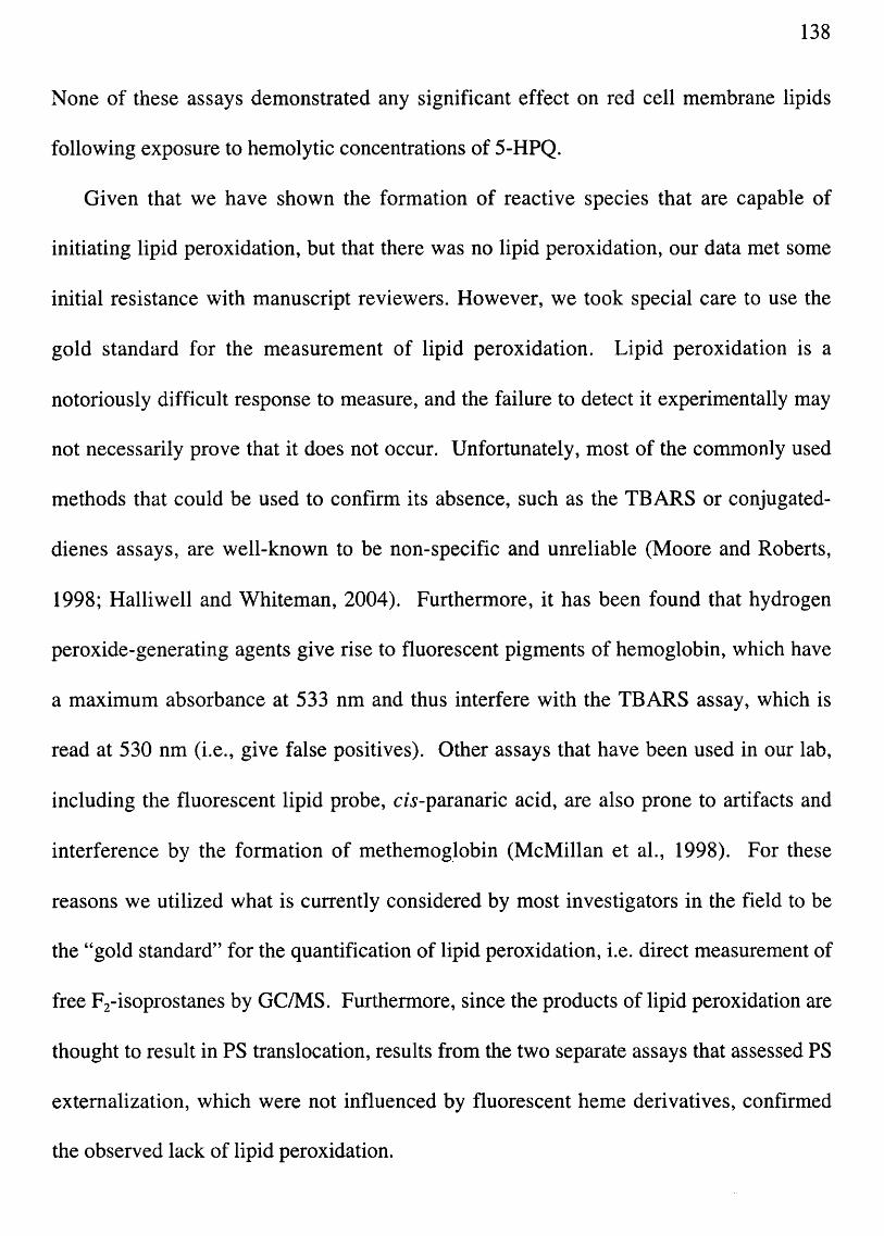
138
None of these assays demonstrated any significant effect on red cell membrane lipids
following exposure to hemolytic concentrations of 5-HPQ.
Given that we have shown the formation of reactive species that are capable of
initiating lipid peroxidation, but that there was no lipid peroxidation, our data met some
initial resistance with manuscript reviewers. However, we took special care to use the
gold standard for the measurement of lipid peroxidation. Lipid peroxidation is a
notoriously difficult response to measure, and the failure to detect it experimentally may
not necessarily prove that it does not occur. Unfortunately, most of the commonly used
methods that could be used to confirm its absence, such as the TBARS or conjugated
dienes assays, are well-known to be non-specific and unreliable (Moore and Roberts,
1998; Halliwell and Whiteman, 2004). Furthermore, it has been found that hydrogen
peroxide-generating agents give rise to fluorescent pigments of hemoglobin, which have
a maximum absorbance at 533 nm and thus interfere with the TBARS assay, which is
read at 530 nm (i.e., give false positives). Other assays that have been used in our lab,
including the fluorescent lipid probe, cis-paranaric acid, are also prone to artifacts and
interference by the formation of methemog.lobin (McMillan et aI., 1998). For these
reasons we utilized what is currently considered by most investigators in the field to be
the "gold standard" for the quantification of lipid peroxidation, i.e. direct measurement of
free F2-isoprostanes by Ge/Ms. Furthermore, since the products of lipid peroxidation are
thought to result in PS translocation, results from the two separate assays that assessed PS
extemalization, which were not influenced by fluorescent heme derivatives, confirmed
the observed lack of lipid peroxidation.

139
The apparent contradiction between previously published results of oxidant induced
lipid peroxidation and our own "controversial" data can be reconciled by considering the
origin of oxidative stress. The high reactivity of hydroxyl radicals causes them to react in
very close proximity to where they are formed. Previously, lipid alterations from
oxidative stress were assessed following the addition of peroxides, such as H20 2, cumene
hydroperoxide and t-butyl hydroperoxide to red cell suspensions. These reactive species
encounter the membrane prior to any other cellular component and can be considered to
cause an oxidative stress from the "outside-in". Alternatively, quinoneimine-, quinone-,
or hydroxylamine-type compounds must first penetrate into the cytoplasm before they
redox cycle and generate ROS from the "inside-out". Thus reactive species are more
likely to react with cytoplasmic components in close proximity, i.e., hemoglobin, rather
than membrane lipids.
In agreement with this hypothesis, exposure to 5-HPQ was found to cause profound
alterations to the red cell hemoglobin and the protein cytoskeleton, principally in the form
of disulfide-linked hemoglobin adducts (Figs. 4.4, 4.5, 4.6 and 4.7). The concentration
response for hemoglobin binding to skeletal membrane proteins correlated very strongly
with the hemolytic response for aSH-normal cells, and when aSH was depleted with
DEM prior to treatment with 5-HPQ, the pattern of hemoglobin binding was also
remarkably similar to the hemolytic response for GSH-depleted erythrocytes. The
observation that 5-HPQ treated red cells could be "rescued" from hemolysis by reduction
with DTT, which was shown to release hemoglobin from skeletal membrane proteins,
further supported a direct role for hemoglobin, or at least some redox sensitive protein,
binding to skeletal membrane protein in the hemolytic response from 5-HPQ.

140
These findings are consistent with the hypothesis that 5-HPQ-exposure results in the
formation of hemoglobin thiyl radicals, which first react with and deplete reduced GSH,
and then react with a variety of sulfhydryl-containing structural proteins to form
hemoglobin adducts to integral membrane and cytoskeletal proteins. Although, it could
be that intracellular oxidative damage might directly alter reactive cysteines in Band 3
that leads to "Band 3 clustering" and subsequent with subsequent antibody recognition
(Turrini et aI., 2003). Regardless, additional studies are necessary to determine the
precise internal protein targets that lead to an external signal for splenic sequestration. In
view of the hypothesis that hemoglobin binding to numerous red cell skeletal membrane
proteins is directly involved (Fig. 4.4B), it is likely the case that most of the binding of
Hb would represent a bystander event. From Fig. 4.4A, it appears that members of the
band 3 macrocomplex, specifically ankyrin and band 4.2, might be targets, and future
experiments to identify specific inside-out protein alterations may involve the targeting
hemoglobin for immunoprecipitation with subsequent mass spectral identification of
bound proteins. Alternatively, biotinylation of cell surface amines, with subsequent
avidin-column or gel isolation and identification of red cell surface proteins from 5-HPQ
treated red cells could be used to examine external cell-surface alterations.
Role for macrophages in the uptake of 5-HPQ treated erythrocytes
An examination of the literature reveals that the removal of damaged or senescent red
cells from the circulation by macrophages in the spleen that is often taken for granted
despite the sparcity of specific evidence. Thus, we examined the fate of 5-HPQ-treated
red cells and demonstrated for the first time that they are taken up exclusively by the

141
spleen. Additionally, we demonstrated that it is the resident macrophages that are
specifically responsible for the uptake of red. Furthermore, we developed an in vitro
erythrophagocytosis assay using the J774A.l macrophage cel1line, which, in addition to
suggesting that uptake by macrophages is signal-dependent rather than dependent on
Heinz body trapping within the splenic cords, provides us with a very useful tool that can
be used to identify specific cell-surface alterations that are responsible for macrophage
recognition.
Studies to further validate and characterize our erythrophagocytosis assay are
ongoing, and we are examining its ability to correlate with the specific effects that we
previously observed in vivo. Specifically, depletion of GSH from red cells with DEM
prior to treatment with 5-HPQ was shown to enhance red cell removal from the
circulation of rats in vivo (Fig. 2.10), and we wanted to know whether depletion of GSH
from red cells prior to treatment with 5-HPQ would enhance their erythrophagocytosis by
J774A.l macrophages in vitro as well. Figure 3.7 showed that phagocytosis of red cells
treated with a subhemolytic concentration of 5-HPQ (25JlM) was significantly enhanced
in GSH-depleted versus GSH-normal rat red cells, and preliminary results suggest that
the ability of DTT to "rescue" 5-HPQ-treated erythrocytes in vivo after in vitro exposure
(Fig. 4.8) can also be reproduced using J774A.l macrophages in vitro (Fig. 5.4).
One of several future directions that could be pursued using the erythrophagocytosis
model in vitro involves the use of a fusion protein, which displays the extracellular
domain of CD47 in order to restore proper recognition of "self' to the macrophages and
specifically inhibit phagocytosis. Additionally, use of J774A.l macrophages could
become an important tool with which to study human hemotoxicity. Specifically, it

500
450
....--... 400 E c. (.) 350 .......,. Q) ~ 300 co ....., c.
::> 250 Q) 0) CO 200 .c c. 0 150 ~ (.) CO ~ 100
50
0 Control
* II - DTT
f[il +DTT
20 min 5-HPQ 40 min 5-HPQ
Treatment Groups
142
Fig. 5.4. Effect of DTT on the phagocytosis of rat 51Cr-labeled erythrocytes after exposure to 5-HPQ (50IlM) by J774A.l macrophages in vitro (preliminary data). The red cells were incubated with or without 5mM DTT for 1 hr at 37°C after treatment with 5-HPQ (20 or 40 min), or with vehicle (H20) alone (40 min) at 37°C. The erythrocytes were then washed and allowed to incubate with plated macrophages for 18 hr. After the incubation period, non-ingested red cells were lysed with NH4Cl and the macrophages extensively washed. Erythrophagocytosis was measured as radioactivity in macrophages (cpm). Data points are means ± S.D. (n=6) *, statistically significant, p<O.05.
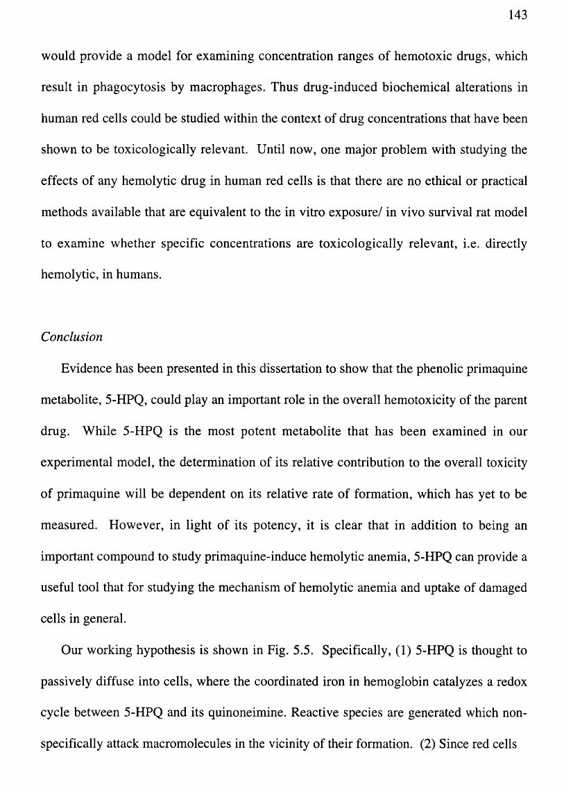
143
would provide a model for examining concentration ranges of hemotoxic drugs, which
result in phagocytosis by macrophages. Thus drug-induced biochemical alterations in
human red cells could be studied within the context of drug concentrations that have been
shown to be toxicologically relevant. Until now, one major problem with studying the
effects of any hemolytic drug in human red cells is that there are no ethical or practical
methods available that are equivalent to the in vitro exposure/ in vivo survival rat model
to examine whether specific concentrations are toxicologically relevant, i.e. directly
hemolytic, in humans.
Conclusion
Evidence has been presented in this dissertation to show that the phenolic primaquine
metabolite, 5-HPQ, could play an important role in the overall hemotoxicity of the parent
drug. While 5-HPQ is the most potent metabolite that has been examined in our
experimental model, the determination of its relative contribution to the overall toxicity
of primaquine will be dependent on its relative rate of formation, which has yet to be
measured. However, in light of its potency, it is clear that in addition to being an
important compound to study primaquine-induce hemolytic anemia, 5-HPQ can provide a
useful tool that for studying the mechanism of hemolytic anemia and uptake of damaged
cells in general.
Our working hypothesis is shown in Fig. 5.5. Specifically, (1) 5-HPQ is thought to
passively diffuse into cells, where the coordinated iron in hemoglobin catalyzes a redox
cycle between 5-HPQ and its quinoneimine. Reactive species are generated which non
specifically attack macromolecules in the vicinity of their formation. (2) Since red cells

Red Cell Membrane
02 Ankyrin
®- Fe++ --...... I ~ 5-HPQ
@- Fe+++ QI
[ROS]
® @-SH 1 8)
"'----~
GSH
®
@)-S-SG
® ?/
Macrophage recognition and uptake
~d3complex Ban
Fig.5.5. Working hypothesis for 5-HPQ-induced hemolytic activiy (see text for detailed explanation). ~
~ ~

145
are packed with hemoglobin, and ROS are formed intracellularly within the cytoplasm,
hemoglobin is likely the primary target of oxidative damage. (3) It is thus thought that
hemoglobin free thiols are activated to reactive thiyl radical intermediates, which are
initially quenched by cellular reduced GSH, but then rapidly overwhelm this defense
mechanism as radicals are formed faster than GSH is able to regenerate. (4) In the
absence of sufficient GSH, hemoglobin thiyl radicals are free to attack other cellular
targets, and our evidence suggests that hemoglobin binds non-specifically to free thiols
on the cytoplasmic side of cytoskeletal and integral membrane proteins. (5) This addition
of large protein adducts likely leads to specific cell surface alterations and/or loss of
normal protein-protein interactions effecting a loss of recognition of "self' by splenic
macrophages and subsequent phagocytosis.
In summary, the data presented in this dissertation has lead to the postulate of a
mechanism by which quinoneimine-type metabolites of primaquine might lead to the
hemolytic removal of red cells. While there are remarkable similarities to the mechanism
of removal induced by hydroxylamine-type metabolites, the mechanism presented in this
dissertation also differs significantly. It is believed that the novel evidence presented
provides additional and important insight into the mechanism( s) of drug-induced
hemolytic anemia normal red-cell senescence. Additionally, we hope that the additional
information provided regarding of the mechanism by which primaquine might mediate its
toxicity will eventually allow for the rational redesign of the primaquine molecule, in
order to significantly lessen its toxicity while maintaining its therapeutic efficacy.

146
REFERENCES
Agarwal S, Gupta UR, Gupta RC, Anand N and Agarwal SS (1988) Susceptibility of glucose-6-phosphate dehydrogenase deficient red cells to primaquine enantiomers and two putative metabolites--I. Effect on reduced glutathione, methemoglobin content and release of hemoglobin. Biochem. Pharmacol. 37:4605-4609.
Allahyari R, Strother A, Fraser 1M and Verbiscar AJ (1984) Synthesis of certain hydroxy analogues of the antimalarial drug primaquine and their in vitro methemoglobinproducing and glutathione-depleting activity in human erythrocytes. J. Med. Chern. 27:407-410.
Anniss AM and Sparrow RL (2002) Expression of CD47 (integrin-associated protein) decreases on red blood cells during storage. Transfusion & Apheresis Science 27:233-238.
Azen EA and Schilling RF (1963) Role of the spleen in acetylphenylhydrazine (APH) anemia in rats. J. Lab. Clin. Med. 62:59-71.
Baird JK, McCormick GJ and Canfield CJ (1986) Effects of Nine Synthetic Putative Metabolites of Primaquine on Activity of the Hexose Monophosphate Shunt in Intact Human Red Blood Cells in Vitro. Biochem. Pharmacol. 35: 1099-1106.
B artosz G (1991) Erythrocyte agIng: physical and chemical membrane changes. Gerontology 37:33-67.
Bates MD, Meshnick SR, Sigler CI, Leland P and Hollingdale MR (1990) In vitro effects of primaquine and
primaquine metabolites on exoerythrocytic stages of Plasmodium berghei. Am. 1. Trap_ Med. Hyg. 42:532-537.
Baty JD, Price Evans DA and Robinson PA (1975) The identification of 6-methoxy-8-aminoquinoline as a metabolite of primaquine in man. Biomed. Mass Spectrom. 2:304-306.
Beutler E (1959) The hemolytic effect of primaquine and related compounds: a review. 1. Hematol.14:103.
Beutler E (1969) Drug-induced hemolytic anemia. Pharmacol. Rev. 21:73-103.

147
Beutler E (1990) Genetics of glucose-6-phosphate dehydrogenase deficiency. Sem. Hematol.27:137-164.
Beutler E (1994) G6PD deficiency. Blood 84:3613-3636.
Beutler E, Dern RJ, Flanagan CL and Alving AS (1955) The hemolytic effect of primaquine. VII. Biochemical studies of drug-sensitive erythrocytes. 1. Lab. Clin. Med. 42:286-295.
Bevers EM, Comfurius P, Dekkers DW, Harmsma M and Zwaal RF (1998) Transmembrane phospholipid distribution in blood cells: control mechanisms and pathophysiological significance. Biological Chemistry 379:973-986.
Bevers EM, Comfurius P, van Rijn JL, Hemker He and Zwaal RF (1982) Generation of prothrombin-converting activity and the exposure of phosphatidylserine at the outer surface of platelets. Eur. 1. Biochem. 122:429-436.
Bjorkman A and Phillips-Howard PA (1990) Drug-resistant malaria: mechanisms of development and inferences for malaria control. [see comment]. Transactions of the Royal Society of Tropical Medicine & Hygiene 84:323-324.
Bolchoz LJ, Budinsky RA, McMillan DC and Jollow DJ (2001) Primaquine-induced hemolytic anemia: formation and hemotoxicity of the arylhydroxylamine metabolite 6-methoxy-8-hydroxylaminoquinoline. 1. Pharmacol. Exp. Ther. 297:509-515.
Bolchoz LJ, Gelasco AK, Jollow DJ and McMillan DC (2002a) Primaquine-induced hemolytic anemia: formation of free radicals in rat erythrocytes exposed to 6-methoxy-8-hydroxylaminoquinoline.l. Pharmacal. Exp. Ther. 303:1121-1129.
Bolchoz LJ, Morrow JD, McMillan DC and Jollow DJ (2002b) Primaquine-induced hemolytic anemia: effect of 6-methoxy-8-hydroxylaminoquinoline on rat erythrocyte sulfhydryl status, membrane lipids, cytoskeletal proteins and morphology. 1. Pharmacal. Exp. Ther. 303:141-148.
Borges A and Desforges JF (1967) Studies of heinz body formation. Acta Haematologica 37: 1-10.

148
Boulard Y, Landau I, Miltgen F, Ellis DS and Peters W (1983) The chemotherapy of rodent malaria, XXXIV. Causal prophylaxios Part III: Ultrastructural changes induced in exo-erythrocytic schizonts of Plasmodium yoelii yoelii by primaquine. Ann. Trop. Med. Parasit. 77:555-568.
Bowman ZS, Oatis JE, Jr., Whelan JL, Jollow OJ and McMillan OC (2004) Primaquineinduced hemolytic anemia: susceptibility of normal versus glutathione-depleted rat erythrocytes to 5-hydroxyprimaquine. J. Pharmacol. Exp. Ther. 309:79-85.
Bradshaw TP, McMillan ~C, Crouch RK and Jollow OJ (1995) Identification of free radicals produced in rat erythrocytes exposed to hemolytic concentrations of phenylhydroxylamine. Free Rad. Bioi. Med. 18:279-285.
Bradshaw TP, McMillan DC, Crouch RK and Jollow OJ (1997) Formation of free radicals and protein mixed disulfides in rat red cells exposed to dapsone hydroxylamine. Free Rad. Bioi. Med. 22: 1183-1193.
Bratosin 0, Mazurier J, Tissier JP, Estaquier J, Huart JJ, Ameisen JC, Aminoff D and Montreuil J (1998) Cellular and molecular mechanisms of senescent erythrocyte phagocytosis by macrophages. A review. Biochimie 80: 173-195.
Bratosin 0, Mazurier J, Tissier JP, Slomianny C, Estaquier J, Russo-Marie F, Huart JJ, Freyssinet JM, Aminoff D, Ameisen JC and Montreuil J (1997) Molecular mechanisms of erythrophagocytosis. Characterization of the senescent erythrocytes that are phagocytized by macrophages. Comptes Rendus de L'Academie des Sciences Serie Iii, Sciences de la Vie 320:811-818.
Bruce LJ, Beckmann R, Ribeiro ML, Peters LL, Chasis JA, Oelaunay J, Mohandas N, Anstee OJ and Tanner MJ (2003) A band 3-based macrocomplex of integral and peripheral proteins in the RBC membrane. Blood. 101:4180-4188.
Bruce LJ, Ghosh S, King MJ, Layton OM, Mawby WJ, Stewart GW, Oldenborg P-A, Delaunay J and Tanner MJA (2002) Absence of C047 in protein 4.2-deficient hereditary spherocytosis in man: an interaction between the Rh complex and the band 3 complex. Blood 100:1878-1885.
Chadburn A (2000) The spleen: anatomy and anatomical function. Semin. Hematol. 37: 13-21.

149
Clark lA, Hunt NH, Cowden WB, Maxwell LE and Mackie EJ (1984) Radical-mediated damage to parasites and erythrocytes in Plasmodium vinckei infected mice after injection of t-butyl hydroperoxide. Clin. Exp. Immunol. 56:524-530.
Cocco RE and Ucker DS (2001) Distinct modes of macrophage recognition for apoptotic and necrotic cells are not specified exclusively by phosphatidylserine exposure. Mol. Bioi. Cell 12:919-930.
Cordes W (1926) Experiences with plasmochin in malaria (preliminary reports). 15th Annual Report, United Fruit Co. (Med. Dept.):66-71.
Dahl KN, Parthasarathy R, Westhoff CM, Layton DM and Discher DE (2004) Protein 4.2 is critical to CD47-membrane skeleton attachment in human red cells. Blood 103:1131-1136.
de Jong K, Geldwerth D and Kuypers FA (1997) Oxidative damage does not alter membrane phospholipid asymmetry in human erythrocytes. Biochemistry 36:6768-6776.
Degowin RL, Eppes RB, Powell RD and Carson PE (1966) The haemolytic effects of diaphenylsulfone (DDS) in normal subjects and in those with glucose-6-phosphate-dehydrogenase deficiency. Bull. World Health Organ. 35:165-179.
Dern RJ, Beutler E and Alving AS (1955) The hemolytic effect of primaquine. V. Primaquine sensitivity as a manifestation of a mUltiple drug sensitivity. 1. Lab. Clin. Med. 45:30-39.
Earle DP, Jr., Bigelow FS, Zubrod CG and Kane CA (1948) Studies on the chemotherapy of the human malarias. IX. Effect of pamaquine on the blood cells of man. 1. Clin. Invest. (Supplement) 27: 121-129.
Eaton JW (1991) Catalases and peroxidases and glutathione and hydrogen peroxide: mysteries of the bestiary. 1. Lab. Clin. Med. 118:3-4.
Elderfield RC, Mertel HE, Mitch RT, Wempen 1M and Werble E (1955) Synthesis of primaquine and certain of its analogs. 1. Am. Chem. Soc. 77:4816-4819.

150
Evelyn KA and Malloy HT (1938) Microdetermination of oxyhemoglobin, methemoglobin, and sulfuemoglobin in a single sample of blood. J. BioI. Chern. 126:655-662.
Fairbanks G, Steck TL and Wallach DF (1971) Electrophoretic analysis of the major polypeptides of the human erythrocyte membrane. Biochemistry 10:2606-2617.
Fletcher K, Price A and Barton P (1984) The pharmacokinetics and biochemical pharmacology of primaquine in rhesus monkeys and rats., in Primaquine: Pharmacokinetics, Metabolism, Toxicity, and Activity (Trigg P ed) pp 49-63, John Wiley & Sons, New York.
Fletcher KA, Barton PF and Kelly JA (1988) Studies on the Mechanisms of Oxidation in the Erythrocyte by Metabolites of Primaquine. Biochem. Pharmacol. 37:2683-2690.
Fridovich I (1975) Superoxide dismutases. Annu. Rev. Biochem. 44: 147.
Gaetani GF, Mareni C, Salvidio E, Galiano S, Meloni T and Arese P (1979) Favism: Erythrocyte metabolism during haemolysis and reticulocytosis. Br. J. Haematol. 43:39-48.
Ganick OJ, Segel GB, Chamberlain J, Hirsch Land Klemperer MR (1977) The effects of splenectomy and glucocorticoids on survival and hepatic uptake of damaged red cells in the mouse. Am. J. Hematol. 2:365-373.
Giles GI, Tasker KM and Jacob C (2001) Hypothesis: the role of reactive sulfur species in oxidative stress. Free Radical Biology & Medicine 31: 1279-1283.
Grossman SJ and Jollow DJ (1988) Role of dapsone hydroxylamine in dapsone-induced hemolytic anemia. J. Pharmacol. Exp. Ther. 244: 118-125.
Grossman SJ, Simson J and Jollow DJ (1992) Dapsone-induced hemolytic anemia: Effect of N-hydroxy dapsone on the sulfuydryl status and membrane proteins of rat erythrocytes. Toxicol. Appl. Pharmacol. 117:208-217.
Halliwell B and Whiteman M (2004) Measuring reactive species and oxidative damage in vivo and in cell culture: how should you do it and what do the results mean? British Journal of Pharmacology 142:231-255.

151
Harel S and Kanner J (1988) The generation of ferryl or hydroxyl radicals during interaction of haemproteins with hydrogen peroxide. Free Radical Res. Commun. 5:21-34.
Harrison J, Jf. and Jollow DJ (1987) Contribution of aniline metabolites to anilineinduced methemoglobinemia. Mol. Pharmacol. 32:423-431.
Harrison JH, Jr. and Jollow DJ (1986) Role of aniline metabolites in aniline-induced hemolytic anemia. 1. Pharmacol. Exp. Ther. 238:1045-1054.
Hempel SL, Buettner GR, O'Malley YQ, Wessels DA and Flaherty OM (1999) Dihydrofluorescein diacetate is superior for detecting intracellular oxidants: comparison with 2' ,7'-dichlorodihydrofluorescein diacetate, 5(and 6)-carboxy-2',7'-dichlorodihydrofluoresein diacetate, and dihydrorhodamine 123. Free Rad. Bioi. Med. 27:146-159.
Howells RE, Peters Wand Fullard J (1970) The chemotherapy of rodent malaria. 13. Fine structural changes observed in the erythrocytic stages of Plasmodium berghei berghei following exposure to primaquine and meoctone. Ann. Trop. Med. Parasitol. 64:203-207.
Idowu OR, Peggins JO and Brewer TG (1995) Side-chain hydroxylation in the metabolism of 8-aminoquinoline antiparasitic agents. Drug Metab. Disp. 23: 18-27.
Jain SK (1984) The accumulation of malony ldialdehyde, a product of fatty acid peroxidation, can disturb aminophospholipid organization in the membrane bilayer of human erythrocytes. 1. of Bioi. Chern. 259:3391-3394.
Jandl JH (1996) Blood: Textbook of Hematology. Little, Brown and Company, Boston.
Jandl JH, Engle LK and Allen DW (1960) Oxidative hemolysis and precipitation of hemoglobin I. Heinz body anemias as an acceleration of red cell aging. 1. Clin. Invest. 39: 1818.
Jallow OJ and McMillan DC (1998) Ethnic variation and genetic susceptibility: Glucose-6-phosphate dehydrogenase deficiency, in Biomarkers. Medical and Workplace Applications (Mendelsohn ML, Mohr LC and Peeters JP eds) pp 227-239, Joseph Henry Press, Washington, DC.

152
J ollow OJ and McMillan DC (2001) Oxidative stress, glucose-6-phosphate dehydrogenase and the red cell. Adv. Exp. Med. Bioi. 500:595-605.
Kain KC and Keystone JS (1998) Malaria in travelers. Epidemiology, disease, and prevention. Infectious Disease Clinics of North America 12:267-284.
Kanner J and Harel S (1985) Initiation of membrane lipid peroxidation by activated metmyoglobin and methemoglobin. Arch. Biochem. Biophys. 237:314-321.
Kay MM (1993) Generation of senescent cell antigen on old cells initiates IgG binding to a neoantigen. Cell Mol. Bioi. 39:131-153.
Kay MM (1994) Regulatory autoantibody and cellular aging and removal. Adv. Exp. Med. Bioi. 347:161-192.
Krishna S, Uhlemann AC and Haynes RK (2004) Artemisinins: mechanisms of action and potential for resistance. Drug Resist. Updat.7:233-244.
Kuypers FA and de Jong K (2004) The role of phosphatidylserine in recognition and removal of erythrocytes. Cellular & Molecular Biology 50: 147-158.
Kuypers FA, Lewis RA, Hua M, Schott MA, Discher D, Ernst JD and Lubin BH (1996) Detection of altered membrane phospholipid asymmetry in subpopulations of human red blood cells using fluorescently labeled annexin V. Blood 87: 1179-1187.
Lii CK and Hung CN (1997) Protein thiol modifications of human red blood cells treated with t-butyl hydroperoxide. Biochimica et Biophysiea Acta 2: 147-156.
Link CM, Theoharides AD, Anders JC, Chung H and Canfield CJ (1985) StructureActivity Relationships of Putative Primaquine Metabolites Causing Methemoglobin Formation in Canine Hemolysates. Toxieol. Appl. Pharmacol. 81: 192-202.
MandaI 0, Moitra PK, Saha Sand Basu J (2002) Caspase 3 regulates phosphatidylserine externalization and phagocytosis of oxidatively stressed erythrocytes. FEBS Lett. 513: 184-188.

153
Maples KR, Eyer P and Mason RP (1990a) Aniline-, phenylhydroxylamine-, nitrosobenzene-, and nitrobenzene-induced hemoglobin thiyl free radical formation in vivo and in vitro. Mol. Pharmacol. 37:311-318.
Maples KR, Jordan SJ and Mason RP (1988) In vivo rat hemoglobin thiyl free radical formation following administration of phenylhydrazine and hydrazine-based drugs. Drug Metab. Disp. 16:799-803.
Maples KR, Kennedy CH, Jordan SJ and Mason RP (1990b) In vivo thiyl free radical formation from hemoglobin following administration of hydroperoxides. Archives of Biochemistry & Biophysics 277:402-409.
McMillan DC, Bolchoz LJ and Jollow DJ (2001) Favism: effect of divicine on rat erythrocyte sulfhydryl status, hexose monophosphate shunt activity, morphology, and membrane skeletal proteins. Toxicological Sciences 62:353-359.
McMillan DC, Bradshaw TP, Hinson JA and Jollow DJ (1991) Role of metabolites in propanil-induced hemolytic anemia. Toxicol. Appl. Pharmacol. 110:70-78.
McMillan DC, Jensen CB and Jollow DJ (1998) Role of lipid peroxidation in dapsoneinduced hemolytic anemia. 1. Pharmacol. Exp. Ther. 287:868-876.
McMillan DC, Simson JV, Budinsky RA and Jollow DJ (1995) Dapsone-induced hemolytic anemia: effect of dapsone hydroxylamine on sulfhydryl status, membrane skeletal proteins and morphology of human and rat erythrocytes. 1. Pharmacol. Exp. Ther. 274:540-547.
Mihaly GW, Ward SA, Edwards G, Nicholl DD, L'eorme M and Breckenridge AM (1985) Pharmacokinetics of primaquine in man. I. Studies of the absolute bioavailability abd effects of dose size. Br. 1. Clin. Pharmacol. 19:745-750.
Mihaly GW, Ward SA, Edwards G, Orme MLE and Breckenridge AM (1984) Pharmacokinetics of primaquine in man; identification of the carboxylic acid derivative as a major plasma metabolite. Br. 1. Clin. Pharmacal. 17:441-446.
Moore K and Roberts LJ (1998) Measurement of lipid peroxidation. Free Rad. Res. 28:659-671.

154
Morrow JD and Roberts LJ, 2nd (1999) Mass spectrometric quantification of F2-isoprostanes in biological fluids and tissues as measure of oxidant stress. Methods Enzymol.300:3-12.
Murakami K and Mawatari S (2003) Oxidation of hemoglobin to methemoglobin in intact erythrocyte by a hydroperoxide induces formation of glutathionyl hemoglobin and binding of alpha-hemoglobin to membrane. Archives of Biochemistry & Biophysics 417:244-250.
Ni Y, Xu Y and Wang M (1992) Rat liver microsomal and mitochondrial metabolism of primaquine in vitro. Acta Pharmacologica Sinica 13:431-435.
Oldenborg P, Zheleznyak A, Fang Y, Lagenaur C, Gresham H and Lindberg F (2000) Role of CD47 as a marker of self on red blood cells. Science 288:2051-2054.
Oldenborg P-A, Gresham HD and Lindberg FP (2001) CD47-SIRPa regulates Fcy and complement receptor-mediated phagocytosis. J. Exp. Med. 193:855-862.
Oldenborg PA (2004) Role of CD47 in erythroid cells and in autoimmunity. Leukemia & Lymphoma 45:1319-1327.
Passon PG and Hultquist DE (1972) Soluble cytochrome b5 reductase from human erythrocytes. Biochem. Biophys. Acta. 275:62.
Peters W (1984) The chemotherapy of rodent malaria XXXVI. Part IV. The activity of a new 8-aminoquinoline, WR 225,448 against exo-erythrocytic schizonts of Plasmodium yoelii yoelii. Ann. Trop. Med. Parasit. 78:467-478.
Peters W (1987) Chemotherapy and drug resistance in malaria. Academic Press, London.
Rifkind RA (1966) Destruction of injured red cells in vivo. Amer. J. Med. 41:711-723.
Rossi R, Barra D, Bellelli A, Boumis G, Canofeni S, Di Simplicio P, Lusini L, Pascarella Sand Amiconi G (1998) Fast-reacting thiols in rat hemoglobins can intercept damaging species in erythrocytes more efficiently than glutathione. Journal of Biological Chemistry 273: 19198-19206.

155
Ruwende C, Khoo SC, Snow RW, Yates SN, Kwiatkowski D, Gupta S, Warn P, Allsop CE, Gilbert SC, Peschu N, Newbold CI, Greenwood BM, Marsh K and Hill AV (1995) Natural selection of hemi- and heterozygotes for G6PD deficiency in Africa by resistance to severe malaria. Nature 376:246-249.
Shanks GD, C. KK and S. KJ (2001) Malaria chemoprophylaxis in the age of drug resistance. II. Drugs that may be available in the future. Clin. Infec. Dis. 33:381-385.
Strother A, Fraser 1M, Allahyari R and Tilton BE (1981) Metabolism of 8-aminoquinoline antimalarial agents. Bull. World Health Org. 59:413-425.
Tarlov AR, Brewer GJ, Carson PE and Alving AS (1962) Primaquine sensitivity. Arch. Int. Med. 109:137-162.
Tizianello A, Pannacciulli I, Ajmar F and Salvivio E (1968) Sites of destruction of red cells in G-6-PD deficient caucasians and in phenylhydrazine treated patients. Scand. J. Haemat. 5:116-128.
Toma E, Thome A, Singer J, Raboud J, Lemieux C, Trottier S, Bergeron MG, Tsoukas C, Falutz J, Lalonde R, Gaudreau C and Therrien R (1998) Clindamycin with primaquine vs. Trimethoprim-sulfamethoxazole therapy for mild and moderately severe Pneumocystis carinii pneumonia in patients with AIDS: a multicenter, double-blind, randomized trial (CTN 004). CTN-PCP Study Group. Clin. Infec. Dis. 27:524-530.
Tracy JW and Webster LT (2001) Drugs used in the chemotherapy of protozoal infections: Malaria, in Goodman & Gilman's The Pharmacological Basis of Therapeutics (Limbird LE ed) pp 1069-1095, McGrawl Hill, New York.
Turrini F, Giribaldi G, Carta F, Mannu F and Arese P (2003) Mechanisms of band 3 oxidation and clustering in the phagocytosis of Plasmodium falciparum-infected erythrocytes. Redox Report 8:300-303.
Tyurina YY, Shvedova AA, Kanai K, Tyurin VA, Kommineni C, Quinn PJ, Schor NF, Fabisiak JP and Kagan VE (2000) Phospholipid signaling in apoptosis: peroxidation and externalization of phosphatidylserine. Toxicol. 148:93-101.

156
van den Berg JJM, Op den Kamp JAF, Lubin BH, Roelofsen Band Kuypers FA (1992) Kinetics and site specificity of hydroperoxide-induced oxidative damage in red blood cells. Free Rad. BioI. Med. 12:487-498.
Van Rooijen N, Kors N, vd Ende M and Dijkstra CD (1990) Depletion and repopulation of macrophages in spleen and liver of rat after intravenous treatment with liposome-encapsulated dichloromethylene diphosphonate. Cell Tissue Res. 260:215-222.
Van Rooijen N and Sanders A (1994) Liposome mediated depletion of macrophages: mechanism of action, preparation of liposomes and applications. 1. Immunol. Methods 174:83-93.
Van Rooijen N and Van Kesteren-Hendrikx E (2003) "In vivo" depletion of macrophages by liposome-mediated "suicide". Methods Enzymol. 373:3-16.
Vasquez-Vivar J and Augusto 0 (1990) ESR detection of free radical intermediates during autoxidation of 5-hydroxyprimaquine. Free Rad. Res. Commun. 9:383-389.
Vasquez-Vivar J and Augusto 0 (1992) Hydroxylated metabolites of the antimalarial drug primaquine. Oxidation and redox cycling. 1. BioI. Chem. 267:6848-6854.
Vasquez-Vivar J and Augusto 0 (1994) Oxidative activity of primaquine metabolites on rat erythrocytes in vitro and in vivo. Biochem. Pharmacol. 47:309-316.
Wernsdorfer WH and McGregor I (1988) Malaria: Principles and Practice of Malariology. Churchill Livingstone, New York.
White NJ (1998) Drug resistance in malaria. Br. Med. Bull. 54:703-715.
Wiesner J, Ortmann R, Jomaa Hand Schlitzer M (2003) New antimalarial drugs. A!}.8!1Yf}!}dj! Ch!!!}j!~ !!!f!.!!JE!i2!J31 Ed_. in E1]~lish 42_:5274_-5293_.
Angewandle Cliemze. rnrernauonaT EC[ rrr e-rrf«f"rrtr~f£(£t-Yf~S.
Yoshida A and Lin M (1973) Regulation of glucose-6-phosphate dehydrogenase activity in red blood cells from hemolytic and nonhemolytic variant subjects. Blood
41:877-891.

157
Zwaal RF and Schroit AJ (1997) Pathophysiologic implications of membrane phospholipid asymmetry in blood cells. Blood 89:1121-1132.