Embed Size (px)
Citation preview

APPLIED AND ENVIRONMENTAL MICROBIOLOGY, Apr. 2006, p. 2837–2848 Vol. 72, No. 40099-2240/06/$08.00�0 doi:10.1128/AEM.72.4.2837–2848.2006Copyright © 2006, American Society for Microbiology. All Rights Reserved.
Molecular Characterization of Subject-Specific Oral Microflora duringInitial Colonization of Enamel
Patricia I. Diaz,1† Natalia I. Chalmers,1,2 Alexander H. Rickard,1 Colin Kong,1‡ Craig L. Milburn,1§Robert J. Palmer, Jr.,1 and Paul E. Kolenbrander1*
Oral Infection and Immunity Branch, National Institute of Dental and Craniofacial Research, National Institutes of Health, Bethesda,Maryland,1 and Department of Biomedical Sciences, University of Maryland School of Dentistry, Baltimore, Maryland2
Received 8 August 2005/Accepted 7 January 2006
The initial microbial colonization of tooth surfaces is a repeatable and selective process, with certainbacterial species predominating in the nascent biofilm. Characterization of the initial microflora is thefirst step in understanding interactions among community members that shape ensuing biofilm develop-ment. Using molecular methods and a retrievable enamel chip model, we characterized the microbialdiversity of early dental biofilms in three subjects. A total of 531 16S rRNA gene sequences were analyzed,and 97 distinct phylotypes were identified. Microbial community composition was shown to be statisticallydifferent among subjects. In all subjects, however, 4-h and 8-h communities were dominated by Strepto-coccus spp. belonging to the Streptococcus oralis/Streptococcus mitis group. Other frequently observedgenera (comprising at least 5% of clone sequences in at least one of the six clone libraries) wereActinomyces, Gemella, Granulicatella, Neisseria, Prevotella, Rothia, and Veillonella. Fluorescence in situhybridization (FISH) confirmed that the proportion of Streptococcus sp. sequences in the clone librariescoincided with the proportion of streptococcus probe-positive organisms on the chip. FISH also revealedthat, in the undisturbed plaque, not only Streptococcus spp. but also the rarer Prevotella spp. were usuallyseen in small multigeneric clusters of cells. This study shows that the initial dental plaque community ofeach subject is unique in terms of diversity and composition. Repetitive and distinctive communitycomposition within subjects suggests that the spatiotemporal interactions and ecological shifts thataccompany biofilm maturation also occur in a subject-dependent manner.
More than 700 different bacterial phylotypes are found inthe oral cavity, as determined by 16S rRNA gene sequencing(1). The initial colonizers of tooth surfaces are a specificsubset of the oral microflora (24). Of these bacteria, thosethat colonize the clean enamel surface independently ofother bacteria possess mechanisms for attachment to theacquired salivary pellicle covering the enamel (32) and theability to metabolize salivary components as the sole nutri-tional source (27). Alternatively, some bacterial species par-ticipate in consortia that are able to attach to enamel andestablish as an initial community requiring metabolic inter-actions among their members (18).
Several studies have identified streptococci as the predomi-nant colonizers of early enamel biofilms (17, 24). Nyvad andKilian (24) characterized the cultivable microflora colonizingenamel pieces exposed to the oral cavity. Streptococci wereshown to compose about 63% (mean value of samples fromfour individuals) of bacteria isolated after 4 h of plaque for-mation and 86% of bacteria isolated after 8 h. A variety ofother bacteria such as veillonellae and actinomyces were also
reported to be present. However, this study was performed ata time when rapid PCR-based taxonomic characterization ofbacterial communities was not available. As a consequence, themicroflora did not include uncultivated organisms, and manyof the rarer isolates were not assigned an identity but ratherwere placed into broad groups such as gram-negative cocci.Another study reporting the molecular characterization of theinitial microflora employed the “checkerboard” DNA-DNAhybridization technique (38) using DNA probes for 40 culturedbacterial species to analyze the early supragingival plaque of 15healthy individuals (17). That study also identified Streptococ-cus spp., in particular Streptococcus mitis and Streptococcusoralis, as the predominant early colonizers but was, at its out-set, limited in range to 40 culturable organisms. Despite theseefforts to characterize initial communities, the broad-basedmolecular taxonomic information that would eliminate limitsimposed by culturability is not available. Furthermore, theapplication of fluorescence in situ hybridization (FISH) toobtain simultaneous taxonomic and positional information onin situ early biofilms is necessary; cell-to-cell interactions be-tween species might determine subsequent biofilm character-istics.
This study used 16S rRNA gene sequencing and a retriev-able enamel chip model (28) to characterize the microflorapresent at 4 h and 8 h of plaque development in three individ-uals. FISH was used to visualize the biofilm architecture of themixed-species communities while confirming the dominance insitu of streptococci in initial dental plaque bacterial popula-tions.
* Corresponding author. Mailing address: National Institutes ofHealth/NIDCR, Building 30, Room 310, 30 Convent Dr., Bethesda,MD 20892-4350. Phone: (301) 496-1497. Fax: (301) 402-0396. E-mail:[email protected].
† Present address: Department of Periodontology, School of Den-tistry, University of North Carolina, Chapel Hill, N.C.
‡ Present address: University of Connecticut School of Dental Med-icine, Farmington, Conn.
§ Present address: College of Dental Medicine, Medical Universityof South Carolina, Charleston, S.C.
2837

MATERIALS AND METHODS
In vivo biofilm development on enamel chips. Details on fabrication of enamelchips and use in healthy human subjects have been published previously (28).Briefly, enamel pieces (1.5 mm wide by 1.5 mm long by 1 mm deep) were cutfrom extracted human third molars, cleaned in an ultrasonic bath (model 1510;Branson, Danbury, CT), sterilized with ethylene oxide, and affixed in custom-fabricated acrylic stents with dental wax. Two bilateral mandibular stents (span-ning the posterior buccal surface from the first premolar to first molar), each ofwhich contained two chips, were carried intraorally by each of three subjects.One stent was placed for 4 h and the other was placed for 8 h. No intake of foodor liquids (other than water) was allowed except during a lunch period whenstents were removed and stored in a humid denture cup at 37°C. Subjects (ages,21, 29, and 47 years) were recruited through protocol 98-D-0116 at the NationalInstitutes of Health. Criteria for enrollment included systemic health, absence ofperiodontitis or other pathology of the oral tissues, nonsmoker status, and norecord of antibiotic usage in the 3 months prior to enrollment.
PCR amplification, cloning, and sequencing of 16S rRNA genes. After re-trieval from the mouth, each enamel chip was placed in 500 �l of sterile phos-phate-buffered saline (PBS) and agitated at 225 rpm for 30 s to release theunattached bacteria present in the coat of saliva surrounding the chip. Afterwashing, each chip was placed in 20 �l sterile water and biofilm cells werereleased by 20-min mild sonication (model 1510 bath; Branson Ultrasonics Cor-poration, Danbury, CT). Five microliters of water containing released cells wasmixed with 5 �l of Microlysis solution (The Gel Company, San Francisco, CA),followed by rapid heating and cooling of the sample according to the manufac-turer’s instructions. This template solution was then subjected to 10 min ofheating at 100°C, as preliminary experiments revealed that in the absence of thisstep Actinomyces spp. were incompletely lysed. Specifically, when a mixturecontaining equal numbers of Streptococcus gordonii DL1 and Actinomycesnaeslundii T14V cells was used as a template for PCR, only S. gordonii 16S rRNAgene sequences were obtained unless the sample was heated at 100°C for 10 min,after which S. gordonii and A. naeslundii sequences were retrieved at the samefrequency (data not shown).
The entire 10 �l of lysed cells was used as template in a 50-�l PCR mixturecontaining Ex Taq buffer (TaKaRa Bio, Inc., Shiga, Japan), 400 �M each de-oxynucleoside triphosphate, 2.5 U Ex Taq Hot Start DNA polymerase (TaKaRaBio, Inc.) and 0.6 �M each primer. Universal primers D88 and E94 (30) wereused to amplify approximately 1,500 bp of the 16S rRNA gene. PCRs wereinitially denatured at 94°C for 3 min, followed by 30 cycles, each consisting ofdenaturation at 94°C for 30 s, annealing at 50°C for 30 s, and extension at 72°Cfor 2 min, with a final extension step at 72°C for 10 min. PCR products were gelpurified (gel purification kit; QIAGEN, Valencia, CA) and ligated into pCR 2.1(Invitrogen, Carlsbad, CA) which was transformed into OneShot Top10 chem-ically competent Escherichia coli (Invitrogen). After selection (ampicillin, 100�g/ml) and blue/white screening, 150 transformants were taken from the samplesof subject 1, and 100 transformants were taken from the samples of subjects 2and 3. EcoRI digestion of plasmid DNA was used to determine colonies con-taining inserts of the right size (�1,500 bp). Purified plasmid DNA was se-quenced in a 3730 DNA analyzer (Applied Biosystems, Foster City, CA). PrimersD88 and F15 (30) were used to sequence approximately 500 bp near the 5� endof the 16S rRNA gene and, when required, the primers B34, E94, F16, F17, F18,F20, and F22 (30) were additionally used to sequence the complete 16S rRNAgene.
All manipulations were carried out in a Plexiglas-enclosed PCR/UV workstation (Coy Laboratory Products, Grass Lake, Mich.). Negative controls in-cluded a PCR with no added template (water control) and a PCR using templateDNA released from an enamel chip fixed on a stent but not placed intraorally(enamel chip control). For the negative controls, the PCR cycle was modified toinclude 40 cycles of denaturation, annealing, and extension. We sequenced atotal of 30 clones obtained for each water and enamel chip control for a total of60 sequences of environmental contaminants. These were used to determineenvironmental contaminants in our samples from each subject.
Phylogenetic analysis of insert sequences. Sequences were assembled usingSeqman II from the Lasergene 6 package (DNASTAR, Inc., Madison, WI).BLAST (2) and FASTA (31) searches were performed for initial identification,and possible chimeric sequences were checked using the RDP-II chimera checktool (6). A sequence was assigned a species identity when it shared �98%identity with a published sequence of a cultured organism in the database.Phylogenetic placement of each sequence was confirmed by construction ofphylogenetic trees using closely related reference sequences obtained fromEMBL and GenBank. Prior to the construction of phylogenetic trees, unambig-uous sequences were aligned using CLUSTAL_X (40). Partial 16S rRNA gene
sequences of 440 nucleotides were used to construct phylogenetic trees by meansof neighbor-joining and maximum-likelihood methods using TREECON (42)and DNAML from the Phylip package (11). All reference sequences used in thetree are from validated bacterial species: each has been cultured, characterized,deposited with at least two recognized culture collections, and named under theprocedure described in the Bacteriological Code, 1990 revision (37). Trees wereviewed using TREEVIEW (version 1.66) and text was edited in CorelDraw 12.Although 100 or 150 colonies were analyzed at each time point from eachsubject, the data reflect only clones with a positive insert and, of these, only thosesequences that were not chimeric or environmental contaminants.
Phylotype determination. A square similarity matrix was produced for eachlibrary of sequences and for all libraries compiled with DNAdist from the Phylippackage (11) and the Kimura two-parameter model of nucleotide substitution.Sequences were grouped into phylotypes using the software DOTUR (for dis-tance-based operational taxonomic unit and richness) (33) and the furthest-neighbor assignment algorithm. Other studies that have sequenced greater por-tions of the 16S rRNA gene have used a cutoff of 99% identity among sequencesto define a phylotype (8, 30). We used a more conservative phylotype definitionof 98% because the sequenced region constituted the most variable one-third ofthe 16S rRNA gene. The partial 16S rRNA gene sequences that possessed �98%identity to 16S rRNA gene sequences in EMBL and GenBank databases werecompletely sequenced, reanalyzed, and compared to database sequences usingBLAST and FASTA search algorithms. Complete 16S rRNA gene sequencesthat still possessed �98% identity to any known database sequences were de-scribed as novel phylotypes.
Estimation of phylotype diversity. Good’s coverage estimation was calculatedas [1 � (n/N)] � 100, where n is the number of singleton sequences and N is thetotal number of sequences for the clone library analyzed. Estimates of phylotyperichness were calculated according to the abundance-based coverage estimator(ACE) (5) and the bias-corrected Chao1 estimator (4). Collector’s curves ofobserved and estimated richness (plots of the number of observed and estimatedphylotypes as a function of the number of clones sequenced) were calculated inDOTUR (33). The Shannon-Weaver diversity index (21) was also calculated inDOTUR.
Statistical comparisons of 16S rRNA gene libraries. -LIBSHUFF (34, 35)was used to determine if DNA libraries differed between subjects and betweentime points. A square similarity matrix constructed in DNAdist (11) using theJukes and Cantor model of nucleotide substitution was used as the input file.Critical P values for individual comparisons were determined based on thecorrection for multiple comparisons provided by -LIBSHUFF to attain anexperiment-wide type I error rate of 5%. P values for each library comparisonwere estimated by 10,000 random permutations of sequences between libraries.
-LIBSHUFF was also used to evaluate interexperiment (days T0 and T21) andintrasubject (chip 1 versus chip 2) variability. Four-hour biofilms from subject 1were used for these analyses. To evaluate variability between libraries obtainedfrom chips carried simultaneously but amplified independently (intrasubject;chip 1 versus chip 2), we compared 25 sequences obtained from each chip at thegenus level by conducting a statistical analysis of the difference between librarieswith -LIBSHUFF. To evaluate interexperiment variability, a 4-h biofilm fromsubject 1 was compared to that sampled 3 weeks later using 40 sequences fromeach time point. No significant differences were found among libraries; there-fore, sequences were pooled with new sequences obtained from subject 1 andused in our overall analysis.
FISH. Enamel chips were removed from the stent and fixed at 4°C for 3 h with4% paraformaldehyde in phosphate-buffered saline. Chips were washed in 50%ethanol. For permeabilization, each chip was attached to glass slides with paraffinwax and treated with 25 �l lysozyme (70,000 U ml�1 in 100 mM Tris/HCl [pH7.5], 5 mM EDTA; Sigma, St. Louis, MO) for 10 min at 37°C in a humidatmosphere. Chips were dehydrated in a series of ethanol washes (50, 80, and100%; 3 min each wash) and exposed to 10 �l of hybridization buffer (0.9 MNaCl, 20 mM Tris/HCl [pH 7.5], 0.01% sodium dodecyl sulfate, 25% formamide)containing 100 ng of probe. The slides were incubated at 46°C for 90 min in ahumid atmosphere. After hybridization, chips were washed first in buffer (20 mMTris/HCl [pH 7.5], 5 mM EDTA, 0.01% sodium dodecyl sulfate, and 159 mMNaCl) for 15 min at 48°C and then in ice-cold water and stained with 1-�g/mlacridine orange. The oligonucleotide probe STR405 (29, 41) was labeled withAlexa 546 and used to identify all Streptococcus spp. The specificity of the probewas tested against Streptococcus vestibularis ATCC 49124, Streptococcus parasan-guinis ATCC 15912, Streptococcus mitis ATCC 49456, Streptococcus mutansATCC 700610, Streptococcus oralis ATCC 10557, Streptococcus sanguinis ATCC10556, Streptococcus gordonii ATCC 51656, Streptococcus gordonii ATCC 49818,Streptococcus gordonii ATCC 10558, Streptococcus salivarius ATCC 25975, Acti-nomyces naeslundii T14V, Fusobacterium nucleatum ATCC 10953, Prevotella
2838 DIAZ ET AL. APPL. ENVIRON. MICROBIOL.

intermedia ATCC 15032, Prevotella oralis ATCC 33269, Prevotella melaninogenicaATCC 25845, Veillonella atypica ATCC 17744, Veillonella dispar ATCC 17748,Veillonella parvula ATCC 10790, Granulicatella elegans ATCC 700633, Granuli-catella adiacens ATCC 49175, and Abiotrophia defectiva ATCC 49176. All Strep-tococcus spp. had a positive reaction, whereas species of other genera had anegative reaction, with the exception of Abiotrophia defectiva, which showed aweakly positive signal. The oligonucleotide probe PRV392 (5�-GCACGCTACTTGGCTGG-3) was designed with ARB software (19) and labeled with Alexa546. The specificity of the probe was tested against Streptococcus parasanguinisATCC 15912, Streptococcus mitis ATCC 49456, Streptococcus mutans ATCC700610, Streptococcus oralis ATCC 10557, Streptococcus sanguinis ATCC 10556,Streptococcus gordonii ATCC 49818, Streptococcus salivarius ATCC 25975, Acti-nomyces naeslundii T14V, Fusobacterium nucleatum ATCC 10953, Prevotellaintermedia ATCC 15032, Prevotella oralis ATCC 33269, Prevotella melaninogenicaATCC 25845, and Porphyromonas gingivalis W50 ATCC 53978. All Prevotella spp.had a positive reaction, whereas other genera had a negative reaction. The probeEUB338 (3) was labeled with Alexa 633 and used as a positive control. All labeledprobes were purchased from Operon Biotechnologies, Inc., Huntsville, AL.
Microscopy. Chips were examined using a 63�/0.9 numerical aperture (NA)water-immersible lens on a Leica TCS/SP2 confocal microscope (Leica Microsys-tems, Malvern, PA). All images presented are maximum projections of the entireconfocal image stack, which was collected simultaneously into three channels. Nine238-�m by 238-�m images from different fields were collected at 63� (low magni-fication) and nine 79.4-�m by 79.4-�m images were collected at 3� electronic zoom(high magnification). Image stacks were acquired with axial spacing of 0.5 �m or 1.0�m.
Image analysis and determination of Streptococcus proportions on enamelchips. Low-magnification images were analyzed by using IMAQ ImageBuilder(National Instruments, Austin, TX), while high-magnification images werecounted manually. For software analysis, red-green-blue (RGB) color maximumprojections of the image stacks were manually processed in Photoshop to removedebris and enamel autofluorescence and then converted to grayscale. The gray-scale images were manually thresholded, and particle analysis results were fil-tered to remove particles of �0.77 �m2 in area (an area slightly less than that ofa 1-�m-diameter coccus). The total area of particles in each channel was calcu-lated, and the percentage of STR405-positive area relative to the total EUB338-positive area or acridine orange-stained area was determined.
Nucleotide sequence accession numbers. Partial and complete 16S rRNA genesequences were deposited in GenBank under accession numbers DQ016669 toDQ016967 and DQ346213 to DQ346444.
RESULTS
Reproducibility of 16S rRNA amplification results. Se-quences obtained from 4-h biofilms from subject 1 were usedto evaluate variability between libraries obtained from chipscarried simultaneously but sampled independently. Limitedchip-to-chip variability was seen in the proportion of Strepto-coccus spp. (60% of sequences were identified as Streptococcusspp. on chip 1 and 65% on chip 2) and in the proportion ofVeillonella spp. (9% of sequences were identified as Veillonellaspp. on chip 1 and 10% on chip 2), while the variability in theproportions of another major non-Streptococcus genus, Pre-votella spp., was greater (11% of sequences were identified asPrevotella spp. on chip 1 and 1% on chip 2). -LIBSHUFFanalysis indicated that the two libraries (composed of 25 se-quences each) were not different [P(X versus Y) 0.2859; P(Yversus X) 0.1792; X and Y represent libraries as shownbelow (see Fig. 3)].
To evaluate the repetitive nature of initial colonization inthe enamel chip model over time, a total of 40 sequences froman enamel chip carried for 4 h by subject 1 was compared to thesame number of sequences obtained from an enamel chipcarried for 4 h by the same subject 3 weeks later. The percent-age of Streptococcus sequences was 60% at the first samplingand 68% at the later sampling. Veillonella sequences made up7% (first sampling) and 14% (later sampling), while Prevotella
spp. comprised 2% (first sampling) and 5% (later sampling).Furthermore, -LIBSHUFF analysis found no difference be-tween the two libraries [P(X versus Y) 0.1285; P(Y versus X) 0.10734). These observations suggested that the compositionof initial plaque within an individual is reestablished and con-sistent over short periods of time (3 weeks).
Environmental contaminants in PCR. Clones obtained fromthe amplification of the water control and enamel chip controlwere sequenced and used to assess contamination within thesubject libraries. Contaminant or transient sequences fromboth sources were similar: an uncultured �-proteobacterium,Pseudomonas spp., Sphingomonas spp., Caulobacter spp., Bre-vundimonas vesicularis, an uncultured �-proteobacterium, Co-mamonas spp., and Delftia acidovorans. Neither control yieldedsequences identifiable as belonging to resident oral organisms.
We sequenced 150 colonies from 4-h and 8-h libraries ofsubject 1. At 4 h, we found that 17 sequences (13%) corre-sponded to environmental contaminants and 4 sequences (3%)were chimeric sequences. At 8 h, 10 sequences (7%) were ofenvironmental contaminants, and 6 sequences (4%) were chi-meric sequences. One hundred colonies were sequenced fromsubject 2 libraries. At 4 h, 49 sequences (49%) were of envi-ronmental contaminants, and 5 sequences (5%) were chimericsequences. At 8 h, we found 23 environmental contaminants(23%) and 4 (4%) chimeric sequences. In subject 3, 100 colo-nies were sequenced at each time point, with 18% of sequencesrepresenting environmental contaminants and 5% of se-quences being chimeric at 4 h of sampling. At 8 h, the propor-tion of environmental contaminants was 22%, and the propor-tion of chimeric sequences was 6%. It is evident that theproportion of sequences derived from environmental contam-inants varied among subjects and was greatest in subject 2, inparticular, in the 4-h library. These results correlated withthose of FISH experiments in which biomass also variedgreatly among subjects (Fig. 1). In subject 2, very few bacteriawere found on the surface of the enamel chip at 4 h of colo-nization (data not shown), whereas at 8 h, small clusters of cells(two to four cells) were evident (Fig. 1B). In contrast, biofilmsfrom subject 1 typically showed abundant colonization by smallto medium (�10 cells) clusters of cells at 4 h (data not shown)and a broad distribution of larger clusters of cells at 8 h (Fig.1A). Therefore, it is likely that the proportion of environmen-tal contaminants in each library correlated inversely with theamount of template available for PCR amplification.
Coverage of 16S rRNA gene libraries and microflora diver-sity at 4 h and 8 h of enamel colonization. The microflorapresent at 4 h and 8 h of colonization on enamel chips carriedintraorally by three subjects yielded 531 nonchimeric se-quences and 97 distinct phylotypes. DOTUR analyses ofpooled 4-h and 8-h libraries from each subject showed thefollowing results: 66 distinct phylotypes in subject 1, 23 phylo-types in subject 2, and 32 phylotypes in subject 3. Table 1presents the number of phylotypes obtained from each subjectat each time point. The Good’s coverage estimates reflectcoverage of roughly 90% in five of the six clone libraries.Coverage in the 8-h library from subject 1 was 80%. Thecommunity was more diverse in subject 1 than in subjects 2and 3, as demonstrated by the number of phylotypes and theShannon-Weaver diversity index. Although the sampling effortwas higher for subject 1, the coverage (in particular at 8 h) was
VOL. 72, 2006 MICROFLORA OF EARLY ENAMEL BIOFILMS 2839

the lowest and was consistent with the higher diversity of thesample. Collector’s curves (Fig. 2) for each clone library depictthe differences in phylotype richness among libraries and thedifferent results in coverage. The number of unseen phylotypesis represented by the gap between the observed phylotypes andthe number of phylotypes estimated by Chao1 and ACE. Inmost cases, this gap decreased towards the end of sampling.However, for the 8-h library from subject 1, sampling stoppedwhile the gap was still increasing. We estimated that the ge-netic distance had to be relaxed to 4% (from 2%) difference toobtain a stable ACE and Chao1 richness estimates in the 8-hlibrary of subject 1. Despite the low sampling effort for thelibraries of subjects 2 and 3, high coverage and stable richnessestimates were obtained because the libraries exhibited lowdiversity.
Predominant genera present at 4 h and 8 h of colonizationand differences in microflora composition among subjects.Consistent with the results of cultivable-flora studies (12, 24),the present results report streptococci as constituting 66% to80% of the microflora at 4 h and 8 h of biofilm development(Table 2) and S. mitis/S. oralis as the predominant streptococciin the three subjects (Table 3). However, interindividual vari-ation was seen in the prevalence and proportions of other
genera, with the most prominent differences seen in the generaAbiotrophia, present only in subject 1; Corynebacterium,present only in subject 2; Neisseria, present at higher levels insubject 2; Rothia, absent in subject 2 and present at higherlevels in subject 3 than in subject 1; Prevotella, absent in subject2 and present at higher levels in subject 1 than in subject 3; anduncultured species from the class Clostridia, found only insubject 1. Furthermore, -LIBSHUFF pairwise comparisons ofpooled libraries (4 h and 8 h) from each subject revealed thatall the libraries were statistically different from one another(P � 0.0001). When we made pairwise comparisons amonglibraries without pooling 4-h and 8-h samples together (Fig. 3),we found that the 4-h library from subject 3 differed from allthe other 4-h libraries. On the contrary, the 4-h library fromsubject 2 was found to be a subset of the 4-h library of subject1, a result that indicates the phylogenies in the subject 2 libraryare contained in the library from subject 1, but the librariesdiffered, probably because of the greater diversity found insubject 1. At 8 h, the same results were observed in that the 8-hlibrary from subject 2 was a subset of the 8-h library fromsubject 1. The comparison of the 8-h library from subject 1 tothat of subject 3 resulted again in a statistically significantdifference, as was observed at 4 h. However, the 8-h libraryfrom subject 3 was similar to the 8-h library from subject 2.
The 531 partial 16S rRNA gene sequences were comparedto each other, and those that possessed �98% identity to oneanother were grouped and classified as phylotypes. Ninety-seven phylotypes were derived, and their phylogenetic related-ness to 16S rRNA gene sequences of previously describedcultured organisms was established by the generation of max-imum-likelihood phylogenetic trees (Fig. 4). Sixty-eight phylo-types possessed sequences most closely related to 16S rRNAgene sequences from gram-positive species (Fig. 4A) and 29phylotypes were most closely related to 16S rRNA gene se-quences from gram-negative species (Fig. 4B). Only 10 phylo-types were found in all three subjects, while 19 phylotypes werepresent in two of the three subjects. The remaining 68 phylo-
FIG. 1. Confocal micrographs (238 by 238 �m) of 8-h plaque development on enamel chips in subject 1 (A) and subject 2 (B). Cells weresimultaneously labeled with the kingdom-level bacterial probe EUB338 (red) and the genus-level Streptococcus probe STR405 (green). Panels showthe overlay of the two images. Notice the difference in biomass between the two samples but the similar proportion of Streptococcus spp. (yellow,colocalization of the two probes). Bar, 40 �m.
TABLE 1. Phylotype richness and calculated coverage anddiversity of each library
Subject Clonelibrary (h)
No.clones in
library
No. ofphylotypesobserved
Good’scoverage
(%)
Shannon-Weaverdiversity indexa
1 4 129 40 87 3.28 0.178 134 45 80 3.39 0.17
2 4 46 13 89 2.10 0.308 73 14 93 2.08 0.23
3 4 77 23 88 2.61 0.268 72 14 92 1.83 0.29
a A higher Shannon-Weaver diversity index is associated with higher diversity.Each value is given as the mean the upper and lower bound to the 95%confidence interval (33).
2840 DIAZ ET AL. APPL. ENVIRON. MICROBIOL.

types were unique to a subject. Subject 1 had 44 unique phy-lotypes, subject 2 had 11 unique phylotypes, and subject 3 had13 unique phylotypes, resulting in 34 to 63% of phylotypescomprising the initial community found in a specific subjectthat was unique to that subject. This result highlights the dis-tinct and subject-specific initial microflora composition.
Table 3 shows the subject-specific abundance of Streptococ-cus spp. present on the enamel chips carried by the threesubjects at 4 h and 8 h of colonization. It was not possible todistinguish by 16S rRNA gene sequencing between S. mitis andS. oralis, or among S. salivarius, S. vestibularis, and Streptococ-cus thermophilus, as their sequences did not diverge suffi-ciently. Nevertheless, interindividual variation was seen in the
prevalence and proportions of these Streptococcus groups. Forexample, the proportion of S. mitis/S. oralis sequences obtainedfrom subject 1 was 27%, while in subjects 2 and 3 the sequencesin the S. mitis/S. oralis group constituted a much higher per-centage (70 to 100% in subject 2 and 55 to 85% in subject 3).Moreover, subject 1, with a more diverse microflora, also har-bored a higher number of different Streptococcus spp. than, forexample, subject 2.
Interindividual differences were also seen in the presenceand proportions of the phylotypes belonging to the predomi-nant genera (those comprising at least 5% of clone sequencesin at least one clone library). The majority of Veillonella clonedsequences were closely related to V. parvula/V. dispar, which
FIG. 2. Collector’s curves of observed and estimated (ACE and Chao1) phylotype richness as a function of the number of clones. These curvesdepict the change in richness values as the number of clones in the library increased and are used to assess the sufficiency of the sampling effortfor each specific library. A sufficient number of clones was sequenced in all libraries except in the 8-h library from subject 1, in which the gapbetween the observed and estimated phylotype curves had not closed when sampling stopped.
VOL. 72, 2006 MICROFLORA OF EARLY ENAMEL BIOFILMS 2841

cannot be differentiated by their 16S rRNA gene sequences.Few clones of Veillonella spp. were identified as V. atypica, andthey were present only in subject 1. Only two phylotypes iden-tified as Gemella were observed, the most abundant beingGemella hemolysans, identified in all the subjects. Gemella san-guinis was obtained from two subjects and at lower frequencythan G. hemolysans. Phylotypes belonging to the Granulicatella
genus were observed in subjects 1 and 2 and were closelyrelated to G. elegans and G. adiacens. Rothia was observed intwo subjects and the phylotypes were closely related to those ofRothia mucilaginosa and Rothia dentocariosa. Some divergencewas seen in Actinomyces phylotypes closely related to A.naeslundii, present in all subjects, while Actinomyces odonto-lyticus was present only in subject 1. Difficult to classify by 16SrRNA-based taxonomy (36), Neisseria spp. of the Neisseriapharyngis/Neisseria flava/Neisseria mucosa/Neisseria sicca groupwere found in all subjects. Subjects 2 and 3 had phylotypesclosely related to N. subflava, but only subject 2 had phylotypesof the Neisseria meningitidis/Neisseria cinerea group (for exam-ple, sequence 8.14 in Fig. 4B). Subject 1 possessed the highestnumber of clones of Prevotella, some of which were closelyrelated to P. melaninogenica, Prevotella veroralis, or Prevotellasalivae. However, some Prevotella sequences obtained could notbe assigned any species identity and belong to uncultured Pre-votella spp. An overall tree depicting a representative sequence ofeach phylotype obtained is presented in Fig. 4.
Population shifts occurring from 4 h to 8 h in each subject.-LIBSHUFF pairwise comparisons of 4-h and 8-h libraries(Fig. 3) revealed that the 4-h and 8-h libraries of subject 1 weresimilar. The 4-h and 8-h libraries of subject 2 were also similar,while the 8-h library in subject 3 was a subset of the 4-h library.These results suggest that only some species were retained andflourished in the biofilm in subject 3, which correlated with thedecreased diversity seen at 8 h with this subject (Table 1).
TABLE 2. Microflora present at 4 and 8 h of enamel colonization
% of sequences ata:
Phylum Genus
Subject 1 Subject 2 Subject 3
4 h(n 129)
8 h(n 134)
4 h(n 46)
8 h(n 73)
4 h(n 77)
8 h(n 72)
Firmicutes Streptococcus 65.9 67.9 67.4 79.5 66.2 73.6Veillonella 8.5 8.2 2.2 None 5.2 2.8Gemella 3.9 3.0 None 4.1 2.6 15.3Abiotrophia 1.6 0.7 None None None NoneGranulicatella None 1.5 6.5 4.1 None NonePeptostreptococcus 0.8 None None None None NoneMogibacterium None 0.7 None None None NoneMegasphaera None 0.7 None None None NoneSelenomonas 0.8 0.7 None None None NoneOribacterium 0.8 None None None None NoneUnculturedb 4.7 2.2 None None None None
Actinobacteria Actinomyces None 1.5 None 1.4 7.8 NoneCorynebacterium None None None 4.1 None NoneRothia 0.8 1.5 None None 13.0 NoneAtopobium 0.8 None None None None None
Proteobacteria Neisseria 2.3 1.5 15.2 5.5 1.3 1.4“Candidatus Burkholderia” 1.6 0.7 4.3 None None NoneHaemophilus None None 4.3 1.4 None 1.4
Bacteroidetes Prevotella 7.0 7.5 None None 2.6 NonePorphyromonas 0.8 1.5 None None None 1.4Fusobacterium None None None None 1.3 NoneBacteroides None None None None None 1.4Unculturedc None None None None None 2.8
a Data represent percentages of sequences that clustered within each genus after phylogenetic analysis. n, number of clones sequenced from each sample.b Sequences of uncultured Firmicutes were all placed within the Clostridia class after phylogenetic analysis.c Sequences of uncultured Bacteroidetes were placed under the class Flavobacteria after phylogenetic analysis.
TABLE 3. Species of Streptoccocus present at 4 and 8 h ofenamel colonization
% of sequences for indicated subject ata:
SpeciesSubject 1 Subject 2 Subject 3
4 h 8 h 4 h 8 h 4 h 8 h
S. oralis/S. mitis 26.7 23.1 70.0 100.0 54.9 84.9S. salivarius/S. vestibularis/
S. thermophilus4.7 14.3 None None None None
S. infantis 14.0 11.0 6.7 None 17.7 9.4S. sanguinis 15.1 6.6 16.7 None 7.8 NoneS. parasanguinis 3.5 13.2 None None None NoneS. gordonii 1.2 6.6 None None 7.8 NoneS. anginosus 3.5 6.6 None None None NoneS. bovis 1.2 None None None None NoneS. cristatus None None None None None 3.8Other 30.2 18.7 6.7 None 11.8 1.9
a Data represent the percentages of sequences from samples that after phylo-genetic analysis were �98% similar to the sequence of a validated Streptococcusreference strain. Sequences with �98% similarity to any known Streptococcusspp. were left unclassified and appear in the table as “other.”
2842 DIAZ ET AL. APPL. ENVIRON. MICROBIOL.

Uncultured early colonizers and new phylotypes. We iden-tified 67 sequences that could not be assigned to a species bythe criteria utilized in this study (�98% identity to a namedorganism in the database). These sequences were thereforeclassified as sequences of uncultured microorganisms. Amongthese sequences we found eight novel phylotypes (sequenceswith �98% identity to any sequence in the database, includingsequences originated from clones). Most of the unculturedorganisms were found in subject 1, and libraries of subject 2yielded the smallest number of uncultured bacteria. Theseobservations correlate with the difference in diversity seen be-tween the two subjects.
FISH for quantification of Streptococcus spp. at 4 h of colo-nization of enamel chips. To validate the PCR results, we useda genus-level probe to determine the percentage of strepto-cocci on enamel chips at 4 h of colonization in subject 1.Manual counting of nine high-magnification images, covering1% of the enamel chip surface, indicated that Streptococcuscells were 73% 18% of the number of acridine orange-stained cells and 80% 19% of the EUB338-positive cells.Automated counting of nine low-magnification images, cover-ing 23% of the area of an enamel chip, indicated that the areaof STR405-positive particles was 72% 17% of the acridineorange-stained biomass and 73% 19% of the EUB338-pos-itive biomass. A comparison of these results with the percent-age of Streptococcus clones obtained at 4 h from subject 1(Table 2) suggests that the PCR-based methodology offered anaccurate representation of the relative abundance of strepto-cocci.
Intergeneric juxtaposition in undisturbed plaque. FISH alsoprovided visualization of intact biofilm architecture in subject1. At 4 h, the enamel surface was dotted with small clusters thattypically contained Streptococcus spp. in contact with bacterianot reactive with the Streptococccus probe (Fig. 5A and B). At
8 h, surface coverage increased, and multigeneric clusters wereprominent. Examples of such multigeneric clusters containingPrevotella spp. in intimate contact with unidentified bacteriaare presented in Fig. 5C and D. The detection of numericallysmall bacterial constituents of supragingival plaque, such asPrevotella spp., underscores the power of FISH for revealingintergeneric associations.
DISCUSSION
The aim of this study was to use molecular techniques tocharacterize oral bacterial communities after 4 h and 8 h ofenamel colonization. We found that the early dental plaquemicroflora varies on a subject-specific basis. More than two-thirds of the observed phylotypes were unique to a specificsubject. Subject-specific variation is commonly overlooked, be-cause studies typically pool samples from different subjects (17,20, 24). However, a recent extensive characterization of thehuman intestinal microflora reported major differences in mo-lecular community composition and diversity among three in-dividuals (8). Perhaps interindividual variation in microflora ofthe digestive tract, including the oral cavity, could be attributedto differences in host factors that modulate colonization of anindividual by a specific set of species. We postulate that mem-bers of a specific community have adapted to each other and tothe host, thus creating interrelationships among communityparticipants that ensure spatiotemporal repeatability and sta-bility of the microbial community composition.
We found 10 phylotypes were common to all three subjects.The closest validated sequences to these phylotypes are Neis-seria pharyngis, Gemella hemolysans (two phylotypes), Strepto-coccus thermophilus, Streptococcus oralis (three phylotypes),Streptococcus infantis, and Streptococcus sanguinis (two phylo-types) (Fig. 4). These phylotypes might constitute a core group
FIG. 3. -LIBSHUFF comparisons of 16S rRNA gene sequence libraries from 4-h and 8-h biofilms in subjects 1, 2, and 3. Significant P valuesafter correction for an experiment-wide type 1 error rate of 5% appear in boldface type and correspond to those values of �0.0017. Libraries aredistinct if both X versus Y and Y versus X are statistically significant. If X versus Y is significant but Y versus X is not, then Y is a subset of X.If X versus Y is not significant but Y versus X is significant, then X is a subset of Y. Comparisons of the pooled combination of 4-h and 8-h samplesof each subject with the pooled combination of each other subject are not presented; they resulted in significant values (P 0.0000) for allcomparisons (subject 1 versus subject 2, subject 2 versus subject 3, and subject 1 versus subject 3).
VOL. 72, 2006 MICROFLORA OF EARLY ENAMEL BIOFILMS 2843

2844 DIAZ ET AL. APPL. ENVIRON. MICROBIOL.
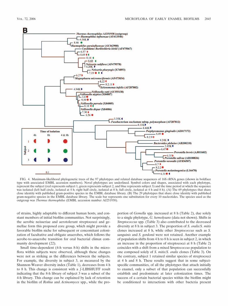
of strains, highly adaptable to different human hosts, and con-stant members of initial biofilm communities. Not surprisingly,the aerobic neisseriae and aerotolerant streptococci and ge-mellae form this proposed core group, which might provide afavorable biofilm niche for subsequent or concomitant coloni-zation of facultative and obligate anaerobes, which follows theaerobe-to-anaerobe transition for oral bacterial climax com-munity development (22).
Small time-dependent (4-h versus 8-h) shifts in the micro-flora within subjects were observed, although these changeswere not as striking as the differences between the subjects.For example, the diversity in subject 3, as measured by theShannon-Weaver diversity index (Table 1), decreases from 4 hto 8 h. This change is consistent with a -LIBSHUFF resultindicating that the 8-h library of subject 3 was a subset of the4-h library. This change can be explained by lack of retentionin the biofilm of Rothia and Actinomyces spp., while the pro-
portion of Gemella spp. increased at 8 h (Table 2), due solelyto a single phylotype, G. hemolysans (data not shown). Shifts inStreptococcus spp. (Table 3) also contributed to the decreaseddiversity at 8 h in subject 3. The proportion of S. oralis/S. mitisclones increased at 8 h, while other Streptococcus such as S.sanguinis and S. gordonii were not retained. Another exampleof population shifts from 4 h to 8 h is seen in subject 2, in whichan increase in the proportion of streptococci at 8 h (Table 2)coincides with a shift from a mixed Streptococcus population toone composed solely of S. mitis/S. oralis clones (Table 3). Onthe contrary, subject 1 retained similar species of streptococciat 4 h and 8 h. These results suggest that in some subject-specific communities, of all the phylotypes that attach initiallyto enamel, only a subset of that population can successfullyestablish and predominate at later colonization times. Thesuccess of a certain bacterial species within the biofilm mightbe conditioned to interactions with other bacteria present
FIG. 4. Maximum-likelihood phylogenetic trees of the 97 phylotypes and related database sequences of 16S rRNA genes (shown in boldfacetype with associated EMBL accession numbers). Novel phylotypes are underlined. Symbol colors and shapes, associated with each phylotype,represent the subject (red represents subject 1, green represents subject 2, and blue represents subject 3) and the time period at which the sequencewas isolated (left half circle, isolated at 4 h; right half circle, isolated at 8 h; full circle, isolated at 4 h and 8 h). (A) The 69 phylotypes that shareclose identity with published gram-positive species in the EMBL database library. (B) The 29 phylotypes that share close identity with publishedgram-negative species in the EMBL database library. The scale bar represents one substitution for every 10 nucleotides. The species used as theoutgroup was Thermus thermophilus (EMBL accession number AJ251938).
VOL. 72, 2006 MICROFLORA OF EARLY ENAMEL BIOFILMS 2845

within the same community. Our results indicate that subject-specific communities follow distinct patterns of development.Strain-level 4-h to 8-h shifts within each species might alsooccur, but the methods used here would not detect suchchanges. For example, the present results show stable propor-tions of veillonellae between 4 and 8 h in subject 1 (Table 2),whereas a recent study in our laboratory revealed that, duringthis time, shifts occur within the veillonella population at thestrain level in this subject in such a way that specific veillonellastrains seen in moderate proportions at 4 h flourish at 8 h,while others are not retained (25). These changes exemplifythe rapid dynamics of early community development and theneed for the utilization of multiple approaches to characterizebacteria at different taxonomic levels when studying commu-nity changes.
The majority of the sequences reported here correspondedto previously cultivated microorganisms, and only 13% of thesequences and 31% of the phylotypes corresponded to uncul-tured microorganisms, a proportion lower than that found insubgingival plaque, where approximately 50% of phylotypes/
species have not yet been cultured (16, 30). This result is notsurprising, however, because the greatest proportion of earlycolonizers are streptococci, which are highly amenable to cul-tivation. The coaggregation properties of streptococci favorthem as initial colonizers, compared to other oral bacteria.Streptococci are the only oral bacteria that exhibit extensiveintrageneric coaggregation (15). In addition, streptococci pos-sess numerous adhesins that recognize receptors in the ac-quired pellicle that coats the enamel (7, 9, 32, 39). Theseproperties of adhesion likely offer an advantage to streptococcithat confers their dominance as the initial colonizing bacteria.Once bound to enamel, the streptococci act as a nascent sur-face for recognition and binding by other streptococci as wellas by species of other genera, such as Actinomyces, Fusobacte-rium, Haemophilus, Prevotella, and Veillonella (14), which leadsto generation of multigeneric communities.
The present study also reports the use of FISH in conjunc-tion with a retrievable enamel chip model. The intersection ofthese techniques allows taxonomic identification while visual-izing architecture and spatial relationships among community
FIG. 5. Confocal micrographs of typical multigeneric clusters of cells found on enamel chips at 4 h (A and B) and 8 h (C and D) of plaquedevelopment. Cells were simultaneously labeled with all-bacterium-specific EUB338 probe (red) and either the Streptococcus-specific STR405probe (A and B) (green) or the Prevotella-specific PRV392 probe (C and D) (green). (A and B) Unidentified bacterial cells (EUB338 reactive; red)juxtaposed with Streptococcus cells (EUB338 and STR405 reactive; red � green yellow). (C and D) Unidentified bacterial cells (EUB338reactive; red) in association with Prevotella cells (EUB338 and PRV392 reactive; red �green yellow). Scale bar for all images, 5 �m.
2846 DIAZ ET AL. APPL. ENVIRON. MICROBIOL.

members. The occurrence of small clusters of genetically dis-tinct cells in close contact seems to be the most common formof colonization during initial plaque formation (26). Closeproximity among early colonizing cells suggests that coaggre-gation plays a role in vivo in establishing multigeneric micro-communities where metabolic interactions among membersmight determine community development. Coaggregation andmetabolic interactions occurring among some of the speciesseen as part of the early microflora of subjects in this studyhave been previously observed in a series of in vitro and in vivoexperiments conducted in our laboratory (10, 14, 26, 27). Con-sidering that streptococci are the principal group of the earlycolonizing bacteria, some of the less predominant generamight be retained by physical or metabolic interactions withstreptococci. One example is Veillonella spp., which are knownto coaggregate with streptococci (13) and establish a foodchain where veillonellae obtain energy from the metabolism oflactic acid produced by the streptococci as an end product ofcarbohydrate fermentation (23) and where veillonellae signalthe streptococci to increase expression of amylase (10). Acti-nomyces are also known to interact with streptococci in initialmicrocommunities in vivo (26), and results from in vitro stud-ies indicate that a mutualistic relationship exists between thesetwo species (27). One of the interesting features of the com-position of these early communities is the presence of cells withvery diverse metabolic requirements. For example, the pres-ence of aerobic organisms such as Neisseria spp. and Rothiaspp. contrasts with the presence of anaerobic species such asPorphyromonas spp. and Prevotella spp. The predominance ofaerobic and facultative bacteria in early colonizing biofilms isconsistent with the long-held hypothesis that initial coloniza-tion occurs by aerobic and aerotolerant bacteria, which arereplaced by anaerobic and less aerotolerant species (22). Thepresent study found, however, that anaerobic microorganismssuch as prevotellae and porphyromonads can be detected withaerobic and facultative species such as gemellae, neisseriae,and streptococci. The prevotellae and porphyromonads mightinhabit initial communities in low proportions until biofilmdevelopment ensures a proper anaerobic environment for theirproliferation. A question for future studies is how bacterialcells interact metabolically within a cluster, as such interactionsmight drive reestablishment of the community between dailyoral hygiene procedures. We propose that repetitive spatio-temporal interactions and ecological shifts in subject-specificcommunities are integral to the organization of stable naturalplaque biofilms.
ACKNOWLEDGMENT
This research was supported by the Intramural Research Program ofNIDCR, NIH.
REFERENCES
1. Aas, J. A., B. J. Paster, L. N. Stokes, I. Olsen, and F. E. Dewhirst. 2005.Defining the normal bacterial flora of the oral cavity. J. Clin. Microbiol.43:5721–5732.
2. Altschul, S. F., T. L. Madden, A. A. Schaffer, J. Zhang, Z. Zhang, W. Miller,and D. J. Lipman. 1997. Gapped BLAST and PSI-BLAST: a new generationof protein database search programs. Nucleic Acids Res. 25:3389–3402.
3. Amann, R. I., B. J. Binder, R. J. Olson, S. W. Chisholm, R. Devereux, andD. A. Stahl. 1990. Combination of 16S rRNA-targeted oligonucleotideprobes with flow cytometry for analyzing mixed microbial populations. Appl.Environ. Microbiol. 56:1919–1925.
4. Chao, A. 1984. Non-parametric estimation of the number of classes in apopulation. Scand. J. Stat. 11:265–270.
5. Chao, A., and S. M. Lee. 1992. Estimating the number of classes via samplecoverage. J. Am. Stat. Assoc. 87:210–217.
6. Cole, J. R., B. Chai, T. L. Marsh, R. J. Farris, Q. Wang, S. A. Kulam, S.Chandra, D. M. McGarrell, T. M. Schmidt, G. M. Garrity, and J. M. Tiedje.2003. The Ribosomal Database Project (RDP-II): previewing a new au-toaligner that allows regular updates and the new prokaryotic taxonomy.Nucleic Acids Res. 31:442–443.
7. Demuth, D. R., Y. Duan, W. Brooks, A. R. Holmes, R. McNab, and H. F.Jenkinson. 1996. Tandem genes encode cell-surface polypeptides SspA andSspB which mediate adhesion of the oral bacterium Streptococcus gordonii tohuman and bacterial receptors. Mol. Microbiol. 20:403–413.
8. Eckburg, P. B., E. M. Bik, C. N. Bernstein, E. Purdom, L. Dethlefsen, M.Sargent, S. R. Gill, K. E. Nelson, and D. A. Relman. 2005. Diversity of thehuman intestinal microbial flora. Science 308:1635–1638.
9. Egland, P. G., L. D. Du, and P. E. Kolenbrander. 2001. Identification ofindependent Streptococcus gordonii SspA and SspB functions in coaggrega-tion with Actinomyces naeslundii. Infect. Immun. 69:7512–7516.
10. Egland, P. G., R. J. Palmer, Jr., and P. E. Kolenbrander. 2004. Interspeciescommunication in Streptococcus gordonii-Veillonella atypica biofilms: signal-ing in flow conditions requires juxtaposition. Proc. Natl. Acad. Sci. USA101:16917–16922.
11. Felsenstein, J. 1989. PHYLIP—Phylogeny Inference Package (version 3.2).Cladistics 5:164–166.
12. Frandsen, E. V., V. Pedrazzoli, and M. Kilian. 1991. Ecology of viridansstreptococci in the oral cavity and pharynx. Oral Microbiol. Immunol. 6:129–133.
13. Hughes, C. V., R. N. Andersen, and P. E. Kolenbrander. 1992. Character-ization of Veillonella atypica PK1910 adhesin-mediated coaggregation withoral Streptococcus spp. Infect. Immun. 60:1178–1186.
14. Kolenbrander, P. E., R. N. Andersen, D. S. Blehert, P. G. Egland, J. S.Foster, and R. J. Palmer, Jr. 2002. Communication among oral bacteria.Microbiol. Mol. Biol. Rev. 66:486–505.
15. Kolenbrander, P. E., R. N. Andersen, and L. V. Moore. 1990. Intragenericcoaggregation among strains of human oral bacteria: potential role in pri-mary colonization of the tooth surface. Appl. Environ. Microbiol. 56:3890–3894.
16. Kroes, I., P. W. Lepp, and D. A. Relman. 1999. Bacterial diversity within thehuman subgingival crevice. Proc. Natl. Acad. Sci. USA 96:14547–14552.
17. Li, J., E. J. Helmerhorst, C. W. Leone, R. F. Troxler, T. Yaskell, A. D.Haffajee, S. S. Socransky, and F. G. Oppenheim. 2004. Identification of earlymicrobial colonizers in human dental biofilm. J. Appl. Microbiol. 97:1311–1318.
18. Liljemark, W. F., C. G. Bloomquist, B. E. Reilly, C. J. Bernards, D. W.Townsend, A. T. Pennock, and J. L. LeMoine. 1997. Growth dynamics in anatural biofilm and its impact on oral disease management. Adv. Dent. Res.11:14–23.
19. Ludwig, W., O. Strunk, R. Westram, L. Richter, H. Meier, Yadhukumar, A.Buchner, T. Lai, S. Steppi, G. Jobb, W. Forster, I. Brettske, S. Gerber, A. W.Ginhart, O. Gross, S. Grumann, S. Hermann, R. Jost, A. Konig, T. Liss,R. Lussmann, M. May, B. Nonhoff, B. Reichel, R. Strehlow, A. Stamatakis,N. Stuckmann, A. Vilbig, M. Lenke, T. Ludwig, A. Bode, and K. H. Schleifer.2004. ARB: a software environment for sequence data. Nucleic Acids Res.32:1363–1371.
20. Mager, D. L., L. A. Ximenez-Fyvie, A. D. Haffajee, and S. S. Socransky. 2003.Distribution of selected bacterial species on intraoral surfaces. J. Clin. Peri-odontol. 30:644–654.
21. Magurran, A. E. 1988. Ecological diversity and its measurement. PrincetonUniversity Press, Princeton, N.J.
22. Marsh, P., and M. V. Martin. 1999. Oral microbiology, 4th ed. Wright,Oxford, United Kingdom.
23. Mikx, F. H., and J. S. van der Hoeven. 1975. Symbiosis of Streptococcusmutans and Veillonella alcalescens in mixed continuous cultures. Arch. OralBiol. 20:407–410.
24. Nyvad, B., and M. Kilian. 1987. Microbiology of the early colonization ofhuman enamel and root surfaces in vivo. Scand. J. Dent. Res. 95:369–380.
25. Palmer, R. J., Jr., P. I. Diaz, and P. E. Kolenbrander. Unpublished data.26. Palmer, R. J., Jr., S. M. Gordon, J. O. Cisar, and P. E. Kolenbrander. 2003.
Coaggregation-mediated interactions of streptococci and actinomyces de-tected in initial human dental plaque. J. Bacteriol. 185:3400–3409.
27. Palmer, R. J., Jr., K. Kazmerzak, M. C. Hansen, and P. E. Kolenbrander.2001. Mutualism versus independence: strategies of mixed-species oral bio-films in vitro using saliva as the sole nutrient source. Infect. Immun. 69:5794–5804.
28. Palmer, R. J., Jr., R. Wu, S. Gordon, C. G. Bloomquist, W. F. Liljemark, M.Kilian, and P. E. Kolenbrander. 2001. Retrieval of biofilms from the oralcavity. Methods Enzymol. 337:393–403.
29. Paster, B. J., I. M. Bartoszyk, and F. E. Dewhirst. 1998. Identification of oralstreptococci using PCR-based, reverse-capture, checkerboard hybridization.Methods Cell Sci. 20:223–231.
30. Paster, B. J., S. K. Boches, J. L. Galvin, R. E. Ericson, C. N. Lau, V. A.Levanos, A. Sahasrabudhe, and F. E. Dewhirst. 2001. Bacterial diversity inhuman subgingival plaque. J. Bacteriol. 183:3770–3783.
VOL. 72, 2006 MICROFLORA OF EARLY ENAMEL BIOFILMS 2847
31. Pearson, W. R., and D. J. Lipman. 1988. Improved tools for biologicalsequence comparison. Proc. Natl. Acad. Sci. USA 85:2444–2448.
32. Scannapieco, F. A. 1994. Saliva-bacterium interactions in oral microbialecology. Crit. Rev. Oral Biol. Med. 5:203–248.
33. Schloss, P. D., and J. Handelsman. 2005. Introducing DOTUR, a computerprogram for defining operational taxonomic units and estimating speciesrichness. Appl. Environ. Microbiol. 71:1501–1506.
34. Schloss, P. D., B. R. Larget, and J. Handelsman. 2004. Integration of mi-crobial ecology and statistics: a test to compare gene libraries. Appl. Environ.Microbiol. 70:5485–5492.
35. Singleton, D. R., M. A. Furlong, S. L. Rathbun, and W. B. Whitman. 2001.Quantitative comparisons of 16S rRNA gene sequence libraries from envi-ronmental samples. Appl. Environ. Microbiol. 67:4374–4376.
36. Smith, N. H., E. C. Holmes, G. M. Donovan, G. A. Carpenter, and B. G.Spratt. 1999. Networks and groups within the genus Neisseria: analysis ofargF, recA, rho, and 16S rRNA sequences from human Neisseria species. Mol.Biol. Evol. 16:773–783.
37. Sneath, P. H. A. 1992. International code of nomenclature of bacteria:bacteriological code, 1990 revision. ASM Press, Washington, D.C.
38. Socransky, S. S., C. Smith, L. Martin, B. J. Paster, F. E. Dewhirst, and A. E.Levin. 1994. “Checkerboard” DNA-DNA hybridization. BioTechniques 17:788–792.
39. Takahashi, Y., K. Konishi, J. O. Cisar, and M. Yoshikawa. 2002. Identifi-cation and characterization of hsa, the gene encoding the sialic acid-bindingadhesin of Streptococcus gordonii DL1. Infect. Immun. 70:1209–1218.
40. Thompson, J. D., T. J. Gibson, F. Plewniak, F. Jeanmougin, and D. G.Higgins. 1997. The CLUSTAL_X windows interface: flexible strategies formultiple sequence alignment aided by quality analysis tools. Nucleic AcidsRes. 25:4876–4882.
41. Thurnheer, T., R. Gmur, E. Giertsen, and B. Guggenheim. 2001. Automatedfluorescent in situ hybridization for the specific detection and quantificationof oral streptococci in dental plaque. J. Microbiol. Methods 44:39–47.
42. van de Peer, Y., and R. de Wachter. 1993. TREECON: a software packagefor the construction and drawing of evolutionary trees. Comput. Appl. Bio-sci. 9:177–182.
2848 DIAZ ET AL. APPL. ENVIRON. MICROBIOL.


![PROGRAM OF LIBERAL STUDIES JUNIOR READING LIST PLS …24 Goethe, Faust, [1832], Part II, Act V only 25 Austen, Pride and Prejudice [1813], Chaps. 1-42 26 Austen, Pride and Prejudice,](https://img.dokumen.tips/doc/110x75/60bdc2377888f060f2423ead/program-of-liberal-studies-junior-reading-list-pls-24-goethe-faust-1832-part.jpg)