Embed Size (px)
Citation preview

Gerodontology. Volume 5. Number I. 1986, ISSN 0734-0664^1986 Beech Hill Enterprises, Inc.
Prevention of Root Caries
33
ERNEST NEWBRUN, D.M.D.,
Primary prevention of root caries should focus on preventing periodontitis and the concomitant loss of gingival attachment.This requires a regimen of plaque control consisting of scrupulous oral hygiene, supplemented, if necessary, by antimicrobialagents. Once gingival recession has occurred, available data from human and animal studies indicate that to prevent root caries,patients should limit their dietary sucrose intake both in amount and frequency. The cornerstone of any preventive regimenfor patients at high risk for caries is some mode of topical fluoride therapy. No controlled clinical data exist that show oneagent (sodium fluoride versus stannous fluoride) or one vehicle (gel versus rinse) as more effective than another. When useddaily at home, these lopical fluoride agents reduce caries in patients with xerostomia. Some reports claiming efficacy are anec-dotal, but ethical considerations preclude the use of an untreated control group. As no studies exist documenting an effectof topical fluoride in controlling root caries per se, current recommendations are based on extrapolation from studies of thesexerostomic patients. Limited studies, both in humans and animals, indicate that drinking fluoridated water helps in reducingroot caries. Progression of early root caries lesions can be arrested by a combination of mechanical/cbemical therapy, recon-touring and smoothing the roots, and applying topical fluoride to these surfaces.
ETIOLOGY
To prevent a disease on a rational rather than simplyan empirical basis, it is necessary to understand the fac-tors in its etiology. Our understanding of coronal carieshas advanced considerably. We know a great deal aboutthe specific oral flora associated with smooth surface andfissure lesions, the transmissibility of the flora, and themechanisms involved in bacterial adherence and the for-mation of dental plaque. Substantial evidence exists toascribe a unique role to dietary sucrose in determiningthe numbers of cariogenic organisms able to colonize cor-onal surfaces. By applying all this information, majorprogress has been made in reducing the prevalence of cor-onal caries, particularly in children in developed coun-tries IGIass, 1982].
Although root caries has afflicted man since ancienttimes, we know less about its etiology, pathologicalmechanisms, and prevention than we do about coronalcaries INewbrun et a!., 1984]. We do know that a prere-quisite for the disease is a susceptible root surface, whichmeans that no root caries can take place unless there hasbeen loss of periodontal attachment with or withoutgingival recession. Primary prevention of root cariestherefore involves prevention of gingivitis and periodon-titis. This requires a cooperative patient who is sufficientlymotivated and skilled to remove microbial plaque fromthe teeth, primarily by mechanical means. Unfortunate-ly, evidence from clinical practice and group studies in-dicates that the technical skill, time, effort and per-severence required to continually maintain oral cleanlinessexceed the ability of the average patient [Loe. 1970]. Asthey get older, therefore, most adults will experience someloss of attachment and will be at risk of developing rootcaries.
MICROBIOLOGY AND ANTIMICROBIALS
Root caries is undoubtedly of microbial origin.However, whether a specific organism is responsible forhuman root caries is far from clear (Table 1). In earlystudies, root caries was induced in hamsters infected withfilamentous bacteria that were subsequently identified asActinomyces viscosus T6 [Keyes, 1959; Jordan & Keyes,1964]. Similarly, Actinomyces naeslundii was shown tocause root caries in gnotobiotic rats [Socransky et al.,1970]. These studies influenced later investigations of themicroflora from human lesions of root caries [Jordan &Hammond, 1972; Syed et al., 1975] that implicated Ac-tinomyces species. A. viscosus is a dominant organism,constituting 40% to 50% of the cultivable flora of rootsurface caries plaque [Syed et al., 1975]. This may weilbe because Actinomyces species colonize better on rootsurfaces than on enamel [Kmet et al., 1985]. In fact, ina recent comparison of Actinomyces sp. on carious andnon-carious root surfaces, it was concluded that factorsother than the presence of Actinomyces sp. in dentalplaque are important in the etiology of human root sur-face caries IBryan et al., 1985]. A better way to discrim-inate between healthy and diseased root surfaces is bycounts of Streptococcus mutans and lactobacilli, eventhough these organisms represent a smaller proportionof the plaque flora [Ellen et al., 1984]. 5. mutans caninduce root caries in rodents fed a high-sucrose diet (Table2). My reason for digressing to consider the specificmicroflora will become more apparent when 1 discuss thepotential role of dietary control in the prevention of rootcaries. However, given the undisputed microbial originof root caries, suitable antimicrobial therapy should alsobe considered as a potential means of preventing such le-sions. Surprisingly few attempts have been made to study
'The author wishes lo thank Ms. E. Leash for her careful editing of this manuscript.
the Department of Slomatology, HSW-604, University of California San Francisco, San Francisco CA 94143, U.S.A.

34 NEWBRUN
TABLE 1
Root Caries in Humans: Predominant Microflora
Predominatu Flora(% CFU)
Plaque (P)Lesion (L)Saliva (S) Comment
Investigators[Year]
A. viscosus, A. naeslundii, A. odontolyiicus,A. eriksonii. Rothia deniocariosa; outer layer
5. mutans, enterococci & Actinomyces sp., outersurface; aerobic diphtheroids {Arthrobacterl) deepin lesions
A. viscosus (47) & S. mutans (30) or A. viscosus(41) & S. sanguis (48)
Streptococci (70), Actinomyces sp. (30), A.odontoiyticus, A. viscosus, A. israelii, A.naeslundii
Unidentified
Streptococci (39), S. mutans (22), Acdnomvces sp,(23), S. mitis (10)
Unidentified (80). A. viscosus (6), A. naeslundii(2), streptococci (5)
A. viscosus (13), A. naeslundii (2)
L Qualitative data only.
L Qualitative data only. Why aerobes deep inlesion?
P Quantitative data for anaerobic flora only. Rootcaries in absence of S. mutans.
P Quantitative data for aerobic and anaerobic flora.Pooled plaque (coronal and radicular) excludinglesion.
S Semiquantitative data only. Lactobacilli countscorrelated with root caries.
P Quantitative data for early to advanced rootcaries. Fluorescent antibody technique showedS. mutans. Actinomyces sp. and Biftdobacterium sp.
P Quantitative data for both carious and noncariousroot surfaces. S. mutans and lactobacilli countscorrelated with caries.
P Quantitative data for both carious and noncariousroot surfaces. No difference in proportions ofActinomyces sp.
Jordan &Hammond [1972]
Sumney & Jordan[1974]
Syed et al. (1975]
Hill et al. [1977]
Ravald and Hamp[1981)
Brown et al.[1983]
Ellen e( al. [1984]
Bryan et al,[1985]
the effect of antimicrobials on the development of rootcaries in animals or humans.
ROLE OF DIET AND DIETARY CONTROL
Root caries has been noted in the teeth of ancient skullsof North American Indians [Leigh. 1925], Fiji Islanders[Chappel, 1927], Hawaiians [Keene, 1974], pre-Colum-
bian Peruvians [Stewart, 1931], and early Anglo-SaxonsICorbett & Moore, 1971; Hardwick, 1960; Miles, 1969].Generally these teeth had few, if any, coronal caries. Theusual explanation offered for this difference in cariesprevalence between ancient man and modern man livingin an industrial society is that primitive diets contain farless sucrose. Sugar cane was not cultivated in Hawaii200-500 years ago, nor was it cultivated in the pre-
TABLE 2
Root Caries in Animal Models: Predominant Microflora and Diet
Animal Modeland Species
PredominantFlora (Strain) Diet Investigators (Year)
Conventional animals
Syrian hamster
Albino hamster
Rice ratGolden hamster
Monoinfected animalsS.D. rat'
S.D. rat
S.D. ratS.D. rat
S.D. rat
S.D. rat
S.D. rat
A. viscosus (T6)
A. viscosus (MlOO, K04), A. naeslundii (N16)Enterococci, Actinobacillus. diphtheroids
A. viscosus. A. naeslundii
S. mutans (GS5)5. 5ff//vc7r/w5-"Iike" (SS2)A. viscosus, Actinomyces sp.
A. naestundii (1)5. salivarius-"hkc" (lA)
A. viscosus (RC45), A. naeslundii (RC15)
A. viscosus, A. naeslundii, S. mutans.S. sanguis, B. cereus
20002000°
2000
2000^
5852000 or 585
2000
20002000 or 585
20002000
Keyes & Jordan [1964]Jordan et al. [1972]Socransky et al. [I960], Doff et al. [1977]
Behbehani & Jordan [1980]
Gibbons et aL [1966]Gibbons & Banghart [1968]Jordan et al. [1972]Socransky et al. [1970]Kelstrup & Gibbons [1970]Jordan & Hammond [1972]Crawford et al. [1977]
"Modified by substituting 56% corn starch for sucrose.''2(X)0 or modified by substituting 56% glucose for sucrose.'S.D. =Sprague-DawIey.

PREVENTION OF ROOT CARIES 35
Columbian Americas. Starcbes sucb as taro root and cornwere a staple of these primitive diets. In certain present-day primitive populations, the prevalence of root carieshas also been found to exceed tbe prevalence of coronalcaries. By the time tbey reach 30-39 years of age, nativesof Lufa, Papua, New Guinea have more root caries thancoronal caries [Scbamscbula et al., 1974]. The principalfood of this population is sweet potatoes, supplementedwith a variety of vegetables, fruits, nuts, and occasionallywith meat [Schamschula et a l , 1972]. Based on theseepidemiological findings in ancient man and primitivesocieties. Banting and Courtright [1975] concluded thatroot caries can occur in populations whose diets have littlesucrose.
Data from human studies on the effect of diet on rootcaries are sparse indeed. Tbe Vipeholm dental caries studyis one of the few in which dietary factors were relativelywell controlled. In those groups receiving sugar sup-plements between meals (22 caramels or 24 toffees daily)the frequency of root caries was high; in tbe toffee group,root caries accounted for 25% of all lesions. However,these groups were also slightly older than the other testgroups [Gustafsson et al., 1954].
More recently, Hix and O'Leary [1976] reported thatin patients with periodontal disease, both treated and un-treated, those wbo had the most root caries gave a diethistory indicating significantly more exposures to fer-mentable carbohydrates per week. Unfortunately, no at-tempt was made to quantify the amounts or types ofsugars ingested; only the frequency of ingestion of "read-ily fermentable carbohydrate" was scored.
Virtually all the animal studies that have induced rootcaries in either conventional or monoinfected rodentshave used a high-sucrose diet. In the original experimentswith hamsters, Keyes [1959] used a high-sugar diet 2000(sucrose 56%, skim milk powder 28%, whole meal flour6%, yeast 4%). Indeed, when the diet was changed toa commercial laboratory chow, the plaque mineralizedand root caries did not progress [Keyes & Jordan, 1964].Root caries was induced in rats monoinfected with Strep-tococcus salivarius-"\\ke" strains SS2 or lA when theywere fed diet 2000, whereas monoinfected rats fed diet585, a coarse-particle diet (sucrose 25%, whole milkpowder 30%, yellow hominy grits 42%), only developedfissure caries [Gibbons & Banghart, 1968; Kelstrup& Gib-bons, 1970]. However, in earlier studies with a morevirulent organism, S. mutans GS5, root caries as well ascoronal caries was obtained with monoinfected rats feddiet 585 [Gibbons et al., 1966]. Wben rice rats were feddiets containing no sucrose (MIT 300) or low sucrose(5%, MIT 305), little or no root caries developed, whereasthose fed a high-sucrose diet (56% diet 2000, 67% MIT200) developed extensive root caries lesions (Figure 1)[Rosen, 1984]. Similarly, wben different foods (bread,potato chips, raisins, toffee, or SLS, a mixture of starch/lactalbumin/sucrose) were compared for their ability tocause root caries in rice rats, there was a direct relation-ship between root caries scores and the total sugar con-tent of the test food item [De Palmaet al., 1983]. Calcula-tion of the correlation coefficient (r^O.99) shows it to
30 T
2 5 ••
2 0 •CO
a: 15 +
1 0 •
5 •
0
26
0 3
0 5 56« SUCROSE
FIGURE 1. Relationship between root caries and dietarysugar. Rice rats were fed diets MIT 300 (no sucrose), MIT305 (5% sucrose), 2000 (56% sucrose) and MIT 200 (67%sucrose). Data from Rosen [1984].
be highly significant (Figure 2).How can we reconcile these apparently contradictory
observations concerning the role of sucrose in the etiologyof root caries? In primitive populations, past or present,sucrose was not and is not a major dietary component,as it is in developed countries (about 20% of the totalcaloric intake). However, there is always some sucrosepresent, even in unprocessed fruits and vegetables. Thismay be sufficient to account for the root caries that hasbeen seen. Furthermore, solid quantitative epidemiologi-cal data on the extent and frequency of these lesions arepractically nonexistent, particularly for the primitivepopulations. Therefore it is not possible to compare ob-jectively the root caries indices of such populations. If
60 PC ^ POTATO CHIPS8 = BREADSIS - STAPrH/LACTAlBUMIN/SUCROSE
oc
o
ut—
OOa:
40
R = TAlSINS
r 0 99
IIJ
TOTAL SUGAR IN DIET (%)
FIGURE 2. Root caries scores in rice rats fed diets of dif-fering sugar content. Regression line plotted from datafor starch/lactalbumin/sucrose, toffee (2 kinds), andraisins. Data from DePalma et al. [1983],

36 NEWBRUN
we consider the findings of the animal study more close-ly (Figure 1) we see that some root caries is induced onthe low sucrose (5%) diet. Similarly, diet 585 (25% su-crose) induced root caries in rats monoinfected with S.mutans.
Accordingly, data from most animal and some humanstudies on the role of diet, as well as from microbiologicalstudies implicating S. mutans (which needs sucrose inorder to colonize tooth surfaces), indicate that root carieswill increase with the amount and frequency of sugar in-take [Newbrun, 1984]. Conversely, to prevent or reduceroot caries, patients should restrict their dietary sucrose,both in amount and frequency of ingestion. These dietaryrecommendations are supported by, but not solely depen-dent on, what is already known about prevention of cor-onal caries.
ROLE OF FLUORIDES
Systemic Fluoride
The concentration of fluoride in the outer layer of theroot surface increases with increasing levels of fluoridein the water supply as well as with advancing age [Yoonet al., 1960; Brudevold et al., 1960; Nakata et al., 1972;Stepnick et al., 1975; Banting & Stamm, 1979]. Labora-tory studies indicate that the solubility of the root sur-face in acid decreases with increased fluoride uptake[Shannon et al., 1976]. These observations suggest thatfluoride could play a role in the prevention of root caries.
Information on how systemic ingestion of fluoride af-fects root caries in adults is limited. The only epidemi-ological data available indicate that lifelong residence ina fluoridated community is associated with a highlysignificant reduction in the prevalence of root caries orroot fillings at all ages. Stamm and Banting [1980] com-pared root caries and root fillings in 465 adults (meanage 42.8) residing in Woodstock, Ont. (0.1 ppm) withthose in 502 adults (mean age 40) in Stratford, Ont. (1.6ppm F). The mean number of decayed or filled root sur-faces was 1.36 and 0.64, respectively. Furthermore, asshown in Figure 3, the percentage of the population withroot caries in each age group over 20 years is consistent-ly lower in those adults who have lived in a fluoridatedcommunity [Banting, 1984]. These data clearly refute theoften cited canard of antifluoridationists that waterfluoridation does not benefit adults. Similarly, as il-lustrated in Figure 4, root caries in rats is also significantlyreduced by systemic administration of fluoride in thedrinking water. Rats given 0, 4.5 or 45 ppm F developedroot surface caries scores of 19.5, 11.8 and 0.35, respec-tively [Rotihe et al., 1977].
These reports describing the effects of systemic fluoridein humans and rats point to an inhibiting effect on rootcaries which could also be partially due to topical action.However, the information is limited in scope and has onlybeen reported in preliminary form. The epidemiologicalevidence compares only two levels of fluoride in the watersupply and does not include a population drinking an op-timal concentration. Carefully designed studies are still
67.5
18-19 20-29 30-39 40-49 50-59 60+
AGE OROUP
FIGURE 3. Relationship between fluoride in drinkingwater and percentage of population with root caries atvariousages. Solid bars = Woodstock, Ont., 0.1 ppm F;dotted bars = Stratford, Ont., 1.6 ppm F. Data fromStamm & Banting [1980].
needed to clarify the relation between fluoride concen-tration in the water supply and prevalence of root caries.
Topical Fluoride
Caries-preventive treatment in patients at high risk forroot caries may involve oral hygiene instruction anddietary advice, but the cornerstone of any preventiveregimen for these patients includes some mode of fluoridetherapy. One such high-risk group consists of those whosesalivary flow has been reduced or eliminated altogether,whether by pathological changes in the salivary glandsor by their exposure to therapeutic irradiation. General-ly, two forms of fluoride treatment have been advocatedand tested for this population. One form is an acid orneutral gel containing 1.1% sodium fluoride; it provides5000 ppm fluoride and is applied in the mouth in acustom-fitted soft plastic tray for 5 to 10 minutes eachday [Daly et al., 1972; Dreizen et al., 1976; Johansen &Olsen, 1979]. The other is a 0.4% stannous fluoride gelproviding 1000 ppm fluoride [Westcott et al., 1975]. For
19.5
6 8 10 12 14ROOT CARIES SCORE
16 18 20
FIGURE 4. Relationship between fluoride in the drinkingwafer and roof caries in rice rafs fed a high-sucrose diet.Dafa from Rotilie et a!. [977].
PREVENTION OF ROOT CARIES 37
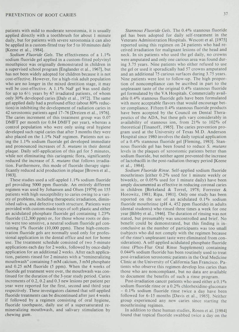
patients with mild to moderate xerostomia, it is usuallyapplied directly with a toothbrush for about 1 minutedaily, but for patients with severe xerostomia it can alsobe applied in a custom-fitted tray for 5 to 10 minutes daily[Keene et al.. 1984].
Sodium Fluoride Gels. The effectiveness of a 1.1 *Vosodium fluoride gel applied in a custom-fitted polyvinylmouthpiece was originally demonstrated in children ina supervised school program [Englander et al., 1967], buthas not been widely adopted for children because it is notcost-effective. However, for a high-risk adult populationwho are no longer in the mixed dentition stage, it maywell be cost-effective. A 1.1% NaF gel was used dailyfor up to 6'/2 years by 67 irradiated patients, of whomonly 30% developed caries [Daly et al., 1972]. The samegel applied daily had a profound effect (about 80% reduc-tion) in inhibiting the development of radiation caries in42 xerostomic patients aged 17-76 [Dreizen et al., 1976].The caries increment of this treatment group was 0.07DMFT per month (or 0.84 DMFT per year), whereas acontrol population who were only using oral hygienedeveloped such rapid caries that after 3 months they werealso placed on the 1.1% NaF regimen. Patients not us-ing the 1.1% sodium fluoride gel developed immediateand pronounced increases of S. mulans in their dentalplaque. Daily self-application of this gel for 5 minutes,while not eliminating this cariogenic flora, significantlyreduced the increase of 5. mutans that follows irradia-tion. Furthermore, this mode of fluoride therapy signi-ficantly reduced acid production in plaque [Brown et al.,1983].
These studies used a self-appled 1.1% sodium fluoridegel providing 5000 ppm fluoride. An entirely differentregimen was used by Johansen and Olsen [1979] on 155patients with high susceptibility to caries owing to a vari-ety of problems, including therapeutic irradiation, dimin-ished saliva, and defective tooth structure. Patients wereprovided with custom-made trays of soft plastic and eitheran acidulated phosphate fluoride gel containing 1.23%fluoride {12,300 ppm) or, for those whose roots or den-tin were too sensitive, a neutral sodium fluoride gel con-taining 1% fluoride (10,000 ppm). These high-concen-tration fluoride gels are normally used only for profes-sional application in the dental office and not for homeuse. The treatment schedule consisted of two 5-minuteapplications each day for 2 weeks, followed by once-dailyapplication for an additional 2 weeks. After each applica-tion, patients rinsed for 2 minutes with a "remineralizingmouthwash" containing 5 mM calcium, 3 mM phosphateand 0.25 mM fluoride (5 ppm). When the 4 weeks offluoride gel treatment were over, the mouthwash was con-tinued for the duration of the 3-year study period. Cariesincrements of 0.2, 0.2 and 0.3 new lesions per patient peryear were reported for the flrst, second and third yearrespectively. These investigators claimed that self-appliedfluoride treatments can be discontinued after just 4 weeksif followed by a regimen consisting of oral hygiene,fluoride toothpaste, daily use of a supersaturated re-mineralizing mouthwash, and salivary stimulation bychewing gum.
Stannous Fluoride Gels. The 0.4% stannous fluoridegel has been adopted for daily self-treatment in theVeterans Administration Hospitals. Wescott et al. [1975]reported using this regimen on 24 patients who had re-ceived irradiation for malignant lesions of the head andneck. In six patients who used the gel daily, no crownswere amputated and only one carious area was found dur-ing 3.75 years. Nine patients who either refused to usethe gel or used it sporadically had 57 crowns amputatedand an additional 75 carious surfaces during 3.75 years.Nine patients were lost to follow-up. The high propor-tion of noncompliance can be ascribed in part to theunpleasant taste ofthe original 0.4% stannous fluoridegel formulated by the VA Hospitals. Commercially avail-able 0.4% stannous fluoride gels have been formulatedwith more acceptable flavors that would encourage bet-ter compliance. Fifteen 0.4% stannous fluoride productshave been accepted by the Council on Dental Thera-peutics of the ADA, but these gels vary considerably inavailability of stannous ion, from 21% to 102% oftheoretical [Tinanoff, 1985]. The caries prevention pro-gram used at the University of Texas M.D. AndersonHospital since 1980 involves the daily topical applicationof a 0.4% stannous fluoride gel [Fleming, 1983]. Stan-nous fluoride gel has been found to reduce S. mutanslevels in the plaques of xerostomic patients better thansodium fluoride, but neither agent prevented the increaseof lactobacilli in the post-radiation therapy period [Keeneet al., 1984].
Sodium Fluoride Rinse. Self-applied sodium fluoridemouthrinses [either 0.2% used for 1 minute weekly orbiweekly, or 0.05% used for 1 minute daily) have beenamply documented as effective in reducing coronal cariesin children [Birkeland & Torrel, 1978; Forrester &Horowitz, 1981; Ripa, 1981]. Only one study has beenreported on the use of an acidulated 0.1% sodiumfluoride mouthrinse {pH 4, 452 ppm tluoride) in adults(dental students) who rinsed three times a week for oneyear [Bibby et al., 1946]. The duration of rinsing was notstated, but presumably was uncontrolled and brief. Nobenefit could be demonstrated, but this finding is in-conclusive as the number of participants was too small(subjects who did not comply with the regimen becauseof the rinse's unpleasant taste were eliminated from con-sideration). A self-applied acidulated phosphate fluoriderinse (Phos-Flur Oral Rinse Supplement) containing0.044% sodium tluoride has been routinely used to treatpost-irradiation xerostomic patients in the Oral MedicineClinic at the University of California San Francisco. Pa-tients who observe this regimen develop less caries thanthose who are noncompliant, but no data are availableto document the benefits of such a rinse program.
Post-irradiation cancer patients who used either a 0.1%sodium fluoride rinse or a 0.2% chlorhexidine-gluconate- 0.1% sodium fluoride rinse twice a day have beenfollowed for 6-15 months [Davis et al., 1985]. Neithergroup experienced any new caries since starting themouthrinsing regimen.
In addition to these human studies, Rosen et al. [1984]found that topical fluoride swabbed twice a dav on the

38 NEWBRUN
molar teeth of rats also significantly inhibited root sur-face caries in comparison with a water control. The agentstested were a sodium fluoride solution (5000 ppm F), asodium fluoride-dentifrice slurry (500 ppm F) and water(0 ppm F). The corresponding root caries scores were10.4, 15.5. and 24.1.
The dose-response relationship for topical fluorides isunclear. In a fluoride dentifrice study on children, therewas a dose-response caries-preventive effect with sodiumfluoride dentifrices containing 250, 750 and 1000 ppm offluoride [Reed, 1973]. However, more recently Koch etal. [1982] claimed no difference in caries prevention be-tween children who used a dentifrice with 250 ppmfluoride and children using dentifrices with 1000 ppmfluoride. Without direct clinical testing, it is therefore notpossible to predict whether the 1.1% sodium fluoride gel(5000 ppm fluoride) would be more effective than a 0.4%stannous fluoride gel (1000 ppm fluoride) or a 0.05%sodium fluoride rinse (226 ppm fluoride) (Newbrun,1985]. In vitro tests indicate that other forms of fluoride,such as acidulated phosphate fluoride and titaniumtetrafluoride, promote greater uptake of fluoride by rootsurfaces than does sodium fluoride [Hals et al., 1981].
In a comprehensive review of the literature on the useof topical fluorides to prevent dental caries in adults,Swango [1983] noted that few of the studies cited abovehave been replicated. It is therefore difficult to discerna trend of success for any given regimen. There has beenno direct comparison of the efficacy of a 1.1% sodiumfluoride gel with a 0.4% stannous fluoride gel, althoughthese two vehicles are the ones most frequently advocated.There are no adequate existing studies on the effect oftopical fluoride procedures in inhibiting the developmentof root caries [Swango, 1983; Banting, 1984].
The efficacy of topical fluoride therapy for preventingroot caries in normal adults or geriatric patients may ormay not be as spectacular as for radiation caries [Bill-ings et al., 1985]. In xerostomic patients the clearance offluoride from the oral cavity would be delayed, allowinga longer reaction time between the topical agent and theroot surface. Fluoride uptake by enamel is diffusion-controlled and increases with time of exposure [Joyston-Bechal et al., 1973]; presumably the mechanism of up-take is similar for root surfaces. The specific role offluoride in remineralization of lesions on the root sur-face is discussed elsewhere in this symposium [Mellberg,1986].
MECHANICAL AND CHEMICAL TREATMENTOF ROOT SURFACES
The measures discussed thus far, namely, preventionof periodontitis by personal oral hygiene, use of anti-microbial agents, avoidance of sucrose-containing be-tween-meal snacks, drinking of optimally fluoridatedwater, and the appropriate use of topical fluoride agents,are primary and secondary preventive measures. They areaimed at reducing either the occurrence of root caries orits prevalence in a population [Nikiforuk, 1985]. Oncean initial lesion exists, tertiary prevention aims at limiting
its further development. Banting and Ellen [1978] recom-mended treating incipient root surface lesions simply byusing a diamond stone to render them smooth and self-cleansing, then applying topical fluorides to the surface.Acting on this suggestion, researchers in two recentstudies have used mechanical smoothing of the root sur-face followed by chemical treatment in efforts to arrestincipient or shallow lesions. At the University of Texasin Houston, investigators classified root caries as follows:Grade I, incipient; Grade II, shallow surface defect, somepigmentation; Grade III, deep lesion; and Grade IV,pulpal involvement. Grade I lesions were treated bytopical fluoride gel alone (1.0% sodium fluoride useddaily in custom-fitted trays). Grade II lesions were eitherrecontoured, smoothed and treated with fluoride, ortreated only with fluoride. After 24 months, 14/17 GradeI lesions showed no clinical changes (visual or tactile);3/17 progressed to Grade II, and were arrested by treat-ment. All Grade II lesions (16/16) that were treatedmechanically and with fluoride were clinically sound [Bill-ings et al., 1985]. The investigators concluded that con-servative debridement, cleansing, and smoothing of theroot surface is a reasonable alternative to placing arestoration in shallow lesions. In a similar study at theUniversity of Alabama, root caries lesions were ex-cavated, recontoured, and treated with 0.5% I2-1% KIand 1.2% sodium fluoride at 0, 9 and 16 days. In addi-tion, patients used 0.2% sodium fluoride mouthrinsedaily [Al-Joburi et al., 1982]. In both these studies, levelsof 5. mutans on the root surface lesions were significantlyreduced below prefreatment levels.
CONCLUSIONS
Our knowledge of the etiology, epidemiology andprevention of root caries is limited, particularly in com-parison with our understanding and ability to prevent cor-onal caries. In part, this is because adults and the elderlyare not nearly as accessible for surveys and clinical trialsas are schoolchildren. We know that personal oralhygiene requiring scrupulous plaque control can preventperiodontitis and attachment loss, thereby avoiding plac-ing root surfaces at risk. However, once recession has oc-curred, we think that antimicrobials should help inpreventing root caries, but little experimental data existto support this contention. We also think that limitingdietary sucrose intake, both in amount and frequency,will lessen the risk of root caries. This is based primarilyon animal experimentation; practically no human studieshave been conducted to test this hypothesis. We believethat ingestion of optimally fluoridated water will reducethe prevalence of root caries, again based partly on animaltesting and on one preliminary report on humans. Weconsider that some form of daily self-application oftopical fluoride is useful m preventing the onset of rootcaries lesions, based mostly on observed reduction ofcaries in the extremely high risk case of xerostomia; butwe do not know which fluoride agent or vehicle is mosteffective. Results of preliminary clinical trials suggest thatinitial root caries lesions can be arrested by a combina-
PREVENTION OF ROOT CARIES 39
lion of mechanical/chemical therapy, recontouring andsmoothing the roots, and applying topical fluoride. Whatcan we recommend with respect to chnical prevention ofroot caries when existing data are inadequate or in-complete? How certain do we have to be before decidinga regimen is beneficial? When faced with patients at risk,dentists need to intervene and instruct their patients,drawing partly on what is known about preventing rootcaries and also, to a large extent, on what is known aboutcontrolling coronal caries. Fortunately, these recommen-dations are not too disparate; what works for coronalcaries prevention seems to work for root caries preven-tion. More definitive answers will be forthcoming in thenext few years.
REFERENCES
Al-Joburi, W., Legler. D.. & Jamison, H.: Root caries:control of lesions by iodine-fluoride therapy. Journalof Dental Research, 61 (Special Issue); 340, abstractno. 1459, 1982.
Banting. D.W.: Dental caries in the elderly. Gerodon-tology, 3: 55-61. 1984.
Banting, D.W., & Courtright, P.N.: The distribution andnatural history of carious lesions on the roots of teeth.Journal of the Canadian Dental Association, 41:45-49, 1975.
Banting, D.W., & Ellen, R.P.: Carious lesions on theroots of teeth: A review for the general practitioner.Journal of the Canadian Dental Association, 42:496-502. 1976.
Banting, D.W., & Stamm, J.W.: Effect of age and lengthof residence in a fluoridated area on root surfacefluoride concentration. Clinical Preventive Dentistry,1: 7-10, 1979.
Behbehani, M.J., & Jordan, H.V.: Comparative coloni-zation of human Actinomyces species in hamstersunder different dietary conditions. Journal ofPeriodontal Research, 15: 395-404, 1980.
Bibby, B.G., Zander. H.A.. McKelleget, M., & Labun-sky. B.: Preliminary reports on the effect on dentalcaries of the use of sodium fluoride in a prophylacticcleaning mixture and in a mouthrinse. Journal of Den-tal Research, 25: 207-211, 1946.
Billings. R.J., Brown, L.R., & Kaster, A.G.: Contem-porary treatment strategies for root surface dentalcaries. Gerodontics, 1: 20-27, 1985.
Birkeland, J.M., & Torrell, P.: Caries-preventive fluoridemouthrinses. Caries Research, 12 (Suppl. 1): 38-51,1978.
Brown, L.R., Billings, R.J., O'Neill, P.A., Wheatcroft,M.G., & Kaster, A.G.: Microbiological comparisonsof carious and noncarious root and enamel tooth sur-faces. Journal of Dental Research, 62 (Special Issue):295, abstract no. 1137, 1983.
Brudevold, F., Steadman, L.T., & Smith, F.A.: Inorganicand organic components of tooth structure. Annals ofthe New York Academy of Sciences, 85: 119-132,1960.
Bryan. A.R., Reynolds, H.S., & Zambon, J.J.: Preva-
lence of Actinomyces sp. in human root surface caries.Journal of Dental Research, 64 (Special Issue): 192,abstract no. 162. 1985.
Chappel. H.G.: Jaws and teeth o\~ ancient Hawaiians.Memoirs Bernice P. Bishop Museum, 9: 251-268,1927.
Corbett, M.E., & Moore, W.J.: Distribution of caries inancient British populations. Journal of DentalResearch, 50: 663, 1971.
Daly. T.H.. Drane, J.B.. & MacComb, W.S.: Manage-ment of problems of the teeth and jaws in patientsundergoing irradiation. American Journal of Surgery,124: 539-542, 1972.
Davis, J., Harper, D.S.. & Hurst. P.S.: NaF andchlorhexidine mouthrinses for prevention of post-irradiation oral disease. Journal of Dental Research,64 (Special Issue): 206. abstract no. 287, 1985.
De Palma, J.. Rosen, S., & Harper, D.S.: Specific foodsas etiological factors in bone loss and root caries. Jour-nal of Dental Research, 62 (Special Issue): 295,abstract no. 1136, 1983.
Doff, R.S., Rosen, S., & App, G.: Root surface cariesin the molar teeth of rice rats. II. Quantitation of le-sions induced by high sucrose diet. Journal of DentaiResearch, 56: 1111-1114, 1977.
Dreizen, S., Brown, L.R., Daly, T., & Drane, J.B.:Prevention of xerostomia-related dental caries in ir-radiated cancer patients. Journal of Dental Research,56: 99-104, 1976.
Ellen, R.P., Banting, D.W., & Fillery, E.: Microbiologi-cal assessment of root caries risk in a chronicallyhospitalized population. Journal of Dental Research63 (Special Issue): 218, abstract no. 430, 1984.
Engiander, H.R., Keyes, P.H., & Gestwicki, M.: Clinicalanticaries effect of repeated topical sodium fluorideapplications by mouth pieces. Journal of the AmericanDental Association, 75: 638-644, 1967.
Fleming, T.J.: Use of topical fluoride by patients receiv-ing cancer therapy. Current Problems in Cancer, 7:37-41. 1983.
Forrester, D.J., & Horowitz, H.S.: Individual topicalfluoride therapy. In D.J. Forester, M.L. Wagner, &J. Fleming (Eds.), Pediatric Dental Medicine.Philadelphia: Lea & Febiger, pp. 320-332, 1981.
Gibbons, R.J., & Banghart, S.: Induction of dental caries ingnotobiotic rats with a Ievan-forming streptococcus anda streptococcus isolated from subacute bacterial endo-carditis. Archives of Oral Biology, 13: 297-308, 1968.
Gibbons, R.J., Berman, K.S., Knoettner, P., & Kap-simalis, B.: Dental caries and periodontal bone lossin gnotobiotic rats infected with capsule-forming strep-tococci of human origin. Archives of Oral Biologv,11: 549-650, 1966.
Glass, R.L. (Ed.): The first international conference onthe declining prevalence of dental caries. Journal ofDental Research, 61 (Special Issue): 1304-1383, 1982.
Gastafsson, B.E., Quensel, C.E., Lanke, L.S., Lund-qvist, C , Grahnen, H., Bonow, B.E., & Krasse. B.:The Vipeholm dental caries study. The effect of dif-ferent levels of carbohydrate intake on caries activity

40 NEWBRUN
in 436 individuals observed for five years. Ada Odon-tologica Scandinavia, 11: 232-364, 1954.
Hals, E., Tveit, A.B., Totdal, B., & Isrenn, R.: Effectof NaF, TiF4 and APF solutions on root surfaces invitro, with special reference to uptake of F. CariesResearch, 15; 468-476, 1981.
Hardwick, J .L.: The incidence and distribution of cariesthroughout the ages in relation to Englishmen's diet.British Dental Journal, 108: 9-17, 1960.
Hill, P.E., Knox, K.W., Schamschula, R.G., & Tabua,J.: The identification and enumeration of Actinomycesfrom plaque of New Guinea indigenes. CariesResearch, 11: 327-335, 1977.
Hix, J.O., & O'Leary, T.J.: The relationship betweencemental caries, oral hygiene status and fermentablecarbohydrate intake. Journal of Periodontology, 47:398-404, 1976.
Johansen, E., & Olsen, T.: Topical fluoride in the preven-tion and arrest of dental caries. In E. Johansen, D.R.Taves, & T.O. Olsen (Eds.), Continuing Evaluationof the Use of Fluorides. A.A.A.S. Selected Sym-posium 11. Boulder, Colorado: West View Press, pp.61-110, 1979.
Jordan, H.V., & Hammond, B.F.: Filamentous bacteriaisolated from human root surface caries. Archives ofOral Biology, 17: 1333-1342, 1972.
Jordan, H.V., & Keyes, P.H.: Aerobic, gram-positive,filamentous bacteria as etiological agents of ex-perimental periodontal disease in hamsters. Archivesof Oral Biology, 9: 401-414, 1964.
Jordan, H.V., Keyes, P.H., & Bellack, S.: Periodontallesions in hamsters and gnotobiotic rats infected withActinomvces of human origin. Journal of Periodon-tal Research, 7: 21-28. 1972'.'
Joyston-Bechal, S., Duckworth, R., & Braden, M.: Themechanism of uptake of '^F by enamel from sodiumfluoride and acidulated phosphate fluoride solutionlabelled with '^F. Archives of Oral Biology, 18:1077-1089, 1973.
Keene, H.J.: Dental caries in ancient and modern Hawaii.Journal of the Hawaii Dental Association, 7: 9-14,1974.
Keene, H.J., Fleming, T.J., Brown, L.R., & Dreizen, S.:Lactobacilli and S. mutans in cancer patients usingfluoride gels. Journal of Dental Research 63 (SpecialIssue): 281, abstract no. 429, 1984.
Kelstrup, J., & Gibbons, R.J.: Induction of dental cariesand alveolar bone loss by a human isolate resemblingStreptococcus salivarius. Caries Research, 4: 360-377,1970.
Keyes, P.H.: Dental caries in a Syrian hamster. VIII. Theinduction of rampant caries activity in albino andgolden hamsters. Journal of Dental Research, 38:525-533, 1959.
Keyes, P.H., & Jordan, H.V.: Periodontal lesions in theSyrian hamster. III. Findings related to an infectiousand transmissible component. Archives of OralBiology, 9: 377-400, 1964.
Kmet, P., Boyar, R., & Bowden, G.: Microbial coloniza-tion of exposed root surfaces and enamel. Journal of
Dental Research 64 (Special Issue): 331, abstract no.1411, 1985.
Koch, G., Peterson, L.G., Kling, E., & Kling, L.: Effectof 250 and 1000 ppm fluoride dentifrice on caries.Swedish Dental Journal, 6: 233-238, 1982.
Leigh, R.W.: Dental pathology of Indian tribes of variedenvironmental and food conditions. American Jour-nal of Physical Anthropology, 8: 179-199, 1925.
Loe, H.: A review of the prevention and control of plaque.In W.D. McHugh (Ed.), Dental Plaque. Edinburgh:E. & S. Livingstone, pp. 259-270, 1970.
Mellberg, J: Demineralization and remineralization ofroot surface caries. Gerodontology, 5: this issue, 1986.
Miles, A.E.W.: The dentition of the Anglo-Saxons. Pro-ceedings of the Royal Society of Medicine, 62:1311-1315, 1969.
Nakata, T.M., Stepnick, R.J., & Zipkin, L: Chemistryof human dental cementum: The effect of age andfluoride exposure on the concentration of ash, fluor-ide, calcium, phosphorus, and magnesium. Journal ofPeriodontology, 43: 115-124, 1972.
Newbrun, E.: Diet and dental caries: symposium over-view. In B. Guggenheim (Ed.), Cariology Today.Basel: Karger, pp. 340-352, 1984,
Newbrun, E. Impact of fluorides on root caries and rootdentinal sensitivity. In S.H.Y. Wei (Ed.), Clinical Usesof Fluorides. Philadelphia: Lea&Febiger, pp. 93-102,1985.
Newbrun, E., Armitage, G., Daniels, T.E., Greenspan,D., Leash, E., & Robertson, P.B.: Root caries.California Dental Association Journal, 12: 68-73,1984.
Nikiforuk, G.: Understanding Dental Caries. 2. Preven-tion: Basic and Clinical Aspects: Basel: Karger, pp.5-6, 1985.
Ravald, N., & Hamp, S.E.: Prediction of root surfacecaries in patients treated for advanced periodontaldisease. Journal of Clinical Periodontology, 8: 400,1981.
Reed, M.W.: Clinical evaluation of three concentrationsof sodium fluoride in dentifrices. Journal of theAmerican Dental Association, 87: 1101-1404, 1973.
Ripa, L.W.: Fluoride rinsing: what dentists should know.Journal of the American Dental Association, 102:477-481, 1981.
Rosen, S.: Alveolar bone loss and root caries in rice ratson diets containing various concentrations of sucrose.In J.J. Hefferren, W.A. Ayer, H.M. Koehler, & C.T.McEnery (Eds.), Foods, Nutrition and Dental Health.Chicago: American Dental Association, 4: 217-221,1984.
Rosen, S., Beck, F.M., <& Beck, E.X.: Effect of sodiumfluoride dentifrice on root surface caries. Journal ofDental Research, 63 (Special Issue): 238, abstract no.609, 1984.
Rotilie, J.A., McDanicl, T., & Rosen, S.: Root surfacecaries in the molar teeth of rice rat. 111. Inhibition ofroot surface caries by fluoride. Journal of DentalResearch, 56: 1408, 1977.
Schamschula, R.G., Barmes, D.E., Keyes, P.H., &

PREVENTION OF ROOT CARIES 41
Gulbinat, W.: Prevalence and interrelationships ofroot surface caries in Lufa, Papua, New Guinea. Com-munitv Dentistry and Oral Epidemiology, 2: 295-304,1974.
Schamschula, R.G., Keyes, P.H., & Hornabrook. R.W.:Root surface caries in Lufa, New Guinea. I. Clinicalobservations. Journal of the American Dental Associa-tion, 85: 603-608, 1972.
Shannon, LL., Buchanan, W.E.. & Mahan, C.J.: In vitrotreatment of human root surfaces with fluorides. Jour-nal of Public Health Dentistry, 36: 201-206, 1976.
Socransky, S.S., Hubersak, C , & Propas, D.: Inductionof periodontal destruction in gnotobiotic rats by ahuman oral strain of Actinomyces naeslundii. Archivesof Oral Biology, 15: 993-995. 1970.
Socransky, S.S.. MacDonald, J.B., & Sawyer, S.J.:Quantitative studies of the bacterial flora of theperiodontium in rice rats. Archives of Oral Biologv,2: 104-110, 1960.
Stamm, J.W.. & Banting, D.W.: Comparison of rootcaries prevalence in adults with life-long residence influoridated and non-fluoridated communities. Jour-nal of Dental Research, 59 (Special Issue A): 405,abstract no. 552, 1980.
Stepnick, R.J., Nakata, R.M., & Zipkin. L: The effectsof age and fluoride exposure on fluoride, citrate and
carbonate component of human cementum. Journalof Periodoniology, 46: 45-50. 1975.
Stewart. T.D.: Dental caries in Peruvian skulls. AmericanJournal of Physical Anthropology, 15:315-326, 1931.
Sumney, D.L.. & Jordan. H.V.: Characterization of bac-teria isolated from human root surface carious lesions.Journal of Dental Research, 53: 343-351. 1974.
Swango, P.A.: The use of topical fluorides to preventdental caries in adults: A review of literature. Journalof the American Dental Association, 107: 447-450,1983.
Syed, S.A.. Loesche, W.J.. Pape. H.L.. &. Grenier. E.:Predominant cultivable flora isolated from humanroot surface plaque. Infection and Immunity, 11:727-731, 1975.
Tinanoff, N. Stannous fluoride in clinical dentistry. InS.H.Y. Wei (Ed.), Clinical Uses of Fluorides.Philadelphia: Lea & Febiger. pp. 25-34, 1985.
Wescott. W.B.. Starcke. E.N.. & Shannon, LL.: Chemi-cal protection against post-irradiation dental caries.Oral Surgery, 40: 709-719, 1975.
Yoon, S.H., Brudevold, F.. Gardner, D.E., & Smith,F.A.: Distribution of fluoride in teeth from areas withdifferent levels of fluoride in the water supply. Jour-nal of Dental Research, 39: 845-856. 1960.