Embed Size (px)
Citation preview

REVIEW ARTICLE
Preventable deaths after injury: why are thetraditional 'vital' signs poor indicators of bloodloss?R. A. LITTLE,1 E. KIRKMAN,1 P. DRISCOLL,2 J. HANSON2 & K. MACKWAY-JONES3
'North Western Injury Research Centre and 2Department of Emergency Medicine, University ofManchester Medical School, Manchester and 3Department of Emergency Medicine, Accident andEmergency, Manchester Royal Infirmary, Manchester
The concept of preventable deaths1 has stimulateddebate about the identification and best ways oiavoiding such undesirable outcomes. A constantfeature of the debate has been the finding of unsus-pected and/or underestimated haemorrhage as amajor contributor to such deaths.2- Why is thepresence of occult bleeding so difficult to diagnose?In many undergraduate text books of physiology itcan be read that an increase in heart rate (HR) isalmost invariably an accompaniment of severehaemorrhage and a tachycardia is one of the mostvaluable indicators of concealed bleeding. Thewidely acclaimed Advance Trauma Life Support(ATLS) courses emphasize the importance of pro-gressive increases in HR and decreases in arterialblood pressure as the hallmarks of hypovolaemicshock.5 Also systolic blood pressure is included inthe Revised Trauma Score (RTS),6 the basis of thephysiological assessment of the injured patientincorporated into TRISS methodology.7The continuing problem with the diagnosis of
haemorrhage should lead to a questioning of therelationship between pulse rate and blood pressureand the magnitude of blood loss. Even during theFirst World War it was noted that traumatic shockwas not always accompanied by a tachycardia8 andthat 'blood pressure is of assistance in judgingblood volume only when it is below a certain point,for there may be a considerable reduction of bloodvolume without any appreciable drop in the pres-sure'.9 When blood pressure was recorded at veryshort intervals after the patient had been woundedtwo groups could be identified; hypertensive and
Based on a lecture given by R.A.L. at the conference -A& E Medicine and the Health of the Nation. RoyalCollege of Physicians, London, 14-15 December, 1993.
hypotensive groups with 'practically none' occupy-ing an intermediate (normal) group.10 The occur-rence of hypertension in injured patients was alsonoted in a study of 100 air-raid casualties during theSecond World War.11 In this study all the patientswere 'shocked' and they were categorized on initialobservation as hypertensive (9%), normotensive(28%) or hypotensive (63%). The normotensivegroup had suffered severe injuries and the mortalityrate was 25%. The hypotensive group (systolicblood pressure <100 mmHg) was divided into thosewith a slow pulse rate (<70 beats min-1, 14%), nor-mal pulse rate (43%) or rapid pulse rate (>100beats min-1, 43%). Thus, only 27% of thosepatients observed within the first hours of injury hadhypotension and tachycardia.
Grant and Reeve12 summarized a large amountof war-time civilian and military data on the relation-ship between systolic blood pressure and blood vol-ume. In 68 out of 70 patients with a systolic bloodpressure above 100mmHg, blood volume was asystolic pressure below 100mmHg, blood volumewas below 70% of normal. They concluded thatafter wound size (assessed as hand-fulls of tissuedamage) systolic blood pressure was the most use-ful sign of blood loss. Pulse rate, colour, tempera-ture, restlessness, thirst, dyspnoea and sweatingwere of little individual value as indicators of theseverity of shock. In a separate review of war-timeexperience it was noted that 'even patients judgedto be in severe shock can have a pulse rate as lowas 60 beats min-'.13 The limited value of pulse ratein the assessment of blood loss after trauma con-firmed previous findings.' l6A series of studies in health volunteers of the
effects of 'simple' blood loss demonstrated a slighttachycardia (seldom exceeding 100 beats min1)
1995 Blackwell Science Ltd
Journal ofAccident andEmergencyMedicine 199512,1-14
Correspondence:R. A. LittleNorth WestemInjury ResearchCentre, Universityof Manchester,Stopford Building,Oxford Road,ManchesterM13 9PT, UK
on October 4, 2021 by guest. P
rotected by copyright.http://em
j.bmj.com
/J A
ccid Em
erg Med: first published as 10.1136/em
j.12.1.1 on 1 March 1995. D
ownloaded from

12
10 'Q
I,.-4 cc
Fig. 1. Changes in HR, systolic arterial bloodpressure, cardiac output, peripheral resistance andright atrial pressure during a controlled haemorrhage(venesection) and subsequent faint in a humanvolunteer (from 43).
and a well-maintained blood pressure up to lossesof approximately 1 L.1719 However when lossesexceeded 1 L syncope with a fall in blood pressure,bradycardia and vasodilation in skeletal musclewere frequently recorded (Fig. 1).19 These findingssuggested a biphasic heart rate response to bloodloss (i.e. a tachycardia followed by a bradycardia)and seemed to have been forgotten until the mid1980s.20 Secher and his colleagues in Copen-hagen reported bradycardia and hypotension insevere but reversible haemorrhagic shock inhumans.2122 A feature of the patient in whom sucha bradycardia was noted was that the haemorrhagewas 'simple' (i.e. from ruptured varices, penetratinginjury to a major vessel etc.) and not accompaniedby concomitant direct tissue injury. These authorsemphasized that the bradycardia was reversed bytransfusion and they suggested that it was medi-ated by a reflex arising in the heart. More recentlythe biphasic HR response to 'simple' blood loss hasbeen confirmed although it was noted that the mag-
nitude of the bradycardia seemed to be attenuatedin the presence of concomitant tissue injury23 (Fig.2a).
It seems that it is now appropriate to review thereflexes involved in the cardiovascular response toinjury and to consider how they might be modifiedby injury. If this information can be applied to clini-cal situations and initial resuscitation rates can beimproved then it will contribute to achieving the tar-gets set in the Government proposals Health of theNation, to reduce road casualties by one third bythe year 2000.
REFLEXES INVOLVED IN THERESPONSE TO HAEMORRHAGE
In explaining the biphasic response to blood lossthree reflexes need to be considered; the arterialbaroreceptor reflex, the reflex elicited by activationof cardiac C-fibre afferents and the arterialchemoreflex.
550 Saline1(a)snn -
CK
400 -
350 -
550
__. 500-
E 450.0
400
350
Bicuculline
0 10 20 30 40
Vol Haemorrhage (%BV)
Fig. 2. Effects of progressive haemorrhage of 40%estimated total blood volume (%BV) at 2%BV min-' infour groups of anaesthetized rats treated with either(a) saline or (b) bicuculline injected into the fourthcerebral ventricle. (-) 'Simple' haemorrhage; (o)haemorrhage on a background of bilateral hindlimbischaemia (used as the model of injury). Values aremean ± SEM.
1995 Blackwell Science Ltd, Joumal ofAccident and Emergency Medicine 12, 1-14
R. A. Littleetal.
[1*
0.o 4 * 12 16MINUTES
2
:.!BARCREFLOC 'DEPRESSOR'
REFLEX
... ...
I... 0
on October 4, 2021 by guest. P
rotected by copyright.http://em
j.bmj.com
/J A
ccid Em
erg Med: first published as 10.1136/em
j.12.1.1 on 1 March 1995. D
ownloaded from

The arterial baroreceptor reflex
This reflex is thought to be responsible for the main-tenance of arterial blood pressure following the lossof 10-15% of the blood volume. This reflex nor-
mally minimizes moment to moment variations inblood pressure around a given 'set-point', whichitself can be altered.24 The baroreceptor endingsare located in the medio-adventitial border of thearterial wall in parts of the arterial system with a
specialized elastic structure, mainly in the aorticarch and carotid sinus. The baroreceptors them-selves are slow-adapting mechanoreceptors whichrespond to the degree of stretch of the arterial wallproduced by the intraluminal pressure, rather thanto the intraluminal pressure itself.25 Furthermore,the baroreceptors are 'rate-sensitive' and can
therefore respond to the rate of change of arterialblood pressure as well as to its absolute level.26Consequently, they can respond to a change inpulse pressure as well as to changes in mean pres-
sure.
Thus, as pulse pressure diminishes during haem-orrhage there is a decrease in baroreceptor afferentactivity, even in the absence of a fall in mean arte-rial pressure. This change in baroreceptor afferentactivity is signalled to the brain via the vagus nerve
(from the aortic arch baroreceptors) and the sinusnerve, a branch of the glossopharyngeal nerve,
(from the carotid sinus baroreceptors).27 The effer-ent limb of the baroreceptor reflex is carried in thevagus and sympathetic nerves to the heart, and inthe sympathetic vasoconstrictor nerves to the bloodvessels.27 When the baroreceptors are unloadedfollowing a haemorrhage there is a resultant reflexwithdrawal of vagal-cardiac and an enhancement ofsympathocardiac activity. This leads to a tachycar-dia and an increase in the activity of the sympa-
thetic vasoconstrictor fibres leading to an increasein total peripheral resistance. It should be empha-sized that the activation of the sympathetic supplyto the various vascular beds is not uniform, withsome experiencing a more intense vasoconstrictionthan others.27 The activity of the baroreceptor reflexat this time is augmented by a concomitantincrease in its sensitivity.28 The mechanism of thisincrease in sensitivity is unknown although it mayresult from increases in the plasma levels of vaso-
pressin and renin activity which occur after haemor-rhage.2930 Furthermore, the balance of pressures
across the microvascular endothelium is changedin such a way that fluid moves from the extravascu-lar to the intravascular compartment. As a conse-
quence of these haemodynamic changes, any
haemorrhage-induced falls in arterial blood pres-
sure are minimized or prevented in the face oflosses of up to 10-15% of the blood volume.Hence, the baroreceptor reflex serves to maintainblood flow to tissues critically dependent on oxygen
delivery (e.g. brain) at the expense of flow to otherorgans (e.g. skeletal muscle) where oxygen deliv-ery is less critical, at least in the short-term.
However, as blood loss exceeds 20% of bloodvolume, blood pressure falls dramatically. This isnot because of a sudden failure of the baroreceptorreflex28 or the imminent demise of the heart, butrather is because of the activation of a secondreflex - possibly that elicited by activation of cardiacvagal C-fibre afferents.
The cardiac vagal C-fibre afferents
The cardiac vagal C-fibres form the afferent path-way from a group of receptors located mainly in theleft ventricular myocardium. These receptors can
be activated by mechanical and/or chemical means
(e.g. prostaglandin E2)31 and lead to a profoundreflex bradycardia, hypotension and reduction inskeletal muscle and renal vascular resistance:32'33the 'cardiac reflex'. The bradycardia results fromincreased vagal efferent activity to the heart, whilethe reduction in vascular resistance is becauseof a withdrawal of sympathetic vasoconstrictortone.3233 Following a severe haemorrhage (>20%of the blood volume) it has been postulated that themechano-sensitive receptor endings of the cardiacvagal C-fibre afferents are stimulated by deforma-tions of the ventricular wall as the heart contractsvigorously around an incompletely filled chamber.34This is supported by the finding that sectioning thecervical vagi can reverse the bradycardia in experi-
mental animals, and that the bradycardia and fall inblood pressure are attenuated markedly in animals,which are deficient in afferent C-fibres.35 Further-more, the reduction in renal sympathetic vasocon-
strictor activity can be prevented by the instillationof procaine into the pericardial sac to block theafferent pathway.'3637 However, more recent stud-ies have questioned the precise nature of the affer-ent pathway mediating the 'depressor response
associated with severe haemorrhage, since theinstillation of procaine into the pericardial sac may
have more wide-ranging effects than simply block-ing the cardiac neural pathways.38 In addition a
similar 'depressor response has been reported in
conscious dogs, which had been subjected to
1995 Blackwell Science Ltd, Joumal of Accident and Emergency Medicine 12, 1-14
Cardiovascularresponses tohaemorrhageand injuly
3
on October 4, 2021 by guest. P
rotected by copyright.http://em
j.bmj.com
/J A
ccid Em
erg Med: first published as 10.1136/em
j.12.1.1 on 1 March 1995. D
ownloaded from

cardiac denervation or acute cardiac nerve block-ade.39'40 Furthermore, a case report of sympatho-inhibition, following the infusion of a vasodilatoragent in a cardiac transplant patient with no ventric-ular innervation, led the authors to suggest that'stimulation of ventricular afferents is not the onlymechanism that can trigger sympatho-inhibitionduring hypovolaemic hypotension'.41 Finally, veryrecent evidence suggests that aspects of thecentral nervous pathways mediating the 'cardiacreflex' may be different to those involved in the'depressor' response associated with severe haem-orrhage. However, regardless of these questionsrelating to the nature of the afferent pathway, itmust be stressed that there is very strong evidencesuggesting that the 'depressor' response associ-ated with severe haemorrhage is reflex in nature,and that the efferent limb of the reflex involves bothincreased vagal activity to the heart and reducedsympathetic tone to vascular beds, e.g. the kidney.
Since the efferent limb mediating the cardio-inhibitory component of the depressor reflex iscarried in the vagus nerve (see above), it is not sur-prising that the bradycardia associated with severehaemorrhage can be prevented by treatment withatropine in both humanS42 43and experimental ani-mals.35 However, the administration of atropineunder these circumstances is not to be recom-mended (unless there is very severe bradycardia orasystole), since it has been suggested that the'depressor' reflex may serve to protect the heart byreducing cardiac work at a time when coronaryblood flow is compromised. Indeed there have beenreports that the administration of atropine in thesesituations can jeopardize the patient's survivalchances." The logical treatment is to restore bloodvolume and hence reduce the activation of thereflex, whereupon the bradycardia should correctitself.
The arterial chemoreceptor reflex
The third reflex of importance in the cardiovascularresponse to haemorrhage is the arterial chemore-ceptor reflex. The arterial chemoreceptors arefound in the carotid and aortic bodies, close to thecarotid sinus and aortic arch, respectively. Theyrespond to changes in oxygen tension, a fall in oxy-gen tension increasing chemoreceptor afferentactivity. In addition, increases in carbon dioxide ten-sion and falls in arterial blood pH increase the sen-sitivity of the arterial chemoreceptors to hypoxia.Stimulation of arterial chemoreceptors produces an
increase in respiration46 while the primary cardio-vascular effects are a vagally-mediated bradycar-dia and a vasoconstriction in, for example, skeletalmuscle, which results from increased sympatheticvasoconstrictor tone.47 This pattern of response issubsequently modified by the increased respiratoryactivity, which tends to inhibit both the vagal activityto the heart and the sympathetic vasoconstrictoractivity.48
Following a severe haemorrhage the arterialchemoreceptors are activated as a result of areduction in blood flow through the carotid and aor-tic bodies secondary to the fall in arterial bloodpressure, and to sympathetic vasoconstriction inthe bodies themselves49'50 mediated by the localrelease of both noradrenaline and its co-transmitterneuropeptide Y.51 Therefore, during the hypoten-sive phase of a severe haemorrhage, stimulation ofthe arterial chemoreceptors may prevent arterialblood pressure falling even further52 and may beresponsible for the increase in respiration followingsevere haemorrhage.53 Since an increase in respi-ratory activity has been shown to reduce the reflexbradycardia produced by stimulation of cardiac C-fibre afferents33 it is possible that the enhanced res-piratory activity seen following a severehaemorrhage may attenuate the bradycardia seenunder these circumstances. This interactionbetween the respiratory and cardiovascularresponses to chemoreceptor stimulation may alsohave further implications for the treatment of injuredpatients. For example, procedures such as intuba-tion which inhibit respiratory activity can unmask adangerous bradycardia.54 The role of the chemore-ceptors in helping to maintain blood pressure will, ofcourse, be increased in the injured patient with tho-racic injuries which may impair pulmonary function.
THE CARDIOVASCULAR RESPONSETO 'INJURY'
In direct contrast to haemorrhage, tissueinjury/ischaemia produces an increase in arterialblood pressure accompanied by a tachycardia.5>57The increase in arterial blood pressure whichaccompanies 'injury' is largely mediated by anincrease in sympathetic outflow to the vasculatureand a consequent increase in total peripheral resis-tance. Thus the 'injury'-induced pressor response isunaffected by the complete cardiac autonomicblockade, but is abolished by the a-adrenoceptorantagonist phentolamine.58 This intense sympa-thetically-mediated vasoconstriction induced by
1995 Blackwell Science Ltd, Joumal ofAccident and Emergency Medicine 12, 1-14
R. A. Littleetal.
4
on October 4, 2021 by guest. P
rotected by copyright.http://em
j.bmj.com
/J A
ccid Em
erg Med: first published as 10.1136/em
j.12.1.1 on 1 March 1995. D
ownloaded from
Cardiovascular 'injury' could lead to a reduction in blood flow to vitalresponses to organs such as the gut and kidney, and possiblyhaemorrhage lead to ischaemic damage of those organs59 henceand injury contributing to the pathophysiology of the response
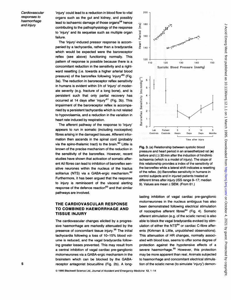
to 'injury' and its sequelae such as multiple organfailure.The 'injury'-induced pressor response is accom-
panied by a tachycardia, rather than a bradycardiawhich would be expected were the baroreceptorreflex (see above) functioning normally. Thispattern of response is possible because there is aconcomitant reduction in the sensitivity and a right-ward resetting (i.e. towards a higher arterial bloodpressure) of the baroreflex following 'injury'60 (Fig.3a). The reduction in baroreceptor reflex sensitivityin humans is evident within 3 h of 'injury' of moder-ate severity (e.g. fracture of a long bone), and ispersistent such that only partial recovery hasoccurred at 14 days after 'injury'61 (Fig. 3b). Thisimpairment of the baroreceptor reflex is accompa-nied by a persistent tachycardia which is not relatedto hypovolaemia, and a reduction in the variation inheart rate induced by respiration.The afferent pathway of the response to 'injury'
appears to run in somatic (including nociceptive)fibres arising in the damaged tissues. Afferent infor-mation then ascends in the spinal cord (probablyvia the spino-thalamic tract) to the brain.60 Little isknown of the precise mechanism of the reduction inthe sensitivity of the baroreflex. However, recentstudies have shown that activation of somatic affer-ent AB fibres can lead to inhibition of baroreflex sen-sitive neurones within the nucleus of the tractussolitarius (NTS) via a GABA-ergic mechanism.62Furthermore, it has been argued that the responseto injury is reminiscent of the visceral alertingresponse of the defence reaction63 and that similarpathways are involved.
THE CARDIOVASCULAR RESPONSETO COMBINED HAEMORRHAGE ANDTISSUE INJURY
The cardiovascular changes elicited by a progres-sive haemorrhage are markedly attenuated by thepresence of concomitant tissue injury.35 The initialtachycardia following a loss of 10-15% blood vol-ume is reduced, and the vagal bradycardia follow-ing greater losses prevented. This may result froma central inhibition of vagal cardiac pre-ganglionicmotorneurones via a GABA-ergic mechanism in thebrainstem which can be blocked by the GABA-
5 receptor antagonist bicucuilline (Fig. 2b). A long-
200 -
I.
~ ~ ~ ~ 7
oa
00
10}.' co ,
* Wood Pressure
Iv ..
s. a;
fn
r
0J* i;rt : ;.: i- ! st 7 tq r,"'-v | C ; J 9 ' ; ) > 4 ' ; | 4 }i-y .li; < 9 +, v i^ ; r<; t i; 1 . Iir:>
:.
L Pam 5 3 14 50 Ilpi 0tWSr&, 400 Days -Das MenUs
Fig. 3. (a) Relationship between systolic bloodpressure and heart period in an anaesthetized rat (-)before and (o) 30 min after the induction of hindlimbischaemia (which is a model of injury). The slope ofthis relationship provides a index of the sensitivity ofthe baroreflex while a lateral shift indicates a resettingof the reflex. (b) Baroreflex sensitivity in humans incontrol subjects and in injured patients treated atdifferent times after injury (ISS range 9-17; median9). Values are mean ± SEM. (From 61.)
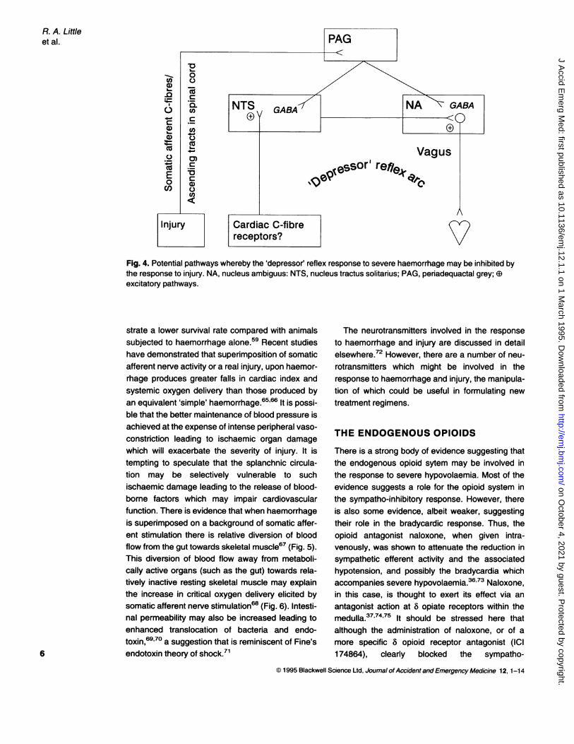
lasting inhibition of vagal cardiac pre-ganglionicmotorneurones in the nucleus ambiguus has alsobeen demonstrated following electrical stimulationof nociceptive afferent fibres64 (Fig. 4). Somaticafferent stimulation (e.g. of the sciatic nerve) is alsoable to block the vagal bradycardia evoked by stim-ulation of either the NTS64 or cardiac C-fibre affer-ents (Kirkman & Little, unpublished observations).This attenuation of HR changes, normally associ-ated with blood loss, seems to offer some degree ofprotection against the hypotensive effects of asevere haemorrhage.35 However, this protectionmay be more apparent than real. Animals subjectedto haemorrhage and concomitant electrical stimula-tion of the sciatic nerve (to simulate 'injury') demon-
© 1995 Blackwell Science Ltd, Joumal of Accident and Emergency Medicine 12, 1-14
I
IA.
tin.4
.I
on October 4, 2021 by guest. P
rotected by copyright.http://em
j.bmj.com
/J A
ccid Em
erg Med: first published as 10.1136/em
j.12.1.1 on 1 March 1995. D
ownloaded from
_)
4 C)
n- co_ *m
C .'0) (1)L- 4-.
01)
cnO4
enE
COI.
Vagus,eSsor' re /efl
0vo
Fig. 4. Potential pathways whereby the 'depressor' reflex response to severe haemorrhage may be inhibited bythe response to injury. NA, nucleus ambiguus: NTS, nucleus tractus solitarius; PAG, periadequactal grey; @excitatory pathways.
strate a lower survival rate compared with animalssubjected to haemorrhage alone.59 Recent studieshave demonstrated that superimposition of somaticafferent nerve activity or a real injury, upon haemor-rhage produces greater falls in cardiac index andsystemic oxygen delivery than those produced byan equivalent 'simple' haemorrhage.65,66 It is possi-ble that the better maintenance of blood pressure isachieved at the expense of intense peripheral vaso-
constriction leading to ischaemic organ damagewhich will exacerbate the severity of injury. It istempting to speculate that the splanchnic circula-tion may be selectively vulnerable to suchischaemic damage leading to the release of blood-borne factors which may impair cardiovascularfunction. There is evidence that when haemorrhageis superimposed on a background of somatic affer-ent stimulation there is relative diversion of bloodflow from the gut towards skeletal muscle67 (Fig. 5).This diversion of blood flow away from metaboli-cally active organs (such as the gut) towards rela-tively inactive resting skeletal muscle may explainthe increase in critical oxygen delivery elicited bysomatic afferent nerve stimulation68 (Fig. 6). Intesti-nal permeability may also be increased leading toenhanced translocation of bacteria and endo-toxin,69 70 a suggestion that is reminiscent of Fine's
6 endotoxin theory of shock.71
The neurotransmitters involved in the response
to haemorrhage and injury are discussed in detailelsewhere.72 However, there are a number of neu-
rotransmitters which might be involved in theresponse to haemorrhage and injury, the manipula-tion of which could be useful in formulating new
treatment regimens.
THE ENDOGENOUS OPIOIDS
There is a strong body of evidence suggesting thatthe endogenous opioid sytem may be involved inthe response to severe hypovolaemia. Most of theevidence suggests a role for the opioid system inthe sympatho-inhibitory response. However, thereis also some evidence, albeit weaker, suggestingtheir role in the bradycardic response. Thus, theopioid antagonist naloxone, when given intra-venously, was shown to attenuate the reduction insympathetic efferent activity and the associatedhypotension, and possibly the bradycardia whichaccompanies severe hypovolaemia.3673 Naloxone,in this case, is thought to exert its effect via an
antagonist action at 8 opiate receptors within themedulla.37'74'75 It should be stressed here thatalthough the administration of naloxone, or of a
more specific 8 opioid receptor antagonist (ICI174864), clearly blocked the sympatho-
1995 Blackwell Science Ltd, Joumal ofAccident and Emergency Medicine 12, 1-14
R. A. Liffleetal.
on October 4, 2021 by guest. P
rotected by copyright.http://em
j.bmj.com
/J A
ccid Em
erg Med: first published as 10.1136/em
j.12.1.1 on 1 March 1995. D
ownloaded from

fid. bidb.sc*b
emP emulXS @. \+ B' \
0a:.*trfr
.40"
so0-
of.sgAdeta -,oaae-t;i3b on]l1 nwo uoJno'mt tcts t
-s1e. iotqe,3je THZ2 oflI'1 ar4t atalina"t it1 Iin6 I
*i rtl3 bns epnIsd
linS
Fig. 5. Blood flow and oxygen delivery to thesplanchnic and skeletal muscle vascular bedsbefore(C) and after the loss of 30% estimated totalblood volume at 1% min-1 (EH) in two groups ofanaesthetized pigs: those subjected to haemorrhagealone, and those subjected to haemorrhage on
background of somatic afferent stimulation (SANS) as
a model of injury. Values expressed as a percentageof those recorded during the control phase, andpresented as mean ± SEM.
the normal 'physiological' response to severe
haemorrhage75 although the use of ,u receptor ago-nists, e.g. fentanyl and alfentanyl,79 may provide a
pharmacological means of inhibiting the depressoreffect of a severe haemorrhage. Whether thiswould be beneficial or detrimental (bearing in mindthe potential protective effects of this depressorreflex, and the consequences of blocking it)remains to be seen.
Since 'injury' is also known to activate theendogenous opioid system, and cell bodies whichsynthesize and release enkephalins are found inthe peri-aqueductal grey80 (Fig. 4), an area knownto be involved in the cardiovascular response to'injury', it is not surprising to learn that naloxone can
modify the response to 'injury'. Thus, it has beendemonstrated in experimental studies that theadministration of naloxone, either centrally or intra-venously, can prevent or reverse the reduction in
baroreflex sensitivity produced by 'injury'.81 Con-versely, some of the effects of 'injury' on the barore-flex can be mimicked by the central administrationof a long-lasting analogue of met-enkephalin (d-ala2-met5-enkephalinamide).82
Thus, it can be seen that both opioid agonistsand antagonists can modify the cardiovascularresponse to haemorrhage and 'injury'. Clearly, theeffects are complex, emphasizing the complexity of
the underlying neural control mechanisms. Furtherstudies are required to evaluate the role of individ-ual opiate receptor subtypes in these responses,
inhibition,7476 the effects of 8 antagonism on thebradycardia were less clear. This may be because ofthe relatively small bradycardic response to severe
hypovolaemia even in the absence of 8 antagonismin these studies, which were conducted on rabbits.Since 8 opioid receptor antagonism appears to blockboth the vagal and the sympathetic component of theresponse to severe hypovolaemia, it is likely that theendogenous opioids are important early in the reflexpathway, before the two limbs diverge. One likelyarea is the NTS,75 which contains a dense popula-tion of 8 opioid receptors.77'78
In addition to the 8 opioid receptors, the and thereceptors also appear to be capable of modifying
the response to severe hypovolaemia. Thus, andv receptor agonists can also prevent the reflex sym-patho-inhibition (and possibly the bradycardia)seen during severe hypovolaemia.7 76 However, it
7 is unlikely that the ,u opioid receptor participates in
16 -
I
_' 12 -
c._
8-
a
4-
I
Control BrachialStimulation
Fig. 6. Critical oxygen delivery (the point at whichoxygen consumption becomes dependent uponsupply and the tissues become ischaemic) in twogroups of anaesthetized dogs: control, and thosesubjected to somatic afferent (brachial) nerve
stimulation to simulate injury. Values are medians andinterquartile ranges.
nC 1 M99 RiarkwIll RriAnfr I trI .IhimaI nf Arritnnt and Fmornon,v Modkin. 12 1-14
Cardiovascularresponses tohaemorrhageand injury
-
ft
9
on October 4, 2021 by guest. P
rotected by copyright.http://em
j.bmj.com
/J A
ccid Em
erg Med: first published as 10.1136/em
j.12.1.1 on 1 March 1995. D
ownloaded from

Fig. 7. Effects of a progressivesimple haemorrhage (2%estimated total bloodvolume min-1) on heart rate in twogroups of anaesthetized ratstreated with either saline (-), or
fI.THOUT methiothepin (o) injected into the9 fourth cerebral ventricle. A third
group was treated withmethiothepin but not subjected tohaemorrhage (o). Values aremean ± SEM.
* 0|t -§'' 10' 20 30 40
emorq'gs {@(ZBVI
and the resultant haemodynamic changes and theirconsequences evaluated before any attempt attreatment are made with these agents.
5-HYDROXYTRYPTAMINE
There have been suggestions that this agent maybe involved in the central nervous pathways medi-ating the response to severe haemorrhage. Thus,Bogle et al.83 demonstrated that a response whichincludes the 'cardiac reflex' elicited by activation ofcardiac-vagal C-fibre afferents could be blocked bymethiothepin (a 5HT receptor antagonist) givencentrally. Furthermore, blockade of the 5-HT sys-tem, either with p-chlorophenylalanine (PCPA,which blocks the synthesis of 5-HT), or with the 5-HT receptor antagonist, methysergide, has beenreported to prevent or reverse both the sympatho-inhibitory and the bradycardic response to severeblood loss, while leaving baroreflex control intact.84Although the 5-HT 'blocking' agents were givenintravenously in this latter study, their sites of actionare likely to be central, since any effect of 5-HT onthe ventricular receptors is mediated via 5-HT3receptors,85 where methysergide has little activ-ity.86
However, the effects of 5HT 'blockade' on theresponse to severe blood loss are equivocal, sinceother studies have failed to show that PCPA blocksthe depressor response to severe haemorrhage87and have suggested that the action of agents, e.g.methysergide, do not occur because of blockade of5HT but rather because of agonism at 5HT recep-tors.88 Thus, Evans et al.88 concluded that therewas no evidence of a physiological role for 5HT in
the depressor response to severe haemorrhage.Recent data from our own laboratories indicate thatthe 5HT receptor antagonist methiothepin wasunable to attenuate the reflex bradycardia whichaccompanies severe haemorrhage89 (Fig. 7) unlikeits effects on the 'cardiac reflex', suggesting that thetwo responses may be mediated via different cen-tral nervous pathways. Indeed, our studies indicatethat 5HT antagonism may potentiate the depressorresponse to severe haemorrhage. Thus, our find-ings support and extend those of Evans et al.;88activation of central 5HT receptors does not appearnecessary for the mediation of the depressorresponse associated with severe haemorrhage. Incontrast 5HT could have a 'physiological' role inproviding a tonic inhibition of this response.Thus, 5-HT and its receptors may provide
another avenue for pharmacological manipulationof the response to haemorrhage and injury,although, because of the complexity of the system,this possibility lies some way ahead.
EFFECTS OF ETHANOL
Ethanol is a drug taken socially, often precedingtraumatic events, e.g. automobile accidents. Theeffects of ethanol on the cardiovascular response tohaemorrhage and 'injury' are therefore of interest.Recent studies have shown that moderately raisedblood levels of ethanol (100-200 mg%) can exacer-bate the 'injury'-induced reduction in baroreflexsensitivity.90 Explaining the action of ethanol on thecardiovascular response to injury is potentially sim-ple. It has been reported that ethanol may potenti-ate the effects of endogenous GABA at both the
1995 Blackwell Science Ltd, Journal ofAccident and Emergency Medicine 12, 1-14
R. A. Littleetal.
8
.1 N.B. MatMothopin Ai
i. hato-z.:.:. .... .... ....
.4
on October 4, 2021 by guest. P
rotected by copyright.http://em
j.bmj.com
/J A
ccid Em
erg Med: first published as 10.1136/em
j.12.1.1 on 1 March 1995. D
ownloaded from

Cardiovascularresponses tohaemorrhageand injury
NTS and the vagal outflow nuclei.91 Since theresponse to injury is thought to inhibit the baroreflexvia a GABA-ergic mechanism within the brainstemnuclei, it is hardly surprising that ethanol augmentsthe baroreflex-inhibitory effects of jury.
rhage in the anaesthetized pig.93 Thus Si seems tobe a simple, non-invasive means of evaluating car-diac function during acute hypovolaemia.
STROKE DISTANCE
SHOCK INDEX
The obvious shortcomings of blood pressure andHR for assessing blood loss from the circulationstimulated the development of the Shock Index (Si- calculated by dividing HR by the systolic bloodpressure).92 It was predicted that combining thechanges in HR and blood pressure would give amore sensitivie indication of blood loss and a lowflow state than either variable used alone. It wasfound that there was a direct relationship betweenSI and the magnitude of blood loss in a group ofpatients with either gastro-intestinal bleeding, openwounds or intra-abdominal thoracic bleeding follow-ing blunt trauma.92 Our own studies have confirmedthe value of SI and suggested that it may be a moresensitive indicator of physiological status afterinjury than the more commonly used RTS. Thus, ina group of 160 trauma patients with a normal RTS,69 had an abnormal Sl.93Shock Index (SI) has been further validated in a
number of experimental studies. For example,despite the markedly biphasic HR response to sim-ple haemorrhage in the rat, a progressive increasein SI was found as the magnitude of blood lossincreased93 (Fig. 8). A negative relationshipbetween Si and both left ventricular stroke volumeand cardiac output has been shown during haemor-
14
12r-i
C. S. .1 . i .nsr n.k . .....s. ......----Ef --m
Another non-invasive assessment of left ventricularstroke volume (LVSV) can be obtained by the mea-surement of stroke distance (SD) using continuouswave Doppler ultrasound via an ultrasonic probeplaced in the suprasternal notch.94 Technically, thisis a very simple procedure requiring no more train-ing than that needed to record blood pressure bysphygnomanometry. In a recent study in our labora-tory, blood loss was simulated by pooling blood inthe lower body using a negative pressurechamber.95 A progressive haemorrhage was simu-lated by progressively increasing lower body nega-tive pressure (LBNP). Mean arterial blood pressuredid not change significantly at any level of LBNP(Fig. 9), unless the subject became pre-syncopal(Fig. 1 Oa,b). Overall, pulse rate and shock index didnot change at the first level of LBNP, but didincrease progressively thereafter (Fig. 10) until pre-syncope, which occurred in a few subjects, where-upon HR fell dramatically and SI either continued torise (Fig. 1 Oa) or fell slightly (Fig. 1 Ob) when thebradycardia and hypotension were extreme. Bycontrast SD fell significantly at each level of LBNP(Figs 9 & 10). However, with very marked reduc-tions in HR there was a small increase in stroke dis-tance, presumably related to improved cardiacfilling as a result of the increased diastolic fillingtime. Thus, SD provides an effective early indicator
Fig. 8. Effect of progressivehaemorrhage (2% estimated totalblood volume min-1 on shockindex in two groups ofanaesthetized rats: (-) simplehaemorrhage, (o) haemorrhageon the background of bilateralhind-limb ischaemia ('injury'). ForHR data for the same animals, seeFig. 2a. Values are mean ± SEM.
.`
"e .ybo8
).- .$Vii
Vol Haemorrhage (XBV)1995 Blackwell Science Ltd, Joumal ofAccident and Emergency Medicine 12, 1-14
9
on October 4, 2021 by guest. P
rotected by copyright.http://em
j.bmj.com
/J A
ccid Em
erg Med: first published as 10.1136/em
j.12.1.1 on 1 March 1995. D
ownloaded from

110
100 -
2
> 80-
70 -
90 -
T 80-
E 70
m 60-
50 -
6 -
0.8
9-
I'o 0.7-=EE
0.6-E- 0.5 -
0.4 -
30 -
25
00a-20-
cn
15 -
1n_%-
0 10 20 30 40 50Lower Body Suction (mmHg)
0 10 20 30 40 50Lower Body Suction (mmHg)
Fig. 9. Effects of progressive lower body suction on mean arterial blood pressure (MBP), heart rate (HR), centralvenous pressure (CVP), shock index (SI) and stroke distance (SD) in eight healthy volunteers. Presyncopal datawere excluded. Values are mean ± SEM.
80 -
= E.8 40120100 -
, 8s0 -
E 60-- 40 -
20 -
T0.8-E 0.7 -
_nE'I 0.6j.ie 0.5
25 -
- 20 -02E(flu
15 -
10 --
0 10 20 30 40 50
Lower Body Suction (mmHg)
_- I
0 10 20 30 40 50
Lower Body Suction (mmHg)
Fig. 10. Effects of progressive lower body suction on heart rate (HR), mean arterial blood pressure (MBP), shockindex (SI) and stroke distance (SD) in two healthy male volunteers (a,b) who became presyncopal during suction
10 at 50 mmHg.
1995 Blackwell Science Ltd, Journal ofAccident and-Emergency Medicine 12, 1-14
R. A. Littleetal.
E
ECL
4 -
2 -
0--2
100 -
X~CZ 801.060
100 -
80 -I2 E 60 1
40 -
T- 1.5 -cm
EU; E 1.0_-
'E_= 0.5 -
20
-15aM E 1
10
5
I I } -AI
0- -
on October 4, 2021 by guest. P
rotected by copyright.http://em
j.bmj.com
/J A
ccid Em
erg Med: first published as 10.1136/em
j.12.1.1 on 1 March 1995. D
ownloaded from
Cardiovascularresponses tohaemorrhageand injury
of reductions in LVSV as a consequence ofreduced central blood volume and venous return.These changes in SD can be detected before alter-ations in the classical vital signs - HR and bloodpressure.
CONCLUSIONS
An increase in the understanding of cardiovascularresponses to haemorrhage and injury will improvethe interpretation of the injured patient's vital signs.The introduction of both the use of Si and measure-ments of stroke distance will provide simple, non-invasive means for detecting changes in cardiacoutput. Such developments will lead to a reductionin preventable trauma deaths.
REFERENCES
1. Cales R.H. & Trunkey D.D. (1985) Preventabletrauma deaths a review of trauma care system devel-opment. Journal of the American Medical Association254, 1059-1063.
2. West J.G., Trunkey D.D. & Lim R.C. (1979) Systemsof trauma care. Study of two countries. Archives ofSurgery 114,455-460.
3. Anderson I.D., Woodford M., DeDombal F.T. & IrvingM. (1988) Retrospective study of 1000 deaths frominjury in England and Wales. British Medical Joumal296, 1305-1308.
4. Stochetti N., Pagliarini G., Gennari M., Baldi G., Ban-chini E., Campari M., Bacchi M. & Zuccoli P. (1994)Trauma care in Italy: evidence of in-hospital pre-ventable deaths. Journal of Trauma 356, 401-405.
5. American College of Surgeons Committee onTrauma (1993) Advanced Trauma Life SupportCourse. American College of Surgeons, Chicago.
6. Champion H.R., Sacco W.J., Copes W.S., GannD.S., Gennarelli T.A. & Flanagan W.E. (1989) A revi-sion of trauma score. Journal of Trauma 29,623-629.
7. Boyd C.R., Tolson M.A. & Copes W.S. (1987) Evalu-ating trauma care: the TRISS method. Journal ofTrauma 27, 370-378.
8. Cowell E.M. (1919) The initiation of wound shock.Special report series. Medical Research Commit-tee 25, 99-108.
9. Robertson O.H. & Bock A.V. (1919) Memorandum onblood volume after haemorrhage. Special reportseries. Medical Research Committee 25, 213-244.
10. Fraser J. & Cowell E.M. (1919) A clinical study of theblood pressure in wound conditions. Special reportseries. Medical Research Committee 25, 49-71.
11. Grant R.T. & Reeve E.B. (1941) Clinical observationson air-raid casualties. British Medical Joumal 2,293-297.
12. Grant R.T. & Reeve E.B. (1951) Observations on thegeneral effects of injury in man with special referenceto wound shock. Special report series. MedicalResearch Committee 277.
13. Beecher H.K. (ed.) (1952) The Physiologic Effects ofWounds pp. 34-35. Board for the Study of theSeverely Wounded, The Medical Department of theUnited States Army, Washington D.C.
14. Keith N.M. (1919) Blood volume changes in woundshock and primary haemorrhage. Special reportseries. Medical Research Committee 27, 3-16.
15. Evans E.l., Hoover M.J., James G.W. & Alm T. (1994)Studies on traumatic shock: I - Blood volumechanges in traumatic shock. Annals of Surgery 119,64-85.
16. Emerson C.P. & Ebert R.V. (1945) A study of shock inbattle casulaties. Annals of Surgery 122, 745-772.
17. Ebert R.V., Stead E.A. & Gibson J.G. (1941)Response of normal subjects to acute blood loss.Archives of Intemal Medicine 68, 578-590.
18. Shenkin H.A., Chenery R.H., Govons S.R., HardyJ.D. & Fletcher A.G. (1944) On the diagnosis ofhaemorrhage in man. American Journal of MedicalScience 208, 421-436.
19. Barcroft H. & Edholm O.G. (1945) On the vasodilata-tion in human skeletal muscle during part haemor-rhagic fainting. Journal of Physiology 104, 161-175.
20. Secher N.H. & Bie P. (1985) Bradycardia duringreversible haemorrhagic shock - a forgotten observa-tion. Clinical Physiology 5, 315-323.
21. Secher N.H., Jensen K.S., Werner C., Warberg J. &Bie P. (1984) Bradycardia during severe butreversible hypovolemic shock in man. CirculatoryShock 14, 267-274.
22. Sander-Jensen K., Secher N.H., Bie P., Warberg J. &Schwartz T.W. (1986) Vagal slowing of the heart dur-ing haemorrhage: observations from 20 consecutivehypotensive patients. British Medical Joumal 292,364-366.
23. Driscoll P. (1994) MD Thesis (submitted). Universityof Leeds.
24. Cowley A.W., Liard J.F. & Guyton A.C. (1973) Role ofbaroreceptor reflex in daily control of arterial bloodpressure and other variables in dogs. CirculationResearch 32, 564-576.
25. Angell-James J.E. (1971) The effects of changes inextramural, 'intrathoracic', pressure on aortic-archbaroreceptors. Joumal of Physiology 214, 89-103.
26. Angell-James J.E. & Daly M.de B. (1970) Comparisonof the reflex vasomotor responses to separate andcombined stimulation of the carotid sinus and aorticarch baroreceptors by pulsatile and non-pulsatilepressures in the dog. Journal of Physiology 209,257-293.
27. Kirchheim H. (1976) Systemic arterial baroreceptorreflexes. Physiological Reviews 56, 100-177.
28. Little R.A., Randall P.E., Redfern W.S., Stoner H.B. &11
© 1995 Blackwell Science Ltd, Joumal ofAccident and Emergency Medicine 12, 1-14
on October 4, 2021 by guest. P
rotected by copyright.http://em
j.bmj.com
/J A
ccid Em
erg Med: first published as 10.1136/em
j.12.1.1 on 1 March 1995. D
ownloaded from
R. A. Littleetal.
Marshall H.W. (1984) Components of injury (haemor-rhage and tissue ischaemia) affecting cardiovascularreflexes in man and rat. Quarterly Journal of Experi-mental Physiology 69, 753-762.
29. Kirkman E. & Scott E.M. (1983) The effect of changesin plasma renin activity on the baroreceptor reflex arcin the cat. Joumal of Physiology 342, 73-74.
30. Cowley A.W., Merril D., Osborn J. & Barber B.J.(1984) Influence of vasopressin and angiotensin onbaroreflex in the dog. Circulation Research 54,163-172.
31. Baker D.G., Coleridge H.M. & Coleridge J.C.G.(1979) Vagal afferent C-fibres from the ventricle. In:Hainsworth R., Kidd C. & Linden R.J. (eds) CardiacReceptors, pp. 117-137. Cambridge UniversityPress, Cambridge.
32. Oberg B. & Thor6n P. (1973) Circulatory response tostimulation of left ventricular receptors in the cat. ActaPhysiologica Scandinavica 88, 8-22.
33. Daly M.de B., Kirkman E. & Wood L.M. (1988) Cardio-vascular responses to stimulation of cardiac recep-tors in the cat and their modification by changes inrespiration. Journal of Physiology 407, 349-362.
34. Oberg B. & Thoren P. (1972) Increased activity in leftventricular receptors during haemorrhage on occlu-sion of the caval veins in the cat. A possible cause ofthe vasovagal reaction. Acta Physiologica Scandi-navica 85, 164-173.
35. Little R.A., Marshall H.W. & Kirkman E. (1989) Atten-uation of the acute cardiovascular responses tohaemorrhage by tissue injury in the conscious rat.Quarterly Journal of Experimental Physiology 74,825-833.
86. Burke S.L. & Dorward P.K. (1988) Influence ofendogenous opiates and cardiac baroreceptors onrenal nerve activity during haemorrhage in consciousrabbits. Journal of Physiology 402, 9-27.
37. Evans R.G., Ludbrook J. & Potocnik S.J. (1989)Intracisternal naloxone and cardiac nerve blockadeprevent vasodilatation during simulated haemorrhagein awake rabbits. Journal of Physiology 409, 1-14.
38. Hayes I.P., Evans R.G. & Ludbrook J. (1992) Pre-sumptive pulmonary neural blockade followingintrapericardial procaine in conscious rabbits. Pro-ceedings of the Australian Physiological Society 23,66.
39. Shen Y.T., Knight D.R., Thomas J.X. & Vatner S.F.(1990) Relative roles of cardiac receptors and arterialbaroreceptors during haemorrhage in consciousdogs. Circulation Research 66, 397-405.
40. Shen Y.T., Cowley A.W. & Vatner S.F. (1991) Rela-tive roles of cardiac and arterial baroreceptors invasopressin regulation during haemorrhage in con-scious dogs. Circulation Research 68, 1422-1436.
41. Scherrer U., Vissing S., Morgan B., Hanson P. &Victor R.G. (1990) Vasovagal syncope after infusionof a vasodilator in a heart-transplant recipient. New
England Joumal of Medicine 322, 602-604.42. Lewis T. (1932) Vasovagal syncope and the carotid
sinus mechanism. British Medical Journal i, 873-876.43. Barcroft H., Edholm O.G., McMichael J. & Sharpey-
Schafer E.P. (1944) Posthaemorrhagic fainting.Study by cardiac output and forearm flow. Lancet i,489-491.
44. Barriot P., Riou B. & Buffat J.-J. (1987) Pre-hospitalmanagement of severe haemorrhagic shock. In:Vincent J.L. (ed.) Update in Intensive Care and Emer-gency Medicine, Vol. 3, pp. 377-384. Springer-Verlag, Berlin.
45. Biscoe T.J., Purves M.J. & Sampson S.R. (1970) Thefrequency of nerve impulses in single carotid bodychemoreceptor afferent fibres recorded in vivo withintact circulation. Joumal of Physiological 208,121-131.
46. Heymans C. & Neil E. (1958) Reflexogenic Areas ofthe Cardiovascular System. Churchill Livingstone,London.
47. Daly M.de B. (1983) Peripheral arterial chemorecep-tors and the cardiovascular system. In: Acker H. &O'Regan R.G. (eds) Physiology of the PeripheralArterial Chemoreceptors, pp. 325-393. Elsevier Sci-ence Publishers, Amsterdam.
48. Spyer K.M. (1984) Central control of the cardiovascu-lar system. Recent Advances in Physiology 10,163-200.
49. Daly M.de B., Lambertsen C.J. & Schweitzer A.(1954) Observations on the volume of blood flow andoxygen utilization of the carotid body in the cat. Jour-nal of Physiology 125, 67-89.
50. Acker H. & O'Regan R.G. (1981) The effects of stimu-lation of autonomic nerves on carotid body blood flowin the cat. Journal of Physiology 315, 99-1 10.
51. Potter E.K. & McCloskey D.l. (1987) Excitation ofcarotid body chemoreceptors by neuropeptide-Y.Repsiratory Physiology 67, 357-365.
52. Kenney R.A. & Neil E. (1951) The contribution of aor-tic chemoreceptor mechanisms to the maintenanceof arterial blood pressure of cats and dogs afterhaemorrhage. Joumal of Physiology 112, 223-228.
53. D'Silva J.L., Gill D. & Mendel D. (1966) The effects ofacute haemorrhage on respiration in the cat. Joumalof Physiology 187, 369-377.
54. Angell-James J.E. & Daly M.deB. (1975) Someaspects of upper respiratory tract reflexes. Acta Oto-larnygologica 79, 242-252.
55. Alam M. & Smirk F.H. (1937) Observations in manupon a blood pressure raising reflex arising from thevoluntary muscles. Journal of Physiology 89,372-383.
56. Alam M. & Smirk F.H. (1938) Observations in manupon a pulse accelerating reflex from the voluntarymuscles of the legs. Journal of Physiology 92,167-177.
57. Howard J.M., Artz C.P. & Stahl R.R. (1955) The12
© 1995 Blackwell Science Ltd, Journal ofAccident and Emergency Medicine 12, 1-14
on October 4, 2021 by guest. P
rotected by copyright.http://em
j.bmj.com
/J A
ccid Em
erg Med: first published as 10.1136/em
j.12.1.1 on 1 March 1995. D
ownloaded from

Cardiovascularresponses tohaemorrhageand injury
hypertensive response to injury. Annals ofSurgery 141, 327-336.
58. Redfern W.S. (1981) Effects of limb ischaemia on thecardiac component of the baroreceptor reflex in theunanaesthetized rat - afferent, central and efferentmechanisms. PhD Thesis, University of Manchester.
59. Overman R.R. & Wang S.C. (1947) The contributoryrole of the afferent nervous factor in experimentalshock: sublethal haemorrhage and sciatic nerve stim-ulation. American Journal of Physiology 148,289-295.
60. Redfern W.S., Little R.A., Stoner H.B. & MarshallH.W. (1984) Effect of limb ischaemia on blood pres-sure and the blood pressure-heart rate reflex in therat. Quarterly Journal of Experimental Physiology 69,763-769.
61. Anderson I.D., Little R.A. & Irving M.H. (1990) Aneffect of trauma on human cardiovascular control:baroreflex suppression. Joumal of Trauma 30,974-982.
62. McMahon S.E., McWilliam P.N., Robertson J. & KayeJ.C. (1992) Inhibition of carotid sinus baroreceptorneurones in the nucleus tractus solitarius of theanaesthetized cat by electrical stimulation of hindlimbafferent fibres. Journal of Physiology 452, 224.
63. Quest J.A. & Gebber G.L. (1972) Modulation ofbaroreceptor reflexes by somatic afferent nerve stim-ulation. American Journal of Physiology 222,1251-1 259.
64. Wang Q., Guo X.-Q. & Li P. (1988) The inhibitoryeffects of somatic input on the excitatory responsesof vagal cardiomotor neurones to stimulation of thenucleus tractus solitarius in rabbits. BrainResearch 439, 350-353.
65. Rady M.Y.A., Kirkman E., Cranley J.J. & Little R.A.(1993) A comparison of the effects of skeletal muscleinjury and somatic afferent nerve stimulation on theresponse to hemorrhage in anesthetized pigs. Jour-nal of Trauma 35, 756-761.
66. Rady M.Y.A., Little R.A., Edwards J.D., Kirkman E. &Faithfull S. (1991) The effect of nociceptive stimula-tion on the changes in hemodynamics and oxygentransport induced by hemorrhage in anesthetizedpigs. Journal of Trauma 31, 617-621.
67. Mackway-Jones K., Foex B., Kirkman E. & Little R.A.(1994) Modification of the haemodynamic responseto blood loss by somatic afferent stimulation. Shock 2(Supplement), 17.
68. Kirkman E., Zhang H., Sapen H., Vincent J.L. & LittleR.A. (1994) Effects of 'injury' on critical oxygen deliv-ery: a potential haemodynamic mechanism? Shock 2(Supplement), 16.
69. Wilmore D.W., Smith R.J., O'Dwyer S.T., JackobsD.O., Ziegler T.R. & Wang X.-D. (1988) The gut: acentral organ after surgical stress. Surgery 104,917-923.
70. Deitch E.A. (1990) Intestinal permeability is increased
in burn patients shortly after injury. Surgery 107,411-416.
71. Fine J. (1961) Endotoxins in traumatic shock. Federa-tion Proceedings 20, Supplement 9, 166-172.
72. Kirkman E. & Little R.A. (1994) Cardiovascular regu-lation during hypovolaemic shock - central integra-tion. In: Secher N.H., Pawelczyk J.A. & Ludbrook J.(eds) Blood Loss and Shock, pp. 61-75. EdwardArnold, London.
73. Ludbrook J. & Rutter P.C. (1988) Effect of naloxoneon haemodynamic responses to acute blood loss inunanaesthetized rabbits. Journal of Physiology 400,1-14.
74. Evans R.G., Ludbrook J. & van Leeuwen A.F. (1989)Role of central opriate receptor subtypes in the circu-latory responses of awake rabbits to graded cavalocclusions. Joumal of Physiology 419, 15-31.
75. Evans R.G. & Ludbrook J. (1990) Effects of mu-opioidreceptor agonists on circulatory responses to simu-lated haemorrhage in conscious rabbits. British Jour-nal of Pharmacology 100, 421-426.
76. Evans R.G. & Ludbrook J. (1991) Chemosensitivecardiopulmonary afferents and the haemodynamicresponse to simulated haemorrhage in consciousrabbits. British Journal of Pharmacology 102,533-539.
77. Dashwood M.R., Muddle J.R. & Spyer K.M. (1988)Opiate receptor subtypes in the nucleus tractus soli-tarii of the cat: the effect of vagal section. EuropeanJournal of Pharmacology 155, 85-92.
78. May C.N., Dashwood M.R., Whitehead C.J. & Math-ias C.J. (1989) Differential cardiovascular and respi-ratory responses to central administration of selectiveopioid agonists in conscious rabbits: correlation withreceptor distribution. British Journal of Pharmacology98, 903-913.
79. Schadt J.C. & Ludbrook J. (1991) Hemodynamic andneurohumoral responses to acute hypovolemia inconscious mammals. American Journal of Physiology260, H305-318.
80. Uhl G.R., Goodman R.R., Kuhar M.J., Childers S.R. &Snyder S.H. (1979) Immunohistochemical mappingof enkephalin containing cell bodies, fibres and nerve
terminals in the brain stem of the rat. BrainResearch 166, 75-94.
81. Eltrafi A., Kirkman E. & Little R.A. (1989) Reversal ofthe injury induced reduction in baroreflex sensitivityby naloxone in the conscious rat. British Journal ofPharmacology 96, 145.
82. Little R.A., Jones R.O. & Eltraifi A.E. (1988) Cardio-vascular reflex function after injury. In: Bond R.F.,Adams H.R. & Chaudry I.H. (eds) Perspectives in
Shock Research. Progress in Clinical and BiologicalResearch, pp. 191-200. Liss, New York.
83. Bogle R.G., Pires J.G. & Ramage A.G. (1990) Evi-dence that central 5-HT1 A-receptors play a role in the13
© 1995 Blackwell Science Ltd, Joumal of Accident and Emergency Medicine 12, 1-14
on October 4, 2021 by guest. P
rotected by copyright.http://em
j.bmj.com
/J A
ccid Em
erg Med: first published as 10.1136/em
j.12.1.1 on 1 March 1995. D
ownloaded from

R. A. Littleet al.
von Bezold-Jarisch reflex in the rat. British Journal ofPharmacology 100, 757-760.
84. Morgan D.A., Thoren P., Wilczynski E.A., Victor R.G.& Mark A.L. (1988) Serotonergic mechanisms medi-ate renal sympathoinhibition during severe hemor-rhage in rats. American Journal of Physiology 255,H496-502.
85. Kay I.S. & Armstrong D.J. (1990) Phenylbiguanidenot phenyldiguanide is used to evoke the pulmonarychemoreflex in anaesthetized rabbits. ExperimentalPhysiology 75, 383-389.
86. Peroutka S.J. (1988) 5-Hydroxytryptamine receptorsubtypes: molecular, biochemical and physiologicalcharacterization. Trends in Neuroscience 11,496-500.
87. Evans R.G., Kapoor V. & Ludbrook J. (1992) A CNSserotonergic mechanism in acute centralhypvolaemia in conscious rabbits? Journal of Cardio-vascular Pharmacology 19, 1009-1017.
88. Evans R.G., Haynes J.M. & Ludbrook J. (1993)Effects of 5-HT-receptor and a2-adrenoceptor ligandson the haemodynamic response to acute centralhypovolaemia in conscious rabbits. British Joumal ofPharmacology 109, 37-47.
89. Kirkman E., Shiozaki T. & Little R.A. (1994) Methio-thepin does not attenuate the bradycardia associatedwith severe haemorrhage in the anaesthetized rat.British Journal of Pharmacology 112, 1 1 1.
90. Kirkman E., Marshall H.W., Banks J.R. & Little R.A.(1994) Ethanol augments the baroreflex inhibitoryeffects of sciatic nerve stimulation in the anaes-thetized dog. Experimental Physiology 79, 81-91.
91. Varga K. & Kunos G. (1990) Ethanol inhibition ofbaroreflex bradycardia: role of brainstem GABAreceptors. British Journal of Pharmacology 101,773-775.
92. Allgower M. & Burri C. (1967) Shock Index. DeutscheMadijinische Wochenschrift43, 1948-1950.
93. Little R.A., Gorman D. & Allgower M. (1990) Theshock index revisited. In: Vincent J.L. (ed.) Update inIntensive Care and Emergency Medicine, Vol. 10, pp.505-513. Springer-Verlag, Berlin.
94. Rawles J.M. (1989) Stroke distance - an improvedmeasure of cardiovascular function. Scottish MedicalJournal 34, 394-398.
95. Hoeyweghen R.S., Hanson J.M., Kirkman E., HoranM.A. & Little R.A. (1995) Cardiovascular response tograded lower body negative pressure in young andelderly man. Journal of Physiology 479, 14.
14
© 1995 Blackwell Science Ltd, Joumal ofAccident and Emergency Medicine 12, 1-14
on October 4, 2021 by guest. P
rotected by copyright.http://em
j.bmj.com
/J A
ccid Em
erg Med: first published as 10.1136/em
j.12.1.1 on 1 March 1995. D
ownloaded from