Embed Size (px)
Citation preview
7/29/2019 Prevalence of left heart contrast in healthy, young, asymptomatic humans at rest breathing room air
http://slidepdf.com/reader/full/prevalence-of-left-heart-contrast-in-healthy-young-asymptomatic-humans-at 1/8
Respiratory Physiology & Neurobiology 188 (2013) 71–78
Contents lists available at SciVerse ScienceDirect
Respiratory Physiology & Neurobiology
journa l homepage: www.elsevier .com/ locate / resphysiol
Prevalence of left heart contrast in healthy, young, asymptomatic humans at restbreathing room air
Jonathan E. Elliott a, S. Milind Nigam a, Steven S. Laurie a, Kara M. Beasley a, Randall D. Goodmanb, Jerold A. Hawn a,b, Igor M. Gladstonea,c, Mark S. Chesnuttd, Andrew T. Loveringa,∗
a University of Oreg on,Department of Human Physiology, Eugene, OR,USAb OregonHeart andVascular Institute, RiverBend, Springfield, OR, USAc Department o f Pediatr ics, OregonHealth& Science University,Portland, OR,USAd Dotter Interventional Institute’sHereditary Hemorrhagic Telangiectasia Centerof Excellence,Oregon Health & Science University, Portland, OR, USA
a r t i c l e i n f o
Article history:
Accepted 23 April 2013
Keywords:
Transthoracic saline contrast
echocardiography
Patent foramen ovale
Right-to-left shunt
Intrapulmonary arteriovenous anastomoses
a b s t r a c t
Our purpose was to report the prevalence of healthy, young, asymptomatic humans who demonstrate left
heart contrast at rest, breathing room air. We evaluated 176 subjects (18–41 years old) using transtho-
racic saline contrast echocardiography. Left heart contrast appearing ≤3 cardiac cycles, consistent with
a patent foramen ovale (PFO), was detected in 67 (38%) subjects. Left heart contrast appearing >3 car-
diac cycles, consistent with the transpulmonary passage of contrast, was detected in 49 (28%) subjects.
Of these 49 subjects, 31 were re-evaluated after breathing 100% O2 for 10–15min and 6 (19%) contin-
ued to demonstrate the transpulmonary passage of contrast. Additionally, 18 of these 49 subjects were
re-evaluated in the upright position and 1 (5%) continued to demonstrate the transpulmonary passage
of contrast. These data suggest that ∼30% of healthy, young, asymptomatic subjects demonstrate the
transpulmonary passage of contrast at rest which is reduced by breathing 100% O2 and assuming an
upright body position.
© 2013 Elsevier B.V. All rights reserved.
1. Introduction
In the last decade, a number of studies using intravenously
injected contrast in combination with echocardiography have
investigated the relationships between the transpulmonary pas-
sage of contrast during exercise (and other conditions) and several
physiologically important functions, including pulmonary gas
exchange efficiency (Lovering et al., 2008a; Stickland and Lovering,
2006; Stickland et al., 2004, 2006) and pulmonary pressure reg-
ulation (La Gerche et al., 2010; Lalande et al., 2012; Stickland
et al., 2004). The majority, but not all, of these studies (Eldridge
et al., 2004; Elliott et al., 2011; Kennedy et al., 2012; Laurie et al.,
2012, 2010; Lovering et al., 2008a,b; Stickland et al., 2004) using
intravenously injected contrast sought to exclude subjects for the
presence of an intracardiac pathway (i.e. PFO) because it is known
that the prevalence of PFO has been reported in various popu-
lations to be ∼25–40% and therefore should be a fairly common
∗ Corresponding author at: Department of Human Physiology, University of Ore-
gon, 1240Universityof Oregon, Eugene,OR 97403-1240,USA.Tel.:+1 5413460831;
fax: +1 541 346 2841.
E-mail addresses: [email protected], [email protected] (A.T. Lovering).
finding (Hagenet al., 1984;Marriott etal.,2013;Woods etal., 2010).
Neglecting to exclude subjects for a PFO could result in ambigu-
ous results as one would be unable to determine if the presence
of left heart contrast was the result of contrast passage via a PFO
or via an intrapulmonary pathway. Thus, excluding subjects with
a PFO would ensure that any potential relationships between the
transpulmonary passage of contrast and the physiological variable
in question would be specific to contrasttraveling through an intra-
pulmonary pathway, rather than a PFO.
Less well accepted is a rationale for screeningand eitherinclud-
ing or excluding subjects without PFO but who demonstrate the
transpulmonary passage of contrastat rest breathing room air prior
to any intervention. Clearly, if subjects demonstrate the transpul-
monary passage of contrast prior to the intervention (i.e. while at
rest breathing room air) there is no baseline without the transpul-
monary passage of contrast on which to form conclusions. Hence,
this is onerationale forscreening subjects andexcludingthosewho
demonstrate thetranspulmonary passage of contrastat restbreath-
ing room air. Recent work by Woods et al. is to our knowledge
the only report of the prevalence of the transpulmonary passage
of saline contrast in humans at rest (Woods et al., 2010). In this
study it was determined that 29/104 (28%) subjects demonstrated
the transpulmonary passage of contrast at rest, however this was
1569-9048/$ – seefrontmatter © 2013 Elsevier B.V. All rights reserved.
http://dx.doi.org/10.1016/j.resp.2013.04.019
7/29/2019 Prevalence of left heart contrast in healthy, young, asymptomatic humans at rest breathing room air
http://slidepdf.com/reader/full/prevalence-of-left-heart-contrast-in-healthy-young-asymptomatic-humans-at 2/8
72 J.E. Elliott et al./ Respiratory Physiology & Neurobiology188 (2013) 71–78
Table 1
Anthropometric and demographic data of all 176 subjects.
Clear, n= 60 Transpulmonary, n= 49 PFO, n= 67
Height (cm) 175 ± 8 174 ± 10 172 ± 11
Weight (kg) 71 ± 12 68 ± 12 69 ± 12
BMI (kg/m2 ) 23.1 ± 2.8 22.6 ± 2.9 23.2 ± 2.8
Age (yrs) 23 ± 4 24 ± 4 23 ± 3
Female (%) 43 47 51
Values areexpressed as mean± standard deviation.
not an asymptomatic population as the authors were investigating
the potential relationship between left heart contrast and migraine
headache. Also noted in the work by Woods et al. the transpul-
monary passage of contrast in healthy humans at rest who do
not have a PFO is conventionally assumed to be an uncommon
finding. This is presumably because the prevalence of otherwise
healthy, asymptomatic humans who demonstrate the transpul-
monary passage of contrast is not well established. Previous work
done in our laboratory using transthoracic saline contrast echocar-
diography (TTSCE) in healthy humans has meticulously screened
for and excluded subjects with PFO, and for subjects without PFO
who demonstrate the transpulmonary passage of contrast at rest
breathing room air (Elliott et al., 2011; Laurie et al., 2012, 2010).Accordingly, our laboratory has accumulated a large data set of
healthy, young, asymptomatic human subjects and is in a unique
position to aidin establishingthe prevalence of thetranspulmonary
passage of contrast at rest breathing room air in this population.
Thus, the first aim of this study is to report a retrospective analysis
of these data.
In addition, it has recently been demonstrated that breathing
100% O2 prevents or significantly reduces the transpulmonary pas-
sage of contrast during exercise in healthy human subjects (Elliott
et al., 2011; Lovering et al., 2008b). Furthermore, Stickland and
colleagues reported that subjects who demonstrated the transpul-
monarypassage of contrast at rest breathing room airin the supine
position (2/8), no longer did so after adopting an upright posi-
tion (Stickland et al., 2004). Thus, if breathing 100% O2 or changesin body positioning in healthy human subjects at rest prevented
the detection of the transpulmonary passage of contrast, then this
could potentially alter the findings from TTSCE studies in subjects
breathing supplemental O2 or in various imagingpositions. Accord-
ingly, the second aim of this study was to determine if breathing
100% O2 or standing in the upright position would prevent the
detection of the transpulmonary passage of contrast in a subset
of healthy, young, asymptomatic human subjects at rest.
2. Methods
This study includes the retrospective analysis of echocardi-
ographic screening data collected at the Cardiopulmonary and
Respiratory Physiology Laboratory at the University of Oregon,between 2008 and 2012. This includes data from 179 healthy,
asymptomatic, non-smoking subjects between the ages of 18 and
41 without a self-reported history of cardiopulmonary disease
(Table 1). All179 subjects have enrolled inan ongoing or completed
study within our laboratory, and each subject provided verbal and
writteninformedconsent priorto participation. All studies received
approval from the University of Oregon Committee for the Pro-
tection of Human Subjects Institutional Review Board and were
conducted according to the Declaration of Helsinki.
The comprehensive echocardiographic assessment each subject
underwent was performed to identify and exclude subjects from
participating in studies within our lab on the basis of previously
undetected cardiopulmonary disease, PFO, or if they demonstrated
the transpulmonary passage of contrast at rest breathing room
air. Accordingly, prior work published from our laboratory only
includes subjects without cardiopulmonary disease, PFO or the
transpulmonary passage of contrast at rest breathing room air
(Elliott et al., 2011; Laurieet al., 2012,2010). However,regardless of
subjects inclusion or exclusion from prior work, the current report
is inclusive of all healthy, asymptomatic subjects (i.e. without
known lung disease) who have undergone this echocardiographic
assessmentin ourlab. Allsonography wasperformed usinga Philips
Sonos 5500, by a registered diagnostic cardiac sonographer in both
adult and pediatric echocardiography with 25 years of experience
using TTSCE. Of the 179 healthy subjects screened within our lab-
oratory between 2008 and 2012, we identified and excluded one
female subject with a small pericardial effusion and two male
subjects with bicuspid aortic valves. The remaining 176 subjects
with normal ventricular function, valvular function, great vessels,
pericardium, and without evidence of myocardial ischemia or con-
genital heart disease are included in this analysis.
2.1. Transthoracic saline contrast echocardiography
Initial agitated saline contrast studies were performed with
subjects breathing room air and reclined at 45◦ in the left lat-
eral decubitus position where a clear apical, four-chamber view
was obtained. Care was taken to optimally visualize all four cham-bers, interatrial septum and delineate myocardial and valvular
structures by individually adjusting the receiver gain settings.
Each saline contrast injection was created by manually agitating
3 ml of sterile saline with 1 ml of room air for 15s between two
10ml syringes connected in parallel to two 3-way stopcocks. The
saline–air microbubble suspension was then immediately injected
in a constant, forceful manner into a peripheral antecubital vein
via an IV catheter (20–22 G). This mixture of salineand air provides
excellent right sidedcontrast(Fig.1). Followingopacificationof the
right atrium and ventricle, the subsequent 20 cardiac cycles were
recorded at >30frames/s for further analysis.
The appearance of ≥1 microbubble in the left atrium or ventri-
cle in any frame during the subsequent 20 cardiac cycles served as
the criterion that subjects were either positive for an intracardiacright-to-left shunt (i.e. PFO) or demonstrated the transpulmonary
passage of contrast (Freeman and Woods, 2008; Hlastala and Van
Liew, 1975; Kjeldsen et al., 1999; Meerbaum, 1993; Meltzer et al.,
1981; Nanthakumar et al., 2001; Roelandt, 1982; Tsujino and
Shima, 1980; Woods et al., 2010; Y ang et al., 1971a,b). Saline con-
trast injections were performed during normal breathing, as well
as immediately following the release of a Valsalva maneuver in
order to transiently elevate right atrial pressure and create condi-
tions optimal for thedetection of an intracardiacright-to-left shunt.
Effective Valsalva maneuvers were confirmed by a transient left-
ward shift of the interatrial septum. Valsalva maneuvers do not
increase left heart contrast in the absence of a PFO. An intracar-
diac right-to-left shunt was suspected if microbubbles appeared in
the left heart ≤3 cardiac cycles following right heart opacification(Cabanes et al., 2002; Di Tullio et al., 1992; Lamy, 2002). More-
over, in all subjects careful color flow Doppler interrogation of the
interatrial septum in multiple imaging planes was performed and
additional evidence for an atrial septal defect (ASD) was ruled out,
such as a dilated coronary sinus or right-heart chamber dilation
and a normal and separate tricuspid annular plane from the mitral
annulus was confirmed. The transpulmonary passage of contrast
was positive if contrast appeared in the left heart >3 cardiac cycles
following right heart opacification (Hlastala and Van Liew, 1975;
Meerbaum, 1993; Meltzer et al., 1981; Roelandt, 1982; Tsujino and
Shima, 1980; Yang et al., 1971a,b).
Following every saline contrast injection, the recording was
meticulously reviewed frame-by-frame to ensure microbub-
bles were accurately diff erentiated from myocardial or valvular
7/29/2019 Prevalence of left heart contrast in healthy, young, asymptomatic humans at rest breathing room air
http://slidepdf.com/reader/full/prevalence-of-left-heart-contrast-in-healthy-young-asymptomatic-humans-at 3/8
J.E. Elliott et al. / Respiratory Physiology & Neurobiology188 (2013) 71–78 73
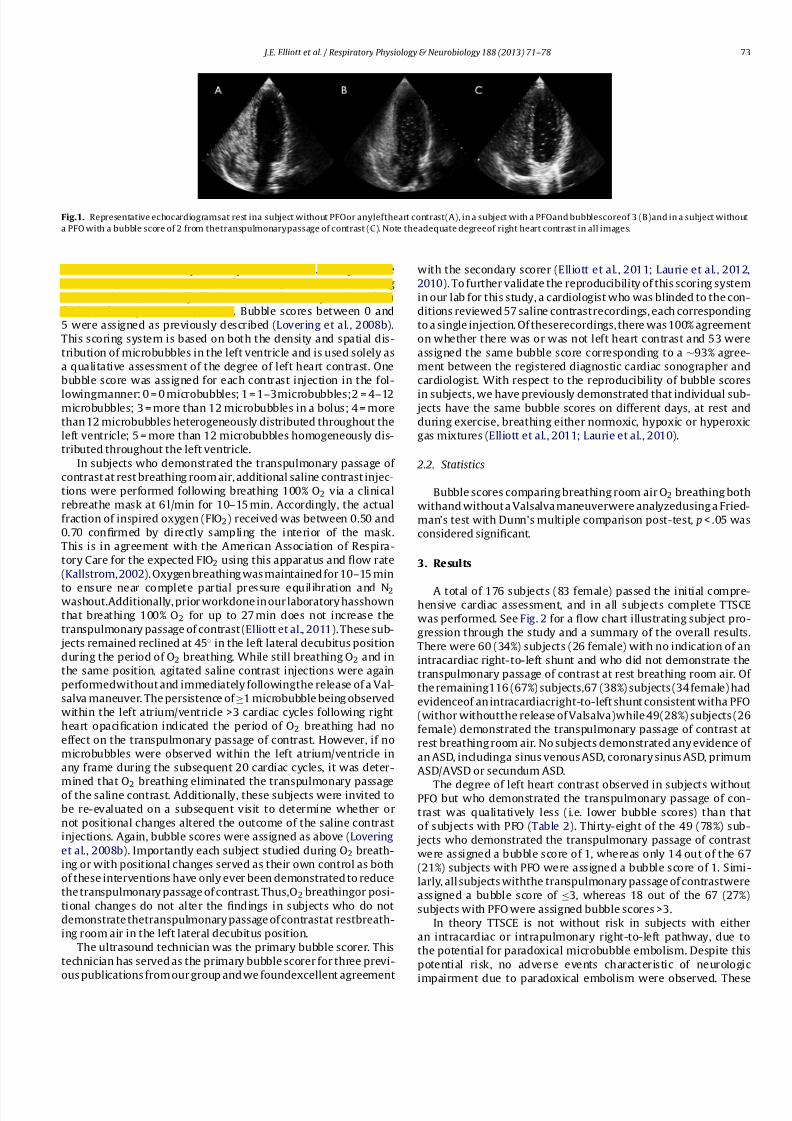
Fig.1. Representative echocardiogramsat rest ina subject without PFOor anyleftheart contrast(A), in a subject with a PFOand bubblescoreof 3 (B)and in a subject without
a PFO with a bubble score of 2 from thetranspulmonarypassage of contrast (C). Note theadequate degreeof right heart contrast in all images.
structuresas well as from potentialpseudocontrast. Multiple saline
contrast injections were made in each subject as needed, during
each condition, to clarify if there was uncertainty and confirm
findings from previous injections. Bubble scores between 0 and
5 were assigned as previously described (Lovering et al., 2008b).
This scoring system is based on both the density and spatial dis-
tribution of microbubbles in the left ventricle and is used solely as
a qualitative assessment of the degree of left heart contrast. One
bubble score was assigned for each contrast injection in the fol-
lowingmanner: 0 = 0 microbubbles; 1 = 1–3microbubbles;2 = 4–12microbubbles; 3 = more than 12 microbubbles in a bolus; 4 = more
than 12 microbubbles heterogeneously distributed throughout the
left ventricle; 5 = more than 12 microbubbles homogeneously dis-
tributed throughout the left ventricle.
In subjects who demonstrated the transpulmonary passage of
contrast at rest breathing room air, additional saline contrast injec-
tions were performed following breathing 100% O2 via a clinical
rebreathe mask at 6 l/min for 10–15 min. Accordingly, the actual
fraction of inspired oxygen (FIO2) received was between 0.50 and
0.70 confirmed by directly sampling the interior of the mask.
This is in agreement with the American Association of Respira-
tory Care for the expected FIO2 using this apparatus and flow rate
(Kallstrom,2002). Oxygen breathing was maintained for 10–15 min
to ensure near complete partial pressure equilibration and N2washout.Additionally, prior workdone in our laboratory hasshown
that breathing 100% O2 for up to 27 min does not increase the
transpulmonary passage of contrast (Elliott et al., 2011). These sub-
jects remained reclined at 45◦ in the left lateral decubitus position
during the period of O2 breathing. While still breathing O2 and in
the same position, agitated saline contrast injections were again
performedwithout and immediately followingthe release of a Val-
salva maneuver. The persistence of ≥1 microbubble being observed
within the left atrium/ventricle >3 cardiac cycles following right
heart opacification indicated the period of O2 breathing had no
effect on the transpulmonary passage of contrast. However, if no
microbubbles were observed within the left atrium/ventricle in
any frame during the subsequent 20 cardiac cycles, it was deter-
mined that O2 breathing eliminated the transpulmonary passageof the saline contrast. Additionally, these subjects were invited to
be re-evaluated on a subsequent visit to determine whether or
not positional changes altered the outcome of the saline contrast
injections. Again, bubble scores were assigned as above (Lovering
et al., 2008b). Importantly each subject studied during O2 breath-
ing or with positional changes served as their own control as both
of these interventions have only ever been demonstrated to reduce
the transpulmonary passage of contrast. Thus,O2 breathingor posi-
tional changes do not alter the findings in subjects who do not
demonstrate thetranspulmonary passage of contrastat restbreath-
ing room air in the left lateral decubitus position.
The ultrasound technician was the primary bubble scorer. This
technician has served as the primary bubble scorer for three previ-
ous publications from our group and we foundexcellent agreement
with the secondary scorer (Elliott et al., 2011; Laurie et al., 2012,
2010). To further validate the reproducibility of this scoring system
in our lab for this study, a cardiologist who was blinded to the con-
ditions reviewed 57 saline contrastrecordings, each corresponding
to a single injection. Of theserecordings, there was 100% agreement
on whether there was or was not left heart contrast and 53 were
assigned the same bubble score corresponding to a ∼93% agree-
ment between the registered diagnostic cardiac sonographer and
cardiologist. With respect to the reproducibility of bubble scores
in subjects, we have previously demonstrated that individual sub- jects have the same bubble scores on different days, at rest and
during exercise, breathing either normoxic, hypoxic or hyperoxic
gas mixtures (Elliott et al., 2011; Laurie et al., 2010).
2.2. Statistics
Bubble scores comparing breathing room air O2 breathing both
withand without a Valsalva maneuverwere analyzedusing a Fried-
man’s test with Dunn’s multiple comparison post-test, p< .05 was
considered significant.
3. Results
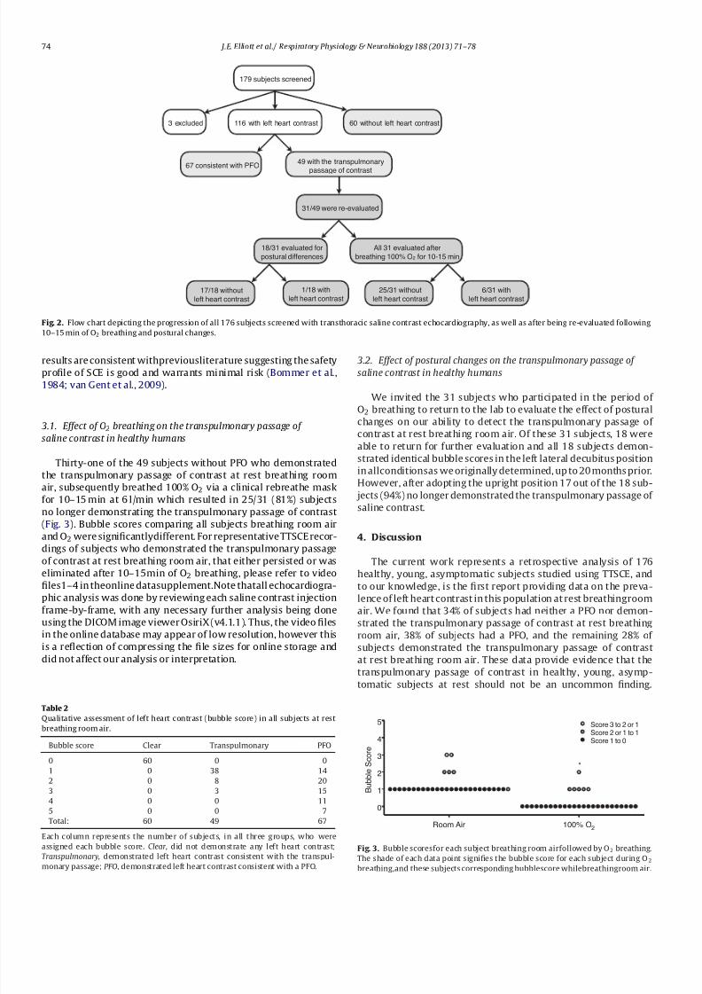
A total of 176 subjects (83 female) passed the initial compre-hensive cardiac assessment, and in all subjects complete TTSCE
was performed. See Fig. 2 for a flow chart illustrating subject pro-
gression through the study and a summary of the overall results.
There were 60 (34%) subjects (26 female) with no indication of an
intracardiac right-to-left shunt and who did not demonstrate the
transpulmonary passage of contrast at rest breathing room air. Of
the remaining116 (67%) subjects,67 (38%) subjects (34 female) had
evidenceof an intracardiacright-to-left shunt consistent witha PFO
(withor withoutthe release of Valsalva)while 49(28%) subjects (26
female) demonstrated the transpulmonary passage of contrast at
rest breathing room air. No subjects demonstrated any evidence of
an ASD, includinga sinus venous ASD, coronary sinus ASD, primum
ASD/AVSD or secundum ASD.
The degree of left heart contrast observed in subjects withoutPFO but who demonstrated the transpulmonary passage of con-
trast was qualitatively less (i.e. lower bubble scores) than that
of subjects with PFO (Table 2). Thirty-eight of the 49 (78%) sub-
jects who demonstrated the transpulmonary passage of contrast
were assigned a bubble score of 1, whereas only 14 out of the 67
(21%) subjects with PFO were assigned a bubble score of 1. Simi-
larly, all subjects withthe transpulmonary passage of contrastwere
assigned a bubble score of ≤3, whereas 18 out of the 67 (27%)
subjects with PFO were assigned bubble scores >3.
In theory TTSCE is not without risk in subjects with either
an intracardiac or intrapulmonary right-to-left pathway, due to
the potential for paradoxical microbubble embolism. Despite this
potential risk, no adverse events characteristic of neurologic
impairment due to paradoxical embolism were observed. These
7/29/2019 Prevalence of left heart contrast in healthy, young, asymptomatic humans at rest breathing room air
http://slidepdf.com/reader/full/prevalence-of-left-heart-contrast-in-healthy-young-asymptomatic-humans-at 4/8
74 J.E. Elliott et al./ Respiratory Physiology & Neurobiology188 (2013) 71–78
179 subjects screened
3 excluded 116 with left heart contrast 60 without left heart contrast
67 consistent with PFO49 with the transpulmonary
passage of contrast
31/49 were re-evaluated
18/31 evaluated for
postural differences
All 31 evaluated after
breathing 100% O2 for 10-15 min
25/31 without
left heart contrast
6/31 with
left heart contrast17/18 without
left heart contrast
1/18 with
left heart contrast
Fig. 2. Flow chart depicting the progression of all 176 subjects screened with transthoracic saline contrast echocardiography, as well as after being re-evaluated following
10–15 min of O2 breathing and postural changes.
results are consistent withpreviousliterature suggesting the safety
profile of SCE is good and warrants minimal risk (Bommer et al.,
1984; van Gent et al., 2009).
3.1. Effect of O 2 breathing on the transpulmonary passage of
saline contrast in healthy humans
Thirty-one of the 49 subjects without PFO who demonstrated
the transpulmonary passage of contrast at rest breathing room
air, subsequently breathed 100% O2 via a clinical rebreathe mask
for 10–15 min at 6 l/min which resulted in 25/31 (81%) subjects
no longer demonstrating the transpulmonary passage of contrast
(Fig. 3). Bubble scores comparing all subjects breathing room airand O2 were significantlydifferent. For representative TTSCE recor-
dings of subjects who demonstrated the transpulmonary passage
of contrast at rest breathing room air, that either persisted or was
eliminated after 10–15min of O2 breathing, please refer to video
files1–4 in theonline datasupplement.Note thatall echocardiogra-
phic analysis was done by reviewing each saline contrast injection
frame-by-frame, with any necessary further analysis being done
using the DICOM image viewer OsiriX (v4.1.1). Thus, the video files
in the online database may appear of low resolution, however this
is a reflection of compressing the file sizes for online storage and
did not affect our analysis or interpretation.
Table 2
Qualitative assessment of left heart contrast (bubble score) in all subjects at rest
breathing room air.
Bubble score Clear Transpulmonary PFO
0 60 0 0
1 0 38 14
2 0 8 20
3 0 3 15
4 0 0 11
5 0 0 7
Total: 60 49 67
Each column represents the number of subjects, in all three groups, who were
assigned each bubble score. Clear , did not demonstrate any left heart contrast;
Transpulmonary, demonstrated left heart contrast consistent with the transpul-
monary passage; PFO, demonstrated left heart contrast consistent with a PFO.
3.2. Effect of postural changes on the transpulmonary passage of
saline contrast in healthy humans
We invited the 31 subjects who participated in the period of
O2 breathing to return to the lab to evaluate the effect of postural
changes on our ability to detect the transpulmonary passage of
contrast at rest breathing room air. Of these 31 subjects, 18 were
able to return for further evaluation and all 18 subjects demon-
strated identical bubble scores in the left lateral decubitus position
in allconditionsas we originally determined, up to 20 months prior.
However, after adopting the upright position 17 out of the 18 sub-
jects (94%) no longer demonstrated the transpulmonary passage of
saline contrast.
4. Discussion
The current work represents a retrospective analysis of 176
healthy, young, asymptomatic subjects studied using TTSCE, and
to our knowledge, is the first report providing data on the preva-
lence of left heart contrast in this population at rest breathingroom
air. We found that 34% of subjects had neither a PFO nor demon-
strated the transpulmonary passage of contrast at rest breathing
room air, 38% of subjects had a PFO, and the remaining 28% of
subjects demonstrated the transpulmonary passage of contrast
at rest breathing room air. These data provide evidence that the
transpulmonary passage of contrast in healthy, young, asymp-
tomatic subjects at rest should not be an uncommon finding.
Room Air 100% O2
0
1
2
3
4
5
Score 2 or 1 to 1Score 3 to 2 or 1
Score 1 to 0
*
B u b b l e
S c o r e
Fig. 3. Bubble scoresfor each subject breathing room airfollowed by O2 breathing.
The shade of each data point signifies the bubble score for each subject during O2
breathing,and these subjects corresponding bubblescore whilebreathingroom air.
7/29/2019 Prevalence of left heart contrast in healthy, young, asymptomatic humans at rest breathing room air
http://slidepdf.com/reader/full/prevalence-of-left-heart-contrast-in-healthy-young-asymptomatic-humans-at 5/8
J.E. Elliott et al. / Respiratory Physiology & Neurobiology188 (2013) 71–78 75
In addition, in subjects without PFO but who demonstrated the
transpulmonary passage of contrast at rest breathing room air, O2
breathing for 10–15 min andstandingin the upright position elim-
inated the transpulmonary passage of contrast in 81% and 94% of
subjects, respectively.
4.1. Prevalence of patent foramen ovale in healthy humans
The prevalence of PFO is reported to be ∼30% of the general
population, but varies between ∼25% and 40% depending on the
method of detectionand ageof subjects, i.e.saline contrastechocar-
diography or via a probe in autopsy studies (Hagen et al., 1984;
Marriott et al., 2013; Woods et al., 2010). The current work reports
a 38% prevalence of PFO in our subject population using TTSCE. In
subjects with PFO it is arguably not possible to definitively deter-
mine whether or not observing left heart contrast in >3 cardiac
cycles post right heart opacification is a result of the intracar-
diac pathway or blood flow through an intrapulmonary pathway.
Accordingly, these data demonstrate why screening and excluding
subjects forPFO is importantto do in studies using TTSCE if thegoal
of that study is to detect the transpulmonary passage of contrast
and relate this with some physiologic function.
4.2. Prevalence of the transpulmonary passage of saline contrast
in healthy humans
Although recent literature has suggested that observing the
transpulmonary passage of contrast is a fairly common finding
(Woods et al., 2010; Woods and Patel, 2006), this is the first study
in a large sample size to report the prevalence of healthy, young,
asymptomatic subjects who demonstrate the transpulmonary pas-
sage of contrast at rest breathing room air (∼28%). Previous
reports which include a description of an otherwise, healthy con-
trol population using SCE exist, however they do not represent
an asymptomatic sample. These other examples in the literature
include reports aimedat characterizing symptomatic patient popu-
lations with hereditary hemorrhagic telangiectasia and pulmonary
arteriovenous malformations, and as such used subjects referredto these speciality clinics who failed to meet criteria for hereditary
hemorrhagic telangiectasia as the control population (Gazzaniga
et al., 2009; Gossage and Kanj, 1998; van Gent et al., 2009).
Considering all 176 subjects in the current study were healthy,
asymptomatic, and free of any cardiopulmonary disease, it is
unlikelythatthe 49 (28%)subjects whodemonstrated the transpul-
monary passage of contrast were due to pulmonary arteriovenous
malformations of a pathologic origin (Gossage and Kanj, 1998;
Shovlin and Letarte, 1999). Thus, other more plausible explana-
tions for the transpulmonary passage of contrast at rest could be
the presence of intrapulmonary arteriovenous anastomoses, which
were first reported to exist in healthy human lungs by Tobin, over
60 years ago (Tobin, 1966; Tobin and Zariquiey, 1953; Tobin and
Zariquiey, 1950) or via distendedpulmonary capillaries and/or cor-ner vessels as recently suggested by others (La Gerche et al., 2010;
Lalande etal., 2012).
Although the current work cannot distinguish between
transpulmonary passage of contrast via large diameter (>50m)
intrapulmonary arteriovenous anastomoses (Lovering et al., 2007)
or distention of pulmonary capillaries, work in animals during
exercise (Stickland et al., 2007) and while breathing hypoxic gas
at rest (Bates et al., 2012) demonstrate the transpulmonary pas-
sage of microspheres >25m and 70m in diameter, respectively.
Interestingly, under these same conditions, during exercise and
breathing hypoxic gas at rest, we and others have demonstrated
the transpulmonary passage of saline contrast in healthy humans
who do not demonstrate the transpulmonary passage of contrast
at rest breathing room air (Elliott et al., 2011; Laurie et al., 2010;
Lovering et al., 2008a; Stickland et al., 2004). Conversely, to date,
there is no direct evidence that either pulmonary capillaries or
corner vessels can distend to diameters greater than 20m, even
under pulmonary pressures that are supra-physiologic for humans.
For example at pulmonary pressures up to 73mm Hg in isolated
greyhound lungs the mean capillary diameter was found to be
6.5m and themaximum capillarydiameter measured was 13m
(Glazier et al., 1969; Rosenzweig et al., 1970), which would not be
a sufficient diameter to allow for the passage of a 25
m, 50
m
or 70m microsphere. Likewise, Manohar and Goetz found that
15m microspheres do not traverse the pulmonary circulation of
the exercising Thoroughbred horse, prompting these authors to
conclude that pulmonary capillaries do not distend above 15m,
even at cardiac outputs and pulmonary pressures that far surpass
those in humans, e.g. >200l/min and ∼100 mm Hg, respectively
(Manohar and Goetz, 2005). Of note, the pulmonary capillary
distensibility coefficient (˛, fractional diameter change/mm Hg
pressure) of horses has been calculated to be 0.01, which is at the
lower limit of normal as calculated by Reeves and colleagues for
humans (∼0.02) (Reeveset al., 2005). Insummary,it seems unlikely
that capillary distention can account for these findings in healthy
humans at rest.
4.3. Effect of O 2 breathing on the transpulmonary passage of
saline contrast in healthy humans
In subjects who do not demonstrate the transpulmonary pas-
sage of contrast at rest breathing room air, breathing 100% O2
during exercise prevents or significantly reduces the transpul-
monary passage of contrast (Elliott et al., 2011; Lovering et al.,
2008b). Similarly, breathing 100% O2 for 10–15min in healthy
human subjects eliminated the transpulmonary passage of contrast
in 25 out of the 31 subjects studied, all of which demonstrated
the transpulmonary passage of contrast at rest breathing room
air. In support of these findings is work by Niden and Aviado
which demonstrated that the number of intravenously injected
glass microspheres (up to 420m in diameter) that were recov-
eredfrom thepulmonary venous effluent decreasedin anesthetizeddogs ventilated with 100% O2 (Niden and Aviado, 1956).
Subjects in the current work breathed 100% O2 via a clini-
cal rebreathe mask and as such the actual FIO2 was between
0.50 and 0.70 (Kallstrom, 2002). For this reason, and to allow
adequatetime for nearly complete invivogaspartial pressure equil-
ibration to occur, we chose to evaluate subjects after 10–15 min
of O2 breathing. Indeed, prior work (Lovering et al., 2008b) has
demonstrated O2 breathing to reduce the transpulmonary pas-
sage of contrast in healthy humans during exercise in ∼2min.
This rapid response could potentially be attributed to the actual
FIO2 being 1.0, however, most subjects (5/7) in this prior work
still maintained some degree of left heart contrast (1–3 microbub-
bles), especially at maximal exercise. Therefore, it remains possible
that had subjects continued to exercise while breathing 100%O2, left heart contrast would have eventually been entirely elim-
inated. Additionally, considering the actual FIO2 and duration
of O2 breathing in the current report, it is unlikely that any
potential negative effect of O2 breathing was encountered, such
as an excessive accumulation of reactive oxygen species lead-
ing to CNS toxicity (Dean, 2004). Although it is unknown how
O2 breathing is preventing the transpulmonary passage of con-
trast, previous work has shown that O2 breathing does not affect
the viability/stability of microbubbles created from room air gas.
Accordingly, right heart contrast is unaffected (see videos 1–4
in the online database) and the absence of left heart contrast
does not reflect a disappearance or absence of microbubbles in
the right heart prior to reaching the left heart due to an alter-
ation of in vivo gas bubble dynamics when the external partial
7/29/2019 Prevalence of left heart contrast in healthy, young, asymptomatic humans at rest breathing room air
http://slidepdf.com/reader/full/prevalence-of-left-heart-contrast-in-healthy-young-asymptomatic-humans-at 6/8
76 J.E. Elliott et al./ Respiratory Physiology & Neurobiology188 (2013) 71–78
pressure environment is changed during O2 breathing (Elliott
et al., 2011). In addition, Melsom and colleagues have shown that
hyperoxia causes a significant redistribution of pulmonary blood
flow in sheep (Melsom et al., 1999) suggesting a hyperoxia-
mediated, active control mechanism for the pulmonary vasculature
which may redirect blood flow away from intrapulmonary arte-
riovenous anastomoses. Taken together, we would speculate, as
others have (McMullan et al., 2004) that blood flow through
intrapulmonary arteriovenous anastomoses may be regulated in
a manner similar to the systemic vasculature or ductus arteriosus
whereby hypoxia causes vasodilation and hyperoxia causes vaso-
constriction, thereby preventing the transpulmonary passage of
contrast.
4.4. Effect of body positioning on the transpulmonary passage of
saline contrast in healthy humans
Inadditionto theinspired O2 concentration altering the findings
from TTSCE, body positioning may also be an importantfactor. Pre-
viously, Stickland et al. demonstrated that standing in the upright
position eliminated the transpulmonary passage of contrastin sub-
jects (2/8) who while supine, did demonstrate the transpulmonary
passage of contrast (Stickland et al., 2004). However, standing in
the upright position did not result in the transpulmonary pas-sage of contrast in subjects (6/8) without left heart contrast while
supine (Stickland et al., 2004). Accordingly, we re-examined 18 of
the 49 subjects who demonstrated the transpulmonary passage
of contrast at rest breathing room air in the left lateral decubi-
tus position after adopting an upright position. All 18 subjects had
identical baseline TTSCE results while in the left lateral decubitus
position compared to their previous visit (up to 20 months prior),
and in 17 (94%) of these subjects standing in the upright posi-
tion eliminated the transpulmonary passage of contrast. Although
the precise mechanism remains unknown, going from supine to
the upright position would redistribute pulmonary blood flow
away from the apical portion of the lungs toward the dependent
portion of the lungs, suggesting that perhaps in the upright posi-
tion this redistribution of pulmonary blood flow may explain thesudden elimination of left heart contrast. Interestingly, Tobin and
Zariquiey have previously demonstrated thatintrapulmonary arte-
riovenous anastomoses are located within the apex of the lung,
which may explain the transpulmonary passage of contrast while
supine versus theabsenceof leftheart contrastwhileupright(Tobin
and Zariquiey, 1950).
4.5. Implication of the transpulmonary passage of contrast on
pulmonarymedicine and physiology
The potential physiologic role and/or pathophysiologic impli-
cations of the transpulmonary passage of contrast remain to be
determined. Previous work has suggested that the transpulmonary
passage of contrast is occurring via intrapulmonary arteriove-nous anastomoses which may represent a source of right-to-left
shunt, and thus, may contribute to pulmonary gas exchange effi-
ciency (Lovering et al., 2008a; Stickland et al., 2004; Stickland and
Lovering, 2006). Others have argued that the transpulmonary pas-
sage of contrast may represent pulmonary capillary and/or corner
vessel distention and therefore may be involved with regulating
pulmonary artery pressure (Bryan et al., 2012; La Gerche et al.,
2010; Lalande et al., 2012; Laurie et al., 2012; Stickland et al.,
2004), or influencing pulmonary vascular reserve and minimiz-
ing right ventricular afterload during exercise (La Gerche et al.,
2010; Lalande et al., 2012; Stickland et al., 2004). Others still have
argued that the presence, or absence, of left heart contrast has
limited to no significant physiologic and/or pathophysiologicimpli-
cations (Hopkins et al., 2009; Vogiatzis et al., 2008). Certainly, data
surrounding the potential implications of the transpulmonary
passage of contrast on pulmonary gas exchangeand regulating pul-
monary vascular pressures remains controversial and more work
is neededin this area to eitherconfirm or refute these postulations.
4.6. Limitations
This study exclusively utilized TTSCE and no subjects went on
to further investigation using TEE with agitatedsaline contrast. We
were predominately concerned withdetecting the transpulmonary
passage of contrast,and thus, theoptimal visualization of the inter-
atrial septum that TEE provides was not essential. Because of the
possibility that contrast could be traversing a PFO due to the pres-
sure changes associatedwith normal respiration throughout the 20
cardiac cycle recording, we performedmultipleagitatedsaline con-
trast injections, with and without the use of a Valsalva maneuver
and our 38% prevalence of PFO is evidence of our rigorous inspec-
tion. Indeed this is higher than previous autopsy studies which,
depending on the patients age, detected a 25–34% prevalence of
PFOusingaprobe(Hagenet al., 1984).Of note,similarto the current
work and also using TTSCE, Woods et al. report a 38% prevalence
of PFO in a population of 104 subjects with a history of migraine
(Woods et al., 2010),whileMarriottet al.report a 35% prevalence of
PFO in a population of 1162 subjects referred for TTSCE for a multi-tude of reasons, mostcommonly due to transientischemic attack or
to assess right ventricular function (Marriott et al., 2013). Thus, the
higherthan previously reported prevalence of PFOin ourstudyand
that of Woods et al. and Marriott et al. to previous autopsy reports,
is best explainedby thediff erences in detectiontechniques. Also,in
the current study weused3 ml of salineand 1 ml of air tocreate the
salinecontrast suspension whereas Woods et al.used 9 ml of saline
and 1ml of air, while Marriott et al. used 8.5 ml of saline, 0.5ml of
air, and 1ml of blood. Despite these differences in contrast mixes,
a similar prevalence of PFO was detected in all studies.
When considering the majority of subjects studied demon-
strated low bubble scores, it is important to emphasize our
thorough TTSCE protocol and meticulous analysis. Differentiating
microbubbles from subvalvular structures that are moving in andout of the observational plane can be very difficult, and for this
reason we performed multiple saline contrast injections as nec-
essary, utilized a sonographer with extensive experience as the
primary analyzer and had a cardiologist who was blinded to the
conditions confirm these findings in a subset of 57 saline contrast
injections. Of these recordings there was 100%agreement between
the sonographer and cardiologist for whether there was, or was
not any left heart contrast and 94% agreement on assigning indi-
vidual bubble scores. Furthermore, previous work from our group
demonstrates an excellent degree of intra-subject repeatability on
different days, with respect to bubble scores for a given condition
(Elliott et al., 2011;Laurie et al., 2010). Lastly, we always performed
a baseline, normoxic, saline contrast injection in subjects reclined
in the left lateral decubitus position prior to any intervention toconfirm previous findings in all subjects. This further demonstrates
our ability to repeatedly identify very few microbubbles, often ≤3
microbubbles within each subject.
With respect to the application of our 0–5 bubble scoring sys-
tem to data obtained using TTSCE it deserves to be reiterated that
this scoring system is a 2-dimensional qualitative assessment of
a 3-dimensional volume. Accordingly, this technique only offers a
qualitative assessment of either more or less contrast in the left
ventricle and has not been directly shown to correlate with more
or less blood flow through intrapulmonary arteriovenous anasto-
moses. The application of a scoring system in studies using TTSCE
does however provide researchers with the ability to categorize
subjects based on the qualitative degree of left sided contrast. Per-
haps the greatest strength of TTSCE is the fact that this technique
7/29/2019 Prevalence of left heart contrast in healthy, young, asymptomatic humans at rest breathing room air
http://slidepdf.com/reader/full/prevalence-of-left-heart-contrast-in-healthy-young-asymptomatic-humans-at 7/8

7/29/2019 Prevalence of left heart contrast in healthy, young, asymptomatic humans at rest breathing room air
http://slidepdf.com/reader/full/prevalence-of-left-heart-contrast-in-healthy-young-asymptomatic-humans-at 8/8
78 J.E. Elliott et al./ Respiratory Physiology & Neurobiology188 (2013) 71–78
Reeves, J., Linehan, J., Stenmark, K., 2005. Distensibility of the normal human lungcirculation during exercise.AmericanJournal of Physiology – Lung Cellular andMolecular Physiology 288, L419-L425.
Roelandt, J., 1982. Contrast echocardiography. Ultrasound in Medicine & Biology 8,471–492.
Rosenzweig, D.Y.,Hughes, J.M.,Glazier, J.B., 1970. Effectsof transpulmonaryand vas-cular pressureson pulmonarybloodvolume in isolated lung. Journal of AppliedPhysiology 28, 553–560.
Shovlin, C.L., Letarte, M., 1999. Hereditary haemorrhagic telangiectasia and pul-monary arteriovenous malformations: issues in clinical management andreview of pathogenic mechanisms. Thorax 54, 714–729.
Stickland, M.K., Lovering, A.T., 2006. Exercise-induced intrapulmonary arteri-ovenous shunting and pulmonary gas exchange. Exercise and Sport SciencesReviews 34, 99–106.
Stickland,M.K., Lovering,A.T.,Eldridge,M.W., 2007. Exercise-induced arteriovenousintrapulmonary shunting in dogs. American Journal of Respiratory and CriticalCare Medicine 176, 300–305.
Stickland, M.K., Welsh, R.C., Haykowsky, M.J ., Pet ers en, S.R., Anderson, W.D.,Taylor, D.A., Bouffard, M., Jones, R.L., 2004. Intra-pulmonary shunt and pul-monary gas exchange during exercise in humans. Journal of Physiology 561,321–329.
Stickland,M.K., Welsh, R.C., Haykowsky,M.J., Petersen, S.R.,Anderson, W.D.,Taylor,D.A.,Bouffard, M., Jones, R.L., 2006. Effect of acuteincreasesin pulmonaryvascu-lar pressureson exercisepulmonary gasexchange.Journal ofApplied Physiology100, 1910–1917.
Tobin, C.E., 1966. Arteriovenous shunts in the peropheral pulmonary circulation inthe human lung. Thorax 21, 197–204.
Tobin, C.E., Zariquiey, M.O., 1950. Arteriovenous shunts in the human lung. Pro-ceedings of theSociety forExperimental Biology and Medicine 75, 827–829.
Tobin, C.E., Zariquiey, M.O., 1953. Some observations on the blood supply of thehuman lung. Medical Radiography and Photography 29, 9–19.
Tsujino, T., Shima, A., 1980. The behaviour of gas bubbles in blood subjected to anoscillating pressure. Journal of Biomechanics 13, 407–416.
vanGent,M.W.F.,Post,M.C.,Snijder,R.J.,Swaans,M.J.,Plokker, H.W.M.,Westermann,C.J.J., Overtoom,T.T., Mager, J.J.,2009. Grading of pulmonary right-to-left shuntwith transthoracic contrastechocardiography: does it predict theindication forembolotherapy? Chest 135, 1288–1292.
Vogiatzis, I., Zakynthinos,S., Boushel,R., Athanasopoulos,D., Guenette, J.A.,Wagner,
H.,Roussos, C.,Wagner,P.D.,2008. Thecontributionof intrapulmonaryshuntstothe alveolar-to-arterial oxygen diff erenceduring exercise is very small. Journalof Physiology 586, 2381–2391.
Woods, T.D., Harmann, L., Purath, T., Ramamurthy, S., Subramanian, S., Jackson,S., Tarima, S., 2010. Small- and moderate-size right-to-left shunts identifiedby saline contrast echocardiography are n or mal and unrelated to migraineheadache. Chest 138, 264–269.
Woods,T.D., Patel, A.,2006. A critical review ofpatent foramenovale detectionusingsaline contrast echocardiography: when bubbles lie. Journal of the AmericanSociety of Echocardiography 19, 215–222.
Yang, W.J., Echigo, R., Wotton, D.R., Hwang, J.B., 1971a. Experimental studies of the dissolutionof gasbubbles in whole blood andplasma. I. Stationary bubbles.
Journal of Biomechanics 4, 275–281.Yang, W.J., Echigo, R.,Wotton, D.R., Hwang,J.B.,1971b. Experimental studies of the
dissolution of gas bubbles in whole blood and plasma. II. Moving bubbles orliquids. Journal of Biomechanics 4, 283–288.


![Review Open Access · and 50% in asymptomatic HBsAg carriers, CHB patients, patients with liver cirrhosis and HCC patients, respectively[39]. The prevalence of pre-S mutants varied](https://img.dokumen.tips/doc/110x75/5fce18c3e96ac728524fe043/review-open-access-and-50-in-asymptomatic-hbsag-carriers-chb-patients-patients.jpg)