Embed Size (px)
Citation preview

B
J Trop Med Parasitol. 2014;37:10-9. ORIGINAL ARTICLE
Prevalence and risk factors of Babesia infections in cattle trespassing natural forest areas in Salakpra
Wildlife Sanctuary, Kanchanaburi Province
Patcharathorn Simking1,2,3,4, Nantawan Yatbantoong5, Nantiya Saetiew1,2,3,4, Sinsamut Saengow4, Wanisa Yodsri6, Rattanawat Chaiyarat7,
Sirichai Wongnarkpet8, and Sathaporn Jittapalapong4
1Center for Agricultural Biotechnology, Kasetsart University, Kamphaeng Saen Campus, Nakhon Pathom 73140, Thailand; 2Center of Excellence on Agricultural Biotechnology: (AG-BIO/PERDO-CHE), Bangkok 10900,Thailand; 3Center for Advanced Studies for Agriculture and Food, Kasetsart University Institute for Advanced Studies, Kasetsart University, Bangkok 10900 Thailand” (CASAF, NRU-KU, Thailand); 4Department of Parasitology, Faculty of Veterinary Medicine, Kasetsart University, Bangkok, 10900,
Thailand; 5Department of Large animal and Wildlife Clinical Science, Faculty of Veterinary Medicine, Kasetsart University, Nakhon Pathom 73140; 6Geo-informatics Service and Application Division, Bangkok,
10900, Thailand; 7Wildlife and Plant Research Center, Faculty of Environment and Resource Studies, Mahidol University, Thailand 73170; 8Department of Veterinary Diagnostic Services, Faculty of Veterinary
Medicine, Kasetsart University, Bangkok 10900, Thailand
ABSTRACT ovine babesiosis is a major tick – borne disease in livestock development since this disease has an effect on animal health led to economic losses due to increase mortality and lower production. Babesiosis in cattle is caused by Babesia bovis and B. bigemina and transmitted by tropical cattle tick, Rhipicephalus (Boophilus) microplus. The objective of this study was to investigate the prevalence of Babesia infection in the cattle invading natural forest area of Salakpra Wildlife Sanctuary (SWS) in Kanchanaburi province using molecular assays. A total of 244 cattle blood samples were collected from SWS areas located among 3 districts of Kanchanaburi Province including Mueang, Si Sawat, and Bo Phloi and examined by polymerase chain reaction (PCR) using SBP2 gene to identify B. bovis and rap – 1α gene for B. bigemina. Factors associated with Babesia infection including location, age, sex, herd size, seasonal management, insect abundant, forage density, deworming, domestic pet in household, tick infestations in cattle and barn, and the grazing area management was statistically analyzed. The overall infection of Babesia spp. of cattle in SWS was 46.7% (114/244), comprising B. bovis (5.3%, 13/244), B. bigemina (38.9%, 95/244) and mixed (B. bovis and B. bigemina) (2.4%, 6/244). Mueang District had the highest infection at 47.9% (45/94). The potential significant factors associated with Babesia infections were herd size and age (p<0.05). The high infection of Babesia spp. in SWS indicated the possible risk of the wild to get infections via ticks contaminated in this areas.
Key word: prevalence, B. bovis, B. bigemina, cattle, PCR, Salakpra Wildlife Sanctuary
Correspondence: Sathaporn Jittapalapong, Department of Parasitology, Faculty of Veterinary Medicine, Kasetsart University, Bangkok, 10900, Thailand. E-mail:[email protected]
สมาคมปรสิตวิท
ยาและอายุรศาสตร�เขตร�อน
Available online at http://www.tm.mahidol.ac.th/ptat
Vol. 37 (No. 1) June 201410
THE
JOU
RNA
L O
F TR
OPI
CA
L M
EDIC
INE
AN
D P
ARA
SITO
LOG
Y

INTRODUCTION Babesiosis is a major tick – borne disease of livestock worldwide particularly in tropic and subtropic areas. This disease is caused by Babesia spp. resulting in economic losses of developing countries due to its effect on animal health and production. A distribution of babesiosis is varied on the spreading of cattle tick (R. microplus) in the nearby environment [1]. The conventional diagnosis of bovine babesiosis is based on the identification of parasites in the stained blood smear under the light microscope. However, microscopic examination has low sensitivity and is not practical in epidemiological investigations due to its time consuming and the result depends much on the degree of parasitemia [2]. Currently, the molecular tool has been developed and seems very useful in the epidemiological studies because of its high sensitivity and specificity in a detection of the parasite’s DNA [3-5]. In particular, genetic and antigenic differences were observed among merozoite surface antigens (MSAs) and some merozoite surface antigens, such as MSA-1, MSA-2a1, MSA-2a2, MSA-2b, and MSA-2c, had been well characterized and considered as vaccine candidate antigens [6-8]. Spherical body protein 2 (SBP-2) was also a candidate for many applications. The rap - 1 α gene of B. bigemina is highly conserved and potent in identifying specific Babesia [9]. Bovine babesiosis has been reported in all regions of Thailand since 1980 [10-16]. Kanchanaburi is in the Western Thailand where the largest protected forest areas are located. Salakpra Wildlife Sanctuary (SWS) covers 4 districts of Kanchanaburi where some areas are invaded by the nearby villagers and their animals. The increasing of livestock rearing and urbanization are important factors associated with deforestation and forestry decay [17,18]. The high number of trespassing cattle over forestry areas has affected the food resource and habitats of wildlife. Moreover, these cattle might be the potential risk to threatening the wildlife’s health because they can be potential reservoirs of some
major tick–borne diseases including babesiosis [19,20]. Therefore, the objective of this study was to investigate the molecular prevalence and factors associated with Babesia infections of cattle resided in the Salakpra Wildlife Sanctuary.
MATERIALS AND METHODS1. Blood samples collection and
microscopic examination A total of 244 cattle blood samples were collected from Salakpra Wildlife Sanctuary in Kanchanaburi including Mueang Si Sawat, and Bo Phloi Districts. The sample size was calculated from Winepiscop program using randomization model (Multistage sampling) which was based on total population at 12,000 individual, the expected prevalence of 30%, absolute precision of ±5% and 95% confidence interval. Ten ml of blood was drawn from jugular vein, transferred to sodium citrate tubes and stored at –40ºC until the laboratory analysis. The thin blood smear was used for microscopic examination. The blood smears were stained be Weight & Giemsa stain and detected for the infected erythrocyte under the light microscope.
2. Data Collection Cattle were thoroughly examined for health profiles. The information regarding age, sex, health condition, and location are recorded. In addition, the questionnaire was designed to record these data including herd sized (small: 1–40 cattle/farm; medium: 41–80 cattle/farm; large: >80 cattle/farm), seasonal management (Summer/Raining: roaming/in house), insect abundant in barn (low density: 0–10/cattle; moderate density:11-20/cattle; high density:>20/cattle), Forage density pattern (low density:1–10 trees/5 m2; moderate density:10–20 trees/ 5 m2; high density: over 20 trees/ 5 m2) deworming, domestic pet in household(found or not found), the tick infestation on the animal, the tick existence in barn, and the using of grazing areas (selected or public areas). All factors were statistical analyzed to identify the significant effect on the distribution pattern of Babesia infection among cattle.
Babesia infection in trespassing cattle in Salakpra Wildlife Sanctuary, Kanchanaburi Province
Vol. 37 (No. 1) June 2014 11
THE JO
URN
AL O
F TROPIC
AL M
EDIC
INE A
ND
PARA
SITOLO
GY

3. DNA extraction One hundred µl of each sample was added into microtube and mixed with 500 µl of D-solution. Then, DNA was extracted by using a phenol-chloroform technique and the purified DNA was stored at -20 °C until used. DNA preparation and molecular analysis were processed at the Molecular Laboratory Unit, Department of Veterinary Parasitology, Faculty of Veterinary Medicine, Kasetsart University.
4. PCR and DNA Sequencing For B. bovis identification, cattle blood DNA was assayed with the screening PCR method previously described [1]. The 20 µl reactions contained 1× buffer (10 mM Tris–HCl pH 8.8, 50 mM KCl and 0.1% Triton X-100), 1.5 mM MgCl2, 1.0 pmol of each primer, 0.2 mM of each dNTP, 0.75 unit of Taq DNA polymerase (DyNAzyme, FINNZYMES) and 1 µg of DNA template. The primers BbovisSBP1 (5’ CGAATTCCTGGAAGTGGATCTCATGCAACC–3’) a n d B b o v i s S B P 2 ( 5 ’ A T C T C GAGTCACGAGCACTCTACGGCTTTGCAG–3’) which amplified approximately 1,236 nucleotides of the SBP2 gene are the 1st primer. The PCR conditions were: pre - denaturation at 95°C for 5 min, then 35 cycles of denaturation at 94°C for 1 min, annealing at 64°C for 1 min and extension at 72°C for 2 min and followed by final extension at 72°C for 10 min. The amplified PCR product of 0.5 ml was used for the subsequent nPCR with a limited annealing temperature at 58°C and 1 min at 72°C. in a MyCyclerTM Thermal Cycler (BioRad Laboratories, USA). The primer BBovisSBP2 3(5 ’–CGAATCTAGGCATATAAGGCAT–3’ ) and BbovisSBP2 4 (5’–ATCCCCTC CTAAGGT TGGCTAC – 3’) are the nested primer. For a detection of B. bigemina, PCR assay was modified following the previous study [5]. The 20 µl reaction contained 1× buffer (10 mM Tris–HCl pH 8.8, 50 mM KCl and 0.1% Triton X-100), 1.5 mM MgCl2, 2 pmol of each primer, 0.2 mM of each dNTP, 1.0 unit of Taq DNA polymerase (DyNAzyme, FINNZYMES) and 1 µg of DNA template. The primer Bbigrap – 1α F1 (5’
GAGTCTGCCAAATCCTTAC 3’) and Bbigrap – 1α
R1 (5’ TCCTCTACAGCTGC TTCG 3’) were used to amplified approximately 879 bp of the B. bigemina rap – 1α gene. The PCR conditions were: pre - denaturation at 94ºC for 5 min, then 35 cycles of denaturation at 94ºC for 1 min, annealing at 55ºC for 1 min and extension at 72ºC for 1 min and followed by final extension at 72°C for 10 min. The nested primers were Bbigrap– 1α F2 (5’–AGCT TGCTTTCACAACTG GCC–3’) and Bbigrap – 1α R2 (5’–TTGGTGCTTTGACCG ACGACAT–3’) and the nested PCR protocol was repeated using the same protocol. The nested PCR product was sized at 419 bp. The positive control was the infected cattle gDNA sample for B.bovis and B.bigemina and the negative control was deionized water.
5. Statistical Analysis Number Cruncher Statistical System (NCSS) ver. 2000 (Kaysville, UT) program was used to assess differences in the prevalence and intensity of infection. The univariate analysis was also undertaken to investigate environmental variables correlated with the infection patterns, based on the probability that individual cattle was infected, by using Chi – square calculation which p –value ≤0.05 were considered significant. The multivariate analysis was estimated by using logistic regression models which the Odds – ratios (OR) and 95% interval also provided from this model by using Wald test algorithm.
RESULTS The positive DNA was shown at approximately 580 bp for B. bovis SBP2 gene and 412 bp for B. bigemina rap - 1α gene under UV light (Fig. 1). A total of 114 Babesia-positive cattle among 36 farms was distributed in the SWS areas as shown in Fig. 2. The highest infection was found at Mueang District (47.9%; 45/94) while Sri Sawat had the lowest prevalence (44.0%; 11/25). The overall Babesia infection of cattle in SWS was 46.7% (114/244) for individual prevalence and 88.9% (32/36) for herd prevalence. The prevalence of B. bovis infection is 5.3% (13/244) and was mostly found in Sri Sawat District (16.0%; 4/25). The
Babesia infection in trespassing cattle in Salakpra Wildlife Sanctuary, Kanchanaburi Province
Vol. 37 (No. 1) June 201412
THE
JOU
RNA
L O
F TR
OPI
CA
L M
EDIC
INE
AN
D P
ARA
SITO
LOG
Y

prevalence of B. bigemina infection was 38.9% (95/244) and was frequently distributed in Bo Phloi District (45.6%; 57/125). In addition, the mixed infection between B. bigemina and B. bovis was also found at 2.5% (6/244). The univariate analysis of the associated factors such as location, age, sex, herd size, seasonal management, insect density, forage density, deworming, pet in farm, tick infestation of cattle, the tick existence in house, and the using of grazing areas were shown in Table 1. Some potential factors associated with Babesia infections including herd sizes, age, and forage density division were statistical significance (p<0.05). However, the multivariate analysis using logistic regression models significantly showed that the infections were affected by the age of animal (between 1 – 5 years) and the herd size (40 – 80 cattle/farm) (Table 2).
DISCUSSIONS The result of this study demonstrated that 5.3% and 38.9% of cattle in SWF areas were infected with B. bovis and B. bigemina, respectively. However, the microscopic examination only presented 2.4% of Babesia spp. in the stained blood smear without species identification. Specific genes such as spherical body protein 2 (SBP-2) and rhoptry-associated protein 1 (rap-1) gene are used to improve their specificity and sensitivity
to identify B. bovis and B. bigemina, respectively, being beneficial for epidemiological investigation [21-25]. The genetic variation of subpopulation within B. bovis or B. bigemina was also shown distinct biological characteristic [26]. For the factors associated with Babesia infections, the age of animals was significantly potential related with the infection (p<0.05). In this study, the animal age (between 1 – 5 years) showed the significant resistance against Babesia infection (Odds ratio = 0.32, CI= 0.14 – 0.72, p = 0.005), as shown as 5.4% and 33.3% infection by B. bovis and B. bigemina, respectively. This might be the evidence of passive immunity in the endemic area. After birth, calves have received passive immunity via the colostrum from pre-immunized mothers. Evidently, the young animals have been reported to be more susceptible to Babesia infection than animals without passive immunity [27-28]. Moreover, the older animals (>5 years) probably have the longer exposure to the pathogen and developed the protective immunity compared to the young animals [29]. In the endemic areas, mild Babesia infections of cattle were normally occurred and induced immunity against babesiosis particularly in native animals. This immunity was also correlated with the herd immunity. The differences of actual prevalence among herds were depended on the diagnostic
Fig 1 DNA bands of B. bovis and B.bigemina infection. S1 – S3 : B. bovis positive sample (580 bp), S4 – S6: B.bigemina positive sample (412 bp), N: negative sample, MK: DNA marker 100 pb plus
Babesia infection in trespassing cattle in Salakpra Wildlife Sanctuary, Kanchanaburi Province
Vol. 37 (No. 1) June 2014 13
THE JO
URN
AL O
F TROPIC
AL M
EDIC
INE A
ND
PARA
SITOLO
GY
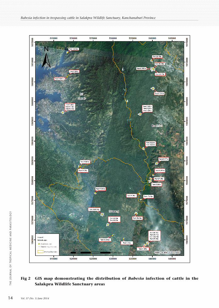
Fig 2 GIS map demonstrating the distribution of Babesia infection of cattle in the Salakpra Wildlife Sanctuary areas
Babesia infection in trespassing cattle in Salakpra Wildlife Sanctuary, Kanchanaburi Province
Vol. 37 (No. 1) June 201414
THE
JOU
RNA
L O
F TR
OPI
CA
L M
EDIC
INE
AN
D P
ARA
SITO
LOG
Y
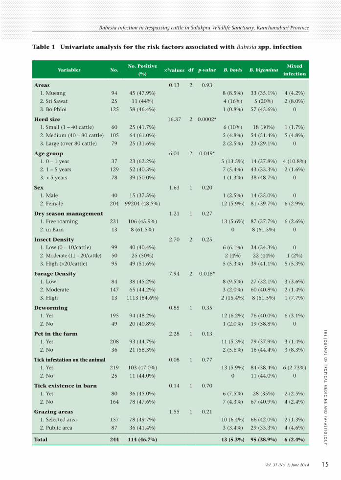
Table 1 Univariate analysis for the risk factors associated with Babesia spp. infection
Variables No.No. Positive
(%)×2values df p-value B. bovis B. bigemina
Mixed
infection
Areas 0.13 2 0.93 1. Mueang 94 45 (47.9%) 8 (8.5%) 33 (35.1%) 4 (4.2%)
2. Sri Sawat 25 11 (44%) 4 (16%) 5 (20%) 2 (8.0%)
3. Bo Phloi 125 58 (46.4%) 1 (0.8%) 57 (45.6%) 0
Herd size 16.37 2 0.0002* 1. Small (1 – 40 cattle) 60 25 (41.7%) 6 (10%) 18 (30%) 1 (1.7%)
2. Medium (40 – 80 cattle) 105 64 (61.0%) 5 (4.8%) 54 (51.4%) 5 (4.8%)
3. Large (over 80 cattle) 79 25 (31.6%) 2 (2.5%) 23 (29.1%) 0
Age group 6.01 2 0.049* 1. 0 – 1 year 37 23 (62.2%) 5 (13.5%) 14 (37.8%) 4 (10.8%)
2. 1 – 5 years 129 52 (40.3%) 7 (5.4%) 43 (33.3%) 2 (1.6%)
3. > 5 years 78 39 (50.0%) 1 (1.3%) 38 (48.7%) 0
Sex 1.63 1 0.20 1. Male 40 15 (37.5%) 1 (2.5%) 14 (35.0%) 0
2. Female 204 99204 (48.5%) 12 (5.9%) 81 (39.7%) 6 (2.9%)
Dry season management 1.21 1 0.27 1. Free roaming 231 106 (45.9%) 13 (5.6%) 87 (37.7%) 6 (2.6%)
2. in Barn 13 8 (61.5%) 0 8 (61.5%) 0
Insect Density 2.70 2 0.25 1. Low (0 – 10/cattle) 99 40 (40.4%) 6 (6.1%) 34 (34.3%) 0
2. Moderate (11 – 20/cattle) 50 25 (50%) 2 (4%) 22 (44%) 1 (2%)
3. High (>20/cattle) 95 49 (51.6%) 5 (5.3%) 39 (41.1%) 5 (5.3%)
Forage Density 7.94 2 0.018* 1. Low 84 38 (45.2%) 8 (9.5%) 27 (32.1%) 3 (3.6%)
2. Moderate 147 65 (44.2%) 3 (2.0%) 60 (40.8%) 2 (1.4%)
3. High 13 1113 (84.6%) 2 (15.4%) 8 (61.5%) 1 (7.7%)
Deworming 0.85 1 0.35 1. Yes 195 94 (48.2%) 12 (6.2%) 76 (40.0%) 6 (3.1%)
2. No 49 20 (40.8%) 1 (2.0%) 19 (38.8%) 0
Pet in the farm 2.28 1 0.13 1. Yes 208 93 (44.7%) 11 (5.3%) 79 (37.9%) 3 (1.4%)
2. No 36 21 (58.3%) 2 (5.6%) 16 (44.4%) 3 (8.3%)
Tick infestation on the animal 0.08 1 0.77 1. Yes 219 103 (47.0%) 13 (5.9%) 84 (38.4%) 6 (2.73%)
2. No 25 11 (44.0%) 0 11 (44.0%) 0
Tick existence in barn 0.14 1 0.70 1. Yes 80 36 (45.0%) 6 (7.5%) 28 (35%) 2 (2.5%)
2. No 164 78 (47.6%) 7 (4.3%) 67 (40.9%) 4 (2.4%)
Grazing areas 1.55 1 0.21 1. Selected area 157 78 (49.7%) 10 (6.4%) 66 (42.0%) 2 (1.3%)
2. Public area 87 36 (41.4%) 3 (3.4%) 29 (33.3%) 4 (4.6%)
Total 244 114 (46.7%) 13 (5.3%) 95 (38.9%) 6 (2.4%)
Babesia infection in trespassing cattle in Salakpra Wildlife Sanctuary, Kanchanaburi Province
Vol. 37 (No. 1) June 2014 15
THE JO
URN
AL O
F TROPIC
AL M
EDIC
INE A
ND
PARA
SITOLO
GY

technique since different genes primers or tests might yield the different prevalence [9]. This protective forestry location (SWS) has 1,277 domestic cattle resided in the 859 km2 [17]. In this study, the result revealed that the medium sized herd (40–80 animals/herd) had more significantly risk effect (Odds ratio = 2.35, CI = 1.07 – 5.18, p = 0.33) to the infection than the large (>80 animals/herd) and small herds (1–40 cattle). This effect might be due to the overcrowded cattle in the herd and the poor management. During the raining season, all of the cattle were roamed in the grazing areas that can be highly contaminated since the temperature and humidity were the essential factors to support the tick survival and population. The annual precipitation of Kanchanaburi was over 1,000 mm (data form Thai Meteorological Department) while the annual precipitation above 700 mm is suitable for enzootic stability [30]. The high humidity (>50%) has influenced the tick biological cycle, leading to the stability of the pathogen in the areas. However, there were no significant differences by seasonal factors in this study. In addition, Babesia cannot be transmitted by the other vectors such as stable flies or tabanus. Therefore, the insect density might not have the effect on Babesia infection in this area. The other factors such as sex, farm management,
and grazing location were not significantly related to the Babesia infection in this study. Evidently, sharing of the grazing location for rearing the cattle might increase the spreading of the cattle ticks, leading to the great risks of transmitted babesiosis [31]. Cattle in SWS areas roamed around the public pasture. Frequently, these animals had been intentionally or unintentionally grazed in the SWS areas. Naturally, this protected forestry area is the habitat of the wildlife such as guar, sambar deer, bantang and elephant [17]. This trespassing cattle combined with available ticks might create the high risk environment for the wildlife. Moreover, the numbers of trespassing cattle in the protected forestry areas is currently increasing. Therefore, the situation was menacing the wildlife’s health. SWS is one of the protected forestry areas that encounter the problem of the cattle trespassing [17]. Most cattle wandered in or nearby the forestry areas since there was no real boundary to limit the trespassing [32]. Some cattle diseases can be transmitted by the invaded cattle to the wildlife or vice versa. Both infected wildlife and cattle can become a carrier with high potential of disease transmission [1,17]. Up to date there are no evidence of the disease transmission between the farms and the wildlife. However, there were a few reports of ticks in animals and environment so that ticks are potential to transmit pathogen from
Table 2 Results of the multivariate analysis of risk factors associated with Babesia infection in SWS areas.
Risk factors β SE β Adjusted ORa (95%CI)b p-valuec
Age group 1. 0 – 1 year - - 1 2. 1 – 5 years -1.138 0.049 0.32 (0.14 – 0.72) 0.005 3. > 5 years -5.01 0.425 0.60 (0.26 – 1.39) 0.23Herd size 1. Small (1 – 40 cattle) - - 1 2. Medium (40 – 80 cattle) 0.856 0.402 2.35 (1.07 – 5.18) 0.033 3. Large (over 80 cattle) -0.360 0.409 0.69 (0.31 – 1.55) 0.37
a OR, odds ratiob CI, confidence intervalc Wald testmulti
Babesia infection in trespassing cattle in Salakpra Wildlife Sanctuary, Kanchanaburi Province
Vol. 37 (No. 1) June 201416
THE
JOU
RNA
L O
F TR
OPI
CA
L M
EDIC
INE
AN
D P
ARA
SITO
LOG
Y

both animals and wildlife. To confirm a route of transmission or carrier, further investigation would be required.
CONCLUSION In this study, the high infection of Babesia (46.7%) might have been concerned as a threat on the wildlife’s health. These results also confirmed that Kanchanaburi was the endemic area of Babesia infection (38.9% B. bigemina and 5.3% B. bovis) compared to the previous report (10.8% B. bigemina and 1.6% B. bovis) in Salakpra Wildlife Sanctuary. However, one of existing is the trespassing of animals from the neighboring community.
ACKNOWLEDGMENTS This study was funded by KURDI and Center for Agricultural Biotechnology (CAB), Kasetsart University, Kamphaeng Saen Campus and Center of Excellence on Agricultural Biotechnology, Science and Technology Postgraduate Education and Research Development Office, Commission on Higher Education, Ministry of Education. (AG-BIO/PERDO-CHE). This work was also supported by the Center for Advanced Studies for Agriculture and Food, Institute for Advanced Studies, Kasetsart University Under the Higher Education Research Promotion and National Research University Project of Thailand, Office of the Higher Education Commission, Ministry of Education, Thailand.
REFERENCES 1 Aboulaila M, Yoloyama N, Igarashi I.
Development and evaluation of two nested PCR assays for the detection of Babesia bovis from cattle blood. Vet Parasitol. 2010; 172: 65-70.
2 Almeria S, Castella J, Ferrer D, Ortuno A, Estrada - Pena A, Gutierrez JF. Bovine piroplasm in Minorca (Balcaric Island, Spain): a comparison of PCR - based and light microscopy detection. Vet Parasitol. 2001; 99: 249-59.
3 Figueroa JV, Cheives LP, Johnson GS, Buening GM. Multiplex polymerase chain reaction
based assay for the detection of Babesia bigemina, Babesia bovis and Anplasma marginale DNA in bovine blood. Vet Parasitol. 1993; 50: 69-81.
4 Oliveira - Sequeira TCG, Oliveira MCS, Araujo Jr JP, Amarante AFT. PCR - based detection of Babesia bovis and Babesia bigemina in their natural host Boophilus microplus and cattle. Int J Parasitol. 2005; 35:105-11.
5 Terkawi MA, Huyen NX, Shinuo C, Inpankaew T, Maklon K, Aboulaila M, et al. Molecular and serological prevalence of Babesia bovis and Babesia bigemina in water buffaloes in the northeast region of Thailand. Vet Parasitol. 2011; 178: 201-7.
6 Saurez CE, Florin - Christensen M, Hines SA, Paler GH, Brown W, McElwain TF. Characterizaion of alellelic variation in the Babesia bovis merozoite surface antigen (MSA - 1) locus and identification of a cross - reactive inhibition - sensitive MSA - 1 epitope. Infect Immun. 2000; 68: 6665-70.
7 Mosqueda J, McElwain TF, Palmer GH. Babesia bovis merozoite surface antigen 2 proteins are expressed on the merozoit and sporozoite surface, and specific antibodies inhibit attachment and invasion of erythrocytes. Infect Immun. 2002; 70: 6448-55.
8 Altangeral K, Sivakumar T, Battsetseg B, Battur B, Ueno A, Igarashi I, et al. Phylogenic relationship of Mongolian Babesia bovis isolates based on the merozoite surface antigen (MSA) - 1, MSA - 2b, and MSA - 2c genes. Vet Parasitol. 2012; 184: 309-16.
9 Cao S, Aboge GO, Terkawi MA, Yu L, Kamyingkird K, Luo Y, et al. Molecular detection and identification of Babesia bovis and Babesia bigemina in cattle in northern Thailand. Parasitol Res. 2012; 111: 1259-66.
10 Ahantarig A, Trinachartvanit W, Milne JR. Tick-borne pathogens and diseases of animals and humans in Thailand. Southeast Asian J Trop Med Public Health. 2008; 38: 1015-32.
11 Iseki H, Zhou L, Kim C, Inpankaew T, Sununta C, Yokoyama N, et al. Seroprevalence of Babesia infections of dairy cows in northern
Babesia infection in trespassing cattle in Salakpra Wildlife Sanctuary, Kanchanaburi Province
Vol. 37 (No. 1) June 2014 17
THE JO
URN
AL O
F TROPIC
AL M
EDIC
INE A
ND
PARA
SITOLO
GY

Thailand. Vet Parasitol. 2010; 170: 193-6. 12 Jittapalapong S, Tipsawek S. Preliminary study
of blood hematology of dairy cows in Nong Pho, Ratchaburi Province. In: Proceedings of 26th Kasetsart University Annual Conference, 1988 Feb 3-5. p.165-9.
13 Nishikawa H, Sarathan N, Tantasuwan D, Neramitmansuk P. Serological survey of trypanosomiasis and babesiosis in cattle and buffaloes in Thailand. In: Proceeding of the Seventh FAVA Congress. 1990. p. 4-7.
14 Phrikanahok N, Bunmalid C, Sarataphan N. Status and prediction of infection rate of tick fever disease among dairy cattle in some province of Thailand. Kasetsart Vet. 2000; 10: 13-23.
15 Tan-Ariya P, Celanond U, Brockelman C. In vitro observation on drug responsiveness of Babesia bovis and on the emergence of drug resistant parasites. J Protozool Res. 1992; 2: 1-9.
16 Vongwatcharadamrong V, Asavametha P, Yuadyong R. Report of babesiosis in native cattle in Nakornsrithammarat. In: Abstracts of the national conference on Agricultural and Biological Sciences 18th session: Veterinary Section. 1980 Jan 28 -30. p. 24.
17 Chaiyarat R, SrikosamataraS. Populations of domesticated cattle and buffalo in the Western Forest Complex of Thailand and their possible impacts on the wildlife community. J Environ Manage. 2009; 90: 1448-53.
18 Gebremedhin B, Pender J, Tesfay G. Collective action for grazing land management in crop - livestock mixed system in the highlands of northern Ethiopia. Agric Syst. 2004; 82: 273-90.
19 Bengis RG. Transfrontier conservation area initatives in Sub - Saharan, Africa: some animal health challenges. In: Osofsky SA, Chleaveland S, Karesh WB, Kock MD, Nyphus PF, Starr L, Yang A. eds. Conservation and Development Interventions at the Wildlife/ Livestock. Interface: Implication for Wildlife, Livestock and Humen Health. Gland, Switzerland: IUCN; 2005:15-19.
20 Schmidt NM, Olsen H, Bildsoe M, Sluydts V, Leirs H. Effects of grazing intensity on small mammal population ecology in wet meadows. Basic App Ecol. 2005; 6: 57-66.
21 McElwain TF, Perryman LE, Davis WC, McGuire TC. Antibodies define multiple proteinswith epitopes exposed on the surface of live Babesia bigemina merozoites. J Immunol. 1987; 138: 2298-304.
22 Figueroa JV, Buening GM, Kinden DA, Green TJ. Identification of common surface antigens among B. bigemina isolates by using monoclonal antibodies. Parasitol. 1990; 100: 161-75.
23 Machado RZ, McElwain TF, Suarez CE, Hines SA, Palmer GH. Babesia bigemina: isolation and characterization of merozoite rhoptries. Exp Parasitol. 1993; 77: 315-25.
24 Suarez CE, McEIwain TF, Echaide I, Toriani de Echaide S, Palmer GH. Interstrain conservation of Babesial RAP-I surface exposed B-cell epitopes despite rap-1 genomic polymorphism. Infect Immun. 1994; 62: 3576-9.
25 Vidotto O, McElwain TF, Machado RZ, Perryman LE, Suarez CE, Palmer GH. Babesia bigemina: identification of B cell epitopes associated with parasitized erythrocytes. Exp Parasitol. 1995; 81: 491-500.
26 Timms P, Stewart NP, de Vos AJ. Study of virulence and vector transmission of Babesia bovis by use of cloned parasites lines. Infect Immun. 1990; 58: 2171-6.
27 James MA. Immunologiy of babesiosis. In: Ristic M. ed. Babesiosis of domestic animal and man. Boca Raton, FL: CRC Press; 1988. pp 119-30.
28 Awad H, Antunes S, Galindo RC, do Rosario VE, de la Fuente J, Domingo A, et al. Prevalence and genetic diversity of Babesia and Anaplasma species in cattle in Sudan. Vet Parasitol. 2011; 181: 146-52.
29 Homer MJ, Aquilar - Defin I, Teleford SR, Krause PJ, Persing DH. Babesiosis. Clin Microb Rev. 2000; 13: 451-69.
30 Barros SL, Madrugal CR, Araújo FR, et al. Serological survey of Babesia bovis, Babesia
Babesia infection in trespassing cattle in Salakpra Wildlife Sanctuary, Kanchanaburi Province
Vol. 37 (No. 1) June 201418
THE
JOU
RNA
L O
F TR
OPI
CA
L M
EDIC
INE
AN
D P
ARA
SITO
LOG
Y

bigemina, and Anaplasma marginale antibodies in cattle from the semi-arid region of the state of Bahia, Brazil, by enzyme-linked immunosorbent assays. Mem Inst Oswaldo Cruz Rio de Janeiro. 2005; 100: 513-7.
31 Simuunza M, Weir W, Courcier E, Tait A, Shiels B. Epidemiological analysis of tick-borne
disease in Zambia. Vet. Parasitol. 2011; 175: 331-42.
32 Nakhasathien S, Stewart - Cox B. Nomination of the Thung Yai - Huai Kha Khaeng Wildlife Sanctuary to be a UNESCO World Heritage Site. Wildlife Conservation Division, Royal Forest Department, Thailand, 1990.
Babesia infection in trespassing cattle in Salakpra Wildlife Sanctuary, Kanchanaburi Province
Vol. 37 (No. 1) June 2014 19
THE JO
URN
AL O
F TROPIC
AL M
EDIC
INE A
ND
PARA
SITOLO
GY


![Hepatitis screening Chinese [Kompatibilitetstilstand] 2014/Oral presentations...collaboration with the Chinese community High prevalence of hepatitis B infections (6%) Low prevalence](https://img.dokumen.tips/doc/110x75/5e175694a1c2c52b8c4f477f/hepatitis-screening-chinese-kompatibilitetstilstand-2014oral-presentationscollaboration.jpg)


