Embed Size (px)
Citation preview

Università degli Studi del Molise Dipartimento di Medicina e di Scienze per la Salute Clinica di Oftalmologia Direttore: Prof. Ciro Costagliola
Roberto dell’Omo
Differences between idiopathic, secondary and diabetic epiretinal membrane

ERM
• TYPE I vitreoschisis collagen between ILM and epiretinal membrane • TYPE II PVD Cleft of the ILM (rare) ILM and ERM are attached
seen in microscopy specimens
ILM collagen
proliferating cells

ERM PATHOLOGY
1) cells of retinal and extraretinal origin 2) extracellular matrix (collagen)
blood vessels, inflammatory cells
Glial cells Hyalocytes Macrophages RPE cells Fibroblasts Myofibroblast

IDIOPATHIC and SECONDARY ERM LIGHT MICROSCOPY
• Glial • Fibrous • Cortical vitreous • Pigment epithelial • Fibroinflammatory • Combinations

• Glial preretinal membrane
ORIGIN discontinuity in the ILM continuity with the optic nerve head
Supportive structures around the nerve Müller cells Retinal astrocytes
ERM
ILM

• Glial preretinal membrane
PATHOGENESIS
Trauma or prior ocular surgery 39 DM or arteriosclerosis 15 Intraocular tumor 3 Retinal detachment 6 Repaired retinal detachment 3
75% of cases with partial or complete PVD

• Fibrous preretinal membranes
Largely hypocellular, multilaminated connective tissue Collagen and rare cells ( quiescent fibroblasts)
PATHOGENESIS Trauma or prior ocular surgery (mostly PVR) 11 DM or hypertensive retinopathy 10
20 MEMBRANES WERE VASCULARIZED

• Cortical vitreous preretinal membranes
Monolayered hypocellular membranes with collagen and infrequent cells
PATHOGENESIS Trauma or prior ocular surgery 8 DM or hypertension 4 No trauma/surgery/ocular disease 10

• Retinal pigment epithelial preretinal membranes
Fibrous tissue (predominant component) with melanin-containg cells arranged in lamellar configuration
PATHOGENESIS Retinal detachment 16 Retinal surgery 5 Chronic uveitis 2 Ocular trauma 7


• Müller cells • hyalocytes • RPE cells
myofibroblast
mechanical tension AGEs PVD
transdifferentiation
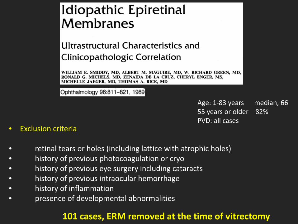
• Exclusion criteria • retinal tears or holes (including lattice with atrophic holes) • history of previous photocoagulation or cryo • history of previous eye surgery including cataracts • history of previous intraocular hemorrhage • history of inflammation • presence of developmental abnormalities
101 cases, ERM removed at the time of vitrectomy
Age: 1-83 years median, 66 55 years or older 82% PVD: all cases

multilaminated basement membrane
RPE cells
1 cell type: 63% of cases Presence of myofibroblast: 63% Blood vessels: 12% Macrophages: 20%

multilaminated basement membrane
RPE cells
1 cell type: 63% of cases Presence of myofibroblast: 63% Blood vessels: 12% Macrophages: 20%
High incidence of RPE cells in idiopathic ERMs
No other cell type in 25% of the cases

multilaminated basement membrane
RPE cells
1 cell type: 63% of cases Presence of myofibroblast: 63% Blood vessels: 12% Macrophages: 20%
High incidence of RPE cells in idiopathic ERMs
No other cell type in 25% of the cases
Data NOT confirmed by subsequent studies

Glial cells and hyalocytes seem to be predominant in ERMs
Hyalocytes
Müller cells
endothelial cells macrophages
a proportion of cells was not labeled with any marker

Fraser-Bell S, et al. Ophthalmology 2003;110:34–40. Ng CH, et al. Ophthalmology 2011;118:694–9.
Studied for years with color fundus images…..
ERM IMAGING
Gass JDM. Macular dysfunction caused by epiretinal membrane contraction. In: Stereoscopic Atlas of Macular Diseases: Diagnosis and Treatment. Vol 2. 4th ed. St Louis, MO: Mosby; 1997:938–950.
Grade 0: Cellophane Maculopathy Grade 1: Crinkled Cellophane Maculopathy Grade 2: Preretinal Macular Fibrosis or Pucker

Nowadays OCT is the gold standard but
multimodal imaging is better
EPIRETINAL MEMBRANES

pre-op post-op
Blue-Fundus AutoFluorescence

cotton-ball sign
abnormalities of the outer retinal bands


disrupted outer retinal bands ???

mfERG P1 amplitude
BCVA

AJO 2017
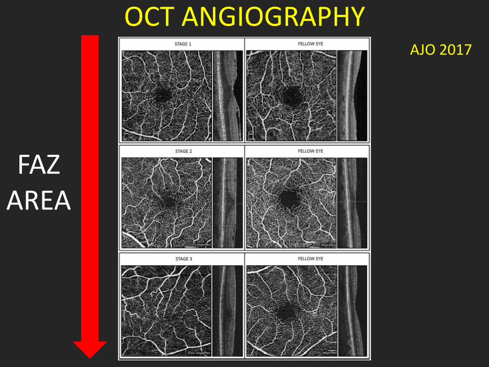
OCT ANGIOGRAPHY
FAZ AREA
AJO 2017

ERM PVR
Müller cells Astrocytes RPE cells

ERM PVR

fingerlike projections
Retina 2014
PVR
Idiopathic

ERM and UVEITIS

2010
2013
2016
B-FAF

Quantiferon test positive for tubercolosis
3 weeks after treatment
B-FAF

ERM and DIABETES Complete PVD is rare and vitreoschisis is common The vitreous cortex is firmly adherent to the ILM Native vitreous collagen forms the scaffold for fibrovascular and fibrocellular proliferation


![Mitchell P Et Al. Ophthalmology 2011;118(4);615-25 [RESTORE]](https://img.dokumen.tips/doc/110x75/55cf984f550346d03396e527/mitchell-p-et-al-ophthalmology-20111184615-25-restore.jpg)