Embed Size (px)
Citation preview

PresentationPresentation
spinal cordspinal cord
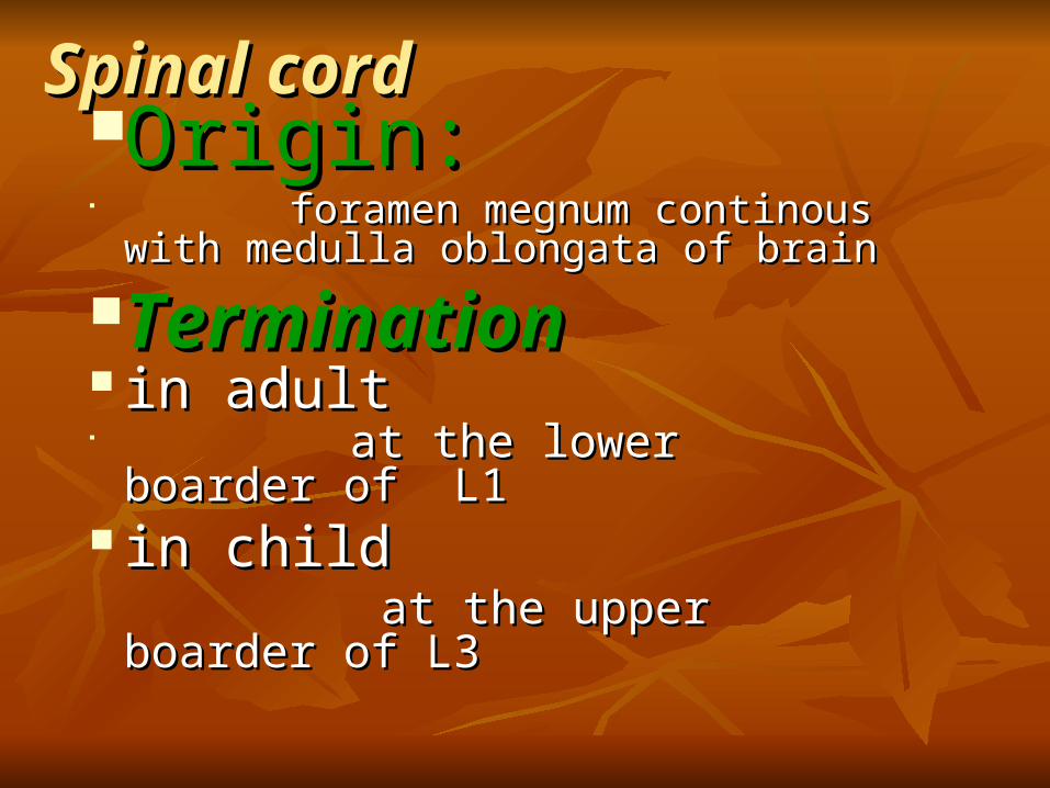
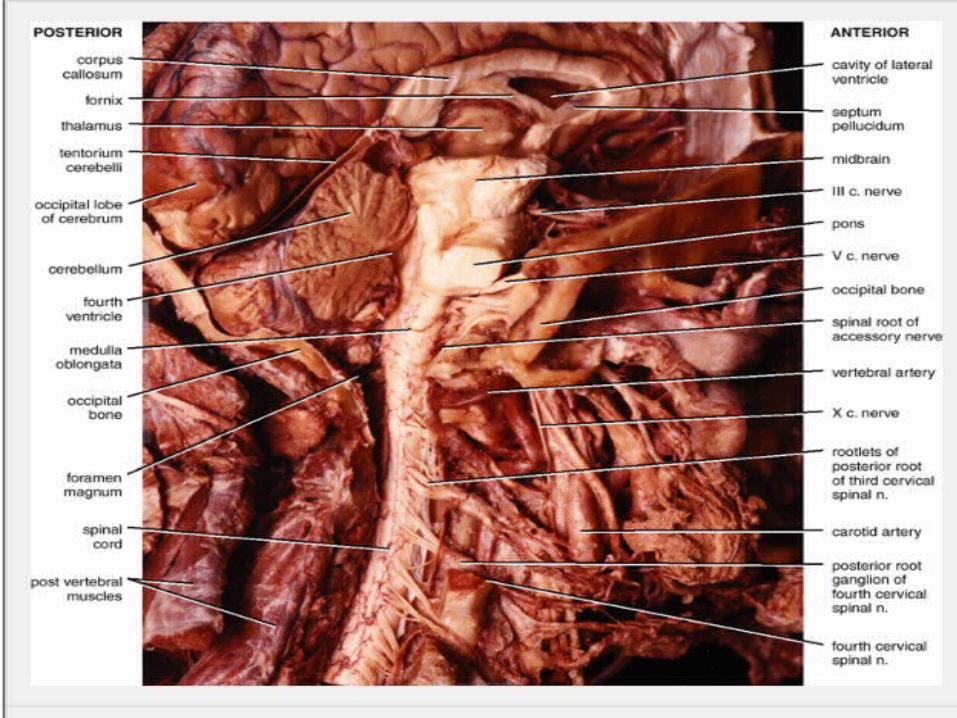
Spinal cordSpinal cordOrigin:Origin:
foramen megnum continous with foramen megnum continous with medulla oblongata of brainmedulla oblongata of brain
TerminationTermination in adultin adult
at the lower boarder of L1at the lower boarder of L1
in childin child at the upper boarder of L3 at the upper boarder of L3

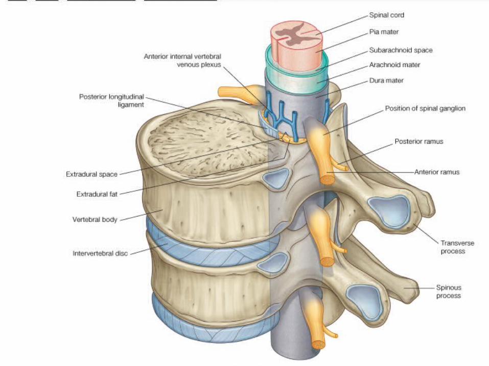
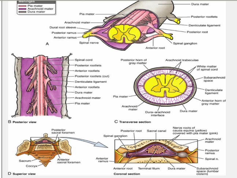
MeningsMeningsThe spinal cord is surrounded by three membranesThe spinal cord is surrounded by three membranes
11 dura materdura mater2 :arachnoid mater2 :arachnoid mater3:pia mater3:pia mater : :
Function’Function’ProtectionProtection Also by cerebrospinal fluid present in the subarachnoid spaceAlso by cerebrospinal fluid present in the subarachnoid space
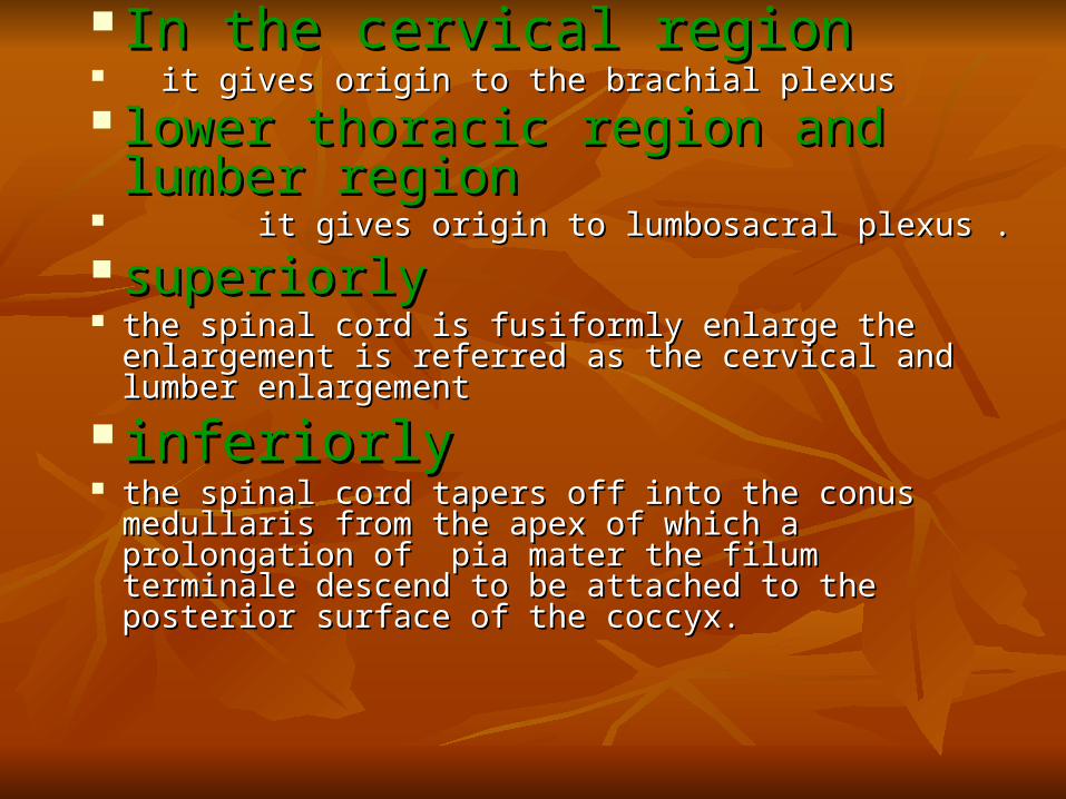
In the cervical regionIn the cervical region it gives origin to the brachial plexus it gives origin to the brachial plexus lower thoracic region and lower thoracic region and
lumber regionlumber region it gives origin to lumbosacral plexus .it gives origin to lumbosacral plexus . superiorlysuperiorly the spinal cord is fusiformly enlarge the enlargement is the spinal cord is fusiformly enlarge the enlargement is
referred as the cervical and lumber enlargement referred as the cervical and lumber enlargement
inferiorly inferiorly the spinal cord tapers off into the conus medullaris from the spinal cord tapers off into the conus medullaris from
the apex of which a prolongation of pia mater the filum the apex of which a prolongation of pia mater the filum terminale descend to be attached to the posterior surface of terminale descend to be attached to the posterior surface of the coccyx. the coccyx.
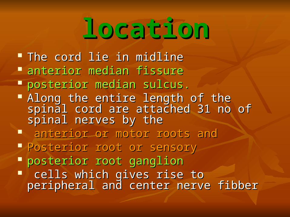
locationlocation The cord lie in midlineThe cord lie in midline anterior median fissure anterior median fissure posterior median sulcus.posterior median sulcus. Along the entire length of the spinal cord are Along the entire length of the spinal cord are
attached 31 no of spinal nerves by theattached 31 no of spinal nerves by the anterior or motor roots andanterior or motor roots and Posterior root or sensoryPosterior root or sensory posterior root ganglionposterior root ganglion cells which gives rise to peripheral and center cells which gives rise to peripheral and center
nerve fibbernerve fibber

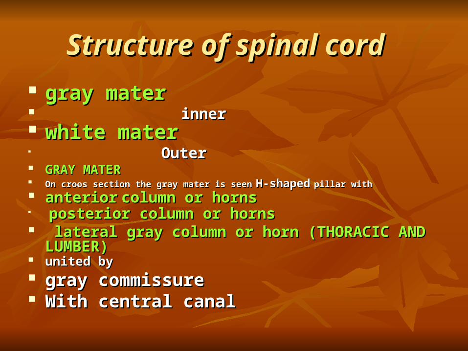
Structure of spinal cordStructure of spinal cord
gray matergray mater innerinner white materwhite mater
OuterOuter
GRAY MATERGRAY MATER On croos section the gray mater is seenOn croos section the gray mater is seen H-shapedH-shaped pillar with pillar with anterioranterior column or hornscolumn or horns
posterior column or hornsposterior column or horns
lateral gray column or horn (THORACIC AND lateral gray column or horn (THORACIC AND LUMBER)LUMBER)
united byunited by
gray commissure gray commissure With central canalWith central canal
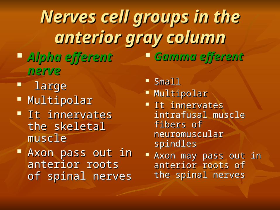
Nerves cell groups in the anterior Nerves cell groups in the anterior gray columngray column
Alpha efferent Alpha efferent nervenerve
large large MultipolarMultipolar It innervates the skeletal It innervates the skeletal
musclemuscle Axon pass out in Axon pass out in
anterior roots of spinal anterior roots of spinal nervesnerves
Gamma efferentGamma efferent
SmallSmall MultipolarMultipolar It innervates intrafusal It innervates intrafusal
muscle fibers of muscle fibers of neuromuscular spindlesneuromuscular spindles
Axon may pass out in Axon may pass out in anterior roots of the anterior roots of the spinal nerves spinal nerves
Nerve cell of the anterior gray column Nerve cell of the anterior gray column is divided into three basic groupsis divided into three basic groups
(1) MEDIAL GROUP(1) MEDIAL GROUP
(2) CENTRAL GROUP(2) CENTRAL GROUP (3) LATERAL GROUP(3) LATERAL GROUP

Medial groupMedial groupEXTENTIONEXTENTION WHOLE SPINAL CORDWHOLE SPINAL CORD innervate innervate muscle of muscle of neck, neck, trunk, trunk, intercostal intercostal abdominalabdominal
(2)Central group:(2)Central group: EXTENTIONEXTENTION
cervical, lumber, sacral segmentscervical, lumber, sacral segments
Three nucleiThree nuclei (a) (a) phrenic nucleusphrenic nucleus(C(C3’4’5)3’4’5)
INERVATEINERVATE DIAGHPHRAMDIAGHPHRAM
(b)(b)accessory nucleusaccessory nucleus) ) (C(C5 OR 6)5 OR 6)
INNERVATIONINNERVATION sternocliedomastoid and trapezius musclesternocliedomastoid and trapezius muscle
(c) lumbosacral nucleus (c) lumbosacral nucleus (L2 TO S1)(L2 TO S1)INNERVATIONINNERVATION unknwon distributionunknwon distribution
Lateral groupLateral group
ExtentionExtention cervical and lumbosacral segmentcervical and lumbosacral segment InnervationInnervation MusclesMuscles (1) upper limb(1) upper limb (2) lower limb(2) lower limb
Nerve cells of the posterior gray Nerve cells of the posterior gray columncolumn
four nerve cell groupfour nerve cell group 1 substantia gelatinosa1 substantia gelatinosa 2 nucleus propius2 nucleus propius 3 nucleus dorsalis (clarks column)3 nucleus dorsalis (clarks column) 4 visceral afferent nucleus4 visceral afferent nucleus First twoFirst two extention extention through out the length of the cord through out the length of the cord other two other two extentionextention lumber and thoracic segmentslumber and thoracic segments

Substantia gelatinosaSubstantia gelatinosa
locationlocation apex of the posterior gray column apex of the posterior gray column composed composed Golgi type 2 neuronGolgi type 2 neuron functionfunction receives afferent fiber associated withreceives afferent fiber associated with pain , pain , temperature temperature touch. touch. Furthermore it receive input from the descending Furthermore it receive input from the descending
fibers from the supraspinal level . fibers from the supraspinal level .
Nucleus propiusNucleus propius
LocationLocation Below s gBelow s g FunctionFunction senses of senses of position position movement (proprioception)movement (proprioception) two points discrimination two points discrimination vibrationvibration
Nucleus dorsalisNucleus dorsalis
LocationLocation base of the posterior gray column base of the posterior gray column extending extending CC8 to 8 to LL3 43 4 FUNCTIONFUNCTION proprioceptive endingsproprioceptive endings
neuromuscular spindles and tendon spindle neuromuscular spindles and tendon spindle
Visceral afferent nucleusVisceral afferent nucleus
LOCATIONLOCATION lateral to the nucleus dorsalislateral to the nucleus dorsalis EXTENTIONEXTENTION TT11 toto L L22
FUNCTIONFUNCTION receiving visceral afferent information receiving visceral afferent information
Nerve cell group lateral gray columnNerve cell group lateral gray column
ExtendExtend TT11 TO S TO S44
CellsCells T1 TO L3T1 TO L3 preganglionic sympathetic nerve fiberpreganglionic sympathetic nerve fiber CELLSCELLS S 2,3,4S 2,3,4 preganglionic parasympathetic preganglionic parasympathetic
fiberfiber
The gray commissure and central The gray commissure and central canalcanal
LOCATIONLOCATION
the anterior and posterior gray columns on each side are connected the anterior and posterior gray columns on each side are connected by a transverse gray commissure so that the gray column r in the by a transverse gray commissure so that the gray column r in the central of the gray commissure is situated central canal.central of the gray commissure is situated central canal.
SuperiorlySuperiorly above this it open into the cavity of the fourth ventricleabove this it open into the cavity of the fourth ventricle continuous with the central canal of the caudal half of the medulla continuous with the central canal of the caudal half of the medulla
oblongataoblongata
InferiorlyInferiorly It is closedIt is closed conus medullaris it expend into the fusiform terminal ventricleconus medullaris it expend into the fusiform terminal ventricle terminate below with in the root of the filum terminaleterminate below with in the root of the filum terminale It is filled with cerebrospinal fluid and is lined with epithelium called It is filled with cerebrospinal fluid and is lined with epithelium called
the ependyma the ependyma
IT resembles letter HIT resembles letter H posterior gray commissureposterior gray commissure The part of the gray commissure that is The part of the gray commissure that is
situated posterior gray canalsituated posterior gray canal
Anterior gray commisureAnterior gray commisure lie anterior to the canallie anterior to the canal

White materWhite mater It is divided into It is divided into anterior lateral anterior lateral posterior white columns or finiculi. posterior white columns or finiculi.
anterior columnanterior column locationlocation lie on each side lie in between the midline and the point of emergence of lie on each side lie in between the midline and the point of emergence of
the anterior nerve root .the anterior nerve root .
lateral columnlateral column locationlocation between the emergence of the anterior nerve root and the entry of the between the emergence of the anterior nerve root and the entry of the
posterior nerve root the posterior nerve root the
posterior columnposterior column locationlocation in between the entry of posterior nerve root and midline in between the entry of posterior nerve root and midline

Structure Structure
compositioncomposition in centrral nervous system the white mater of spinal in centrral nervous system the white mater of spinal
cord consist of a mixture of cord consist of a mixture of nerve fibernerve fiber neuroglia neuroglia blood vessel blood vessel it surrounds the gray mater it surrounds the gray mater its white color is due to the high proportion of its white color is due to the high proportion of
myelinated nerve fibermyelinated nerve fiber

Blood Supply of the Spinal CordBlood Supply of the Spinal Cord The spinal cord receives its arterial supply from three small, The spinal cord receives its arterial supply from three small,
longitudinally running arteries: longitudinally running arteries: the two posterior spinal arteries the two posterior spinal arteries and and one anterior spinal artery. one anterior spinal artery. The posterior spinal arteries, The posterior spinal arteries, which arise either directly or indirectly from the vertebral which arise either directly or indirectly from the vertebral arteries, run down the side of the spinal cord, close to the arteries, run down the side of the spinal cord, close to the attachments of the posterior spinal nerve roots. The anterior attachments of the posterior spinal nerve roots. The anterior spinal arteries, which arise from the vertebral arteries, unite to spinal arteries, which arise from the vertebral arteries, unite to form a single artery, which runs down within the anterior form a single artery, which runs down within the anterior median fissure.median fissure.
The posterior and anterior spinal arteries are The posterior and anterior spinal arteries are reinforced by reinforced by radicular arteriesradicular arteries, which enter the vertebral canal through the , which enter the vertebral canal through the intervertebral foramina.intervertebral foramina.
The veins The veins of the spinal cord drain into the internal vertebral of the spinal cord drain into the internal vertebral venous plexus.venous plexus.

INJURIESINJURIES
Injuries in childrenInjuries in children
Children account for 1-10% of all spinal Children account for 1-10% of all spinal injuries.injuries.
Motor vehicle accidents account for most Motor vehicle accidents account for most injuries, followed by falls and sports.injuries, followed by falls and sports.
Most serious spinal injuries in children involve Most serious spinal injuries in children involve the cervical spine.the cervical spine.
In children less than 8 years of age, most In children less than 8 years of age, most injuries are between the occiput and C2:injuries are between the occiput and C2: Fulcrum of movement located Fulcrum of movement located at C2-3 in childrenat C2-3 in children, ,
C5-6 in adultsC5-6 in adults Significant ligamentous and joint capsule laxitySignificant ligamentous and joint capsule laxity Relatively large head and weak neck musclesRelatively large head and weak neck muscles Horizontal orientation of facet jointsHorizontal orientation of facet joints Incomplete ossicification of odontoid processIncomplete ossicification of odontoid process
Injuries in Injuries in
adultsadults

Terminology Terminology
PlegiaPlegia = complete lesion = complete lesion Paresis Paresis = some muscle strength is preserved= some muscle strength is preserved Tetraplegia Tetraplegia (or quadriplegia)(or quadriplegia)
Injury of the cervical spinal cordInjury of the cervical spinal cord Patient can usually still move his arms using the segments above Patient can usually still move his arms using the segments above
the injury (e.g., in a C7 injury, the patient can still flex his the injury (e.g., in a C7 injury, the patient can still flex his forearms, using the C5 segment)forearms, using the C5 segment)
ParaplegiaParaplegia Injury of the thoracic or lumbo-sacral cord, or cauda equinaInjury of the thoracic or lumbo-sacral cord, or cauda equina
HemiplegiaHemiplegia Paralysis of one half of the bodyParalysis of one half of the body Usually in brain injuries (e.g., stroke)Usually in brain injuries (e.g., stroke)
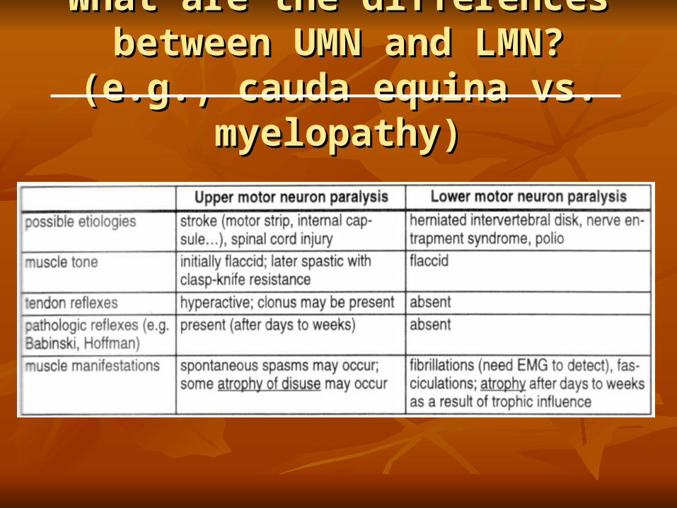
What are the differences between What are the differences between UMN and LMN? (e.g., cauda equina UMN and LMN? (e.g., cauda equina
vs. myelopathy)vs. myelopathy)
Thoracic injuries (T2-L1)Thoracic injuries (T2-L1)
Paraparesis or paraplegiaParaparesis or paraplegia UMN (upper motor neuron) signsUMN (upper motor neuron) signs
Cauda equina injuries (L2 or Cauda equina injuries (L2 or below)below)
Paraparesis or paraplegiaParaparesis or paraplegia LMN (lower motor neuron) signsLMN (lower motor neuron) signs Thigh flexion is almost always preserved to Thigh flexion is almost always preserved to
some degreesome degree

What is the difference between What is the difference between cauda equina and conus medullaris cauda equina and conus medullaris
syndrome?syndrome?