Embed Size (px)
Citation preview
PRIMARY RESEARCH PAPER
Presence and genetic diversity of microcystin-producingcyanobacteria (Anabaena and Microcystis) in Lake Kotokel(Russia, Lake Baikal Region)
Olga I. Belykh • Ekaterina G. Sorokovikova • Galina A. Fedorova •
Oksana V. Kaluzhnaya • Evgeniya S. Korneva • Maria V. Sakirko •
Tatyana A. Sherbakova
Received: 25 October 2010 / Revised: 19 April 2011 / Accepted: 26 April 2011 / Published online: 8 May 2011
� Springer Science+Business Media B.V. 2011
Abstract A survey was conducted for the presence of
cyanobacteria toxins in Lake Kotokel due to a few cases
of Haff disease registered in 2008–2009 caused by
consumption of fish from Lake Kotokel, and wildlife
mortality including large fish kill. The aims of this study
were to determine what cyanotoxins (if any) were
present in the lake, to describe phytoplankton compo-
sition including morphology, density, and species
diversity of cyanobacteria, as well as to evaluate the
trophic state of the lake. Samples were collected from
both nearshore and central sites in August of 2009.
Aphanocapsa holsatica dominated the phytoplankton.
The presence of toxigenic genotypes of Microcystis spp.
and Anabaena lemmermannii was detected by sequenc-
ing of PCR-amplified aminotransferase domain of
microcystin synthetase gene. LR, RR, and YR micro-
cystin (MC) variants were detected with liquid chroma-
tography-UV mass spectrometry. The data do not shed
light on the etiology of Haff disease in Lake Kotokel
region, nevertheless taking into account the recreational
importance of the lake and its direct connection to Lake
Baikal, a necessity to monitor cyanobacteria in these
water bodies is evident. This is the first report on
simultaneous detection of MC-producing genotypes and
MCs in the Lake Baikal region.
Keywords Toxic cyanobacteria � Microcystin �mcyE gene � HPLC � LC/MS � Lake Kotokel �Lake Baikal
Introduction
Cyanobacteria produce a range of secondary bioactive
metabolites, including toxins. One of the best studied
cyanotoxins in freshwater ecosystems is the group of
microcystins (MCs). MCs are cyclic heptapeptides
produced nonribosomally by peptide synthetases, poly-
ketide synthases, and tailoring enzymes. Over 90
variants of MCs are known (Welker & Von Dohren,
2006), the most common of them differ in L-amino acids
and in modification by methyl groups. MCs specifically
inhibit the eukaryotic serine/threonine phosphatases 1
and 2A rendering the hyperphosphorylation of proteins
in the hepatocyte cytoskeleton, which leads to massive
hepatocyte necrosis and pooling of blood into the liver
(MacKintosh et al., 1990). Chronic exposure to sublethal
doses of MCs resulted in endemic cancer of the liver in
one of the provinces of China (Chorus & Bartram, 1999).
Predominant MC-producing cyanobacteria belong
to the Anabaena, Microcystis, Planktothrix, Anabaen-
opsis, Hapalosiphon, and Nostoc genera (Chorus &
Bartram, 1999). Planktonic cyanobacteria, including
Handling editor: Luigi Naselli-Flores
O. I. Belykh (&) � E. G. Sorokovikova �G. A. Fedorova � O. V. Kaluzhnaya �E. S. Korneva � M. V. Sakirko � T. A. Sherbakova
Limnological Institute of the Siberian Branch of RAS,
3, Ulan-Batorskaya St, Irkutsk, Russia 664033
e-mail: [email protected]
123
Hydrobiologia (2011) 671:241–252
DOI 10.1007/s10750-011-0724-2
unicellular colony forming Microcystis and the fila-
mentous, heterocystous Anabaena, commonly form
toxic blooms in highly productive freshwater ecosys-
tems worldwide (Codd et al., 2005). Therefore, many
countries monitor the presence and safe concentrations
of highly toxic MC-LR, which contains leucine and
arginine as variable L-amino acids. The LD50 (lethal
dose, 50%) of MC-LR (given intraperitoneally to mice)
is 50 lg kg-1, MC-RR- (arginine–arginine) and MC-
YR- (tyrosine–arginine) variants are less toxic. The
World Health Organization (1998) has set an advisory
level for MCs in drinking water of 1.0 lg l-1 MC-LR.
Several detection methods for MCs are commonly
used, including high performance liquid chromatog-
raphy (HPLC), immunoassays, and protein phospha-
tase inhibition assays (reviewed in Carmichael & An,
1999). However, liquid chromatography–mass spec-
trometry (LC/MS) provides a greater specificity and
sensitivity (Chorus & Bartram, 1999).
Identification of a gene locus of microcystin
synthetase provided a powerful tool to detect
potential toxigenic species (Nishizawa et al., 1999;
Rouhiainen et al., 2004). MCs producing multien-
zyme complex is encoded by a 55 kb-long gene
cluster containing 10 genes designated mcyA–mcyJ
(Rouhiainen et al., 2004). One of the enzymes, the
aminotransferase (AMT) plays a crucial role in
cyanotoxin producing by transferring amino groups
to the side chain of the Adda moiety (Moffitt &
Neilan, 2004). The gene for AMT domain locating
between microcystin synthetase gene E and nodularin
synthetase gene F has been found suitable for the
detection of MC or nodularin producing Microcystis,
Planktothrix, Anabaena, Nostoc and Nodularia
strains (Jungblut & Neilan, 2006). In phylogenetic
analysis, mcyE sequences from different toxin pro-
ducer genera form their own groups and are not
affected by lateral gene transfer (Rantala et al., 2004).
Thus, the primers for AMT region are suitable for
reliable detection and identification of main cyano-
toxin producers in environmental samples.
Lake Kotokel is situated close to the eastern coast of
Lake Baikal (Eastern Siberia, Fig. 1). In 2008, mass
mortality of fish, birds, and domestic animals was
recorded in Lake Kotokel and its neighborhoods.
Sixteen persons were hospitalized with symptoms of
Haff disease: severe muscular pains, rhabdomyolysis
(rapid breakdown of skeletal muscle), and myoglobi-
nuria. The etiology is not yet known, but there is little
doubt that the disease is due to an unknown, natural
toxin. In June of 2009, the Ministry of Health and Social
Development of Russia forbade eating fish caught in the
lake, as well as bathing in and drinking the water.
Analysis of phytoplankton samples obtained in Lake
Kotokel in August of 2008 revealed the presence of
potentially toxic Microcystis bearing microcystin syn-
thetase gene E, however, the analysis for the appearance
of MCs in the water samples was not performed (Belykh
et al., 2010). Therefore, Lake Kotokel became a subject
of the interdisciplinary investigation.
Lake Kotokel was thoroughly studied in the
1950–1980s due to its recreational and mainly com-
mercial value for the region—the annual fish catch
exceeded 1,200 tons (Kuzmich, 1988). It was a
eutrophic water body, cyanobacteria were the most
abundant phytoplankton group, and Gloeotrichia eh-
inulata was the dominant species (Korde, 1968;
Kuzmich, 1988). Microcystis aeruginosa, M. pulverea,
and Anabaena lemmermannii proliferated less inten-
sively. In summer, the water transparency dropped to
0.6–0.8 m and the water temperature sometimes
reached 25.8�C. Mass development of phytoplankton
in July–August resulted in a decrease of phosphate and
mineral nitrogen concentration; in other seasons, the
content of these nutrients was higher (Kuzmich, 1988).
In Siberia, the first report on finding of microcystin
synthetase genes for potentially toxic M. aeruginosa
and Anabaena sp. was concerned to Ust-Ilim and
Bratsk reservoirs fall within of Lake Baikal
watershed (Tikhonova et al., 2006, 2007). However,
mcy genes have not been detected in any samples
either from the pelagic zone or the shallow areas of
Lake Baikal.
The aim of this work was to comprehensively
investigate the phytoplankton of Lake Kotokel includ-
ing morphology, density, and species diversity of
cyanobacteria, to evaluate the trophic state of the lake,
to measure the concentration of different variants of the
MCs (if they were), as well to find potential toxin
producers by the PCR-amplified mcyE genes.
Materials and methods
Site description
Lake Kotokel is located in the south-central region of
Siberia (Russia), 2 km off the eastern coast of Lake
242 Hydrobiologia (2011) 671:241–252
123
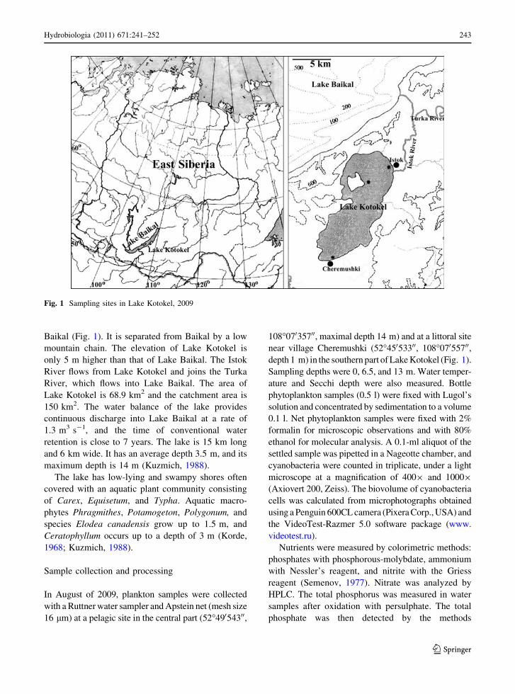
Baikal (Fig. 1). It is separated from Baikal by a low
mountain chain. The elevation of Lake Kotokel is
only 5 m higher than that of Lake Baikal. The Istok
River flows from Lake Kotokel and joins the Turka
River, which flows into Lake Baikal. The area of
Lake Kotokel is 68.9 km2 and the catchment area is
150 km2. The water balance of the lake provides
continuous discharge into Lake Baikal at a rate of
1.3 m3 s-1, and the time of conventional water
retention is close to 7 years. The lake is 15 km long
and 6 km wide. It has an average depth 3.5 m, and its
maximum depth is 14 m (Kuzmich, 1988).
The lake has low-lying and swampy shores often
covered with an aquatic plant community consisting
of Carex, Equisetum, and Typha. Aquatic macro-
phytes Phragmithes, Potamogeton, Polygonum, and
species Elodea canadensis grow up to 1.5 m, and
Ceratophyllum occurs up to a depth of 3 m (Korde,
1968; Kuzmich, 1988).
Sample collection and processing
In August of 2009, plankton samples were collected
with a Ruttner water sampler and Apstein net (mesh size
16 lm) at a pelagic site in the central part (52�49054300,
108�07035700, maximal depth 14 m) and at a littoral site
near village Cheremushki (52�45053300, 108�07055700,depth 1 m) in the southern part of Lake Kotokel (Fig. 1).
Sampling depths were 0, 6.5, and 13 m. Water temper-
ature and Secchi depth were also measured. Bottle
phytoplankton samples (0.5 l) were fixed with Lugol’s
solution and concentrated by sedimentation to a volume
0.1 l. Net phytoplankton samples were fixed with 2%
formalin for microscopic observations and with 80%
ethanol for molecular analysis. A 0.1-ml aliquot of the
settled sample was pipetted in a Nageotte chamber, and
cyanobacteria were counted in triplicate, under a light
microscope at a magnification of 4009 and 10009
(Axiovert 200, Zeiss). The biovolume of cyanobacteria
cells was calculated from microphotographs obtained
using a Penguin 600CL camera (Pixera Corp., USA) and
the VideoTest-Razmer 5.0 software package (www.
videotest.ru).
Nutrients were measured by colorimetric methods:
phosphates with phosphorous-molybdate, ammonium
with Nessler’s reagent, and nitrite with the Griess
reagent (Semenov, 1977). Nitrate was analyzed by
HPLC. The total phosphorus was measured in water
samples after oxidation with persulphate. The total
phosphate was then detected by the methods
Fig. 1 Sampling sites in Lake Kotokel, 2009
Hydrobiologia (2011) 671:241–252 243
123
described above for inorganic phosphate. Determina-
tion of permanganate and dichromate oxidizability
was carried out by titration in an acid medium.
The concentration of chlorophyll a was measured by
spectrophotometry. Samples (0.5 l) were filtered onto
0.45 lm Millipore polycarbonate filters, extracted
using hot methanol, and then analysed with a spectro-
photometer (Bio-Rad SmartSpec Plus, USA).
Microcystin analyses
Net samples of phytoplankton were frozen and thawed
(5 times) and then dried (60�C) prior to toxin
extraction. The dried phytoplankton biomass
(178.2 mg) was extracted twice (10 ml each time)
with methanol by sonication in an ultrasonic bath
(1 h). The combined raw extracts were centrifuged (at
5,263 rpm for 15 min), and the supernatant was dried
in a rotary vacuum evaporator IKA RV 05-ST (Werke
GmbH & Co. KG, Germany). The extract obtained was
redissolved in 0.5 ml of methanol prior to Liquid
chromatography–mass spectrometry analysis. LC/MS
analyses were performed on an Agilent HP1200
chromatographic system that was coupled to an
Agilent time-of-flight mass spectrometer (ESI-TOF)
equipped with an electrospray ionization source oper-
ating in the positive ionization mode. Chromato-
graphic separation of MCs was carried out using a
Zorbax 300 SB C18 column (2.1 9 150 mm, 5 lm)
maintained at 35�C, at a flow rate of 0.3 ml min-1,
with a gradient of 0.1% formic acid in water (solvent
A) and 0.1% formic acid in acetonitrile (solvent B).
The solvent program was as follows: 10% B at time
zero, 10-80% B for 30 min, 100% B for 5 min, and
100% B for 40 min. The injected sample volume was
10 ll, and the detection was performed with a diode
array UV detector at 214, 222, 238, and 330 nm.
Quantification was achieved by measuring the chro-
matographic signals of each MC against calibration
curve produced from ‘‘Microcystins-LR standard’’
(Biosense Laboratories, Norway), as they have the
same extinction coefficients as MC-LR.
Genetic analyses
Amplification, cloning, and sequencing
DNA from the phytoplankton samples was extracted
using ‘‘DNA-sorb’’ extraction kit (InterLabService,
Russia). Mcy gene fragments were PCR amplified
with the HepF and HepR gene specific primers
(Jungblut & Neilan, 2006) in the Peltier Thermal
Cycler (MJ Research, USA). PCR was performed in
50 ll reaction mixtures containing 0.1–0.4 lM of
each primer, 0.25 mM of dNTPs, 2.5 mM of MgCl2,
buffer (20 mM TrisCl, pH 8.4, KCl 40 mM), Taq
DNA polymerase (1.2 units) (Fermentas, MD, USA),
and 10–15 ng of DNA template. Cycling conditions
were as follows: initial denaturation at 95�C for
5 min, then 35 cycles of 95�C for 0.5 min, 55�C for
0.5 min, and 72�C for 1 min followed by 10 min at
72�C to complete the extension. PCR products of
expected size (*470 bp) were gel-purified using the
‘‘PCR Clean-Up Gel Extraction NucleoSpin Extract
II kit’’ (Macherey-Nagel, Germany) and subcloned
into E. coli XL1BL cells using ‘‘InsTAclone’’
(Fermentas, MD, USA). 100 cloned amplicons were
processed for sequencing. Plasmid inserts were
sequenced bidirectionally by using vector-specific
primers on the CEQ 8800 Genetic Analysis System
and using DTSC Quick Start Kit (Beckman Coulter,
USA). All unique sequences from this study have
been deposited in GenBank under accession numbers
GU186843–GU186846.
Phylogenetic analyses
Newly decoded mcyE gene sequences were used in a
tBLASTn search against the NCBI ‘‘nr’’ database
(http://www.ncbi.nlm.nih.gov/BLAST/) to find close
relatives. The most similar reference sequences,
preferably of cultivated strains, were downloaded and
aligned with Kotokel isolate sequences using the
Clustal W program (MEGA software; Tamura et al.,
2007). The data set was analyzed by neighbor-joining
(NJ), maximum likelihood (ML), and Bayesian (BI)
methods to infer trees and estimate branch support.
Evolutionary distances for the NJ tree (MEGA soft-
ware; Tamura et al., 2007) were calculated by the
Kimura-2 parameter model; the tree topology was
evaluated by bootstrap support in 1,000 replicons.
ML calculations were performed using PhyML soft-
ware (Guindon & Gascuel, 2003), and the most
appropriate evolutionary model (GTR ? G) for the
nucleotide sequences was set. The model was
generated by the Model Generator program (http://
distributed.cs.nuim.ie/multiphyl.php). BI tree topol-
ogy was calculated using a Bayesian Markov chain
244 Hydrobiologia (2011) 671:241–252
123
Monte Carlo method implemented by the program
Mr. Bayes v3.1.2. (Ronquist & Huelsenbeck, 2003).
One million generations were run, and the likelihood
function stabilized after 2,000 generations; these trees
were discarded, and every hundredth of the remaining
trees was used to drive the final consensus tree
according to the 50% majority rule.
Results
Environmental parameters
Temperature, Secchi disk transparency, concentra-
tions of total phosphorus, nutrients, permanganate
and dichromate oxidation, and chlorophyll a in the
Lake Kotokel pelagic area are presented in Table 1.
Based on these data, the trophic state of Lake
Kotokel was assigned as eutrophic according to the
Vollenweider & Kerekes classification (1982).
Intracellular microcystin concentration
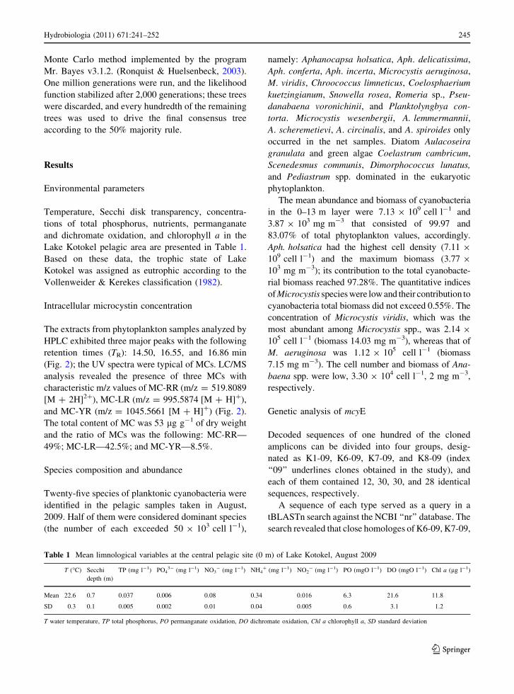
The extracts from phytoplankton samples analyzed by
HPLC exhibited three major peaks with the following
retention times (TR): 14.50, 16.55, and 16.86 min
(Fig. 2); the UV spectra were typical of MCs. LC/MS
analysis revealed the presence of three MCs with
characteristic m/z values of MC-RR (m/z = 519.8089
[M ? 2H]2?), MC-LR (m/z = 995.5874 [M ? H]?),
and MC-YR (m/z = 1045.5661 [M ? H]?) (Fig. 2).
The total content of MC was 53 lg g-1 of dry weight
and the ratio of MCs was the following: MC-RR—
49%; MC-LR—42.5%; and MC-YR—8.5%.
Species composition and abundance
Twenty-five species of planktonic cyanobacteria were
identified in the pelagic samples taken in August,
2009. Half of them were considered dominant species
(the number of each exceeded 50 9 103 cell l-1),
namely: Aphanocapsa holsatica, Aph. delicatissima,
Aph. conferta, Aph. incerta, Microcystis aeruginosa,
M. viridis, Chroococcus limneticus, Coelosphaerium
kuetzingianum, Snowella rosea, Romeria sp., Pseu-
danabaena voronichinii, and Planktolyngbya con-
torta. Microcystis wesenbergii, A. lemmermannii,
A. scheremetievi, A. circinalis, and A. spiroides only
occurred in the net samples. Diatom Aulacoseira
granulata and green algae Coelastrum cambricum,
Scenedesmus communis, Dimorphococcus lunatus,
and Pediastrum spp. dominated in the eukaryotic
phytoplankton.
The mean abundance and biomass of cyanobacteria
in the 0–13 m layer were 7.13 9 109 cell l-1 and
3.87 9 103 mg m-3 that consisted of 99.97 and
83.07% of total phytoplankton values, accordingly.
Aph. holsatica had the highest cell density (7.11 9
109 cell l-1) and the maximum biomass (3.77 9
103 mg m-3); its contribution to the total cyanobacte-
rial biomass reached 97.28%. The quantitative indices
of Microcystis species were low and their contribution to
cyanobacteria total biomass did not exceed 0.55%. The
concentration of Microcystis viridis, which was the
most abundant among Microcystis spp., was 2.14 9
105 cell l-1 (biomass 14.03 mg m-3), whereas that of
M. aeruginosa was 1.12 9 105 cell l-1 (biomass
7.15 mg m-3). The cell number and biomass of Ana-
baena spp. were low, 3.30 9 104 cell l-1, 2 mg m-3,
respectively.
Genetic analysis of mcyE
Decoded sequences of one hundred of the cloned
amplicons can be divided into four groups, desig-
nated as K1-09, K6-09, K7-09, and K8-09 (index
‘‘09’’ underlines clones obtained in the study), and
each of them contained 12, 30, 30, and 28 identical
sequences, respectively.
A sequence of each type served as a query in a
tBLASTn search against the NCBI ‘‘nr’’ database. The
search revealed that close homologes of K6-09, K7-09,
Table 1 Mean limnological variables at the central pelagic site (0 m) of Lake Kotokel, August 2009
T (�C) Secchi
depth (m)
TP (mg l-1) PO43- (mg l-1) NO3
- (mg l-1) NH4? (mg l-1) NO2
- (mg l-1) PO (mgO l-1) DO (mgO l-1) Chl a (lg l-1)
Mean 22.6 0.7 0.037 0.006 0.08 0.34 0.016 6.3 21.6 11.8
SD 0.3 0.1 0.005 0.002 0.01 0.04 0.005 0.6 3.1 1.2
T water temperature, TP total phosphorus, PO permanganate oxidation, DO dichromate oxidation, Chl a chlorophyll a, SD standard deviation
Hydrobiologia (2011) 671:241–252 245
123

and K8-09 sequences belong to Microcystis spp.,
whereas Anabaena spp. are the nearest relatives of the
K1-09 sequence. Pair-wise comparisons of mcyE gene
fragments of cyanobacteria isolated from geographi-
cally distinct freshwater bodies, including Lake
Kotokel, show that Microcystis spp. differ by no more
than 4% of nucleotide changes, whereas the dissimi-
larities in sequences of Anabaena and Microcystis
genera reach up to 21% of the substitutions. K6-09 and
K7-09 mcyE gene sequences share up to 98–99%
identity with the strains of M. viridis NIES-102,
M. aeruginosa NIES-843, M. aeruginosa K-139, and
M. wesenbergii NIES-107 isolated during toxic bloom
from Lakes Kasumigaura and Kawaguchi in Japan.
K8-09 fragment shows 99% similarity with sequences
of some strains of M. aeruginosa (PCC 7806, PCC
7005, UTEX B 2667) isolated from different locations
in Europe and North America. K1-09 shows 100%
homology with many of the sequences of Anabaena-
species found in 9 Scandinavian lakes, for example
Anabaena sp. SYKE 971/6 (Lake Kotojarvi, Finland)
or A. lemmermannii NIVA-CYA 270/1 (Arefjordsvat-
net, Norway).
As all tree-constructing methods resulted in sim-
ilar topologies of the mcyE gene phylogeny, the
unrooted neighbor-joining tree only is shown in
Fig. 3. The branching of mcyE genes corresponds to
highly supported diversification according to
14 15 16 17 18
Time, min
100
150
200
250
300
Ab
sorb
ance
214
238
13
2
A
519 520 521 522
m/z
0
200000
400000
600000
800000
1000000
520,3090
519,8089
520,8100
B
1044 1046 1048 1050
m/z
0
200000
400000
600000
800000
1000000
1045,5661
1046,5685
1047,5713
C
994 996 998 1000
m/z
0
400000
800000
1200000
1600000
995,5874
996,5891
997,5910
D
Fig. 2 HPLC/MS determination of microcystins extracted from
the Lake Kotokel phytoplankton (for experimental details see
text). A HPLC/UV chromatogram of raw extract of microcystins:
1 microcystin-RR (TR = 14.50 min), 2 microcystin-YR (TR =
16.55 min), 3 microcystin-LR (TR = 16.86 min). B–D ESI-TOF
mass spectra for the peaks 1 (B), 2 (C), 3 (D)
246 Hydrobiologia (2011) 671:241–252
123
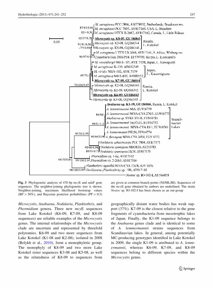
Microcystis, Anabaena, Nodularia, Planktothrix, and
Phormidium genera. Three new mcyE sequences
from Lake Kotokel (K6-09, K7-09, and K8-09
sequences) are reliable examples of the Microcystis
genus. The internal relationships of the Microcystis
clade are uncertain and represented by threefold
polytomies. K6-09 and two more sequences from
Lake Kotokel (K1-08 and K2-08), isolated in 2008
(Belykh et al., 2010), form a monophyletic group.
The monophyly of K8-09 and two more Lake
Kotokel sister sequences K3-08 and K5-08, as well
as the relatedness of K8-09 to sequences from
geographically distant water bodies has weak sup-
port (37%). K7-09 is the closest relative to the gene
fragments of cyanobacteria from mesotrophic lakes
of Japan. Finally, the K1-09 sequence belongs to
the Anabaena genus clade and is identical to some
of A. lemmermannii strains sequences from
Scandinavian lakes. In general, among potentially
MC-producing genotypes identified in Lake Kotokel
in 2009, the single K1-09 is attributed to A. lemm-
ermannii, whereas K6-09, K7-09, and K8-09
sequences belong to different species within the
Microcystis genus.
Fig. 3 Phylogenetic analysis of 470 bp mcyE and ndaF gene
sequences. The neighbor-joining phylogenetic tree is shown.
Neighbor-joining, maximum likelihood bootstrap values
(BP [ 50%), and Bayesian posterior probabilities (PP [ 0.5)
are given at common branch points (NJ/ML/BI). Sequences of
the mcyE gene obtained by authors are underlined. The strain
Nostoc sp. IO-102-I has been chosen as an out-group
Hydrobiologia (2011) 671:241–252 247
123
Discussion
Lake Kotokel is a eutrophic water body, as stated
earlier (Kuzmich, 1988), and confirmed by our data
on the concentration of total phosphorus, content of
chlorophyll a and water transparency. In the summer
of 2009, the concentrations of phosphate and mineral
nitrogen were lower 1.7 and 3.6 times, accordingly, in
comparison with the 1990s (N. M. Pronin, personal
communication). It was probably caused by some
reduced anthropogenic impact on the lake due to the
depletion of agricultural activity in this area. More-
over, in the summer of 2009, the low concentrations
of nutrients in Lake Kotokel might be attributed not
only to the phytoplankton bloom but also to the high
development of aquatic plants, which are diverse in
the lake.
An invasive macrophyte Elodea canadensis,
unlike in most water bodies of Siberia, proliferates
in Lake Kotokel, especially in the coastal zone near
camp sites and residential buildings. This species
facilitates the utilization of the nutrients in the lake,
preventing the transition of the lake to the hypereu-
trophic state (Kuzmich, 1988). It has been shown that
there are competitive relationships between cyano-
bacteria and aquatic macrophytes—the major primary
producers in small eutrophic lakes (Li et al., 2009).
Aquatic plants such as E. canadensis, Ceratophyllum,
and Phragmithes spp. overgrown the lake are known
to accumulate and transform MCs (Pflugmacher
et al., 1999, 2001). Considering that the littoral zone
covered by aquatic plants extends to a depth of 2 m
and occupies 23% of the Lake Kotokel surface
(Kuzmich, 1988), it is possible that macrophytes can
decrease the content of toxins in the water.
Aphanocapsa holsatica (previously known as
Microcystis holsatica) was a dominant species of
planktonic cyanobacteria in the lake in August of
2009 (97.28% of total abundance), as well as in the
summer of the previous year (53% of total abun-
dance, Belykh et al., 2010). This species is nontoxic
(Yasuno et al., 1998) and differs in 16S rDNA
sequence from the Microcystis genus, despite minor
morphometric differences (Neilan et al., 1997).
Previously, the morphological similarity of Aph.
holsatica and Microcystis spp. probably often led to
their misidentification in Lake Kotokel samples. In
August of 2008, besides Aph. holsatica, the poten-
tially toxic M. viridis also dominated in the
phytoplankton of Lake Kotokel (36% of total abun-
dance). Other cyanobacteria species, including
M. aeruginosa, made up less than 5% of the total
number (Belykh et al., 2010). As there were no data
on water chemistry in August of 2008, we can
speculate only about the decreasing of the develop-
ment of Microcystis compared to August of 2009.
Heavy organic input from nearby camp sites and
residential buildings might result in the dominance of
M. viridis as a more resistant beta-mesosaprob to
pollution (Barinova et al., 2006). Its abundance
reduced in 2009 possibly due to the ban of the
recreational use of the lake.
In 2009, the samples were collected in August,
after the bloom. The development of cyanobacteria
was low (3.87 g m-3), and the biomass was 4 times
lower when compared to the maximal biomass
recorded in July 1986 by other authors (Kuzmich,
1988). The chlorophyll a concentration was high due
to abundant development of cyanobacteria (*80% of
total biomass) and also to the impact of green and
diatom algae. Previous results (Korde, 1968;
Kuzmich, 1988) and our survey show that a complex
of species typical of shallow well-warmed produc-
tive water bodies has been developed in Lake
Kotokel over the last 50 years. It is characterized
by a high abundance of diatoms, green algae and
cyanobacteria.
The plankton cyanobacteria of Lake Kotokel like
Microcystis aeruginosa, M. viridis, M. wesenbergii,
A. lemmermannii, A. flos-aquae, and A. circinalis are
known as potential MC producers. We amplified 10
diverse cyanobacterial sequences of microcystin
synthetase genes from Lake Kotokel samples col-
lected during 2 years of our observations (Belykh
et al., 2010): nine of them belonged to Microcystis
genus, the tenth was identical to A. lemmermannii.
The total number of clones containing the Microcys-
tis gene insert was 9 times higher than those with the
Anabaena gene. Similarly, in lakes of Finland where
Anabaena and Microcystis often form hepatotoxic
mass occurrences, Microcystis mcyE gene copy
numbers were more abundant than those of Ana-
baena; both toxic and nontoxic strains of Anabaena
existed, whereas all strains of Microcystis spp. were
toxic (Vaitomaa et al., 2003). The data correlate with
records that Microcystis-species are the most com-
mon and significant MC producers in freshwater
environments. Eighty to ninety percent of the samples
248 Hydrobiologia (2011) 671:241–252
123
from the water bodies of Denmark, Germany, Czech
Republic, and Korea, where Microcystis dominated,
contained MCs (Chorus & Bartram, 1999). Coexis-
tence of toxigenic Microcystis and Anabaena species,
like in Lake Kotokel, was detected in 15% of
mesotrophic and 25% of eutrophic lakes in Finland
(Rantala et al., 2006). In four other examined
Siberian water bodies, the genera occurred as well
(Tikhonova et al., 2006, 2007). In oligotrophic Lake
Baikal and Irkutsk reservoir, cyanobacteria were
nontoxic. In a mesotrophic Bratsk reservoir, the
mcyA genes were found in Anabaena species. In Ust-
Ilim reservoir, which is a mesotrophic also, both
mcyA and mcyE genes of Microcystis genus were
detected (Tikhonova et al., 2006, 2007). Thus,
simultaneous development of toxic cyanobacteria of
the both genera was observed only in the eutrophic
Lake Kotokel.
The total value of potentially toxic Microcystis and
Anabaena-species in Lake Kotokel in August 2009
was low (3.59 9 105 cell l-1 and 23.2 mg m-3) and
similar to that detected in the oligotrophic Lake
Baikal during the warmest season (Belykh et al.,
2007). Nevertheless, the total abundance of cyano-
bacteria in Lake Kotokel was higher than that
recommended by WHO guidelines for recreational
water reservoirs averaging 7.13 9 109 cell l-1. The
recreational use of water is not recommended
in many countries when the concentrations of
cyanobacteria exceed 20 9 106 cell l-1 (Chorus &
Bartram, 1999).
However, the intra-cellular concentrations of MCs
(MC-RR, MC-LR, and MC-YR) were not high in the
Lake Kotokel phytoplankton (53 lg g-1 DW). For
comparison, the highest total concentration of MCs
detected by HPLC in phytoplankton of Kenya water
bodies was 19,822 lg g-1 DW, which is similar to
that found in the cyanobacterial bloom samples
from Germany that contained 14,700 lg g-1 DW
(Jungmann et al., 1996; Ballot et al., 2003). In
general, the average MC concentration in water
during the Microcystis bloom was seven times higher
than that for mass development of Anabaena (Fastner
et al., 1999).
Microcystis and Anabaena strains commonly pro-
duce two or more MC variants simultaneously, which
may be the same for these genera (Sivonen et al.,
1992; Sivonen & Jones, 1999). In the cyanobacterial
blooms caused by more than one toxin-producing
species, several MC types can be detected, but the
maximum contribution to the total is provided by a
few main types of MCs. Of the three MC variants
recorded in Lake Kotokel, the most toxic MC-LR
concentration was slightly lower than that of the least
toxic MC-RR. MC-YR, being eight times more toxic
than MC-RR, was in the minor portion of the
microcystin pool.
The composition and ratio of MCs in Lake
Kotokel were similar to those in field samples and
Microcystis strains from Japan where variants of MC-
LR, -RR, and -YR mainly dominated; however, the
total concentration of MCs during cyanobacterial
bloom reached 2,100 lg g-1 DW (Yasuno et al.,
1998; Sivonen & Jones, 1999). MC-RR was also
more abundant in the majority of Microcystis strains
from Japanese lakes (Yasuno et al., 1998). The same,
three MC types were also detected in Lake Wannsee,
which was dominated by Microcystis (Kurmayer
et al., 2002). In general, MC-LR was proposed to be
the major toxin in bloom samples and strains from
temperate waters of Europe and Canada (Chorus &
Bartram, 1999). Based on this assumption, and on the
data on blooming of Indian ponds and lakes, it was
suggested that the production of MC-RR was more
common under tropical conditions (Ghosh et al.,
2008). The prevalence of MC-RR in temperate Lake
Kotokel conflicts with this conclusion. MC-YR is
generally a minor component of the MC pool, for
example, in the Great Lakes its contribution ranged
between 9–23% of total MCs (Hotto et al., 2007;
Dyble et al., 2008; Allender et al., 2009). The low
content of the dominating, the least toxic MC-RR in
Lake Kotokel was not likely to lead to animal or
human disease, nevertheless deaths were reported in
2008–2009. Apparently, our finding of MCs does not
shed light on the etiology of Haff disease because the
clinical symptoms associated with the hepatotoxic
MCs and those typical for Haff disease are different
(Buchholz et al., 2000). However, although the
etiology of Haff disease is not clear, outbreaks of
the disease usually correlate to cyanobacterial blooms
(Smith, 2000).
It should be noted that Lake Kotokel is similar to
small Japanese lakes in the composition of MCs,
ratios of MC variants, and genetic affinity of
toxigenic Microcystis strains. The Japanese strains
M. wesenbergii NIES-107 and M. viridis NIES-102
contained the same three MC variants and were
Hydrobiologia (2011) 671:241–252 249
123
grouped into the same clade as the K7-09 sequence
from Lake Kotokel. MC-RR dominated in these two
strains, and MC-YR was a minor component (Yasuno
et al., 1998).
The K1-09 sequence from Lake Kotokel is iden-
tical to A. lemmermannii strains from Finnish lakes,
where MC-LR, MC-RR, and their demethylated
forms are produced as dominant MCs (Sivonen
et al., 1992).
Toxic cyanobacterial blooms have been well
documented from over sixty-five countries, including
the European part of Russia (Codd et al., 2005).
However, the data from Asian regions of Russia were
absent. This work and previous studies (Tikhonova
et al., 2006, 2007; Belykh et al., 2010) demonstrate
that toxigenic cyanobacterial mcyA and mcyE genes
present in two water bodies of East Siberia, and both
mcyE gene and toxins have been found in Lake
Kotokel.
One of the factors facilitating the introduction of
toxic cyanobacteria is the spread of MC-producing
genotypes into the nearby shallow lakes and ponds
due to storm-induced water currents or by external
carriers like fishing boats and animals (Hotto et al.,
2007). The direct water connection between Lake
Kotokel, in which toxic cyanobacteria were found,
and Lake Baikal may be a threat to the latter lake,
which holds *20% of the world’s fresh water
reserve. The water temperature at the coastal zone
of Lake Baikal rises to 20�C in the summer, i.e., there
are conditions favorable for dense growth of cyano-
bacteria. The species composition of cyanobacteria in
Lake Kotokel is similar to that described for bays and
sors that are located on the eastern shore of Lake
Baikal (Korde, 1968; Belykh et al., 2007). The
abundance of cyanobacterial plankton is high in these
areas of Lake Baikal. Therefore, the emergence of
toxic genotypes in Lake Baikal is not improbable.
Conclusions
Light microscopy analysis showed the mass develop-
ment of Aphanocapsa holsatica in Lake Kotokel.
Potentially toxic Microcystis and Anabaena species
(M. aeruginosa, M. viridis, M. wesenbergii, A. lemm-
ermannii, A. circinalis, and A. spiroides) were
recorded in small quantities (less than 24 mg m-3).
Toxigenic cyanobacteria of Microcystis and Anabaena
containing the mcyE gene were also present in the lake.
The ratio of MC-RR:-LR:-YR found in Lake Kotokel
was 49:42.5:8.5. Intra-cellular MC concentration in the
phytoplankton was 53 lg g-1 DW. The presence of
toxic cyanobacteria in the lake can pose a serious threat
to the humans living in the Lake Baikal region. Taking
into account the recreational importance of Lake
Kotokel and its direct connection with Lake Baikal, it
is necessary to perform regular assessment of the toxic
cyanobacteria in this lake to prevent cases of mass
poisoning of people and animals in this area.
Acknowledgments We would like to express our gratitude to
Prof. Mikhail Grachev (Limnology Institute, SB RAS) for
assistance in data processing and useful remarks. Two
anonymous referees whose comments contributed to the
improvement of this manuscript also deserve recognition.
This work was supported by the Russian Foundation for Basic
Research, Project No. 10-004-01613a, 09-04-90420-Ukr_f_a,
and MK-1239.2010.4, Project No. VI.51.1.9 (Limnological
Institute SB RAS), and Grant ‘‘Complex Ecological-Biological
Expedition…’’ of SB RAS.
References
Allender, C. J., G. R. LeCleir, J. M. Rinta-Kanto, R. L. Small,
M. F. Satchwell, G. L. Boyer & S. W. Wilhelm, 2009.
Identifying the source of unknown microcystin genes and
predicting microcystin variants by comparing genes
within uncultured cyanobacterial cells. Applied and
Environmental Microbiology 75: 3598–3604.
Ballot, A., S. Pflugmacher, C. Wiegand, K. Kotut & L. Krie-
nitz, 2003. Cyanobacterial toxins in Lake Baringo, Kenya.
Limnologica 33: 2–9.
Barinova, S. S., L. A. Medvedeva & O. V. Anisimova, 2006.
Diversity of algal indicators in the environmental assess-
ment. Pilies Studio, Tel Aviv: 498 pp (in Russian).
Belykh, O. I., G. V. Pomazkina, I. V. Tikhonova & I.
V. Tomberg, 2007. Characteristics of Lake Baikal sum-
mer phytoplankton and autotrophic picoplankton. Inter-
national Journal on Algae 9: 247–263.
Belykh, O. I., I. V. Tikhonova, A. S. Gladkikh, E. G. Soroko-
vikova & Ok. V Kaluzhnaya, 2010. Detection of toxic
Microcystis in Lake Kotokelskoe (Buryatia). Vestnic
Tomsk State University 330: 172–175. (in Russian with
English abstract).
Buchholz, U., E. Mouzin, R. Dickey, R. Moolenaar, N. Sass &
L. Mascola, 2000. Haff Disease: from the Baltic Sea to the
U.S. Shore. Emerging Infectious Diseases 6: 192–195.
Carmichael, W. W. & J. An, 1999. Using an enzyme linked
immunosorbent assay (ELISA) and a protein phosphatase
inhibition assay (PPIA) for the detection of microcystins
and nodularins. Natural Toxins 7: 377–385.
Chorus, I. & J. Bartram (eds), 1999. Toxic Cyanobacteria in
Water: A Guide to Their Public Health Consequences,
Monitoring and Management. E & FN Spon, London.
250 Hydrobiologia (2011) 671:241–252
123
Codd, G. A., S. M. F. O. Azevedo, S. N. Bagchi, M. D. Burch, W.
W. Carmichael, W. R. Harding, K. Kaya & H. C. Utkilen,
2005. CyanoNet: A Global Network for Cyanobacterial
Bloom and Toxin Risk Management. International Hydro-
logolical Programme. Initial situation assessment and rec-
ommendations. UNESCO, Paris: 138.
Dyble, J., G. L. Fahnenstiel, R. W. Litaker, D. F. Millie & P.
A. Tester, 2008. Microcystin concentrations and genetic
diversity of Microcystis in the Lower Great Lakes. Envi-
ronmental Toxicology 23: 507–516.
Fastner, J., U. Neumann, B. Wirsing, J. Weckesser, C. Wied-
ner, B. Nixdorf & I. Chorus, 1999. Microcystins (hepa-
totoxic heptapeptides) in German fresh water bodies.
Environmental Toxicology 14: 13–22.
Ghosh, S. K., P. K. Das & S. N. Bagchi, 2008. PCR-based
detection of microcystin-producing cyanobacterial blooms
from Central India. Indian Journal of Experimental Biol-
ogy 46: 66–70.
Guindon, S. & O. Gascuel, 2003. A simple, fast and accurate
algorithm to estimate large phylogenies by maximum
likelihood. Systematic Biology 52: 696–704.
Hotto, A. M., M. F. Satchwell & G. L. Boyer, 2007. Molecular
characterization of potential microcystin-producing cya-
nobacteria in Lake Ontario embayments and nearshore
waters. Applied and Environmental Microbiology 73:
4570–4578.
Jungblut, A.-D. & A. B. Neilan, 2006. Molecular identification
and evolution of the cyclic peptide hepatotoxins, micro-
cystin and nodularin, synthetase genes in three orders of
cyanobacteria. Archives of Microbiology 185: 107–114.
Jungmann, D., K. U. Ludwichowski, V. Faltin & J. Benndorf,
1996. Field study to investigate environmental factors that
could effect microcystin synthesis of a Microcystis pop-
ulation in the Bautzen reservoir. Internationale Revue der
Gesamten Hydrobiologie 81: 493–501.
Korde, N. V., 1968. Bottom-Sediments Biostratigraphy of Lake
Kotokel. In Galazii G. I., G. A. Dmitriev & A. P. Zhuze
et al. (eds), Mesozoic and Cenozoic Lakes of Siberia.
Nauka, Moscow: 150–170 (in Russian).
Kurmayer, R., E. Dittmann, J. Fastner & I. Chorus, 2002.
Diversity of microcystin genes within a population of the
toxic cyanobacterium Microcystis spp. in Lake Wannsee
(Berlin, Germany). Microbial Ecology 43: 107–118.
Kuzmich, V. N. (ed.), 1988. Bioproductivity of Eutrophic
Lakes Irkana and Kotokel in Basin of Lake Baikal.
Sbornik nauch. trudov GosNIORH 279. Promrybvod,
Leningrad. (in Russian).
Li, D., G. Li, W. Chen & Y. Liu, 2009. Interactions between a
cyanobacterial bloom (Microcystis) and the submerged
aquatic plant Ceratophyllum oryzetorum Kom. Chinese
Journal of Oceanology and Limnology 27: 38–42.
MacKintosh, C., K. A. Beattie, S. Klumpp, P. Cohen & G.
A. Codd, 1990. Cyanobacterial microcystin-LR is a potent
and specific inhibitor of protein phosphatases 1 and 2A
from both mammals and higher plants. FEBS Letters 264:
187–192.
Moffitt, M. C. & B. A. Neilan, 2004. Characterization of the
nodularin synthetase gene cluster and proposed evolution
of cyanobacterial hepatotoxins. Applied and Environ-
mental Microbiology 70: 6353–6362.
Neilan, B. A., D. Jacobs, T. Del Dot, L. L. Blackall, P.
R. Hawkins, P. T. Cox & A. E. Goodman, 1997. rRNA
sequences and evolutionary relationship among toxic and
nontoxic cyanobacteria of the genus Microcystis. Inter-
national Journal of Systematic Bacteriology 47: 693–697.
Nishizawa, T., M. Asayama, K. Fujii, K. Harada & M. Shirai,
1999. Genetic analysis of the peptide synthetase genes for
a cyclic heptapeptide microcystin in Microcystis spp.
Journal of Biochemistry 126: 520–529.
Pflugmacher, S., G. A. Codd & C. E. W. Steinberg, 1999.
Effects of the cyanobacterial toxin microcystin-LR on
detoxication enzymes in aquatic plants. Environmental
Toxicology 14: 111–115.
Pflugmacher, S., C. Wiegand, K. A. Beattie, E. Krause & C.
E. W. Steinberg, 2001. Uptake, effects and metabolism of
cyanobacterial toxins in the emergent reed plant Phrag-
mites australis (Cav.) Trin ex. Steud. Environmental
Toxicology Chemistry 20: 846–852.
Rantala, A., D. P. Fewer, M. Hisbergues, L. Rouhiainen, J.
Vaitomaa, T. Borner & K. Sivonen, 2004. Phylogenetic
evidence for the early evolution of microcystin synthesis.
Proceedings of the National Academy of Science of the
USA 101: 568–573.
Rantala, A., P. Rajaniemi-Wacklin, C. Lyra, L. Lepisto, J.
Rintala, J. Mankiewicz-Boczek & K. Sivonen, 2006.
Detection of microcystin-producing cyanobacteria in
Finnish Lakes with genus-specific microcystin synthetase
gene E (mcyE) PCR and associations with environmental
factors. Applied and Environmental Microbiology 72:
6101–6110.
Ronquist, F. & J. P. Huelsenbeck, 2003. MRBAYES 3:
Bayesian phylogenetic inference under mixed models.
Bioinformatics 19: 1572–1574.
Rouhiainen, L., T. Vakkilainen, B. L. Siemer, W. Buikema, R.
Haselkorn & K. Sivonen, 2004. Genes coding for hepa-
totoxic heptapeptides (microcystins) in the cyanobacte-
rium Anabaena strain 90. Applied and Environmental
Microbiology 70: 686–692.
Semenov, A. D. (ed.), 1977. Manual for Chemical Analysis of
Land Surface Waters. Gidrometeoizdat, Leningrad. (in
Russian).
Sivonen, K. & G. Jones, 1999. Cyanobacterial toxins. In Chorus,
I. & J. Bartram (eds), Toxic Cyanobacteria in Water.
A Guide to Their Public Health Consequences, Monitoring
and Management. E&FN Spon, London: 41–111.
Sivonen, K., M. Namikoshi, W. R. Evans, W. W. Carmichael,
F. Sun, L. Rouhiainen, R. Luukkainen & K. L. Rinehart,
1992. Isolation and characterization of a variety of mi-
crocystins from seven strains of the cyanobacterial genus
Anabaena. Applied and Environmental Microbiology 58:
2495–2500.
Smith, P. T., 2000. Freshwater neurotoxins: mechanisms of
action, pharmacology, toxicology, and impacts on aqua-
culture. In Botana, L. M. (ed.), Seafood and Freshwater
Toxins: Pharmacology, Physiology, and Detection. Mar-
cel Dekker, New York: 583–602.
Tamura, K., J. Dudley, M. Nei & S. Kumar, 2007. MEGA4:
Molecular Evolutionary Genetics Analysis (MEGA)
Software Version 4.0. Molecular Biology and Evolution
24: 1596–1599.
Hydrobiologia (2011) 671:241–252 251
123
Tikhonova, I. V., O. I. Byelykh, G. V. Pomazkina & A.
S. Gladkikh, 2006. Analysis of cyanobacteria from Lake
Baikal and the Ust-Ilim Reservoir for the gene responsible
for microcystin synthesis. Doklady Biological Sciences
409: 425–427.
Tikhonova, I. V., A. S. Gladkikh, O. I. Belykh & E. G. Soroko-
vikova, 2007. Detection of potentially toxic cyanobacteria
in Lake Baikal and reservoirs of Baikal region by molecu-
lar-biology methods. Ecology. ScienceBG Publishing,
Bulgaria.
Vaitomaa, J., A. Rantala, K. Halinen, L. Rouhiainen, P. Tall-
berg, L. Mokelke & K. Sivonen, 2003. Quantitative real-
time PCR for determination of microcystin synthetase E
copy numbers for Microcystis and Anabaena in lakes.
Applied and Environmental Microbiology 69: 7289–7297.
Vollenweider, R. A. & J. Kerekes, 1982. Eutrophication of
Waters. Monitoring Assessment and Control. Organiza-
tion for Economic Co-Operation and Development
(OECD), Paris.
Welker, M. & H. Von Dohren, 2006. Cyanobacterial peptide—
nature’s own combinatorial biosynthesis. FEMS Micro-
biology Reviews 30: 530–563.
World Health Organization WHO, 1998. Guidelines for
Drinking-Water Quality. Addendum to Volume 2. Health
Criteria and Other Supporting Information, 2nd ed. World
Health Organisation, Geneva.
Yasuno, M., Y. Sugaya, K. Kaya & M. M. Watanabe, 1998.
Variations in the toxicity of Microcystis species to Moinamacrocopa. Phycological Research 46: 31–36.
252 Hydrobiologia (2011) 671:241–252
123