Embed Size (px)
Citation preview
2017 1
PRELIMINARY SYNTHESIS OF CALCIUM CARBONATE USING CO2 BUBBLING METHOD FOR BIOMEDICAL APPLICATION N.H. Azzakiroh1 , Z. Hasratiningsih2, I.M. Joni3, A.Cahyanto2
COMPARISON OF THE DIAMETRAL TENSILE STRENGTH OF BONE CEMENT BASED ON CARBONATE APATITE BETWEEN MICRON AND NANO PARTICLES CALCIUM CARBONATE AS A PRECURSOR L. Arianti1 , E. Karlina2, A. Cahyanto2
APATITE CEMENT VERSUS CARBONATE APATITE CEMENT A. Cahyanto1*, M.N. Zakaria2
BIOCERAMICS MATERIAL: LIFTING HOPE IN ENDODONTICS 26M. N. Zakaria1, A. Cahyanto2 26
REVIEW ON BIOCERAMIC NANOFIBER USING ELECTROSPINNING METHOD FOR DENTAL APPLICATION 33N. Djustiana*, Y. Faza, A. Cahyanto 33
MICROLEAKAGE IN COMPOSITE RESTORATION DUE TO THE APPLICATION OF CARBAMIDE PEROXIDE BLEACHING MATERIAL WITH A CONCENTRATION OF 10%, 15% AND 20% 42Renny Febrida1,a* , Elin Karlina1,b , Oksania Wahyuni Putri2,c 42
RECONSTRUCTION PROCEDURE USING ELASTOMER PUTTY MATERIALS, WHAT TYPE TO CHOOSE? 49V. Takarini1, E. Karlina1, R. Febrida1, Z. Hasratiningsih1 49
EFFECT OF BAGGASE FIBER (Saccharum officinarum L) ON FLEXURAL STRENGTH OF COMPOSITE RESIN 56IT. Amirah*, M. Hudiyati**, MC. Negara** 56
THE KNOWLEDGE OF BPJS HEALTH AMONG BANDUNG INFORMAL SECTOR WORKERS AS BPJS HEALTH CARD OWNER 1Agata Ayu Pratiwi, 1Cucu Zubaedah, and 1Sri Susilawati
ORAL HYGIENE INDEX OF QUADRIPLEGIC ATHLETES IN BANDUNG Mochamad Nur Ramadhani1, Riana Wardani2 and Cucu Zubaedah2
1-6
7-11
12-16
17-23
24-31
32-37
38-44
45-51
52-63
64-72

2017 17
Bioceramics material: lifting hope in endodontics
M. N. Zakaria1, A. Cahyanto2
1Department of Endodontology and Operative Dentistry, Study Program of Dentistry, Jenderal Achmad Yani University, Cimahi, Indonesia2Department of Dental Materials Science and Technology, Faculty of Dentistry, Universitas Padjadjaran, Bandung, Indonesia
E-mail : a*[email protected]; [email protected]
ABSTRACT
Achieving good and predictable results in endodontic treatment relates to awareness in science, techniques, material and procedural skills. In vital pulp therapies, the main goal of the treatment is to maintain the vitality of the pulp, as a consequence, the chosen material must provide good sealing and stimulation to the pulp cells to repair it self. Whilst in necrotic pulp, particularly in cases with periapical disease, treatment emphasizes eliminating the source of infection by proper disinfection and creating a hermetic seal in the root canal to induce healing of the periapical tissue. To achieve these goals, endodontic treatment can not be separated from disinfection and repair materials that should have good antimicrobial activity, dimensional stability, good handling characteristics, adequate adherence ability to tooth structure to provide good seal, biocompatible and able to induce tissue repair. However, until this present time, no material available could meet the needs of all ideal dental material properties. Therefore, studies to provide better material are still on going. Bioceramics has been a promising material widely used in medicine, combining biocompatibility and bioactivity. Recent delicate studies also suggested the use of bioceramics in dentistry, which has been promising in stimulating regeneration of broken tooth due to its biocompatibility and bioactive properties. This brief review will focus on advance improvements in dental materials particularly in endodontics using bioceramics including calcium hydroxide, mineral trioxide aggregates (MTA), Biodentine, iRoot SP, and BioAggregate that has been reported by laboratory and clinical studies to improve the quality of endodontic treatment.
Keywords:Bioceramics, endodontics, regeneration, material, biocompatible
PROCEEDING 18
INTRODUCTION
In recent years, the term bioceramics has been commonly mentioned in publications or even dental product advertising, with promising outcome for the used of these materials in medical and dentistry field. In medical and dental perspective, bioceramics, refers to all ceramic materials that are designed for used in medical and dental practice, this includes; alumina and zirconia, bioactive glass, glass ceramics, coatings and composites, hydroxyapatite and resorbable calcium phosphates.1,2 The superior properties of bioceramics are biocompatibility and the ability to conduct hard tissue formation, in other words, these materials have great tissue response and stimulates damaged mineralized tissue regeneration. The superior properties makes these materials remarkable to be applied in orthopedics applications such as artificial hip joint and bone reconstruction materials.3 In dentistry, bioceramics has been used as implant coatings and bone repair materials, more particular in endodontics, a wide range of application has been proposed from pulp capping materials, endodontic sealers, root end restoration material, to furcation or dentin repair materials. This review will present a brief discussion about bioceramics materials proposed in endodontics, from calcium hydroxide, MTA, Biodentin, iRoot SP, BioAggregate cements based on laboratory and clinical studies.
Calcium hydroxideIn endodontics, calcium hydroxide is believed to be the most frequently material used
in clinical practices for the past years. Its biocompatibility to dental tissue, antibacterial properties and its ability to stimulate hard tissue formation has been favorable properties to its used in endodontics. The liberation of hydroxyl and calcium ions was believed to be its basic mechanism of action leading to antibacterial effect (due it high pH) and activation of alkaline phosphates (ALP) involved in hard tissue formation.3 Hard tissue formation is initiated by an inflammation process in the superficial layer of the injured pulp, that leads to superficial necrosis that eventually leads to stimulation of the pulp cells to form a repair reaction.4, 5
Moving from the favorable properties of calcium hydroxide, studies also have reported some limitations of this compound including high solubility, degradation of material over time, weak dentinal barrier formation, poor adhesion property to tooth structure, and long uncontrolled chronic inflammation to the pulp.5,6,7 Lack of adhesion also lead to poor seal which contributes to failure of this material to act as a bacteriometic seal.6 Moderate inflammation with no dentinal bridge formation was also observed in human premolar after 15 days capped with calcium hydroxide (Dycal) and most of the Dycal specimens showed incomplete bridge formation with more inflammation of the pulp tissue.7
In this present time, calcium hydroxide comes in different forms specifically designed for different purpose, an injectable calcium hydroxide commonly used for inter-appointment dressings in root canal treatment, a paste-paste form with base and catalyst to facilitate controlled setting reaction in pulp capping treatment, or in a combination with resin to be light cured and used as liners or pulp capping treatment.
2017 19
Mineral Trioxide Aggregate (MTA)MTA is a multifunctional cement design for multipurpose in endodontics, namely for
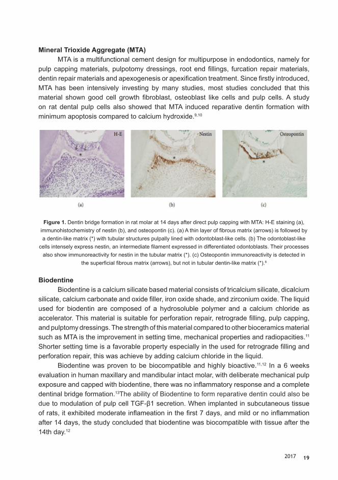
pulp capping materials, pulpotomy dressings, root end fillings, furcation repair materials, dentin repair materials and apexogenesis or apexification treatment. Since firstly introduced, MTA has been intensively investing by many studies, most studies concluded that this material shown good cell growth fibroblast, osteoblast like cells and pulp cells. A study on rat dental pulp cells also showed that MTA induced reparative dentin formation with minimum apoptosis compared to calcium hydroxide.9,10
Figure 1. Dentin bridge formation in rat molar at 14 days after direct pulp capping with MTA: H-E staining (a), immunohistochemistry of nestin (b), and osteopontin (c). (a) A thin layer of fibrous matrix (arrows) is followed by a dentin-like matrix (*) with tubular structures pulpally lined with odontoblast-like cells. (b) The odontoblast-like
cells intensely express nestin, an intermediate filament expressed in differentiated odontoblasts. Their processes also show immunoreactivity for nestin in the tubular matrix (*). (c) Osteopontin immunoreactivity is detected in
the superficial fibrous matrix (arrows), but not in tubular dentin-like matrix (*).8
BiodentineBiodentine is a calcium silicate based material consists of tricalcium silicate, dicalcium
silicate, calcium carbonate and oxide filler, iron oxide shade, and zirconium oxide. The liquid used for biodentin are composed of a hydrosoluble polymer and a calcium chloride as accelerator. This material is suitable for perforation repair, retrograde filling, pulp capping, and pulptomy dressings. The strength of this material compared to other bioceramics material such as MTA is the improvement in setting time, mechanical properties and radiopacities.11 Shorter setting time is a favorable property especially in the used for retrograde filling and perforation repair, this was achieve by adding calcium chloride in the liquid.
Biodentine was proven to be biocompatible and highly bioactive.11,12 In a 6 weeks evaluation in human maxillary and mandibular intact molar, with deliberate mechanical pulp exposure and capped with biodentine, there was no inflammatory response and a complete dentinal bridge formation.13The ability of Biodentine to form reparative dentin could also be due to modulation of pulp cell TGF-β1 secretion. When implanted in subcutaneous tissue of rats, it exhibited moderate inflameation in the first 7 days, and mild or no inflammation after 14 days, the study concluded that biodentine was biocompatible with tissue after the 14th day.12

PROCEEDING 20
BioAggregateBioAggregate is a fine nanoparticle size, material, aluminum-free powder that is mixed
with deionized water to form a bioceramic paste. The powder consists of SiO2 (13.70%), P2O5 (3.92%), CaO (63.50%), and Ta2O5 (17%), the latter was to improve the radioopacitiy of the material (tantalum oxide).14BioAggregate is indicated as pulp capping material, perforation repair as well as root-end fillings, where many in vivo reports concluded that BioAggregate are comparable to MTA.14,15,16
Figure 2. Effects of PMTA, BA, MMTA, and IRM on adhesion in HDPCs by MTT assay and SEM, respectively. An OS containing 50 mg/mL ascorbic acid and 10 mmol/L b-glycerophosphate was used as the positive control. Cells were incubated for 3, 7, and 14 days with the materials. SEM observation
(original magnification, 1000) of HDPCs exposed to various materials was completed 2 days after incubation. Arrows indicate pulp cells. These data findings are representative of 3 independent
experiments. Statistically significant difference compared with each group (P < 0.05).17
As for biocompatibility, on one study comparing BioAggregate (BA) to Micromega MTA (MMTA), ProRoot MTA (PMTA) and Intermediate Restorative Material (IRM) by using human dental pulp cells and concluded that the first three material exhibited equally good biocompatibility whereas IRM showed cytotoxicity (Figure 2).17 BioAggregate also increased the alkaline phosphatase activity, promote mineralization and enhance the expression of osteogenic/odontogenic markers (ALP, osteopontin, osteocalcin, dentin sialophosphoprotein and dentinal matrix protein-1.17,18 Collectively, the biocompatibility, odontogenic potentials, and inflammatory response of BioAggregate and MTA are equal. 17,18,19

2017 21
iRoot SPiRoot SP is another bioceramics material similar to MTA, it composed mainly by
calcium silicates. iRoot SP are injectable, biocompatible, hydrophilic, radiopaque and insoluble and indicated for root canal sealers. In the presence of water, this material can generate calcium silicate hydrates and have highly alkaline pH which provide a detrimental environment for bacterial growth. MTA and iRoot SP also induced human Tooth Germ Stem Cells (hTGSCs) to differentiate into odontoblast like cells, while calxiym hydroxide (Dycal) caused cytotoxicity.20 However according to the the same study, MTA provide more inductive potentiall and hard generate more hard tissuer deposition than iRoot SP.
Figure 3. Immunocytochemistry staining of (a) MTA and odontogenic medium-treated cells, (b) iRoot SP and odontogenic med- ium-treated cells, (c) odontogenic differentiation medium-treated cells (PC) and
(d) undifferentiated hTGSCs (NC) with DSP and COL1A antibodies. Scale bar: 100 lm.20
CONCLUSION
The trend of biocompatible endodontics material has shifted from bioinert to bioactive and biodegradable materials in which the propose materials not only restore the broke part but also to stimulate the body itself to regenerate and fulfill acceptable physical and mechanical properties.
REFERENCES
1. Best SM, Porter AE, Thian ES, Huang J. Bioceramics: past, present and for the future. J Eur Ceram Soc. 2008; 28:1319-1327.
2. Nasseh A. The rise of bioceramics. Endodontic Practice. 2009;2:17-22.3. Ishikawa K. Calcium phosphate cement. In Bioceramics and Their Clinical Application;
PROCEEDING 22
Kokubo, T., Ed.; CRC Press: New York, NY, USA, 2008; pp. 438–463.4. Zakaria MN. Save the pulp: the essential issues on pulp capping treatment. Journal of
Dentomaxillofacial Science. 2016;2:301-305.5. Enkel B, Dupas C, Armengol V, Adou A, Bosco J, Daculsi G, Jean A, Laboux O, LeGeros
RZ, Weiss P. Bioactive materials in endodontics. Expert Rev. Med. Devices. 2008;5(4): 475-494.
6. Cox CF, Subay RK, Ostro E, Suzuki SH. Tunnel defects in dentin bridges: their formation following direct pulp capping. Oper Dent. 1996;21:4–11.
7. Parolia A, Kundabala M, Rao NN, Acharya SR, Agrawal P, Mohan M, Thomas M. A comparative histological analysis of human pulp following direct pulp capping with Propolis, mineral trioxide aggregate and Dycal. Australian Dental Journal. 2010;55: 59–64.
8. Okiji T, Yoshiba K. Reparative Dentinogenesis Induced by Mineral Trioxide Aggregate: A Review from the Biological and Physicochemical Points of View. International Journal of Dentistry. 2009;1-12.
9. Asgary S, Eghbal MJ, Parirokh M, Ghanavati F, Rahimi H. A comparative study of histologic response to different pulp capping materials and a novel endodontic cement. Oral Surg Oral Med Oral Pathol Oral Radiol Endod 2008;106:609–14.
10. Masuda-Murakami Y, Kobayashi M, Wang X, Yamada Y, Kimura Y, Hossain M, Matsumoto K. Effects of mineral trioxide aggregate on the differentiation of rat dental pulp cells. Acta Histochem. 2010 Sep;112(5):452-8.
11. Koubi G, Colon P, Franquin JC, Hartmann A, Richard G, Faure MO, Lambert G. Clinical evaluation of the performance and safety of a new dentine substitute, Biodentine, in the restoration of posterior teeth - a prospective study. Clin Oral Investig. 2013 Jan; 17(1):243-9.
12. Mori GG, Teixeira LM, de Oliveira DL, Jacomini LM, da Silva SRBiocompatibility evaluation of biodentine in subcutaneous tissue of rats. J Endod. 2014 Sep;40(9):1485-8.
13. Nowicka A, Lipski M, Parafiniuk M, Sporniak-Tutak K, Lichota D, Kosierkiewicz A, Kaczmarek W, Buczkowska-Radlińska J. Response of human dental pulp capped with biodentine and mineral trioxide aggregate. J Endod. 2013 Jun; 39(6):743-7.
14. Park JW, Hong SH, Kim JH, Lee SJ, Shin SJ. X-Ray diffraction analysis of white ProRoot MTA and Diadent BioAggregate. Oral Surg Oral Med Oral Pathol Oral Radiol Endod. 2010;109:155–8.
15. El Sayed MA, Saeed MH. In vitro comparative study of sealing ability of Diadent BioAggregate and other root-end filling materials. J Conserv Dent. 2012 Jul-Sep; 15(3): 249–252.
16. Guven EP, Tas PN, Yalvac ME, Sofiev N, Kayahan MB, Sahin F. In vitro comparison of induction capacity and biomineralization ability of mineral trioxide aggregate and a bioceramic root canal sealer. International Endodontic Journal, 46, 1173–1182, 2013.
17. Chang SW, Lee SY, Kum KY, Kim EC. Effects of ProRoot MTA, Bioaggregate, and

2017 23
Micromega MTA on Odontoblastic Differentiation in Human Dental Pulp Cells. J Endod. 2014;40(1):113-118.
18. Yuan Z, Peng B, Jiang H, et al. Effect of bioaggregate on mineral-associated gene 19. expression in osteoblast cells. J Endod 2010;36:1145–8. 20. Yan P, Yuan Z, Jiang H, et al. Effect of bioaggregate on differentiation of human peri-
odontal ligament fibroblasts. Int Endod J 2010;43:1116–21. 21. Guven Y, Tuna EB, Dincol ME, Ozel E, Yilmaz B, Aktoren O. Long-Term Fracture
Resistance of Simulated Immature Teeth Filled with Various Calcium Silicate-Based Materials. Biomed Res Int. 2016; 2016:2863817.