Embed Size (px)
Citation preview

Instructions for use
Title Pregnancy-induced antithrombin deficiency
Author(s) Morikawa, Mamoru; Yamada, Takashi; Yamada, Takahiro; Shimada, Shigeki; Koyama, Takahiro; Cho, Kazutoshi;Minakami, Hisanori
Citation Journal of Perinatal Medicine, 38(4), 379-385https://doi.org/10.1515/JPM.2010.049
Issue Date 2010-07
Doc URL http://hdl.handle.net/2115/47215
Type article (author version)
File Information JPM38-4_379-385.pdf
Hokkaido University Collection of Scholarly and Academic Papers : HUSCAP

Morikawa
Pregnancy-induced antithrombin deficiency
1
[ORIGINAL ARTICLE] 1
Title: Pregnancy-induced antithrombin deficiency 2
Author names and degrees: 3
Mamoru Morikawa, MD. PhD: Correspondence author and data analysis 4
Takashi Yamada, MD. PhD: Personal information management 5
Takahiro Yamada, MD. PhD: Data sampling 6
Shimada Shigeki, MD. PhD: Data sampling 7
Takahiro Koyama, MD: Data sampling 8
Kazutoshi Cho, MD. PhD: Data sampling and adviser of data analysis 9
Hisanori Minakami, MD. PhD: Director and institutional reviewer 10
Affiliation of all authors: Department of Obstetrics 11
Hokkaido University Graduate School of Medicine, Sapporo, Japan 12
13
14
Correspondence to: 15
Mamoru Morikawa, M.D. Ph.D. 16
Department of Obstetrics, 17
Hokkaido University Graduate School of Medicine, 18
Kita-ku N15 W7, Sapporo 060-8638, Japan 19
Phone: +81-11-706-5941, 20
Fax: +81-11-706-7711, 21
E-mail: [email protected] 22
23
24
Manuscript

Morikawa
Pregnancy-induced antithrombin deficiency
2
ABSTRACT 25
OBJECTIVE: Some women exhibit a gradual decline in antithrombin activity during 26
the late stage of pregnancy. This retrospective study was performed to better 27
characterize the laboratory features and water metabolism of such women with 28
pregnancy-induced antithrombin deficiency (PIATD). 29
METHODS: Among 1493 women who gave birth to a singleton infant at our institution, 30
114 women who developed PIATD and/or pregnancy-induced hypertension (PIH) were 31
reviewed with respect to perinatal changes in laboratory variables (hematocrit value, 32
fibrinogen, fibrinogen degradation product, D-dimer, uric acid, aspartate 33
aminotransferase, lactate dehydrogenase) and body weight. PIATD was defined as a 34
gradual decline in antithrombin activity to ≤ 65% of normal activity levels. One 35
hundred and fourteen women with neither PIATD nor pregnancy-induced hypertension 36
(PIH) and matched for the cesarean delivery rate were selected as a control group. 37
RESULTS: Of the 81 women who developed PIH, 19 (23.4%) also developed PIATD. 38
Thirty-three women developed PIATD in the absence of PIH. Coagulation-fibrinolysis 39
was significantly more enhanced and the postpartum reduction in the hematocrit value 40
was significantly larger in women with PIATD, irrespective of the presence or absence 41
of hypertension, than in women without PIATD. The postpartum decrease in body 42
weight was significantly smaller in women with PIATD, irrespective of the presence or 43
absence of hypertension, than in women without PIATD. 44
CONCLUSIONS: A decrease in antithrombin activity can occur in the absence of 45
hypertension. Even in the absence of hypertension, a decreased plasma volume and 46
enhanced coagulation-fibrinolysis seem to be notable characteristics in women with 47
PIATD. The monitoring of antithrombin activity may be helpful for distinguishing 48

Morikawa
Pregnancy-induced antithrombin deficiency
3
pregnant women with these insidious risks. 49
Key words: antithrombin, blood vessel permeability, coagulation-fibrinolysis, 50
dehydration, pregnancy-induced hypertension 51
52

Morikawa
Pregnancy-induced antithrombin deficiency
4
INTRODUCTION 53
Some women develop a gradual decline in antithrombin (AT) activity during the late 54
stage of pregnancy, even in the absence of hypertension [9, 16]. This decline in AT 55
activity continues until the day of or one day after delivery, and a prompt normalization 56
of AT activity occurs postpartum in such patients with pregnancy-induced AT deficiency 57
(PIATD) [9]. Because AT is the most important molecule for the anti-coagulation of the 58
circulating blood, delivery delays may be dangerous in women with PIATD. Indeed, 59
the risk of developing pulmonary embolism increases in women with known risk factors 60
for PIATD, such as preeclampsia [6, 17, 18] or multi-fetal pregnancy [2, 6, 10, 16-18]. 61
Further, women with PIATD are at risk of developing acute fatty liver of pregnancy [2, 62
9]. Indeed, the risk of developing acute fatty liver of pregnancy is reportedly high in 63
women with a known risk factor for PIATD, such as preeclampsia or multi-fetal 64
pregnancy [11]. Because a profoundly decreased AT level is seen in women with acute 65
fatty liver of pregnancy at the time of presentation [2] and because the risk of a perinatal 66
elevation in aspartate aminotransferase increases as the antenatal AT activity decreases 67
[9], the monitoring of AT activity in women who exhibit a gradual decline in AT activity 68
may help to avoid the development of acute fatty liver of pregnancy. These 69
observations suggest that women with PIATD might constitute a high-risk pregnancy 70
group, although the clinical and laboratory features of women with PIATD remain to be 71
studied. 72
We introduced the measurement of AT activity into clinical practice in 2001 because 73
of its possible usefulness in the management of women with complicated pregnancies. 74
Indeed, AT determination resulted in the appropriate interventions in a patient with 75
PIATD [7]. Our preliminary survey of patients with PIATD suggested a decreased 76
Morikawa
Pregnancy-induced antithrombin deficiency
5
plasma volume or dehydration, as reported in women with preeclampsia [14, 15] and in 77
a case with acute fatty liver of pregnancy [8]. Enhanced coagulation-fibrinolysis and 78
coagulopathy are other well known laboratory features of preeclampsia [1, 4] and acute 79
fatty liver of pregnancy [2], respectively. Prenatal shortage of the plasma volume or 80
dehydration in the mother may influence postnatal changes in body weight and 81
hematocrit value. 82
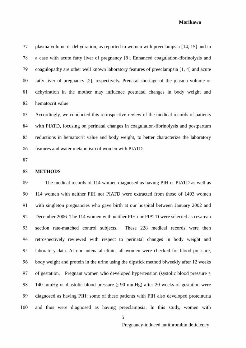
Accordingly, we conducted this retrospective review of the medical records of patients 83
with PIATD, focusing on perinatal changes in coagulation-fibrinolysis and postpartum 84
reductions in hematocrit value and body weight, to better characterize the laboratory 85
features and water metabolism of women with PIATD. 86
87
METHODS 88
The medical records of 114 women diagnosed as having PIH or PIATD as well as 89
114 women with neither PIH nor PIATD were extracted from those of 1493 women 90
with singleton pregnancies who gave birth at our hospital between January 2002 and 91
December 2006. The 114 women with neither PIH nor PIATD were selected as cesarean 92
section rate-matched control subjects. These 228 medical records were then 93
retrospectively reviewed with respect to perinatal changes in body weight and 94
laboratory data. At our antenatal clinic, all women were checked for blood pressure, 95
body weight and protein in the urine using the dipstick method biweekly after 12 weeks 96
of gestation. Pregnant women who developed hypertension (systolic blood pressure ≥ 97
140 mmHg or diastolic blood pressure ≥ 90 mmHg) after 20 weeks of gestation were 98
diagnosed as having PIH; some of these patients with PIH also developed proteinuria 99
and thus were diagnosed as having preeclampsia. In this study, women with 100

Morikawa
Pregnancy-induced antithrombin deficiency
6
preeclampsia were included in the PIH group. This study was approved by the 101
institutional review board of our university hospital, and all the women gave their 102
informed consent to participate in this study. 103
Women with PIH, proteinuria alone (≥ 1+ using the dipstick method), edema alone, 104
excessive weight gain (> 500 g/week), or some other risk factors known to influence 105
obstetrical outcome, such as an advanced age, obesity, and medical complications, were 106
routinely screened for blood abnormalities, including complete blood cell counts, AT 107
activity, serum levels of aspartate aminotransferase (AST), lactate dehydrogenase 108
(LDH), blood urea nitrogen (BUN), and serum levels of creatinine and uric acid (UA). 109
In addition, these laboratory tests were performed routinely one day or immediately 110
before delivery and on postpartum days 1 and 7 for all the patients who underwent 111
cesarean delivery and in some patients with other risk factors. Our institution is a 112
tertiary hospital that admits and manages mainly pregnant women with risk factors who 113
have been referred from an area with approximately 40,000 births each year. Therefore, 114
the cesarean section rate was approximately 45% during the study period and more than 115
80% of the pregnant women who delivered at our institution were screened at least once 116
for AT activity. Pregnant women who exhibited a gradual perinatal decline in AT 117
activity to ≤ 65% of the normal level were diagnosed as having PIATD. Women who 118
were diagnosed as having PIATD antenatally were monitored closely. The body 119
weights of the women who were admitted to our hospital were checked daily until the 120
day of hospital discharge, which routinely occurred on postpartum day 8. 121
The plasma levels of D-dimer and the fibrinogen/fibrin degradation products (FDP) 122
were measured using the latex agglutination assay (Mitsubishi Kagaku Iatron Inc., 123
Tokyo, Japan). The plasma fibrinogen levels were measured using the thrombin clotting 124
Morikawa
Pregnancy-induced antithrombin deficiency
7
time method (Sysmex Co., Kobe, Japan). The AT activity was measured using the 125
chromogenic substrate assay (Daiichi Pure Chemicals Co., Tokyo, Japan). The platelet 126
counts and hematocrit values were determined using an electronic particle counting 127
system (Beckman Coulter Int., Fullerton, CA, USA). The UA level was determined 128
using the uricase-peroxidase (colorimetric) coupled reaction (Serotec Int., Sapporo, 129
Japan). The AST level was determined using the Japan Society of Clinical Chemistry 130
(JSCC) transferable method (Serotec Int., Sapporo, Japan). 131
All data were presented as the mean or median values. Unpaired t-tests, 132
Kruskal-Wallis tests, Mann-Whitney U tests, Wilcoxon tests and ANOVAs were used to 133
analyze the results. χ2 tests and Fisher exact tests were used to compare frequencies. A 134
value of p < 0.05 was considered statistically significant. The statistical software 135
package StatView 5.0 for Macintosh (SAS Institute Inc. Cary, NC, USA) was used for 136
all data analyses. 137
138
RESULTS 139
Among the 114 women diagnosed as having PIH and/or PIATD, 62 women (54.4%) 140
had PIH alone (PIH group, 28 of whom were preeclamptic), 33 women (28.9%) had 141
PIATD alone (PIATD group), and 19 women (16.7%) had both PIH and PIATD 142
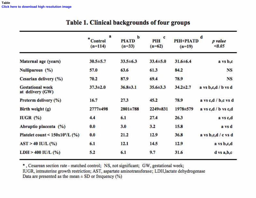
(PIH+PIATD group, 12 of whom were preeclamptic) (Table 1). Thus, the frequency of 143
preeclampsia did not differ significantly between the PIH group and the PIH+PIATD 144
group (45.2% [28/62] vs. 63.2% [12/19], p=0.198). The frequency of PIATD also did 145
not differ significantly between women with gestational hypertension and those with 146
preeclampsia (17.1% [7/41] vs. 30.0% [12/40], p=0.198). The cesarean delivery rate in 147
the control group was matched with the rates in the study groups to enable the 148
Morikawa
Pregnancy-induced antithrombin deficiency
8
comparison of various parameters, therefore, the cesarean delivery rate did not differ 149
significantly among the four groups. The length of gestation, the rate of preterm birth, 150
the birth weight, the rate of intrauterine growth restriction (IUGR), and the rate of 151
abruptio placentae differed significantly among the groups. 152
As expected, the clinical outcomes were significantly worst in the PIH+PIATD 153
group. The PIH group had a significantly shorter duration of gestation, a higher 154
frequency of preterm birth, a smaller infant weight, and a higher frequency of IUGR 155
than the control group. However, these parameters did not differ significantly between 156
the control and PIATD groups, except for the duration of gestation. The frequency of a 157
reduced platelet count < 150 × 109/L and the frequency of an elevated AST level > 40 158
IU/L were significantly higher in the groups with PIH and/or PIATD than in the control 159
group. The frequency of an elevated LDH level > 400 IU/L was significantly higher in 160
the PIH+PIATD group than in the other three groups. 161
AT activity decreased significantly with advancing gestation in all the groups (Table 162
2). AT activity that was already depressed two weeks prior to delivery decreased 163
perinatally to ≤ 65% of normal in the PIATD and PIH+PIATD groups. The nadir of AT 164
activity seen on postpartum day 1 returned to normal on postpartum day 7 in all the 165
groups. 166
The FDP level immediately before delivery was significantly higher in the groups 167
with PIATD than in the control group (Figure 1) and was significantly higher in the 168
PIH+PIATD group than in the PIH group. The D-dimer level immediately before 169
delivery was also significantly higher in the groups with PIATD than in the groups 170
without PIATD. The D-dimer level on postpartum day 1 was significantly higher in the 171
PIATD group than in the control group. The fibrinogen level decreased after delivery 172

Morikawa
Pregnancy-induced antithrombin deficiency
9
in all the groups. The magnitude of the decrease (the difference between the values on 173
-1/0 day and +1 day relative to delivery) was significantly larger in the groups with 174
PIATD (53.2 ± 77.2 mg for PIH+PIATD group, 51.7 ± 83.8 mg for PIATD group) than 175
in the groups without PIATD (20.8 ± 74.9 mg for PIH group, 21.7 ± 57.9 mg for control 176
group), suggesting the peripartum hyperconsumption of fibrinogen in women with 177
PIATD. 178
The serum urate level seemed to be elevated in patients with PIH and/or PIATD; 179
throughout the study period, it was significantly higher in the PIH+PIATD group than 180
in the PIH, PIATD, or control groups and in the PIH group than in the control group 181
(Figure 2). 182
The hematocrit value continued to decrease until 7 days after delivery in the groups 183
with PIATD, but not in the groups without PIATD (Figure 3a). The magnitude of the 184
decreases on postpartum days 1 and 7 were significantly larger in the groups with 185
PIATD than in the groups without PIATD (Figure 3b), suggesting prenatal 186
hemoconcentration as a result of enhanced blood vessel permeability in women with 187
PIATD. 188
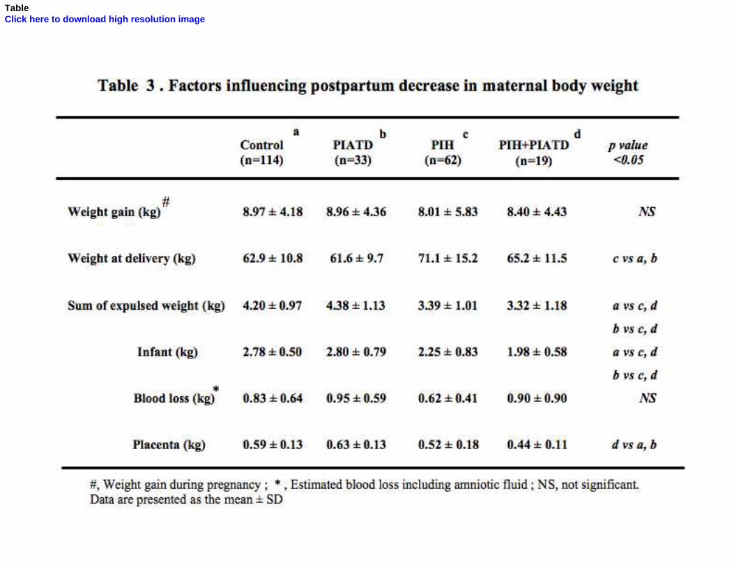
Although the maternal body weight at delivery differed significantly among the 189
four groups, the weight gain during pregnancy did not differ among the groups (Table 190
3). The total weight expulsed during labor (including the infant), the estimated blood 191
loss, and the placenta were significantly larger in the control and PIATD groups than in 192
the groups with PIH, reflecting the larger infants and placentas in the groups with 193
PIATD. However, the postpartum decrease in body weight was significantly smaller in 194
the groups with PIATD than in the control group on postpartum day 3 and was 195
significantly larger in the PIH group than in the control group on postpartum day 7 196

Morikawa
Pregnancy-induced antithrombin deficiency
10
(Figure 4). 197
Eighty-one women with PIH were divided into two groups, 41 with gestational 198
hypertension and 40 with preeclampsia, and a subanalysis of the postpartum changes in 199
hematocrit value and body weight was performed (Figures 5 and 6). Although the 200
magnitude of the decreases in the hematocrit value on postpartum days 1 and 7 was 201
significantly larger in women with preeclampsia than in women with gestational 202
hypertension and normal controls (Figure 5), the postpartum decrease in body weight 203
did not differ significantly among the three groups (Figure 6). 204
205
DISCUSSION 206
In this study, the degree of coagulation-fibrinolysis was consistently more 207
exaggerated in women with PIATD than in women without PIATD. The larger 208
peripartum decrease in fibrinogen supports the enhancement of coagulation-fibrinolysis 209
in these women with PIATD. Exaggerated coagulation-fibrinolysis is a well-known 210
abnormality in patients with PIH [1, 4]. In this study, women with PIH were divided 211
into a PIH+PIATD group and a PIH group according to the presence or absence of 212
PIATD. The former group exhibited a more exaggerated degree of 213
coagulation-fibrinolysis than the latter group. In addition, women with PIATD alone 214
showed a more exaggerated degree of coagulation-fibrinolysis than the women with 215
PIH alone or the control women in this study. Thus, the presence of PIH alone did not 216
have a large effect on coagulation-fibrinolysis in the absence of PIATD. Reduced AT 217
activity in women with PIH is another well-known phenomenon [6, 17, 18], and our 218
study also demonstrated that as many as 19 (23.5%) of 81 women with PIH also had 219
PIATD. These results support the enhanced coagulation-fibrinolysis seen in women 220
Morikawa
Pregnancy-induced antithrombin deficiency
11
with PIH described in earlier reports [4] and reflect the presence of 221
coagulation-fibrinolysis in women with PIATD who are inevitably included amongst 222
women with PIH. 223
This study demonstrated that women with PIATD exhibit a larger decrease in their 224
hematocrit values and a smaller decrease in their body weights postnatally than women 225
without PIATD. An increase in water retention is a normal physiological alteration 226
during pregnancy. Clearly demonstrable pitting edema of the ankles and legs is seen in 227
most pregnant women, especially during the late stages of pregnancy [3]. Edema 228
resulting from the retention of excess water in the interstitial space can be massive in 229
women with PIH, mainly because of the increased blood vessel permeability [5, 12], 230
and usually results in hemoconcetration and a decrease in the circulating plasma volume 231
in patients with PIH [14, 15]. The process involved in the retention of water is reversed 232
by parturition, and excess water in the interstitial space returns into the intravascular 233
space, resulting in a fall in the hematocrit value, and then is excreted as urine. The 234
extent of the postpartum decrease in the hematocrit value may therefore reflect the 235
degree of antenatal hemoconcentration and the decrease in the circulating plasma 236
volume. In this study, women with PIATD, irrespective of the presence or absence of 237
hypertension, showed a larger and sustained decrease in the hematocrit value after 238
delivery, suggesting a more severe antenatal hemoconcentration and decrease in the 239
plasma volume in women with PIATD than in women without PIATD. Although 240
women with PIH showed a relatively high absolute hematocrit value compared with 241
women without PIH antenatally, the changes in the postnatal hematocrit value differed 242
according to the presence or absence of PIATD. Women with PIH alone showed a 243
pattern of change in the hematocrit value similar to that observed in the control women. 244

Morikawa
Pregnancy-induced antithrombin deficiency
12
These results may suggest that the decrease in the plasma volume described in women 245
with PIH in earlier reports [14, 15] may have reflected the plasma volume in women 246
with PIATD who have been included in the group with PIH. 247
The postpartum decrease in body weight is expected to be relatively small in 248
women who have suffered from antenatal dehydration and a decrease in the plasma 249
volume. Indeed, the postpartum decrease in body weight was consistently smaller in 250
women with PIATD irrespective of the presence or absence of hypertension than in 251
women without PIATD, supporting the above-mentioned “severe antenatal 252
hemoconcentration and decrease in plasma volume in women with PIATD”. Women 253
with PIH alone showed the largest decrease in body weight. The reason for this finding 254
may be explained as follows. Because the pattern of change in the hematocrit value in 255
women with PIH alone was similar to that observed in the control women, the women 256
with PIH alone may not have experienced a decrease in the circulating plasma volume 257
but may have had excess water in the interstitial space as a result of increased blood 258
vessel permeability. The intake of water may have had compensated for the leakage of 259
plasma into the extravascular space in these women with PIH alone. This excess water 260
may have returned to the intravascular space after delivery and may have then been 261
excreted promptly as urine (since a shortage in the circulating plasma volume did not 262
exist), resulting in the relatively large postpartum decrease in body weight observed in 263
the women with PIH alone. In contrast, an insufficient intake of water or an 264
inappropriate urine output may have caused a decrease in the total body water in women 265
with PIATD. 266
The magnitude of the postpartum decrease in the hematocrit value was significantly 267
larger in the 40 women with preeclampsia than in the 41 women with gestational 268

Morikawa
Pregnancy-induced antithrombin deficiency
13
hypertension, but the postpartum decrease in body weight did not differ significantly 269
between the two groups. However, the postpartum decrease in body weight differed in 270
the two hypertensive women groups divided by the presence or absence of PIATD 271
(Figure 4). These results suggested that women with preeclampsia may have a larger 272
plasma volume reduction, consistent with the results of an earlier study [14], and further 273
that these women may have had a larger increase in interstitial fluid, compared with 274
women with gestational hypertension. The presence of PIATD, rather than the presence 275
of proteinuria, might be a better predictor of a decrease in interstitial fluid as well as a 276
decrease in circulating plasma in hypertensive pregnant women. 277
In this study, the incidence of PIATD was 23.5% (19/81) in women with PIH (17.1% 278
[7/41] for women with gestational hypertension and 30.0% [12/40] for women with 279
preeclampsia). Although all the women without hypertension were not screened for AT 280
activity, PIATD was detected in 33 (2.3%) out of 1412 women without hypertension. 281
This incidence among women without hypertension might not be representative of the 282
incidence among women with uncomplicated pregnancies, since a considerable number 283
of the women in our study population had risk factors other than hypertension and 284
because the results of our preliminary study (unpublished data) suggested that the 285
incidence of PIATD was 1.0% among women with uncomplicated singleton 286
pregnancies. 287
In conclusion, some women exhibit a gradual perinatal decline in AT activity to ≤ 288
65% of normal, even in the absence of hypertension. Such women with a gradual 289
decline in AT activity to ≤ 65% of normal suffer from exaggerated 290
coagulation-fibrinolysis and may also suffer from a decreased interstitial fluid and 291
circulating plasma volume. The monitoring of AT activity may be helpful in 292

Morikawa
Pregnancy-induced antithrombin deficiency
14
distinguishing women with these insidious risks. 293
294

Morikawa
Pregnancy-induced antithrombin deficiency
15
REFERENCES 295
[1] Cadroy Y, Grandjean H, Pichon J, Desprats R, Berrebi A, Fournie A, Boneu B: 296
Evaluation of six markers of haemostatic system in normal pregnancy and pregnancy 297
complicated by hypertension or pre-eclampsia. Br J Obstet Gynaecol 1993; 100: 416-20 298
[2] Castro MA, Coodwin TM, Shaw KJ, Ouzounian JF, McGehee WG: Disseminated 299
intravascular coagulation and antithrombin III depression in acute fatty liver of 300
pregnancy. Am J Obstet Gynecol 1996; 174: 211-6 301
[3] Cunningham FG, Leveno KJ, Bloom SL, Hauth JC, Gilstrap III LC, Wenstrom KD. 302
Maternal physiology, In; Williams Obstetrics 22nd
ed., McGraw-Hill, New York; 2005, 303
pp. 122–50. 304
[4] DeBoer K, TenCate JW, Sturk A, Borm JJJ, Treffers PE: Enhanced thrombin 305
generation in normal and hypertensive pregnancy. Am J Obstet Gynecol 1989; 160: 306
95-100 307
[5] Haller H, Hempel A, Houmuth V, Mandelkow A, Busjahn A, Maasch C, Drab M, 308
Lindschau C, Jupner A, Vetter K, Dudenhausen J, Luft FC: Endothelial-cell 309
permeability and protein kinase C in pre-eclampsia. Lancet 1998; 351: 945-9 310
[6] Ho C-H, Yang Z-L: The predictive value of the hemostasis parameters in the 311
development of preeclampsia. Thromb Haemos 1992; 67: 214-8 312
[7] Koyama T, Yamada T, Morikawa M, Yamada T, Shimada S, Araki N, Yamamura 313
M, Minakami H: Marked gestational edema as a clinical sign of life-threatening 314
condition. J Obstet Gynaecol Res, in press 315
[8] Minakami H, Kimura K, Kanazawa T, Tamada T, Kaneko K: Acute fatty liver of 316
pregnancy with hyperlipidemia, acute hemorrhagic pancreatitis, and disseminated 317
intravascular coagulation. Asia-oceania J Obstet Gynaecol 1985; 11: 371-6 318
Morikawa
Pregnancy-induced antithrombin deficiency
16
[9] Minakami H, Watanabe T, Izumi A, Matsubara S, Koike T, Sayama M, Moriyama I, 319
Sato I. Association of a decrease in antithrombin III activity with a perinatal elevation in 320
aspartate aminotransferase in women with twin pregnancies: relevance to the HELLP 321
syndrome. J Hepatol. 1999; 30: 603-11. 322
[10] Morikawa M, Yamada T, Kataoka S, Cho K, Yamada H, Suzuki S, Sakuragi N, 323
Minakami H. Changes in antithrombin activity and platelet counts in the late stage of 324
twin and triplet pregnancies. Semin Thromb Hemos 2005; 31: 290-6 325
[11] Riely CA: Acute fatty liver of pregnancy. Semin liver Dis 1987; 7: 47-54 326
[12] Roberts JM, Taylor RN, Musci TJ, Rodgers GM, Hubel CA, McLaughlin MK: 327
Preeclampsia: an endothelial cell disorder. Am J Obstet Gynecol 1989; 161: 1200-4 328
[13] Ros HS, Lichtenstein P, Bellocco R, Petersson G, Cnattingius S: Pulmonary 329
embolism and stroke in relation to pregnancy: How can high-risk women be identified? 330
Am J Obstet Gynecol 2002; 186: 198-203 331
[14] Silver HM, Seebeck MA, Carlson R: Comparison of total blood volume in normal, 332
preeclamptic, and nonproteinuric gestational hypertensive pregnancy by simultaneous 333
measurement of red blood cell and plasma volumes. Am J Obstet Gynecol 1998; 179: 334
87-93 335
[15] Soffronoff EC, Kaufmann BM, Connaughton JF: Intravascular volume 336
determinations and fetal outcome in hypertensive diseases of pregnancy. Am J Obstet 337
Gynecol 1977; 127: 4-9 338
[16] Tsunoda T, Ohkuchi A, Izumi A, Watanabe T, Matsubara S, Sato I, Minakami H: 339
Antithrombin III activity and platelet count are more likely to decrease in twin 340
pregnancies than in singleton pregnancies. Acta Obstet Gynecol Scand 2002; 81: 840-5 341
[17] Weiner CP: The mechanism of reduced antithrombin III activity in women with 342
Morikawa
Pregnancy-induced antithrombin deficiency
17
preeclampsia. Obstet Gynecol 1988; 72: 847-9 343
[18] Weiner CP, Kwaan HC, Xu C, Paul M, Burmeister L, Hauck W: Antithrombin III 344
activity in women with hypertension during pregnancy. Obstet Gynecol 1985; 65: 301-6345
Morikawa
Pregnancy-induced antithrombin deficiency
18
FIGURE LEGENDS 346
Figure 1: Perinatal changes in FDP, D-dimer, and fibrinogen. ○, Control group (n=114); 347
∆, PIH group (n=62); ●, PIATD group (n=33); ▲; PIH+PIATD group (n=19). 348
Significant differences were seen as follows: Control vs. PIATD, PIH+PIATD for FDP 349
immediately before delivery; PIH vs. PIH+PIATD for FDP immediately before 350
delivery; PIH vs. PIATD, PIH+PIATD for D-dimer immediately before delivery; 351
Control vs. PIATD, PIH+PIATD for D-dimer immediately before delivery; Control vs. 352
PIATD for D-dimer on postpartum day 1. 353
354
Figure 2: Perinatal change in serum urate. ○, Control group (n=114); ∆, PIH group 355
(n=62); ●, PIATD group (n=33); ▲; PIH+PIATD group (n=19). Significant differences 356
were seen as follows: PIH+PIATD vs. PIH, PIATD, Control at any point; PIH vs. 357
Control at any point; Control vs. PIATD immediately before delivery. 358
359
Figure 3: Perinatal change in hematocrit value (a) and its net decreases on postpartum 360
days 1 and 7 (b). ○, Control group (n=114); ∆, PIH group (n=62); ●, PIATD group 361
(n=33); ▲; PIH+PIATD group (n=19); *, p < 0.05 between groups; **, p < 0.01 362
between groups. Significant differences were seen as follows (for Fig. 4a): Control vs. 363
PIH at 2 weeks before delivery; Control vs. PIH+PIATD immediately before delivery; 364
PIATD vs. PIH, PIH+PIATD immediately before delivery; PIATD vs. Control, PIH on 365
postpartum day 1; Control vs. PIH on postpartum day 1; PIATD vs. Control, PIH on 366
postpartum day 7; PIH vs. PIH+PIATD on postpartum day 7. 367
368
Figure 4: Postpartum decease in maternal body weight (a) and net decreases on 369

Morikawa
Pregnancy-induced antithrombin deficiency
19
postpartum days 3 and 7 (b). ○, Control group (n=114); ∆, PIH group (n=62); ●, PIATD 370
group (n=33); ▲; PIH+PIATD group (n=19); *, p < 0.05 between groups. The patterns 371
of maternal body weight reduction (a) differed significantly among the four groups, as 372
shown using an ANOVA. 373
374
Figure 5: Perinatal changes in hematocrit values (a) and net decreases on postpartum 375
days 1 and 7 (b). ○, Control group (n=114); ●, Gestational hypertension group (n=41); 376
▲; Preeclampsia group (n=40); *, p < 0.05 between groups; **, p < 0.01 between 377
groups. Significant differences were seen (for Fig. 5a) between the control vs. 378
gestational hypertension group and between the control vs. preeclampsia group before 379
delivery. 380
381
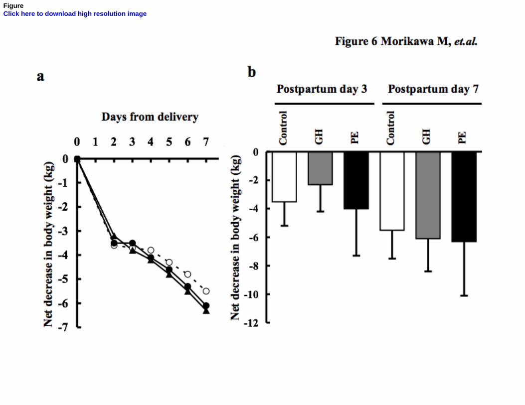
Figure 6: Postpartum deceases in maternal body weight and net decreases on 382
postpartum days 3 and 7 (b). ○, Control group (n=114); ●, Gestational hypertension 383
group (n=41); ▲; Preeclampsia group (n=40). No significant differences were seen 384
among the three groups. 385
386

FigureClick here to download high resolution image

FigureClick here to download high resolution image

FigureClick here to download high resolution image

FigureClick here to download high resolution image

FigureClick here to download high resolution image
FigureClick here to download high resolution image
TableClick here to download high resolution image

TableClick here to download high resolution image
TableClick here to download high resolution image