Embed Size (px)
Citation preview

Preferential Interaction of v-Conotoxins with Inactivated N-typeCa21 Channels
Jonathan W. Stocker,1 Laszlo Nadasdi,2 Richard W. Aldrich,3 and Richard W. Tsien1
1Department of Molecular and Cellular Physiology and 2Howard Hughes Medical Institute, Stanford University, Stanford,California 94305 and 3Neurex Corporation, Menlo Park, California 94025
The selective block of N-type Ca21 channels by v-conotoxinshas been a hallmark of these channels, critical in delineatingtheir biological roles and molecular characteristics. Here wereport that the v-conotoxin-channel interaction dependsstrongly on channel gating. N-type channels (a1B, a2, and b1)expressed in Xenopus oocytes were blocked with a variety ofv-conotoxins, including v-CTx-GVIA, v-CTx-MVIIA, and SNX-331, a derivative of v-CTx-MVIIC. Changes in holding potential(HP) markedly altered the severity of toxin block and the kinet-ics of its onset and removal. Notably, strong hyperpolarization
renders v-conotoxin block completely reversible. These effectscould be accounted for by a modulated receptor model, inwhich toxin dissociation from the inactivated state is ;60-foldslower than from the resting state. Because v-conotoxins actexclusively outside cells, our results suggest that voltage-dependent inactivation of Ca21 channels must be associatedwith an externally detectable conformational change.
Key words: calcium channel; v-conotoxin; inactivation;voltage-dependent binding; modulated receptor hypothesis;N-type
Specific interactions between peptide neurotoxins and voltage-gated channels have been valuable in defining the contributions ofvarious channels to key physiological processes. For example, thev-conotoxins v-CTx-GVIA and v-CTx-MVIIA potently blockN-type Ca21 channels (Kasai et al., 1987; McCleskey et al., 1987;Plummer et al., 1989; Regan et al., 1991) and have been critical fortheir biochemical isolation (McEnery et al., 1991; Sakamoto andCampbell, 1991) and delineation of their diverse functional roles(Nowycky et al., 1985; Hirning et al., 1988; Stanley and Goping,1991; Komuro and Rakic, 1992; Wheeler et al., 1994; Dunlap etal., 1995). v-CTx-GVIA is thought to bind to the outer mouth ofthe N-type Ca21 channel (McCleskey et al., 1987; Ellinor et al.,1994). Because the three-dimensional structures of v-CTx-GVIAand other v-conotoxins are known from nuclear magnetic reso-nance (Pallaghy et al., 1993; Sevilla et al., 1993; Basus et al., 1995;Farr-Jones et al., 1995; Nemoto et al., 1995), analysis of thetoxin-channel interaction might provide valuable informationabout the outer vestibule of the Ca21 channel.
As structural probes, peptide toxins have the potential of re-porting dynamic as well as static aspects of channel structure.Modulation of block by changes in gating is well known forsmaller molecules such as quaternary ammonium compounds
(Armstrong, 1969, 1971), local anesthetics (Hille, 1977;Hondeghem and Katzung, 1977), organic Ca21 channel blockers(Lee and Tsien, 1983; Bean, 1984), and sulfhydryl-reactive re-agents (Yang and Horn, 1995; Larsson et al., 1996; Liu et al.,1996). Peptide toxins offer advantages as probes of voltage-dependent conformational changes because their effects are po-tent and specific and their site of action is unequivocallyextracellular.
Inactivation is an important aspect of Ca21 channel gating,critical for governing the amount of Ca21 entry during repetitivefiring (Carbone and Swandulla, 1990; Pelzer et al., 1990). Al-though the kinetics of voltage-dependent inactivation differs strik-ingly among Ca21 channel types, it is not clear whether Ca21
channel inactivation involves internal or external conformationalchanges or both. One possible internal mechanism is the rapidocclusion of the open channel by a tethered intracellular blockinggroup, as proposed for the N-terminal domain of K1 channels(Zagotta et al., 1990) or the intracellular III–IV loop in Na1
channels (Vassilev et al., 1988; Stuhmer et al., 1989; Catterall,1993). Another possible mechanism is a conformational changenear the external mouth of the Ca21 channel pore, analogous toC-type inactivation of K1 channels, which affects K1 channelinteractions with extracellular Cd21, tetraethylammonium1, andsulfhydryl modifiers (Hoshi et al., 1990; Choi et al., 1991; Lopez-Barneo et al., 1993; Yellen et al., 1994; Baukrowitz and Yellen,1995; Liu et al., 1996). Questions about possible external confor-mational changes in Ca21 channels are of further interest becausesubtype-specific differences in voltage-dependent inactivation ki-netics have been traced to residues in membrane-spanning seg-ment IS6 and nearby extracellular and cytoplasmic regions(Zhang et al., 1994). Here we explore such issues by examining theeffects of a series of v-conotoxins, focusing on the question ofwhether their potency is dependent on the gating state of theN-type Ca21 channel.
Received Nov. 28, 1996; revised Feb. 18, 1997; accepted Feb. 20, 1997.This research was supported by grants from National Institutes of Health (R.W.T.,
R.W.A.) and a training grant in cardiac electrophysiology (National Institutes ofHealth) (J.W.S.). R.W.A. is an investigator of the Howard Hughes Medical Institute.We are grateful to P. T. Ellinor, W. A. Horne, and T. Tanabe, V. Flockerzi, and F.Hofmann for cDNAs for a1B subunits, a2/d subunits, and b1 subunits, respectively.We thank Ilya Bezprozvanny and Xiao-Hua Chen for helpful discussions andRebecca Agin for skilled technical support.
Correspondence should be addressed to Dr. Richard W. Tsien, Department ofMolecular and Cellular Physiology, Beckman Center, B105A, Stanford, CA94305-5426.
Dr. Stocker’s present address: ICAgen, Inc., 4222 Emperor Boulevard, Durham,NC 27703.Copyright © 1997 Society for Neuroscience 0270-6474/97/173002-12$05.00/0
The Journal of Neuroscience, May 1, 1997, 17(9):3002–3013

MATERIALS AND METHODSPreparation of Xenopus oocytes and expression of channels. Ovarian tissuewas removed from anesthetized female Xenopus laevis. Stage V–VI oo-cytes free of follicular cells were obtained after incubation with shakingfor 2 hr in a Ca21-free solution (82.5 mM NaCl, 2 mM KCl, 1 mM MgCl2,and 5 mM HEPES, pH 7.5) containing 1 mg/ml collagenase A as describedpreviously (Sather et al., 1993). The oocytes were washed and transferredto a storage medium (96 mM NaCl, 2 mM KCl, 1.8 mM CaCl2, 1 mMMgCl2, and 5 mM HEPES, pH 7.6, with 2.5 mM sodium pyruvate and theantibiotics gentamycin, penicillin, and streptomycin).
Selected oocytes were then stored at 18°C for several hours or over-night before injection. Expression of calcium channels was achieved byinjection of a1b with a2/d and b1a cRNA that had been dissolved in waterand mixed in approximately equimolar ratios. The cRNAs were synthe-sized in vitro using T7 or SP6 polymerase from corresponding cDNAs, ana1b construct largely derived from human hippocampal cDNA (Ellinor etal., 1994), a2/d (courtesy of Prof. T. Tanabe) and b1a (Ruth et al., 1989).Details about the composition of these subunits are given in the earlierpapers cited.
Electrophysiological recordings and toxin application. Whole-cell Ba21
currents were recorded using a two-electrode voltage-clamp amplifier(model OC-725A, Warner Instruments, Hamden, CT). Current-passingand voltage-measuring electrodes were filled with 3 M KCl and typicallyhad resistances in the range of 1–5 MV. The potential of the bath wasmeasured by a chlorided silver wire immersed in a reservoir filled with 3M KCl connected to the chamber by a 3 M KCl–agar bridge. A chloridedsilver wire placed directly in the recording chamber was used to measurethe amount of current injected by the current electrode.
Ba21 current through the expressed Ca21 channels was induced bystepping the holding potential (HP) to 0 mV for test pulse with a 50 msecduration. Intrapulse duration was varied between 5 sec and 1 min de-pending on the total time of a given experimental run and the timeresolution desired. The current signal was filtered at 500 Hz (FrequencyDevices, Haverhill, MA) and processed using Axon Basic-based programs(Axon Instruments, Foster City, CA). Leak traces were taken using a2P/4 protocol and subtracted off-line.
For current measurements, oocytes were placed in a perfusion solutioncontaining 5 mM Ba(OH)2, 2 mM KOH, 85 mM tetraethylammonium, and5 mM HEPES, with pH adjusted to 7.4 with methanesulfonic acid. Inaddition, all of the solutions contained 0.1 mg/ml cytochrome c tosaturate nonspecific binding sites. During recording, the oocytes wereperfused continuously at a rate of ;0.5 ml/min. The v-conotoxins GVIA(Peninsula Laboratories), MVIIA (The Peptide Institute), MVIIC (SNX-230), TVIA (SNX-185), and SNX-331 (Neurex) were all initially dissolvedin water at the stock concentration of 1.0 mM and stored at 220°C. Freshdilutions of the peptides into the 5 mM Ba21 perfusion solution weremade immediately before use. Application of v-conotoxins was achievedby complete perfusion of the recording chamber with solution containinga given peptide.
RESULTSVoltage-dependent block of N-type calcium channelsby an v-conotoxinDuring the course of characterizing a series of v-conotoxins, wefound evidence that their ability to block N-type Ca21 channels isstrongly voltage-dependent. This was seen most dramatically withSNX-331, the Y13W derivative of the well known v-conotoxinv-CTx-MVIIC (Fig. 1A). The rapid onset and recovery fromblock seen after application and washoff of SNX-331 made itadvantageous for use in this study. The availability of N-type Ca21
channels was assessed by monitoring the peak current elicited bya brief test pulse to 0 mV. The kinetics of the development andremoval of block was strongly affected by the membrane potentialat which the oocyte was maintained (HP). At a strongly hyperpo-larized potential (HP 5 2120 mV), both the onset and recoveryfrom block appeared to follow simple exponential time courses. Incontrast, at a less negative potential (HP 5 270 mV), much morecomplex behavior was seen. In this case, the onset of block bySNX-331 was strikingly biphasic, consisting of an initial rapid
phase, very much like that observed at the strongly negative HP,followed by a much slower phase that required at least 10 min oftoxin exposure to approach completion. Likewise, the removal ofblockade after washoff of toxin at 270 mV was also complex. Asmall fraction of current recovered rapidly, with a time coursesimilar to that found at 2120 mV, but after completion of thisphase, a large portion of the overall current remained inhibited.
The incompleteness of recovery was not caused by rundown orsome irreversible effect of the conotoxin, as illustrated by theprotocol in Figure 1B. After application and removal of SNX-331(5 mM in this case) at the depolarized potential of 270 mV, apartial rapid recovery of current is observed similar to that inFigure 1A. After the initial partial recovery, a change of the HP to2120 mV yielded a dramatic and rapid recovery from the remain-ing block. Relief of the block at the hyperpolarized potentialappears complete in that the larger peak current level obtained atan HP of 2120 mV can be accounted for by the greater availabilityof channels in the resting (R) state at that potential (Fig. 3A). Asa further test of the voltage dependence of block, reapplication ofSNX-331 yielded a block that could be rapidly and completelyremoved after toxin washoff in contrast to the long-lived blockingbehavior observed at the more depolarized potential in the sameoocyte.
Voltage-dependent effects of v-CTx-GVIA andother conotoxinsThis robust voltage-dependent interaction with the N-type cal-cium channel was not only seen with SNX-331. A variety of toxins,including the commonly used GVIA, MVIIA, TVIA, and MVIIC,were tested for their voltage-dependent interaction with Ca21
channels. Application of each of the toxins at an HP of 280 mVgave a rapid block of current (Fig. 2). After washoff, no or verylittle recovery of current was observed for any of the four toxins(Fig. 2); however, after hyperpolarizing the oocyte to an HP of2120 mV, the rate of recovery showed an immediate and dra-matic increase. Although still slow, a nearly complete recovery ofblock was observed for three of the four toxins tested after ;1 hr(Fig. 2). Although the voltage dependence of recovery from blockappears most dramatic with SNX-331 because of its intrinsicallyrapid rate of block and unblock, a wide variety of otherv-conotoxins appear to show some degree of voltage-dependentinteraction with N-type Ca21 channels.
Correlation between degree of channel inactivationand changes in toxin-blocking characteristicsThe influence of membrane depolarization on the degree of blockmight be interpreted in a number of ways. One possibility, basedon well described actions of local anesthetics on Na1 channelsand Ca21 channel blockers on L-type Ca21 channels, is that thestrength of toxin binding depends on the state of the channel, asin the classic modulated receptor hypothesis (Hille, 1977;Hondeghem and Katzung, 1977). In the simplest version of thishypothesis (Fig. 3A, inset), toxin binds more strongly to channelsin the inactivated (I) state than to those in the R state, thus givingrise to a voltage dependence of block. As a test of this idea, welooked for a relationship between the degree of inactivation andthe characteristics of the toxin block. The voltage dependence ofinactivation was determined using a conventional protocol (Fig.3A). As the HP was varied between 2120 mV and 230 mV, thepeak current during a test depolarization to 0 mV graduallydiminished, indicating that an increasing proportion of channelshad become inactivated. A slight degree of inactivation was ob-
Stocker et al. • Ca21 Channel Inactivation Promotes v-Conotoxin Block J. Neurosci., May 1, 1997, 17(9):3002–3013 3003

served at 290 mV, half inactivation was achieved at approxi-mately 270 mV, and complete inactivation was obtained near240 mV (Fig. 3A), in line with previous work on cloned N-typeCa21 channels (Williams et al., 1992; Fujita et al., 1993; Ellinor etal., 1994; Bezprozvanny and Tsien, 1995).
The modulated receptor hypothesis in its simplest form predictsthat the characteristics of toxin block should be governed entirelyby the gating properties of the channel. Accordingly, the timecourse and extent of block should remain unaltered over a voltagerange in which the gating state is not changed. We tested this byexamining properties of toxin block at potentials at which theoccupancy of the R state had reached saturation: 2100 mV and2120 mV (Fig. 3A). As predicted, the characteristics of onset andremoval of toxin block were indistinguishable at these levels of HP
(Fig. 3B). In contrast, at an HP of 270 mV, in which ;50% of thechannels are in the I state, a dramatic difference was observed inboth the blocking and unblocking characteristics. The same con-centration of SNX-331 caused a significantly greater degree ofinhibition, and the block displayed a biphasic onset and onlypartial recovery. Thus, changes in the balance between the R andI states are correlated with differences in toxin block.
The absence of a significant difference between the blockingbehavior at 2100 mV and 2120 mV runs counter to a scenario inwhich the membrane electric field exerts a direct effect on thelocal concentration of toxin near its receptor site. This interpre-tation seems unlikely in any case, because, if anything, depolar-izations should decrease the local concentration of the positivelycharged v-conotoxin, thereby diminishing its blocking effect.
Figure 1. Ca21 channel block by cono-toxin is highly voltage-dependent. A, Theblocking characteristics of SNX-331 (200nM) applied to N-type Ca21 channels ex-pressed in Xenopus oocytes are shown fortwo different HPs: 2120 mV and 270 mV.Test currents were evoked by stepping to 0mV at 20 sec intervals, and peak currentvalues were plotted as a function of time.Sample traces of the test current areshown at the times indicated by lower caseletters. Results from this exemplar oocyteare representative of more than five exper-iments. B, A partial recovery from block isobserved after application and washout ofSNX-331 (5 mM) at the HP of 270 mV,similar to that seen in A, but rapid relief ofthis block occurs after hyperpolarizing theoocyte to an HP of 2120 mV. A secondapplication of SNX-331 at the HP of 2120mV gives a block, which after washoff oftoxin yields rapid and complete recoveryof current. Illustrative results from thisexemplar oocyte are representative ofseven experiments.
3004 J. Neurosci., May 1, 1997, 17(9):3002–3013 Stocker et al. • Ca21 Channel Inactivation Promotes v-Conotoxin Block

Affinity of SNX-331 for the R state of the channelAs a further test of the modulated receptor scheme, we set out tocharacterize the interaction of the toxin with R and I states of thechannel and to determine to what extent these were different.Determination of the affinity of the toxin for the R state wasrelatively straightforward. As a standard procedure, the mem-brane potential was held at 2120 mV, at which the vast majorityof the channels are in the R state, and the blocking effect ofSNX-331 was monitored over a wide range of concentrations (Fig.4). After application of the toxin, the amplitude of the peakcurrent decreased with an exponential time course toward a lower,steady level (Fig. 4A). The percentage of current remaining atvarious toxin concentrations conformed reasonably well to adose–response relationship describing one-to-one block (Fig. 4B).Half-block was attained at 180 nM toxin, giving an approximateestimate of the dissociation constant to the R state (KR). Theapproach to steady state was relatively quick at the negative HP.The rate constants (t21) for toxin block showed a linear depen-dence on toxin concentration, as expected for a first-order reac-tion (Fig. 4C). The slope of this dependence provided an estimateof the on-rate for toxin block (kon), 4 3 104 M21 z sec21, and they-intercept gives an off-rate (koff) of 2.8 3 1022 sec.
Affinity of SNX-331 for the I state of the channelDetermination of the toxin’s affinity for the I state of the channelcannot be made as directly because complete inactivation of thechannels leaves no current to be measured. However, it is possibleto characterize the toxin’s affinity for channels under less extremeconditions and extract an estimate of the dissociation constant forthe I state (KI). According to the modulated receptor hypothesis,the apparent affinity of toxin at any given HP should be theweighted sum of the affinities for the various states (Bean et al.,1983). Considering only R and I states for the moment,
Kapp21 ~V ! 5 h`~V ! z KR
21 1 $ ~1 2 h`~V ! ! z KI21 % (1)
where Kapp(V ) is the KD of toxin for channel, and h`(V ) is thefraction of available channels. Because KR and h`(V ) weredetermined previously, a measurement of Kapp is sufficient todetermine KI. We chose to do this with an HP of 270 mV,where h`(V ) > 0.5.
The potency of the toxin was clearly greater at 270 mV thanat 2120 mV (Fig. 5A). For example, 1 nM SNX-331 reduced thepeak current by at least one-third at the depolarized potential,whereas it had no detectable effect at the more hyperpolarizedpotential (Fig. 4 A). Likewise, inhibition by 10 nM toxin was.60% at 270 mV and ,10% at 2120 mV. Figure 5B shows thedose dependence of the fractional current remaining 20 minafter beginning the application of SNX-331. This duration oftoxin exposure allowed block to come reasonably close tosteady state even at the lowest toxin concentrations but avoidedthe rundown seen in control experiments over much longerperiods. The percentage of block at various conotoxin concen-trations was roughly fit with a theoretical binding curve, yield-ing a value of Kapp(270) of 6 nM. Using Equation 1 and takingh` 5 0.5 and KR 5 180 nM, the calculated value of KI is ;3 nM.This represents an ;60-fold increase in affinity relative to theR state. The estimated change in affinity must be regarded as alower limit, because the toxin-channel interaction may fallshort of equilibrium even after 20 min.
Varying residency in the open state has no detectableeffect on toxin blockHaving found a large difference between R and I states withrespect to the apparent affinity of SNX-331, we went on toconsider the possibility of preferential interactions with the chan-nel’s open state. A series of experiments was conducted to seewhether toxin blocking characteristics might be altered as a resultof modifying the proportion of time the channels spent in theopen state (Fig. 6). Test-pulse durations of 25, 50, or 250 msecwere used during application of SNX-331 (1 mM), with the cycle
Figure 2. Voltage-dependent relief ofblock seen for GVIA, MVIIA, TVIA, andMVIIC. GVIA (A), MVIIA (B), TVIA (C),and MVIIC (D) (all at 5 mM) were appliedto oocytes expressing the N-type Ca21
channels initially held at 280 mV. After 7min of toxin application, washout at thesame HP gave very little recovery of cur-rent. After an initial 7 min of washout at280 mV, the HP of the oocyte was steppedto 2120 mV, which evoked a more rapidrate of current recovery. After what ap-peared to be complete recovery of current,a second application of toxin was given toensure that this recovered current was stillsensitive to block. Results illustrate behav-ior observed in multiple oocytes (n 5 3 foreach toxin).
Stocker et al. • Ca21 Channel Inactivation Promotes v-Conotoxin Block J. Neurosci., May 1, 1997, 17(9):3002–3013 3005

time fixed at 5 sec. Because inactivation was incomplete even aftera 250 msec depolarization, this procedure resulted in a largevariation in the fraction of time spent in the open state (Fig. 6,inset). There was no appreciable difference in the time course ofonset of block or the steady-state degree of block as the test-pulseduration was lengthened 10-fold. Likewise, these properties re-mained unchanged when the frequency of pulses was variedbetween once every 30 sec and once every 5 sec (data notshown). The process of channel inactivation put practical re-strictions on the percentage of the overall duty cycle that couldbe spent at the test potential level. Within these limitations, itis clear that differences in toxin affinity for R and open states ofthe channel could not be detected under the present experi-mental conditions.
Simulation of voltage-dependent block by a modulatedreceptor schemeA critical test of the applicability of the modulated receptorhypothesis is to determine whether it can simulate the kinetics ofthe toxin-channel interaction in a realistic manner (Fig. 7). Weattempted this using the simplest modulated receptor schemecontaining only two gating states, R and I, and the correspondingtoxin-bound states, RT and IT. Our objective was to see whetherthis minimal model could predict the major characteristics ofblock and unblock of v-conotoxins at both hyperpolarized (HP 52120 mV) and depolarized (HP 5 270 mV) potentials. Inasmuch
as possible, the kinetic parameters of the scheme were determinedexperimentally. kon and koff, the rate constants for toxin binding toand dissociation from the the R state, were based on data ob-tained at HP 5 2120 mV (Fig. 4). kon was extracted from theslope of the relationship between t21 and toxin concentration; koff
was estimated as kon z KR, where KR was obtained from theobserved dose dependence (Fig. 4B). The equilibrium between Rand I states was described conventionally by the voltage-dependent rate constants bh and ah for development of andrecovery from inactivation, respectively. bh and ah were con-strained to be equal at HP 5 270 mV, where h` 5 ah/(ah 1 bh)5 0.5 (Fig. 3A). At this HP, the rate of equilibration between Rand I states in Xenopus oocytes was found to be exceedingly slow(t ; 100 sec) [J. W. Stocker and R. W. Tsien, unpublished data;also seen in native channels in dissociated neurons (Jones andMarks, 1989)], and the values of bh and ah were adjusted accord-ingly. At HP 5 2120 mV, ah was set at a value 1000-fold greaterthan bh to describe an equilibrium greatly in favor of the R state.
The higher affinity of toxin for the I state was embodied in theparameters k9on and k9off, which were adjusted to allow simula-tions of onset of block to approximate the experimental data at270 mV (Fig. 5A). For toxin-bound forms of the channel, the rateconstants for inactivation and recovery from inactivation were b9h
and a9h. By the principle of microscopic reversibility, the ability ofthe gating state to affect the toxin interaction must be accompa-nied by a corresponding influence of bound toxin on the gating
Figure 3. Correlation between channel inactivation and blockingcharacteristics of conotoxin. A, A steady-state inactivation curve forthe N-type Ca21 channels expressed in Xenopus oocytes was gener-ated by measuring channel availability as a function of HP. Theholding voltage was stepped from 2120 mV to 230 mV, andchannels were held at each HP for 5 min before giving the test pulse.The peak current at HP 5 2120 mV was normalized to 1 (i.e., 100%of the channels are estimated to be in the R state), and the data werefit by a Boltzmann relationship: I(V ) 5 1/(1 1 exp((V 2 V1/2)/k)),where V1/2 5 272.7 mV and k 5 7.3 mV. The results shown are theaverage of five trials 6 SD. B, The blocking characteristics ofSNX-331 (200 nM) were monitored at three different HPs. With testcurrents being evoked every 10 sec, characteristic onset of andrecovery from block after application and washoff of SNX-331,respectively, was observed for three different oocytes being held atthe HPs of 2120, 2100, and 270 mV.
3006 J. Neurosci., May 1, 1997, 17(9):3002–3013 Stocker et al. • Ca21 Channel Inactivation Promotes v-Conotoxin Block

transition. Accordingly, the scaling that was used to modify theI7 IT transition was also applied to the IT7 RT reaction, andthe steepness of the voltage dependence of h9`' a9h/(a9h 1 b9h)was kept the same as that of h` (Fig. 7, legend). We found that itwas necessary at depolarized potentials to set b9h and a9h to valuesconsiderably slower than bh and ah to account for the dynamicand steady-state characteristics of the toxin block.
With parameters assigned in this way, the minimal modulatedreceptor scheme generates kinetics of channel block and unblock(Fig. 7B) that agree reasonably well with the experimental obser-vations at both HPs (Fig. 7A). The simple behavior at 2120 mVresults from toxin association and dissociation from the R stateonly. The more complex kinetics at HP 5 270 mV can be
understood in terms of the changing occupancy of the four states(Fig. 7D). Both resting and inactivated toxin-bound states (RTand IT) undergo an initial rapid increase in occupancy after toxinapplication at the expense of R and I, accounting for the earlyrapid decrease in current observed experimentally. During sus-tained exposure to toxin, channels accumulate in the IT state,causing a continual depletion of channels from all other states,including the R state (Fig. 7D), thereby causing the slow secondphase of toxin block observed experimentally (Fig. 7A). Afterremoval of toxin, recovery from the IT state occurs only veryslowly because the dissociation of the toxin (off-rate, k9off) and therate of recovery from inactivation (a9h) are both quite slow. Asmall component of rapid recovery is observed, corresponding to
Figure 4. Simple and rapid blockade ofcurrent by SNX-331 at an HP of 2120mV. A, Onset and degree of block by avariety of SNX-331 concentrations cov-ering a 3000-fold range was measuredfor oocytes held at HP 5 2120 mV.Onset of block at each concentrationcould be fit well by a single-exponentialtime course. The steady-state degree ofblock was determined from the fit to asingle-exponential plus a constant. B,The amount of current after steady-state block had been reached was deter-mined as in A, averaged (n 5 5, 6SD),and plotted as a function of toxin con-centration. A good fit to the data wasachieved using the functional form for aone-to-one binding relationship: (1/(11 ([Tx]/KD))), where KD 5 180 nM. C,Rate constants for the exponential timecourse of the onset of block at differenttoxin concentrations were determinedfrom the data in A. The averaged rateconstants at four of the intermediatetoxin concentrations were plotted as afunction of concentration (n 5 5, 6SD)and were fit well by the relationship(t)21 5 kon[Tx] 1 koff, where kon 54.0 3 104 M21 z sec21, and koff 5 2.8 31022 z sec21.
Stocker et al. • Ca21 Channel Inactivation Promotes v-Conotoxin Block J. Neurosci., May 1, 1997, 17(9):3002–3013 3007

the small proportion of channels in the RT state that can quicklymake a direct transition to the R state.
Buildup and recovery of slowly recovering fraction areboth strongly voltage-dependentUntil now, we have focused on experiments in which the samelevel of HP was maintained during toxin exposure and washout.These protocols leave open questions about whether the trap-ping of channels in a long-lasting nonconducting form dependson the gating state of the channels during toxin association,toxin dissociation, or both. To distinguish between these pos-sibilities, we turned to more complex protocols in which toxinwas applied at one level of HP and removed at a sharplydifferent HP. Figure 8 A illustrated an experiment in which theHP was switched between 280 mV and 2120 mV in variouspossible combinations in the presence and absence of appliedSNX-331. When toxin application was carried out at the rela-tively depolarized potential but removed at the strongly nega-
tive potential (trial 1), a rapid and complete recovery of currentensued (compare with recovery after trial 4). Likewise, whentoxin was applied at a hyperpolarized potential followed bywashoff at a depolarized potential (trial 2), the current afterwashoff returned to its pretoxin, depolarized HP level (i.e.,before trial 1) without indication of trapping. These results arein sharp contrast to the prominent trapping observed whentoxin application and washoff were both performed at HP 5280 mV (trial 3).
These experiments provided an additional opportunity for test-ing the validity of the modulated receptor hypothesis with regardto conotoxin block. As shown in Figure 8B, the minimal modelcorrectly simulates all the essential features of the experimentalresults. In the context of the model, strong hyperpolarization isable to alleviate trapping of channels in the IT state because itgreatly speeds the recovery from IT3RT3R. On the other hand,the application of toxin at a hyperpolarized potential allows only
Figure 5. Increase in potency of blockadeof current observed at depolarized poten-tials. A, Blockade of current at an HP of270 mV was monitored by applying a va-riety of SNX-331 concentrations for 20min. Whereas the onset of block at 270mV occurred with a much slower timecourse than that observed at an HP 52120 mV, the degree of block at low dos-ages was also much greater than that ob-served at 2120 mV, indicating a dramaticincrease in the efficacy of block at the de-polarized potential. B, Averaged values ofthe percentage of current remaining after a20 min application of SNX-331 were plot-ted as a function of toxin concentration(n 5 4, 6SD). The data were fit to thefunctional form (1/(1 1 ([Tx]/KD))) toplace an upper limit on the estimate of theapparent affinity of SNX-331 for the chan-nel at an HP of 270 mV.
3008 J. Neurosci., May 1, 1997, 17(9):3002–3013 Stocker et al. • Ca21 Channel Inactivation Promotes v-Conotoxin Block

a very small population of channels to ever reach the IT state, sothat little trapping is found even if the washout is performed at therelatively depolarized potential.
DISCUSSIONThe discovery of a marked potential dependence of v-conotoxinblock of N-type Ca21 channels, not anticipated from earlierstudies, has important implications. As we discuss below, it re-flects the ability of the v-conotoxin to discriminate between rest-ing and inactivated states of the channel, a feature not previouslyreported for a peptide toxin and a Ca21 channel. The enhance-ment of toxin affinity after steady depolarization bears relevanceto biochemical and cell biological experiments in whichv-conotoxins serve as labels of N-type Ca21 channels, and tophysiological studies in which the toxin is used to dissect theircontributions. Finding that a peptide toxin can interact preferen-tially with a voltage-gated Ca21 channel after its inactivationprovides a fresh perspective on the structural changes that accom-pany this form of gating.
Importance of voltage-dependent interaction forstudies using v-conotoxinsMembrane depolarization strongly affected the interaction be-tween N-type Ca21 channels and v-conotoxins. Although thiswas observed most clearly in the case of SNX-331 because of itsrelatively rapid rate of dissociation, a marked voltage depen-dence was a general finding for other members of thev-conotoxin family, including v-CTx-GVIA, v-CTx-MVIIA,v-CTx-MVIIC and v-CTx-TVIA (Fig. 2). In all cases, mem-brane hyperpolarization to strongly negative HPs sharply ac-celerated the removal of block after washout of v-conotoxin,allowing a complete recovery. This is a striking result, becausethe blocking effects of toxins such as v-CTx-GVIA have beenconsidered largely or completely irreversible on the time scaleof electrophysiological experiments. Variations in voltage pro-
tocol may have contributed to the partial recovery in those fewcases in which it has been reported (Aosaki and Kasai, 1989;Plummer et al., 1989; Ellinor et al., 1994).
Our results open up new possibilities for experiments usingv-conotoxins such as v-CTx-GVIA. The ability to removev-conotoxin inhibition at strongly hyperpolarized potentials couldbe very useful in experiments in which reversibility of blockade ofN-type channels is important. An equilibration between toxin andchannel can be characterized on the time scale of electrophysio-logical recordings, making quantitative studies of structural rela-tionships between mutant channels and variant toxins possible (cf.Hidalgo and MacKinnon, 1995). However, our experiments alsoreveal the need for caution in the interpretation of experimentsthat rely on v-conotoxins to define properties of N-type channelsor their physiological contributions. Special care must be takenwhen comparing experimental results acquired under conditionsin which membrane potential varies or is undetermined. Forexample, toxin block in cells with negative resting potentials willshow properties very different from characteristics of toxin bind-ing to depolarized membrane fragments, thus explaining previousdiscrepancies between radioligand binding (picomolar dissocia-tion constants) and electrophysiology (nanomolar IC50s) (for ananalogous case, see Bean, 1984).
Interpretation of the voltage dependence of blockadeThe mechanism of the potential-dependence of v-conotoxin blockwas of considerable interest. It cannot be explained by a move-ment of the toxin’s positive charge along the membrane electricfield, in which case hyperpolarization would promote blockade.The relief of v-conotoxin inhibition by hyperpolarization that wasobserved contrasts with the voltage dependence of a-scorpiontoxin binding to neuronal Na1 channels, which greatly increases inaffinity the more negative the membrane potential (Catterall,1977), and that of tetrodotoxin and saxitoxin block of skeletalmuscle Na1 channels, which is accentuated at negative potentialsin proportion to toxin charge (Satin et al., 1994).
Another scenario for voltage dependence is an electrostaticinteraction between the cationic toxin and the positive charges ofthe voltage-sensing machinery. However, the predicted effectwould be a relief of block with increasing depolarization, onceagain in conflict with the observed voltage dependence. Thus,blockade of N-type channels by v-conotoxins differs from block-ade of P-type Ca21 channels by v-Aga-IVA, which is relieved byvery strong depolarizations, presumably reflecting antagonismbetween toxin binding and activation (Mintz et al., 1992; Randalland Tsien, 1995). Similarly, in Na1 channels, a partially blockingform of m-conotoxin can oppose voltage-dependent activation(French et al., 1996).
Our experiments demonstrated that the characteristics oftoxin block changed in accordance with the degree of inactiva-tion. The potency of block was greatly enhanced at depolarizedHP levels that promote inactivation (Fig. 5); at the otherextreme, over a voltage range in which inactivation was com-pletely removed, the characteristics of block showed no detect-able voltage dependence (Fig. 3). These observations led to theproposal that the conotoxin has greater affinity for inactivatedchannels than for those at rest.
Numerical simulations were carried out to see whether thevoltage and time dependence of v-conotoxin block could beaccounted for by a classical modulated receptor scheme (Figs. 7,8). We focused mainly on a four-state kinetic scheme comprisedof the normal R and I states and their toxin-bound counterparts
Figure 6. Variation of the total amount of time spent in the open statehas no effect on toxin block. Blockade of current after application ofSNX-331 (1 mM) was monitored while varying test-pulse duration. The HPduring the duration of the experiment was 2120 mV, and all test pulseswere to 0 mV. The cycle time between pulses was kept at a constant 5 sec,and the duration of pulses was varied between 25, 50, and 250 msec. Eachpoint represents the average of data from four experiments.
Stocker et al. • Ca21 Channel Inactivation Promotes v-Conotoxin Block J. Neurosci., May 1, 1997, 17(9):3002–3013 3009
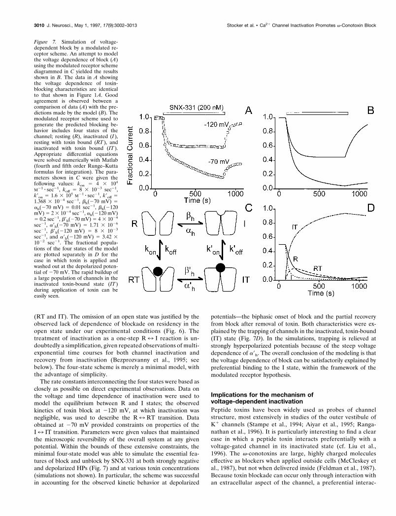
(RT and IT). The omission of an open state was justified by theobserved lack of dependence of blockade on residency in theopen state under our experimental conditions (Fig. 6). Thetreatment of inactivation as a one-step R7 I reaction is un-doubtedly a simplification, given repeated observations of multi-exponential time courses for both channel inactivation andrecovery from inactivation (Bezprozvanny et al., 1995; seebelow). The four-state scheme is merely a minimal model, withthe advantage of simplicity.
The rate constants interconnecting the four states were based asclosely as possible on direct experimental observations. Data onthe voltage and time dependence of inactivation were used tomodel the equilibrium between R and I states; the observedkinetics of toxin block at 2120 mV, at which inactivation wasnegligible, was used to describe the R7 RT transition. Dataobtained at 270 mV provided constraints on properties of theI7 IT transition. Parameters were given values that maintainedthe microscopic reversibility of the overall system at any givenpotential. Within the bounds of these extensive constraints, theminimal four-state model was able to simulate the essential fea-tures of block and unblock by SNX-331 at both strongly negativeand depolarized HPs (Fig. 7) and at various toxin concentrations(simulations not shown). In particular, the scheme was successfulin accounting for the observed kinetic behavior at depolarized
potentials—the biphasic onset of block and the partial recoveryfrom block after removal of toxin. Both characteristics were ex-plained by the trapping of channels in the inactivated, toxin-bound(IT) state (Fig. 7D). In the simulations, trapping is relieved atstrongly hyperpolarized potentials because of the steep voltagedependence of a9h. The overall conclusion of the modeling is thatthe voltage dependence of block can be satisfactorily explained bypreferential binding to the I state, within the framework of themodulated receptor hypothesis.
Implications for the mechanism ofvoltage-dependent inactivationPeptide toxins have been widely used as probes of channelstructure, most extensively in studies of the outer vestibule ofK1 channels (Stampe et al., 1994; Aiyar et al., 1995; Ranga-nathan et al., 1996). It is particularly interesting to find a clearcase in which a peptide toxin interacts preferentially with avoltage-gated channel in its inactivated state (cf. Liu et al.,1996). The v-conotoxins are large, highly charged moleculeseffective as blockers when applied outside cells (McCleskey etal., 1987), but not when delivered inside (Feldman et al., 1987).Because toxin blockade can occur only through interaction withan extracellular aspect of the channel, a preferential interac-
Figure 7. Simulation of voltage-dependent block by a modulated re-ceptor scheme. An attempt to modelthe voltage dependence of block (A)using the modulated receptor schemediagrammed in C yielded the resultsshown in B. The data in A showingthe voltage dependence of toxin-blocking characteristics are identicalto that shown in Figure 1 A. Goodagreement is observed between acomparison of data (A) with the pre-dictions made by the model (B). Themodulated receptor scheme used togenerate the predicted blocking be-havior includes four states of thechannel; resting (R), inactivated (I ),resting with toxin bound (RT ), andinactivated with toxin bound (IT ).Appropriate differential equationswere solved numerically with Matlab(fourth and fifth order Runge–Kuttaformulas for integration). The para-meters shown in C were given thefollowing values: kon 5 4 3 104
M21 z sec21, koff 5 8 3 1023 sec21,k9on 5 1.6 3 105 M21 z sec21, k9off 51.368 3 1024 sec21, bh(270 mV) 5ah(270 mV) 5 0.01 sec21, bh(2120mV) 5 2 3 1024 sec21, ah(2120 mV)5 0.2 sec21, b9h(270 mV) 5 4 3 1024
sec21, a9h(270 mV) 5 1.71 3 1026
sec21, b9h(2120 mV) 5 8 3 1023
sec21, and a9h(2120 mV) 5 3.42 31022 sec21. The fractional popula-tions of the four states of the modelare plotted separately in D for thecase in which toxin is applied andwashed out at the depolarized poten-tial of 270 mV. The rapid buildup ofa large population of channels in theinactivated toxin-bound state (IT )during application of toxin can beeasily seen.
3010 J. Neurosci., May 1, 1997, 17(9):3002–3013 Stocker et al. • Ca21 Channel Inactivation Promotes v-Conotoxin Block

tion with the inactivated channel must mean that inactivation isaccompanied by an externally detectable conformationalchange. The locus of this structural change can be narrowedfurther if v-conotoxin binds near the mouth of the pore, assuggested by several lines of evidence. All four motifs of theN-type Ca21 channel contribute to its interaction with v-CTx-GVIA, but especially motifs I and III, which are believed tooccupy positions diametrically opposite each other across thepore mouth (Ellinor et al., 1994). These results suggest that thetoxin straddles the external mouth of the pore while engaged infavorable interactions with residues in extracellular loops nearthe putative pore-forming region (e.g., IIIS5) (Ellinor et al.,1994). Furthermore, increases in external Ba21 greatly slowv-conotoxin block, consistent with the notion that the toxincomes close to divalent cations bound at the locus of selectivity(Boland et al., 1994; McDonough et al., 1996). Taken together,these observations suggest that the toxin interacts with the
channel near the extracellular pore mouth, suggesting that thisregion harbors a conformational change associated with in-activation. This is reminiscent of the mechanism proposed forC-type inactivation in K1 channels (Choi et al., 1991; Lopez-Barneo et al., 1993; Yellen et al., 1994; Baukrowitz and Yellen,1995, 1996).
An important aim for future experiments is to utilize thefavorable properties of peptide toxins (rigid, well characterizedbackbone structure, many derivatives with extensively modifiedside chains) to obtain a clearer picture of the structural basis ofinactivation. Residues near S6 segments, adjacent to the pore-forming region, are a logical place to start because they appear tobe important in controlling the rate of voltage-dependent inacti-vation in Ca21 channels such as doe-1 and a1A (Zhang et al., 1994;Hering et al., 1996). Although toxin interactions can be mostreadily studied with the large collection of natural and syntheticv-conotoxins known to affect N-type Ca21 channels, any struc-
Figure 8. Both buildup and relief of block from theslowly recovering fraction of channels are stronglyvoltage-dependent. A, A protocol was used to inves-tigate the blocking characteristics of toxin when ap-plication and washout occur at different HPs. First,SNX-331 (1 mM) was applied at an HP 5 280 mV.Washoff of toxin occurred with a concomitant changein the HP to 2120 mV. After recovery of current,SNX-331 was applied again, but now with the oocyteheld at 2120 mV, and washoff after this applicationoccurred at an HP of 280 mV. In the last two seg-ments of this protocol, the same HP is maintainedthroughout toxin application and washoff. Toxin ap-plication and washoff is first done at an HP of 280mV. Full recovery of current is then achieved byhyperpolarizing the cell to 2120 mV, and applicationand washoff of toxin at this hyperpolarized potentialfollows. B, Using the same modulated receptorscheme and parameters as in Figure 7, a model of theexpected peak current levels during the above givenprotocol was generated. C, Plot of the fractional pop-ulation of the I state of the channel with toxin-bound(IT ) as predicted by the modulated receptor scheme.
Stocker et al. • Ca21 Channel Inactivation Promotes v-Conotoxin Block J. Neurosci., May 1, 1997, 17(9):3002–3013 3011

tural insights about inactivation are likely to apply across theentire family of voltage-gated Ca21 channels.
REFERENCESAiyar J, Withka JM, Rizzi JP, Singleton DH, Andrews GC, Lin W, Boyd
J, Hanson DC, Simon M, Dethlefs B, Lee C, Hall JE, Gutman GA,Chandy KG (1995) Topology of the pore-region of a K1 channelrevealed by the NMR-derived structures of scorpion toxins. Neuron15:1169–1181.
Aosaki T, Kasai H (1989) Characterization of two kinds of high-voltage-activated Ca-channel currents in chick sensory neurons. Differentialsensitivity to dihydropyridines and omega-conotoxin GVIA. PflugersArch 414:150–156.
Armstrong CM (1969) Inactivation of the potassium conductance andrelated phenomena caused by quaternary ammonium ion injection insquid axons. J Gen Physiol 54:553–575.
Armstrong CM (1971) Interaction of tetraethylammonium ion deriva-tives with the potassium channels of giant axons. J Gen Physiol58:413–437.
Basus VJ, Nadasdi L, Ramachandran J, Miljanich GP (1995) Solutionstructure of v-conotoxin MVIIA using 2D NMR spectroscopy. FEBSLett 370:163–169.
Baukrowitz T, Yellen G (1995) Modulation of K1 current by frequencyand external [K1]: a tale of two inactivation mechanisms. Neuron15:951–960.
Baukrowitz T, Yellen G (1996) Use-dependent blockers and exit rate ofthe last ion from the multi-ion pore of a K1 channel. Science271:653–656.
Bean BP, Cohen CJ, Tsien RW (1983) Lidocaine block of cardiac sodiumchannels. J Gen Physiol 81:613–642.
Bean BP (1984) Nitrendipine block of cardiac calcium channels: highaffinity binding to the inactivated state. Proc Natl Acad Sci USA81:6388–6392.
Bezprozvanny I, Tsien RW (1995) Voltage-dependent blockade of diversetypes of voltage-gated Ca21 channels expressed in Xenopus oocytes bythe Ca21 channel antagonist mibefradil (Ro40-5967). Mol Pharmacol48:540–549.
Bezprozvanny I, Scheller RH, Tsien RW (1995) Functional impact ofsyntaxin on gating of N-type and Q-type calcium channels. Nature378:623–626.
Boland LM, Morrill JA, Bean BP (1994) v-Conotoxin block of N-typecalcium channels in frog and rat sympathetic neurons. J Neurosci14:5011–5027.
Carbone E, Swandulla D (1990) Neuronal calcium channels: kinetics,blockade and modulation. Prog Biophys Mol Biol 54:31–58.
Catterall WA (1977) Membrane potential-dependent binding of scor-pion toxin to the action potential Na1 ionophore. J Biol Chem23:8660–8668.
Catterall WA (1993) Structure and function of voltage-gated ion chan-nels. Trends Neurosci 16:500–506.
Choi KL, Aldrich RW, Yellen G (1991) Tetraethylammonium blockadedistinguishes two inactivation mechanisms in voltage-activated K1
channels. Proc Natl Acad Sci USA 88:5092–5095.Dunlap K, Luebke JI, Turner TJ (1995) Exocytotic Ca21 channels in
mammalian central neurons. Trends Neurosci 18:89–98.Ellinor PT, Zhang J, Horne WA, Tsien RW (1994) Structural determi-
nants of the blockade of N-type calcium channels by a peptide neuro-toxin. Nature 372:272–275.
Farr-Jones S, Miljanich GP, Nadasdi L, Ramachandran J, Basus VJ(1995) Solution structure of v-conotoxin MVIIC, a high affinity ligandof P-type calcium channels, using 1H NMR spectroscopy and completerelaxation matrix analysis. J Mol Biol 248:106–124.
Feldman DH, Olivera BM, Yoshikami D (1987) Omega conus geogra-phus toxin: a peptide that blocks calcium channels. FEBS Lett214:295–300.
French RJ, Prusak-Sochaczewski E, Zamponi GW, Becker S, KularatnaAS, Horn R (1996) Interactions between a pore-blocking peptide andthe voltage sensor of the sodium channel: an electrostatic approach tochannel geometry. Neuron 16:407–413.
Fujita Y, Mynlieff M, Dirksen RT, Kim M-S, Niidome T, Nakai J,Friedrich T, Iwabe N, Miyata T, Furuichi T, Furutama D, Mikoshiba K,Mori Y, Beam KG (1993) Primary structure and functional expressionof the omega-conotoxin-sensitive N-type calcium channel from rabbitbrain. Neuron 10:585–598.
Hering S, Aczel S, Grabner M, Doring F, Berjukow S, Metterdorfer J,Sinnegger MJ, Striessnig J, Degitar VE, Wang Z, Glossmann H (1996)Transfer of high sensitivity for benzothiazepines from L-type to class A(BI) calcium channels. J Biol Chem 271:24471–24475.
Hidalgo P, MacKinnon R (1995) Revealing the architecture of a K1
channel pore through mutant cycles with a peptide inhibitor. Science268:307–310.
Hille B (1977) Local anesthetics: hydrophilic and hydrophobic pathwaysfor the drug-receptor reaction. J Gen Physiol 69:496–515.
Hirning LD, Fox AP, McCleskey EW, Olivera BM, Thayer SA, Miller RJ,Tsien RW (1988) Dominant role of N-type Ca21 channels in evokedrelease of norepinephrine from sympathetic neurons. Science239:57–61.
Hondeghem LM, Katzung BG (1977) Time- and voltage-dependent in-teractions of antiarrhythmic drugs with cardiac sodium channels. Bio-chim Biophys Acta 472:373–398.
Hoshi T, Zagotta WN, Aldrich RW (1990) Biophysical and molecularmechanisms of Shaker potassium channel inactivation. Science250:533–538.
Jones SW, Marks TN (1989) Calcium currents in bullfrog sympatheticneurons. J Gen Physiol 94:169–182.
Kasai H, Aosaki T, Fukuda J (1987) Presynaptic Ca-antagonist omega-conotoxin irreversibly blocks N-type Ca-channels in chick sensory neu-rons. Neurosci Res 4:228–235.
Komuro H, Rakic P (1992) Selective role of N-type calcium channels inneuronal migration. Science 257:806–809.
Larsson HP, Baker OS, Dhillon DS, Isacoff EY (1996) Transmembranemovement of the Shaker K1 channel S4. Neuron 16:387–397.
Lee KS, Tsien RW (1983) Mechanism of calcium channel blockade byverapamil, D600, diltiazem and nitrendipine in single dialyzed heartcells. Nature 302:790–794.
Liu Y, Jurman ME, Yellen G (1996) Dynamic rearrangement of theouter mouth of a K1 channel during gating. Neuron 16:859–867.
Lopez-Barneo J, Hoshi T, Heinemann SH, Aldrich RW (1993) Effects ofexternal cations and mutations in the pore region on C-type inactivationof Shaker potassium channels. Receptors and Channels 1:61–71.
McCleskey EW, Fox AP, Feldman D, Cruz LJ, Olivera BM, Tsien RW,Yoshikami D (1987) v-Conotoxin: direct and persistent block of spe-cific types of calcium channels in neurons but not muscle. Proc NatlAcad Sci USA 84:4327–4331.
McDonough SI, Swartz KJ, Mintz IM, Boland LM, Bean BP (1996)Inhibition of calcium channels in rat central and peripheral neurons byv-conotoxin MVIIC. J Neurosci 16:2612–2623.
McEnery MW, Snowman AM, Sharp AH, Adams ME, Snyder SH (1991)Purified omega-conotoxin GVIA receptor of rat brain resembles adihydropyridine-sensitive L-type calcium channel. Proc Natl Acad SciUSA 88:11095–11099.
Mintz I, Adams ME, Bean BP (1992) P-type calcium channels in ratcentral and peripheral neurons. Neuron 9:85–95.
Nemoto N, Kubo S, Yoshida T, Chino N, Kimura T, Sakakibara S,Kyogoku Y, Kobayashi Y (1995) Solution structure of v-conotoxinMVIIC determined by NMR. Biochem Biophys Res Commun207:695–700.
Nowycky MC, Fox AP, Tsien RW (1985) Three types of neuronal cal-cium channel with different calcium agonist sensitivity. Nature316:440–443.
Pallaghy PK, Duggan BM, Pennington MW, Norton RS (1993) Three-dimensional structure in solution of the calcium channel blockerv-conotoxin. J Mol Biol 234:405–420.
Pelzer D, Pelzer S, McDonald TF (1990) Properties and regulation ofcalcium channels in muscle cells. Rev Physiol Biochem Pharmacol114:107–207.
Plummer MR, Logothetis DE, Hess P (1989) Elementary properties andpharmacological sensitivities of calcium channels in mammalian periph-eral neurons. Neuron 2:1453–1463.
Randall AD, Tsien RW (1995) Pharmacological dissection of multipletypes of Ca21 channel currents in rat cerebellar granule neurons.J Neurosci 15:2995–3012.
Ranganathan R, Lewis JH, MacKinnon R (1996) Spatial localization ofthe K1 channel selectivity filter by mutant cycle-based structure analy-sis. Neuron 16:131–139.
Regan LJ, Sah DWY, Bean BP (1991) Ca21 channels in rat central andperipheral neurons: high-threshold current resistant to dihydropyridineblockers and v-conotoxin. Neuron 6:269–280.
Ruth P, Rohrkasten A, Biel M, Bosse E, Regulla S, Meyer HE, Flockerzi
3012 J. Neurosci., May 1, 1997, 17(9):3002–3013 Stocker et al. • Ca21 Channel Inactivation Promotes v-Conotoxin Block

V, Hofmann F (1989) Primary structure of the beta subunit of theDHP-sensitive calcium channel from skeletal muscle. Science245:1115–1118.
Sakamoto J, Campbell KP (1991) A monoclonal antibody to the betasubunit of the skeletal muscle dihydropyridine receptor immunoprecipi-tates the brain omega-conotoxin GVIA receptor. J Biol Chem266:18914–18919.
Sather WA, Tanabe T, Zhang J-F, Mori Y, Adams ME, Tsien RW (1993)Distinctive biophysical and pharmacological properties of class A (BI)calcium channel a1 subunits. Neuron 11:291–303.
Satin J, Limberis JT, Kyle JW, Rogart RB, Fozzard HA (1994) Thesaxitoxin /tetrodotoxin binding site on cloned rat brain IIa Na channelsis in the transmembrane electric field. Biophys J 67:1007–1014.
Sevilla P, Bruix M, Santoro J, Gago F, Garcia AG, Rico M (1993)Three-dimensional structure of v-conotoxin GVIA determined by 1HNMR. Biochem Biophys Res Commun 192:1238–1244.
Stampe P, Kolmakova PL, Miller C (1994) Intimations of K1 channelstructure from a complete functional map of the molecular surface ofcharybdotoxin. Biochemistry 33:443–450.
Stanley EF, Goping G (1991) Characterization of a calcium current ina vertebrate cholinergic presynaptic nerve terminal. J Neurosci 11:985–993.
Stuhmer W, Conti F, Suzuki H, Wang X, Noda M, Yahagi V, Kubo H,
Numa S (1989) Structural parts involved in activation and inactivationof the sodium channel. Nature 339:597–603.
Vassilev PM, Scheuer T, Catterall WA (1988) Identification of an intra-cellular peptide segment involved in sodium channel inactivation. Sci-ence 241:1658–1661.
Wheeler DB, Randall A, Tsien RW (1994) Roles of N-type and Q-typeCa21 channels in supporting hippocampal synaptic transmission. Sci-ence 264:107–111.
Williams ME, Brust PF, Feldman DH, Patthi S, Simerson S, Maroufi A,McCue AF, Velicelebi G, Ellis SB, Harpold MM (1992) Structure andfunctional expression of an omega-conotoxin-sensitive human N-typecalcium channel. Science 257:389–395.
Yang N, Horn R (1995) Evidence for voltage-dependent S4 movement insodium channels. Neuron 15:213–218.
Yellen G, Sodickson D, Chen T-Y, Jurman ME (1994) An engineeredcysteine in the external mouth of a K1 channel allows inactivation to bemodulated by metal binding. Biophys J 66:1068–1075.
Zagotta WN, Hoshi T, Aldrich RW (1990) Restoration of inactivation inmutants of Shaker potassium channels by peptide derived from ShB.Science 250:568–571.
Zhang J-F, Ellinor PT, Aldrich RW, Tsien RW (1994) Molecular deter-minants of voltage-dependent inactivation of calcium channels. Nature372:97–100.
Stocker et al. • Ca21 Channel Inactivation Promotes v-Conotoxin Block J. Neurosci., May 1, 1997, 17(9):3002–3013 3013