Embed Size (px)
Citation preview

Proc. Natl. Acad. Sci. USAVol. 89, pp. 3967-3971, May 1992Biochemistry
Preferential hydroxylation by the chemical nuclease meso-tetrakis-(4-N-methylpyridiniumyl)porphyrinatomanganese"I' pentaacetate/KHSO5 at the 5' carbon of deoxyriboses on both 3' sides of threecontiguous AT base pairs in short double-stranded oligonucleotides
(manganese porphyrin/sugar degradation)
MARGUERITE PITI1, GENEVIEVE PRATVIEL, JEAN BERNADOU*, AND BERNARD MEUNIER*Laboratoire de Chimie de Coordination du Centre National de la Recherche Scientifique, 205 route de Narbonne, 31077 Toulouse Cedex, France
Communicated by Jean-Marie Lehn, December 23, 1991 (received for review October 14, 1991)
ABSTRACT Selected double-stranded oligodeoxyribonu-cleotides have been used to probe, at the molecular level, DNAchain breakages induced by the chemical nuclease meso-tetrakis(4-N-methylpyridiniumyl)porphyrinatomanganese"'1pentaacetate/KHSO5. The results show that cleavage selec-tively occurs on the two 3' sides of three contiguous A-T basepairs (an AT triplet). Hydroxylation at 5' carbon of thedeoxyribose targets represents the initial damage on the sugar-phosphodiester backbone and leaves a 3' phosphate and a 5'aldehyde at the ends. The fragments were separated by HPLCand unambiguously identified through chemical and biochem-ical reactions and/or sequencing after enzymatic conversion tomononucleosides. Also studied was the degradation of a 22-nucleotide DNA molecule containing two A-T triplets. Gelelectrophoresis analyses on the corresponding 5'-32P-end-labeled substrate supported the above cleavage specificity andmechanism.
As a result of extensive studies on the molecular aspects ofDNA degradation by bleomycin (1, 2), "chemical nucleases"have been developed (3-5). All these systems are based onredox-active complexes (copper, iron, cobalt, etc.) with highDNA affinity. In many cases DNA strand breaks result froman oxidative attack on deoxyribose units. Whereas bleomy-cin abstracts the hydrogen atom at the C4' position (1, 2),copper-1,10-phenanthroline/hydrogen peroxide hydroxy-lates the anomeric C-H bond and a concomitant base releaseis observed (6). Metalloporphyrins, especially those bearingN-methylpyridinium groups at meso positions, are also effi-cient DNA cleavers when activated by a single oxygen atomdonor such as KHSO5 (7-9); the tris(pyridinium) metallopor-phyrin entity is a cytotoxic DNA cleaving reagent that can beattached to a variety of vectors (intercalating agent, oligo-nucleotide, groove binder, peptide, etc.) (10-13). For exam-ple, in meso-tetrakis(4-N-methylpyridiniumyl)porphyrinato-manganese"' pentaacetate (Mn-TMPyP), the high-valencemanganese-oxo complex can hydroxylate two C-H bonds, atCl' and at C5', accessible from the minor groove. These twolesions are diagnosed by the formation of 5-methylene-2-furanone (5-MF) and furfural (Fur), respectively (14), and thereleased monophosphate termini have been identified by 31PNMR (15). Hydroxylation at C5' has never been reported formetallic redox-active complexes but is well known for neo-carzinostatin (16) and calicheamicin (17).To understand the relationship between the sequence
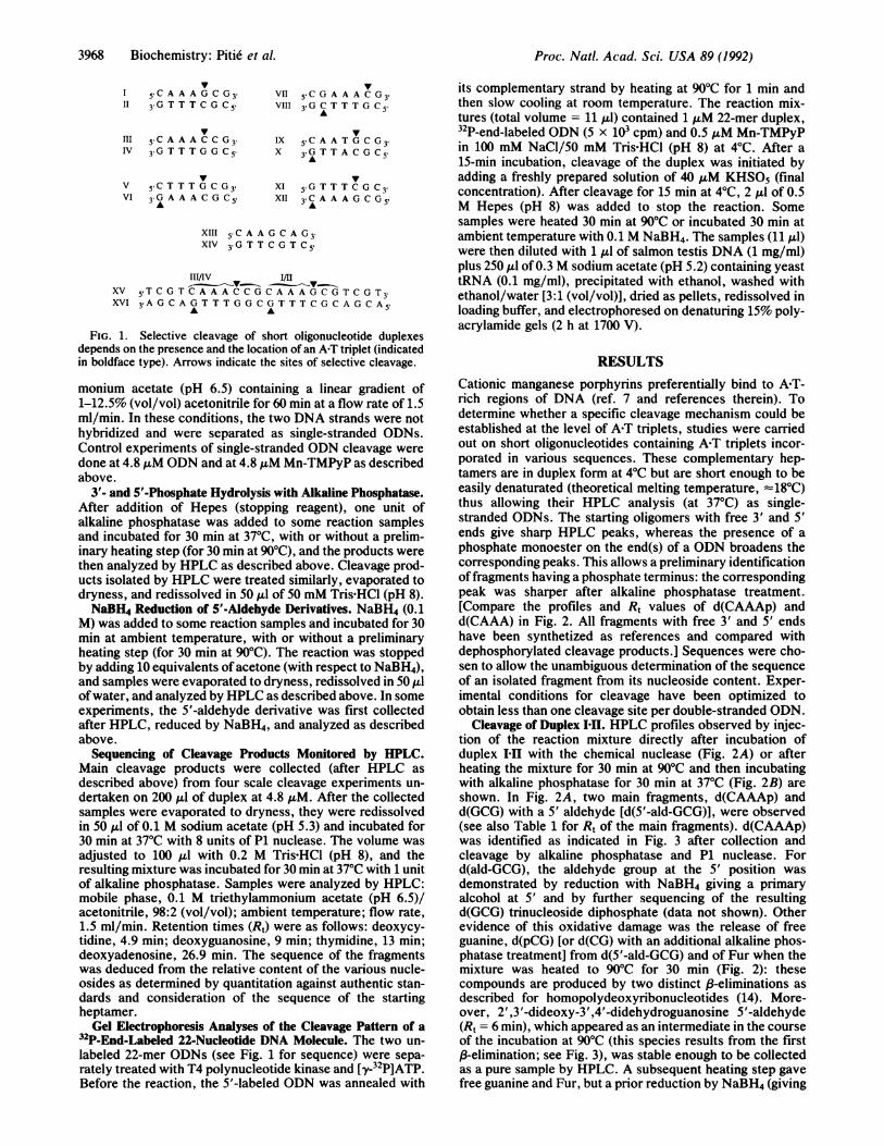
specificity of the DNA cleaver and the selection of the sugartargets accessible from the minor groove, we prepared aseries of seven short duplexes (Fig. 1). Six of them contain
the smallest selective Mn-TMPyP cleavage site consisting ofa trinucleotide sequence having only the bases adenine andthymine-i.e., three contiguous A-T base pairs that we terman A-T triplet (7, 18, 19). Here we report the identification ofthe major attack sites in a double-stranded oligonucleotide atthe C5' position of the sugar units at the two 3' sides of theA-T triplet. All DNA fragments and sugar residues have beenidentified by HPLC by comparison with authentic samplesand analysis offragments obtained by treatment with alkalinephosphatase, P1 nuclease, or NaBH4.
MATERIALS AND METHODSMaterials. Potassium monopersulfate (Curox) was a gift of
Interox. Mn-TMPyP was synthesized in our laboratory (8).Alkaline phosphatase (EC 3.1.3.1, from Escherichia coli,type III), P1 nuclease (EC 3.1.30.1, from Penicillium citri-num), adenine, cytosine, thymine, guanine, 2'-deoxyadeno-sine, 2'-deoxycytidine, thymidine, and 2'-deoxyguanosinewere from Sigma. T4 polynucleotide kinase (EC 2.7.1.78) and[y-32P]ATP were from Amersham. 5-MF was prepared asdescribed (20). Fur and other chemicals were from Aldrich.
Oligodeoxynucleotides (ODNs). ODNs I-XVI and referencefragments were synthesized on a Cyclone Plus DNA synthe-sizer from MilliGen/Biosearch (Novato, CA). Concentra-tions of heptamers and their fragments were determinedaccording to their absorption coefficient (21). The sequenceof the duplex XV-XVI was built to contain both duplexes IIHand IIIIV.HPLC. Analyses were done on a reverse-phase Nucleosil
C18 10 g column eluted in isocratic or gradient mode asindicated. All products were detected at a wavelength of 254nm. In some experiments, each cleavage product was col-lected to test its own chemical or biochemical reactivity.ODN Digestion. All reactions were done on an ice bath, and
reaction mixtures contained 100 mM NaCl and 50 mMTris HCl (pH 8). The two complementary single-strandedODNs were allowed to anneal for 15 min (resulting duplex =4.8 ,.M) before Mn-TMPyP (4.8 tLM) was added to themixture. After another 15-min incubation, cleavage of theduplex was initiated by the addition of a freshly preparedsolution of KHSO5 (96 A.M). Total volume was 50 gl; finalconcentrations are indicated in parentheses. After cleavagefor 5 min at 40C, 5 1.l of 1 M Hepes at pH 8 was added to stopthe reaction (8). The reaction mixture was then directlyanalyzed by HPLC. The column was thermostatically regu-lated at 370C and material was eluted with 0.1 M triethylam-
Abbreviations: Mn-TMPyP, meso-tetrakis(4-N-methylpyridiniumyl)-porphyrinatomanganesel" pentaacetate; 5-MF, 5-methylene-2-furanone; Fur, furfural; ODN, oligodeoxyribonucleotide; Rt, reten-tion time; d(5'-ald-GCG), d(GCG) with a 5' aldehyde.*To whom reprint requests should be addressed.
3967
The publication costs of this article were defrayed in part by page chargepayment. This article must therefore be hereby marked "advertisement"in accordance with 18 U.S.C. §1734 solely to indicate this fact.
Proc. Natl. Acad. Sci. USA 89 (1992)
5C A A A G C G3.C G A A A C G3.II GTTTCGC V 3.G C T T T G C5.
V VHI 5.C A A A C C G3. IX C A A T G C G3.IV 3.G T T T G G C5 X 3G T T A C G C5
V VV 5C T T T G C G3. XI 5G T T T C G C3.VI 3G A A A C G C5 XII 3YCA A A G C G5
XIII 5C A A G C A G3.XIV G T T C G T C5.
IIVIV I/HXV 5.T C G T C A A A C C 0 C A A A G C G T C G T3.XVI 3-AGCAGTTTGGCGTTTCGCAGCA5,
A A
FIG. 1. Selective cleavage of short oligonucleotide duplexesdepends on the presence and the location of an AT triplet (indicatedin boldface type). Arrows indicate the sites of selective cleavage.
monium acetate (pH 6.5) containing a linear gradient of1-12.5% (vol/vol) acetonitrile for 60 min at a flow rate of 1.5ml/min. In these conditions, the two DNA strands were nothybridized and were separated as single-stranded ODNs.Control experiments of single-stranded ODN cleavage weredone at 4.8 ,uM ODN and at 4.8 AM Mn-TMPyP as describedabove.
3'- and 5'-Phosphate Hydrolysis with Alkaline Phosphatase.After addition of Hepes (stopping reagent), one unit ofalkaline phosphatase was added to some reaction samplesand incubated for 30 min at 37°C, with or without a prelim-inary heating step (for 30 min at 90°C), and the products werethen analyzed by HPLC as described above. Cleavage prod-ucts isolated by HPLC were treated similarly, evaporated todryness, and redissolved in 50 ,u1 of 50 mM Tris HCI (pH 8).NaBH4 Reduction of 5'-Aldehyde Derivatives. NaBH4 (0.1
M) was added to some reaction samples and incubated for 30min at ambient temperature, with or without a preliminaryheating step (for 30 min at 90°C). The reaction was stoppedby adding 10 equivalents of acetone (with respect to NaBH4),and samples were evaporated to dryness, redissolved in 50 ,uof water, and analyzed by HPLC as described above. In someexperiments, the 5'-aldehyde derivative was first collectedafter HPLC, reduced by NaBH4, and analyzed as describedabove.
Sequencing of Cleavage Products Monitored by HPLC.Main cleavage products were collected (after HPLC asdescribed above) from four scale cleavage experiments un-dertaken on 200 ,ul of duplex at 4.8 ,M. After the collectedsamples were evaporated to dryness, they were redissolvedin 50 ,ul of 0.1 M sodium acetate (pH 5.3) and incubated for30 min at 37°C with 8 units of P1 nuclease. The volume wasadjusted to 100 ,ul with 0.2 M Tris-HCl (pH 8), and theresulting mixture was incubated for 30 min at 37°C with 1 unitof alkaline phosphatase. Samples were analyzed by HPLC:mobile phase, 0.1 M triethylammonium acetate (pH 6.5)/acetonitrile, 98:2 (vol/vol); ambient temperature; flow rate,1.5 ml/min. Retention times (RJ) were as follows: deoxycy-tidine, 4.9 min; deoxyguanosine, 9 min; thymidine, 13 min;deoxyadenosine, 26.9 min. The sequence of the fragmentswas deduced from the relative content of the various nucle-osides as determined by quantitation against authentic stan-dards and consideration of the sequence of the startingheptamer.
Gel Electrophoresis Analyses of the Cleavage Pattern of a32P-End-Labeled 22-Nucleotide DNA Molecule. The two un-labeled 22-mer ODNs (see Fig. 1 for sequence) were sepa-rately treated with T4 polynucleotide kinase and [y-32P]ATP.Before the reaction, the 5'-labeled ODN was annealed with
its complementary strand by heating at 90'C for 1 min andthen slow cooling at room temperature. The reaction mix-tures (total volume = 11 p.l) contained 1 p.M 22-mer duplex,32P-end-labeled ODN (5 x 10³ cpm) and 0.5 p.M Mn-TMPyPin 100 mM NaCl/50 mM Tris HCl (pH 8) at 40C. After a15-min incubation, cleavage of the duplex was initiated byadding a freshly prepared solution of 40 p.M KHSO5 (finalconcentration). After cleavage for 15 min at 40C, 2 ,ul of 0.5M Hepes (pH 8) was added to stop the reaction. Somesamples were heated 30 min at 900C or incubated 30 min atambient temperature with 0.1 M NaBH4. The samples (11 ILI)were then diluted with 1 p.l of salmon testis DNA (1 mg/ml)plus 250 .l of 0.3 M sodium acetate (pH 5.2) containing yeasttRNA (0.1 mg/ml), precipitated with ethanol, washed withethanol/water [3:1 (vol/vol)], dried as pellets, redissolved inloading buffer, and electrophoresed on denaturing 15% poly-acrylamide gels (2 h at 1700 V).
RESULTSCationic manganese porphyrins preferentially bind to A-T-rich regions of DNA (ref. 7 and references therein). Todetermine whether a specific cleavage mechanism could beestablished at the level of AT triplets, studies were carriedout on short oligonucleotides containing A-T triplets incor-porated in various sequences. These complementary hep-tamers are in duplex form at 4°C but are short enough to beeasily denaturated (theoretical melting temperature, =18°C)thus allowing their HPLC analysis (at 37°C) as single-stranded ODNs. The starting oligomers with free 3' and 5'ends give sharp HPLC peaks, whereas the presence of aphosphate monoester on the end(s) of a ODN broadens thecorresponding peaks. This allows a preliminary identificationof fragments having a phosphate terminus: the correspondingpeak was sharper after alkaline phosphatase treatment.[Compare the profiles and Rt values of d(CAAAp) andd(CAAA) in Fig. 2. All fragments with free 3' and 5' endshave been synthetized as references and compared withdephosphorylated cleavage products.] Sequences were cho-sen to allow the unambiguous determination of the sequenceof an isolated fragment from its nucleoside content. Exper-imental conditions for cleavage have been optimized toobtain less than one cleavage site per double-stranded ODN.
Cleavage of Duplex I-II. HPLC profiles observed by injec-tion of the reaction mixture directly after incubation ofduplex III with the chemical nuclease (Fig. 2A) or afterheating the mixture for 30 min at 90°C and then incubatingwith alkaline phosphatase for 30 min at 37°C (Fig. 2B) areshown. In Fig. 2A, two main fragments, d(CAAAp) andd(GCG) with a 5' aldehyde [d(5'-ald-GCG)], were observed(see also Table 1 for Rt of the main fragments). d(CAAAp)was identified as indicated in Fig. 3 after collection andcleavage by alkaline phosphatase and P1 nuclease. Ford(ald-GCG), the aldehyde group at the 5' position wasdemonstrated by reduction with NaBH4 giving a primaryalcohol at 5' and by further sequencing of the resultingd(GCG) trinucleoside diphosphate (data not shown). Otherevidence of this oxidative damage was the release of freeguanine, d(pCG) [or d(CG) with an additional alkaline phos-phatase treatment] from d(5'-ald-GCG) and of Fur when themixture was heated to 90°C for 30 min (Fig. 2): thesecompounds are produced by two distinct a-eliminations asdescribed for homopolydeoxyribonucleotides (14). More-over, 2',3'-dideoxy-3',4'-didehydroguanosine 5'-aldehyde(Rt = 6 min), which appeared as an intermediate in the courseof the incubation at 90'C (this species results from the first,B-elimination; see Fig. 3), was stable enough to be collectedas a pure sample by HPLC. A subsequent heating step gavefree guanine and Fur, but a prior reduction by NaBH4 (giving
3968 Biochemistry: Pitie' et al.

Proc. Natl. Acad. Sci. USA 89 (1992) 3969
II
Ad(5'-ald-GCG)
,1,,r-
mo-0o¢
I
d(CAAAP)
d(CAAA)
B
Gua
d(CG)
FURIL0 5Time (min) L j
FIG. 2. HPLC profiles of duplex III cleaved with Mn-TMPyP/KHSO5 before (A) and after (B) heating for 30 min at 90TC and thenincubating with alkaline phosphatase for 30 min at 37TC.
2',3'-dideoxy-3',4'-didehydroguanosine; Rt = 7.5 min) inhib-ited this second 8-elimination.As shown in Figs. 1 and 2 and as indicated in Table 1, only
oligomer I was selectively cleaved with a selectivity of=90%. A 90% selectivity indicates that the two main ODNfragments correspond to 90% of the fraction of the startingstrand that has been cleaved. For nonselective strand cleav-age, all fragments generated contribute to the background ofHPLC chromatograms. This is the case for XIIIXIV, whichcan be considered a control duplex with no APT triplet and forsingle-stranded oligomers (data not shown). In the cleavageof III, complementary strand II was randomly cleaved (33%of remaining oligomer but no selective cleavage was ob-served; Table 1), probably after strand dissociation thatfollowed the cleavage of oligomer I.
Cleavage of 7-Mer Duplexes with Various Sequences. Themain features are as follows. (i) All selective breaks inducedon the oligomers resulted from an oxidative attack at C5'.Strand cleavage gave a 3'-phosphate fragment and a 5'-atlehyde fragment, NaBH4 reduction of the aldehyde frag-ment gave the corresponding 5'-OH oligomer, free baseswere only released after heating, and Fur (Rt = 11 min), butnot 5-MF (Rt = 12.5 min), could be detected as the sugarresidue (Fig. 1). (ii) The presence of an A-T triplet is neces-sary to observe a selective cleavage on at least one strand ofthe duplex. With XIIIXIV, which does not contain an A-Ttriplet, no selective cleavage was observed even though
r
starting oligomers were effectively degraded up to 30% (XIV)and 50% (XIII) (Table 1). (iii) The main strand scission alwaysoccurred at the sugar on the 3' side of the APT triplet (i.e., onthe sugar-bearing guanine in I-11, V-VI, and IX-X and cytosinein III-IV, VIIVIII, and XIXII, see Fig. 1). Thus the locationof this triplet is directing the cleavage site. For example, theA-T triplet was shifted by 1 base pair in VI-VIII compared toIIH, both having the same base composition, and the maincleavage site was shifted to cytosine in VI1FVII compared toguanine in I-II. The nature of the 3' side of the base (guanineor cytosine) was not important [see also 1IH and HISIV (Fig.1)]. (iv) When a selective cleavage was taking place on bothstrands of the duplex, the cleavage site on one strand wasalways shifted toward the 3' end by 4 base pairs relatively tothe site of the opposite strand. As shown in Fig. 4 this4-base-pair shift is in good agreement with two 5' hydroxy-lations on opposite DNA strands mediated by the nondiffus-ible manganese-oxo species located in the minor groove ofDNA (19). Cleavage was always higher in the middle of theoligomers than at the end. (v) When scission occurredthrough oxidation ofthe last sugar at the 3' end ofan oligomer(V-VI, IX-X, and XIXII, Fig. 1), 2'-deoxyguanosine 5'-aldehyde (Rt = 7.8 min, for V-VI and IX-X) and 2'-deoxy-cytosine 5'-aldehyde (Rt = 4.6 min, for XFXII) were releasedand identified by reduction with NaBH4 giving 2'-deoxyguanosine and 2'-deoxycytosine, respectively, whichcoeluted on HPLC column with authentic samples. (vi) Themajor sites of cleavage were located preferentially on theA-rich strand (I, III, VII, and IX). However, when theposition of the A-rich sequence was close to the 3' end, themajor site of attack shifted to the T-rich strand in the middleof the duplex (V and XI) (see Table 1). (vii) Control experi-ments on single-stranded oligomers always showed loweroxidation (up to 75% of the oligomer still remained un-changed after 5 min of reaction) and no selectivity wasobserved.
Cleavage of a 5'-32P-End-Labeled 22-Mer ODN. To confirmthe sequence selectivity observed on short ODNs, a 5 _32p_end-labeled 22-mer ODN containing two A-T triplets wasincubated with Mn-TMPyP and KHSO5. As shown in Figs.1 and 5, a very selective breakage was observed at two siteson each strand, always located on the 3' side of the two APTtriplets; this induced a shift of 4 base pairs between thecleavage positions on the two strands. Migration of variousfragments was not sensitive to heat or NaBH4 treatment, inagreement with the presence of 3'-phosphate termini.
DISCUSSIONEarly studies on DNA cleavage by activated cationic man-ganese porphyrins showed that this chemical reagent pro-duces single-strand breaks with no preference for a givenbase but with a marked sequence selectivity giving prefer-
d(CAAAp) AP. Pi + d(CAAA)P1+APo dC + 3 dA
d(CAAA)OP3O 0GuaMn-TMPKHS0.
OP03 d(CG)
Heptamer I
H +
d(CAAA)OP03 0Gua 0Gua!Y03SBA0GILIG
OP03d(CG) OP03d(CG)B
NaBH4I
F0 0Gua 00 + Guaff~-l BE+ FUR
d(pCG) -P-Pi + d(CG) Yi-+ AP, dC + dG
HO 0Gua P+Pd+dHOP3 GPI + AP) dC + 2 dG
OP03d(CG)
FIG. 3. Hydroxylation ofheptamer I by Mn-TMPyP/KHSO5 at C5' ofa deoxyribose and cleavage and identification ofthe resulting products.SB, strand break; BE, ,8-elimination; A, heating step; P1, P1 nuclease; AP, alkaline phosphatase; Pi, inorganic phosphate.
Biochemistry: Pitid et al.

Proc. Natl. Acad. Sci. USA 89 (1992)
Table 1. Main products observed during duplex cleavage by the Mn-TMPyP/KSHO5 system% of NaBH4 % of
Starting remaining 3'-Phosphate reduction selective Characteristicoligomer oligomer fragment 5'-Phosphate product cleavage base released
I 24 (44.5) d(CAAAp) (42.0) d(pCG) (25.0) d(GCG) (30.0) 90 Guanine (4.5)II 33 (48.6) NOIII 12 (46.5) d(CAAAp) (42.0) d(pCG) (25.0) d(CCG) (27.0) 90 Cytosine (3.0)IV 35 (47.0) - NOV 30 (48.7) d(CTTTp) (42.5) d(pCG) (25.0) d(GCG) (30.0) 80 Guanine (4.5)VI 68 (46.3) d(CGCAAAp) (44.5) dG (11.5) 100 NRVII 43 (46.5) d(CGAAAp) (41.5) pdG (11.5) d(CG) (25.0) 100 Cytosine (3.0)VIII 37 (51.0) d(CGTTTp) (44.5) pdG (11.5) d(CG) (25.0) 30 Cytosine (3.0)IX 30 (49.5) d(CAATp) (41.5) d(pCG) (25.0) d(GCG) (30.0) 100 Guanine (4.5)X 40 (48.0) d(CGCATTp) (47.0) dG (11.5) 75 NRXI 22 (53.0) d(GTTTp) (45.0) d(pGC) (24.0) d(CGC) (29.0) 90 Cytosine (3.0)XII 42 (50.0) d(GCGAAAp) (48.0) dC (6.0) 40 NRXIII 50 (47.0) NOXIV 70 (49.5) NOAll oligomerfragments have been identified through sequencing and HPLC analyses using reference fragments (ifnecessary, cleavage products
were first dephosphorylated and then compared with the synthetic fragments). The characteristic base released was observed after heating for30 min at 90°C. The base released is related to the main cleavage site. The NaBH4 reduction product was obtained by reduction ofthe 5'-aldehydeintermediate before the heating step. Numbers shown in parentheses are the Rt values (in min) from HPLC. NO, not observed; NR, no freebase release.
ential cleavage for A-T-rich regions (7, 18, 19). To understandthe possible relationship between this sequence selectivityand the fact that C-H bonds at CS' or C1' are the main targetsof the high-valence manganese-oxo species, we investigatedthe fragmentation of annealed complementary DNA heptam-ers containing an A-T triplet in various positions. StartingODNs, modified short oligonucleotides, nucleosides, freebases, and residual sugars were analyzed by HPLC to obtaininformation in addition to that available from gel electropho-resis, especially on molecular aspects of the cleavage reac-tion.
Cleavage Selectivity Is Due to a Tight Interaction Betweenthe Metalloporphyrin and an A-T Triplet. Mn-TMPyP isspecific for A-T-rich regions containing an A-T triplet. Thusthe AT triplet has to be considered the minimal size of thepreferred binding site for the metalloporphyrin. This selec-tivity is consistent with the relative affinity ofMn-TMPyP forpoly[d(A-T)*d(A-T)] and poly[d(G-C)-d(G-C)] (K = 12 x 104M-1 and 0.2 x 10⁴ M-1, respectively; ref. 22) and can beexplained by electrostatic interactions of the cationic por-phyrin with the more negative potential in the minor grooveof A-T-rich polymers compared to G-C polymers (the weakernegative potential in the latter is in part due to the presenceof the guanine NH2 group) (23). When the metalloporphyrinbinds to an A-T triplet, the interaction is sufficiently strict toinduce specific cleavage at the 3' side that is adjacent to a G-C(or CG) base pair. The specificity of this cleavage has beenidentified by the release of free cytosine and guanine only (I,HI, V, VII, VIII, IX, and XI; Table 1); adenine and thyminewere never detected. Analysis of the resulting short oligo-nucleotides was also consistent with these observations asindicated in Table 1. In the presence of only an A-T doublet(XIIIXIV), the cleavage occurs randomly: no major shortoligomers or free bases could be detected in the reactionmixture due to the low amounts of each. As documented (7,18), when the A-T sequence is longer than a triplet, cleavagenot only affects adjacent G*C or CG base pairs but also basepairs inside the A-T-rich sequence.Occurrence of the Oxidative Attack on the Base Pair Flank-
ing the A-T Triplet. For all duplexes containing an A-T triplet,a selective cleavage site has been demonstrated on at leastone strand at the 3' side on the contiguous base pair. Thestrand break results from an oxidative attack at the CS' of thecorresponding deoxyribose. No significant amounts of cleav-age products resulting from an oxidative attack at the Cl'
could be detected: free bases were not released before theheating step; only Fur as sugar residue was detected (5-MF,the marker for hydroxylation at Cl', was never seen). A shiftof4 base pairs was observed (an attack at C1' on both strandsacross the minor groove should lead to a 2-base-pair shift, anda mixed 1'/5' or 5'/1' attack should lead to a 3-base-pairshift). For a high-valence manganese-oxo porphyrin complexlocated in the minor groove, several C-H bonds are acces-sible as hydroxylation targets: the one at C4', one of the C-Hbonds at C2', the one at Cl', and one of C-H bonds at C5'.In fact these two latter C-H bonds are sufficiently close to beactivated without a large displacement of the activated met-alloporphyrin within the minor groove, but the C-H bonds atCl' and CS' belong to two contiguous deoxyribose units (Fig.3) and their respective oxidation leads to a shift of one morebase toward the 3' end for the 5' hydroxylation compared toa 1' attack.
Since the metalloporphyrin-mediated DNA breaks werestudied on short oligonucleotides, end effects might affect ordisturb the selectivities described above. For example, no
"51D3 39"5' + d5 -al- )--- CG
A A~-
V3AAAEGC5 ----> 0d(CVCAAAP)C~~~~~
+ d(S'-a1d-3 C G) ----> d(pC G)
V5-CTT TGCG3. ----> d(CTTTp) + 2-_z+FUR
VI 3AiAAA6GC5.---->[d(CGCAAAp)j1 L+ d(5'-ald-G)
4 base-pairs shift
FIG. 4. Scission on both strands of duplex VVI shows a 4-base-pair shift toward the 3' ends as shown by cleavage product analysis.
3970 Biochemistry: Pitie' et al.

Proc. Natl. Acad. Sci. USA 89 (1992) 3971
8 9 10 11 12131415 16
_s_ _mvot
amampm_
A_ =~o
._" 4
.0
TG
CTGcG -_AAA
C
GC
C _AA
A
C
FIG. 5. Polyacrylamide gel electrophoresis analysis of the 22-base-pair 5'-32P-end-labeled duplex XV/XVI after treatment withMn-TMPyP/KHSO5. Reaction mixtures contained 1 AsM duplex, 40mM Tris-HCl (pH 8), and 100 mM NaCl. Lanes: 1-10, experimentswith 5'-end-labeled strand XVI; 11-16, experiments with 5'-end-labeled strand XV; 1 + 15 and 2 + 16, Maxam-Gilbert sequencingreaction mixtures (A + G and C + T, respectively); 3, duplex control;4, 0.5 AM Mn-TMPyP; 5, 40 ,uM KHSO5; 6, control heating treat-ment; 7, control NaBH4 treatment; 8 and 11, 0.5 AuM Mn-TMPyP/40,M KHSO5; 9 and 12, 0.5 /LM Mn-TMPyP/40 AM KHSO5 followedby heating treatment; 10 and 13, 0.5 ,M Mn-TMPyP/40 AM KHSO5followed by NaBH4 treatment; 14, 1 AM Mn-TMPyP/100 /LMKHSO5.
cleavage was detected on a guanine located at the 3' side ofthe TT[ strand for 1I.1 and m-IV. To determine possible endeffects and to confirm the HPLC method for studying DNAcleavage on short ONDs, a 22-mer duplex, XV-XVI, was builtthat contained the sequences of duplexes I- and mniv in themiddle (see Fig. 1 for the sequence of XV-XVI and Fig. 5 forcleavage sites). Two asymmetric (4-base-pair shift) cleavagesites are generated due to the presence ofthe two ART triplets.These data unambiguously support the mechanism describedabove for short oligonucleotides. So the absence of cleavageat the guanine located at the 3' side of the iTT strand in II
and IV was only due to an end effect.Why Does an ART Triplet Preference Induce Specific Cleav-
age at the C5' of Deoxyribose? Present observations clearlyindicate that the reagent recognizes local variations in back-bone geometry of the ODNs at least at the triplet level andspecifically activates the C5'-H bond. This result appearsquite different of our previous observations (14) on cleavageof calf thymus DNA, poly(dG)-poly(dC), or poly[d(G-C)-(d(G-C)], where C1'-H bond was mainly activated [Fur,the marker for hydroxylation at C5'-H was always releasedin smaller amounts than 5-MF, the marker for hydroxylationat C1'-H; percent ratio Fur/(5-MF + Fur) < 12%; ref. 14], butis in best agreement with experiments on poly(dA)-poly(dT)or poly[d(A-T)-d(A-T)], where the C5'-H bond is the maintarget [percent ratio Fur/(5-MF + Fur) > 64%; ref. 14]. Thesequence preference described in the present paper and thecorresponding specific mechanism of cleavage can be as-cribed (i) to the more negative potential in the minor grooveofA-T-rich regions (see above) and (ii) to the decreased minorgroove width as narrow as 9 A in A-T-rich sequences,compared with 12 A for B-DNA or even higher values for G-Cpolymers (24-26); these values have to be diminished by 5.8A to account for van der Waals radii of phosphate groups.Thus, because of restricted access to the groove cavity forA-T sequences, the activated metalloporphyrin could onlyhave access to the C5'-H bond that is located at the entranceof the groove and so oxidizes it. In G-C sequences, which
present a wider minor groove, the C1'-H bond, a morereactive tertiary C-H bond (it was also the main target insingle-stranded polynucleotides, ref. 14), could react al-though it was located deeper in the groove.
CONCLUSIONThrough HPLC and gel electrophoresis analyses of shortODNs, activated Mn-TMPyP was shown to very selectivelyattack one C-H bond at the 5' position of sugars contiguousto the 3' sides of an ART triplet. Such transition metalcomplexes, able to cleave DNA at precise sites, are poten-tially potent pharmaceuticals (antitumor or antiviral drugs)and/or selective reagents in molecular biology. The attach-ment of these complexes to vectors could be used to improveboth their selectivity and sequence-specificity as DNA cleav-ers. Knowledge of how DNA breaks are created by cationicmetalloporphyrins should help in the design of other com-pounds that cleave DNA.
We are indebted to Dr. Martine Defais-Villani and Marie-JeannePillaire for making possible gel electrophoresis analyses and helpfuldiscussions during this work. This work was partially supported bygrants from the French agency for AIDS research (ANRS), theRegion Midi-Pyrdndes, and the Association pour la Recherche contrele Cancer (ARC, Villejuif).
1. Hecht, S. M. (1986) Acc. Chem. Res. 19, 383-391.2. Stubbe, J. & Kozarich, J. W. (1987) Chem. Rev. 87, 1107-1136.3. Sigman, D. S. (1990) Biochemistry 29, 9097-9105.4. Moser, H. E. & Dervan, P. B. (1987) Science 238, 645-650.5. Barton, J. K. (1986) Science 233, 727-734.6. Goyne, T. E. & Sigman, D. S. (1987) J. Am. Chem. Soc. 109,
2846-2848.7. Dabrowiak, J. C., Ward, B. & Goodisman, J. (1989) Biochem-
istry 28, 3314-3322.8. Bernadou, J. B., Pratviel, G., Bennis, F., Girardet, M. &
Meunier, B. (1989) Biochemistry 28, 7268-7275.9. Van Atta, R. E., Bernadou, J., Meunier, B. & Hecht, S. M.
(1990) Biochemistry 29, 4783-4789.10. Ding, L., Etemad-Moghadam, G. & Meunier, B. (1990) Bio-
chemistry 29, 7868-7875.11. Ding, L., Etemad-Moghadam, G., Cros, S., Auclair, C. &
Meunier, B. (1991) J. Med. Chem. 34, 900-906.12. Boutorine, A. S., Le Doan, T., Battioni, J. P., Mansuy, D.,
Duprd, D. & Helene, C. (1990) Bioconjugate Chem. 1, 350-356.13. Frolova, E. I., Ivanova, E. M., Zarytova, V. F., Abramova,
T. V. & Vlassov, V. V. (1990) FEBS Lett. 269, 101-104.14. Pratviel, G., Piti6, M., Bernadou, J. & Meunier, B. (1991)
Angew. Chem. Int. Ed. Engl. 30, 702-704.15. Gasmi, G., Pasdeloup, M., Pratviel, G., Pitid, M., Bernadou, J.
& Meunier, B. (1991) Nucleic Acids Res. 19, 2835-2839.16. Goldberg, I. H. (1987) Free Radical Biol. Med. 3, 41-54.17. Zein, N., Sinha, A. M., McGahren, W. J. & Ellestad, G. A.
(1989) Science 240, 1198-1201.18. Ward, B., Skorobogaty, A. & Dabrowiak, J. C. (1986) Bio-
chemistry 25, 6875-6883.19. Ward, B., Skorobogaty, A. & Dabrowiak, J. C. (1986) Bio-
chemistry 25, 7827-7833.20. Grundman, C. & Kober, E. (1955) J. Am. Chem. Soc. 77,
2332-2333.21. Fasman, G. D. ed. (1975) Handbook of Biochemistry and
Molecular Biology, (CRC, Boca Raton, FL), 3rd Ed., Vol. 1, p.589.
22. Ding, L., Bernadou, J. & Meunier, B. (1991) BioconjugateChem. 2, 201-206.
23. Weiner, P. K., Langridge, R., Blaney, J. M., Schaefer, R. &Kollman, P. A. (1982) Proc. Natl. Acad. Sci. USA 79, 3754-3758.
24. Drew, H. R. & Travers, A. A. (1984) Cell 37, 491-502.25. Nelson, H. C. M., Finch, J. T., Luisi, B. F. & Klug, A. (1987)
Nature (London) 330, 221-226.26. Yoon, C., Prive, G. G., Goodsell, D. S. & Dickerson, R. E.
(1988) Proc. Natl. Acad. Sci. USA 85, 6332-6336.
1 2 3 4 5 6 7
G 5ACA-
a
_-O GTTTG en.AG
-0IG,ITT .T
a
Biochemistry: Pitie' et al.